Advertisement

Advertisement

Advertisement

Advertisement

Small Intestine: Anatomy

The small intestine is the longest part of the GI tract, extending from the pyloric orifice of the stomach Stomach The stomach is a muscular sac in the upper left portion of the abdomen that plays a critical role in digestion. The stomach develops from the foregut and connects the esophagus with the duodenum. Structurally, the stomach is C-shaped and forms a greater and lesser curvature and is divided grossly into regions: the cardia, fundus, body, and pylorus. Stomach: Anatomy to the ileocecal junction. The small intestine is the major organ responsible for chemical digestion Digestion Digestion refers to the process of the mechanical and chemical breakdown of food into smaller particles, which can then be absorbed and utilized by the body. Digestion and Absorption and absorption Absorption Absorption involves the uptake of nutrient molecules and their transfer from the lumen of the GI tract across the enterocytes and into the interstitial space, where they can be taken up in the venous or lymphatic circulation. Digestion and Absorption of nutrients. The small intestine is divided into 3 segments: the duodenum, the jejunum, and the ileum. Like the entire GI tract, the walls of the small intestine have several layers: an inner absorptive mucosal layer (which is made up of an epithelium Epithelium The epithelium is a complex of specialized cellular organizations arranged into sheets and lining cavities and covering the surfaces of the body. The cells exhibit polarity, having an apical and a basal pole. Structures important for the epithelial integrity and function involve the basement membrane, the semipermeable sheet on which the cells rest, and interdigitations, as well as cellular junctions. Surface Epithelium: Histology, lamina propria Lamina propria Whipple’s Disease, and muscularis mucosa) and submucosal, muscular, and serosal layers. The arterial supply to the small intestine is via branches of the superior mesenteric artery, and veins Veins Veins are tubular collections of cells, which transport deoxygenated blood and waste from the capillary beds back to the heart. Veins are classified into 3 types: small veins/venules, medium veins, and large veins. Each type contains 3 primary layers: tunica intima, tunica media, and tunica adventitia. Veins: Histology drain into the hepatic portal Hepatic portal Liver: Anatomy system. The small intestine is innervated by the ANS ANS The ans is a component of the peripheral nervous system that uses both afferent (sensory) and efferent (effector) neurons, which control the functioning of the internal organs and involuntary processes via connections with the CNS. The ans consists of the sympathetic and parasympathetic nervous systems. Autonomic Nervous System: Anatomy.

Last updated: Nov 19, 2024

Editorial responsibility: Stanley Oiseth, Lindsay Jones, Evelin Maza

Advertisement

Advertisement

Advertisement

Advertisement

Advertisement

Advertisement

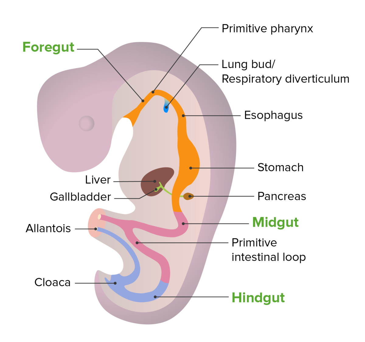
Development

  • Embryologic origins:
  • Development occurs primarily between the 5th and 10th weeks of fetal life
  • Ileum elongates rapidly → forms a U-shaped fold called the primary intestinal loop, which:
    • Grows around the developing superior mesenteric artery (its blood supply)
    • Herniates (protrudes) into the umbilical cord Umbilical cord The flexible rope-like structure that connects a developing fetus to the placenta in mammals. The cord contains blood vessels which carry oxygen and nutrients from the mother to the fetus and waste products away from the fetus. Placenta, Umbilical Cord, and Amniotic Cavity
    • Is connected to the yolk sac Yolk Sac The first of four extra-embryonic membranes to form during embryogenesis. In reptiles and birds, it arises from endoderm and mesoderm to incorporate the egg yolk into the digestive tract for nourishing the embryo. In placental mammals, its nutritional function is vestigial; however, it is the source of intestinal mucosa; blood cells; and germ cells. It is sometimes called the vitelline sac, which should not be confused with the vitelline membrane of the egg. Embryoblast and Trophoblast Development via the vitelline duct Vitelline duct The narrow tube connecting the yolk sac with the midgut of the embryo; persistence of all or part of it in post-fetal life produces abnormalities, of which the commonest is meckel diverticulum. Meckel’s Diverticulum traveling through the umbilical cord Umbilical cord The flexible rope-like structure that connects a developing fetus to the placenta in mammals. The cord contains blood vessels which carry oxygen and nutrients from the mother to the fetus and waste products away from the fetus. Placenta, Umbilical Cord, and Amniotic Cavity
  • As the abdomen develops, the intestines return to the abdominal cavity
  • Undergoes a 270-degree counterclockwise rotation Rotation Motion of an object in which either one or more points on a line are fixed. It is also the motion of a particle about a fixed point. X-rays around the superior mesenteric artery (SMA)  → results in normal anatomic locations for the GI tract

Gross Anatomy

The small intestine is a long tubular structure in the abdomen that is responsible for approximately 90% of nutrient absorption Absorption Absorption involves the uptake of nutrient molecules and their transfer from the lumen of the GI tract across the enterocytes and into the interstitial space, where they can be taken up in the venous or lymphatic circulation. Digestion and Absorption.
General characteristics

  • Connects the stomach Stomach The stomach is a muscular sac in the upper left portion of the abdomen that plays a critical role in digestion. The stomach develops from the foregut and connects the esophagus with the duodenum. Structurally, the stomach is C-shaped and forms a greater and lesser curvature and is divided grossly into regions: the cardia, fundus, body, and pylorus. Stomach: Anatomy at its proximal end to the colon Colon The large intestines constitute the last portion of the digestive system. The large intestine consists of the cecum, appendix, colon (with ascending, transverse, descending, and sigmoid segments), rectum, and anal canal. The primary function of the colon is to remove water and compact the stool prior to expulsion from the body via the rectum and anal canal. Colon, Cecum, and Appendix: Anatomy at its distal end
  • Average length: 6–7 m
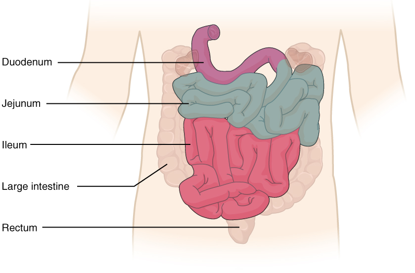
  • Has 3 parts:
    1. Duodenum 
    2. Jejunum
    3. Ileum
  • Fills the majority of the mid and lower abdominal cavity
Small intestine and its parts

Small intestine and its parts

Image: “2417 Small IntestineN” by OpenStax College. License: CC BY 4.0

Duodenum

General characteristics and anatomic relations:

  • The 1st portion of the small intestines
  • Approximately 25 cm in length
  • C-shaped, curving around the head of the pancreas Pancreas The pancreas lies mostly posterior to the stomach and extends across the posterior abdominal wall from the duodenum on the right to the spleen on the left. This organ has both exocrine and endocrine tissue. Pancreas: Anatomy
  • Receives contents from the stomach Stomach The stomach is a muscular sac in the upper left portion of the abdomen that plays a critical role in digestion. The stomach develops from the foregut and connects the esophagus with the duodenum. Structurally, the stomach is C-shaped and forms a greater and lesser curvature and is divided grossly into regions: the cardia, fundus, body, and pylorus. Stomach: Anatomy at its proximal end
  • Empties contents into the jejunum at its distal end
  • Receives secretions from the pancreatic and bile Bile An emulsifying agent produced in the liver and secreted into the duodenum. Its composition includes bile acids and salts; cholesterol; and electrolytes. It aids digestion of fats in the duodenum. Gallbladder and Biliary Tract: Anatomy ducts 

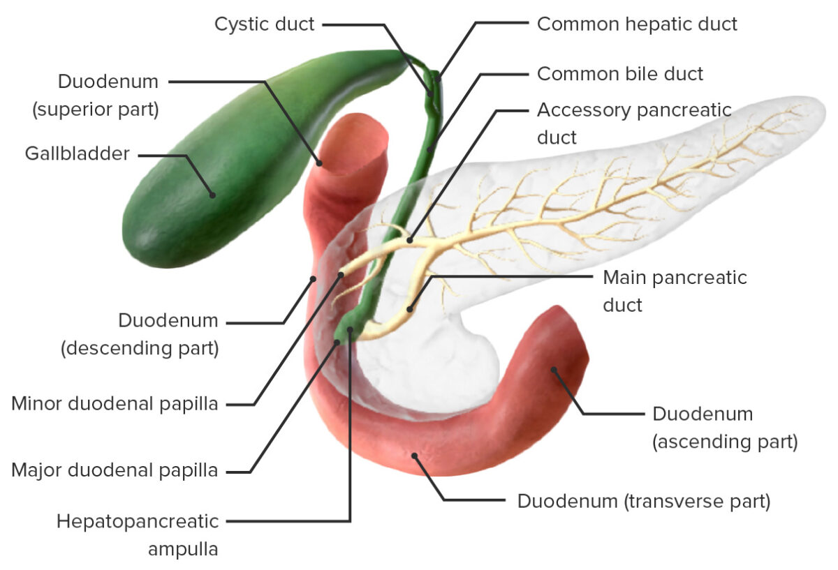
Parts of the duodenum:

The duodenum consists of 4 parts (from proximal to distal):
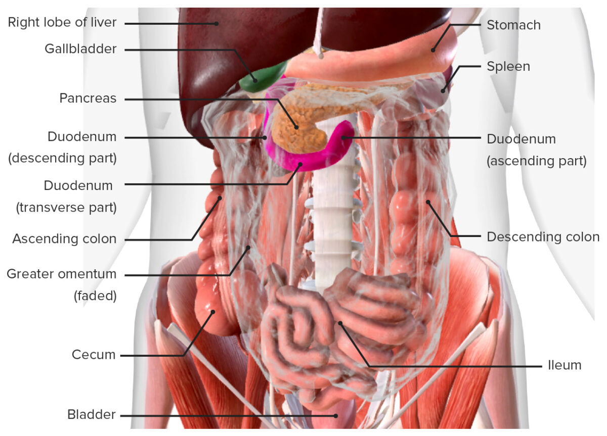
  • Superior: 
    • Begins at the pyloric valve (distalmost portion of the stomach Stomach The stomach is a muscular sac in the upper left portion of the abdomen that plays a critical role in digestion. The stomach develops from the foregut and connects the esophagus with the duodenum. Structurally, the stomach is C-shaped and forms a greater and lesser curvature and is divided grossly into regions: the cardia, fundus, body, and pylorus. Stomach: Anatomy)
    • Located at spinal level L1
    • Intraperitoneal Intraperitoneal Peritoneum: Anatomy
    • Most duodenal ulcers occur in this part
  • Descending:
    • Located at spinal level L1–L3
    • Retroperitoneal Retroperitoneal Peritoneum: Anatomy (duodenum has moved posterior to the peritoneal membrane)
    • Contains connections to the pancreas Pancreas The pancreas lies mostly posterior to the stomach and extends across the posterior abdominal wall from the duodenum on the right to the spleen on the left. This organ has both exocrine and endocrine tissue. Pancreas: Anatomy and bile Bile An emulsifying agent produced in the liver and secreted into the duodenum. Its composition includes bile acids and salts; cholesterol; and electrolytes. It aids digestion of fats in the duodenum. Gallbladder and Biliary Tract: Anatomy duct via the major and minor duodenal papilla
    • Major duodenal papilla:
      • Opening of the hepatopancreatic ampulla (i.e., ampulla of Vater, the combination of the common bile Bile An emulsifying agent produced in the liver and secreted into the duodenum. Its composition includes bile acids and salts; cholesterol; and electrolytes. It aids digestion of fats in the duodenum. Gallbladder and Biliary Tract: Anatomy and main pancreatic ducts) into the duodenal lumen to allow entry of bile Bile An emulsifying agent produced in the liver and secreted into the duodenum. Its composition includes bile acids and salts; cholesterol; and electrolytes. It aids digestion of fats in the duodenum. Gallbladder and Biliary Tract: Anatomy and pancreatic juice Pancreatic Juice The fluid containing digestive enzymes secreted by the pancreas in response to food in the duodenum. Pancreas: Anatomy 
      • Papilla contains the hepatopancreatic sphincter (i.e., sphincter of Oddi), which regulates secretions
    • Minor duodenal papilla:
  • Transverse (sometimes referred to as the horizontal part): 
  • Ascending:
    • Located at spinal level L2–L3
    • Retroperitoneal Retroperitoneal Peritoneum: Anatomy
    • Terminates at the duodenojejunal flexure:
      • Fixed to the posterior abdominal wall Abdominal wall The outer margins of the abdomen, extending from the osteocartilaginous thoracic cage to the pelvis. Though its major part is muscular, the abdominal wall consists of at least seven layers: the skin, subcutaneous fat, deep fascia; abdominal muscles, transversalis fascia, extraperitoneal fat, and the parietal peritoneum. Surgical Anatomy of the Abdomen by the ligament of Treitz Ligament of treitz Gastrointestinal Bleeding
      • Marks the end of the duodenum and beginning of the jejunum

Jejunum and ileum

The jejunum and ileum make up a majority of the small intestine as a long winding tube filling a large portion of the abdominal cavity. Both are completely intraperitoneal Intraperitoneal Peritoneum: Anatomy (within the peritoneal cavity Peritoneal Cavity The space enclosed by the peritoneum. It is divided into two portions, the greater sac and the lesser sac or omental bursa, which lies behind the stomach. The two sacs are connected by the foramen of winslow, or epiploic foramen. Peritoneum: Anatomy).
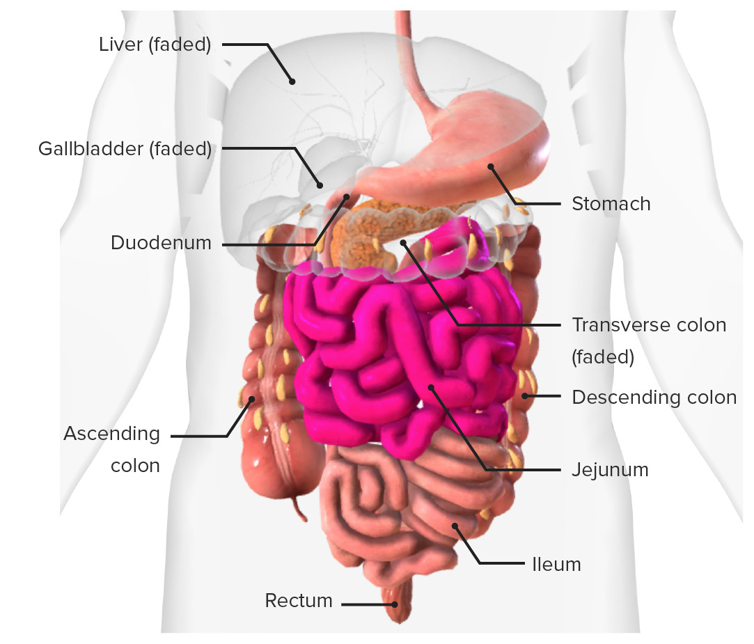
  • Jejunum:
    • Makes up the proximal ⅖ of the small intestine after the duodenum 
    • Approximately 2.5 m in length
    • Begins in the LUQ of the abdomen
    • Progresses inferiorly and medially
  • Ileum:
    • Makes up the distal ⅗ of the small intestines 
    • Approximately 3.5 m in length
    • Primarily in the lower half of the abdomen
    • Ends at the ileocecal junction in the RLQ, where it empties into the cecum Cecum The blind sac or outpouching area of the large intestine that is below the entrance of the small intestine. It has a worm-like extension, the vermiform appendix. Colon, Cecum, and Appendix: Anatomy (1st portion of the large intestine Large intestine The large intestines constitute the last portion of the digestive system. The large intestine consists of the cecum, appendix, colon (with ascending, transverse, descending, and sigmoid segments), rectum, and anal canal. The primary function of the colon is to remove water and compact the stool prior to expulsion from the body via the rectum and anal canal. Colon, Cecum, and Appendix: Anatomy)
  • Mesentery Mesentery A layer of the peritoneum which attaches the abdominal viscera to the abdominal wall and conveys their blood vessels and nerves. Peritoneum: Anatomy:
    • A fold of peritoneum Peritoneum The peritoneum is a serous membrane lining the abdominopelvic cavity. This lining is formed by connective tissue and originates from the mesoderm. The membrane lines both the abdominal walls (as parietal peritoneum) and all of the visceral organs (as visceral peritoneum). Peritoneum: Anatomy that suspends the jejunum and ileum from the posterior abdominal cavity
    • Allows the jejunum and ileum significant mobility within the abdominal cavity
  • Transition between jejunum and ileum:
    • Gradual (as opposed to a discrete anatomic landmark)
    • Different segments are defined by differences in microscopic anatomy noted most prominently when comparing the proximal jejunum and the distal ileum

Microscopic Anatomy

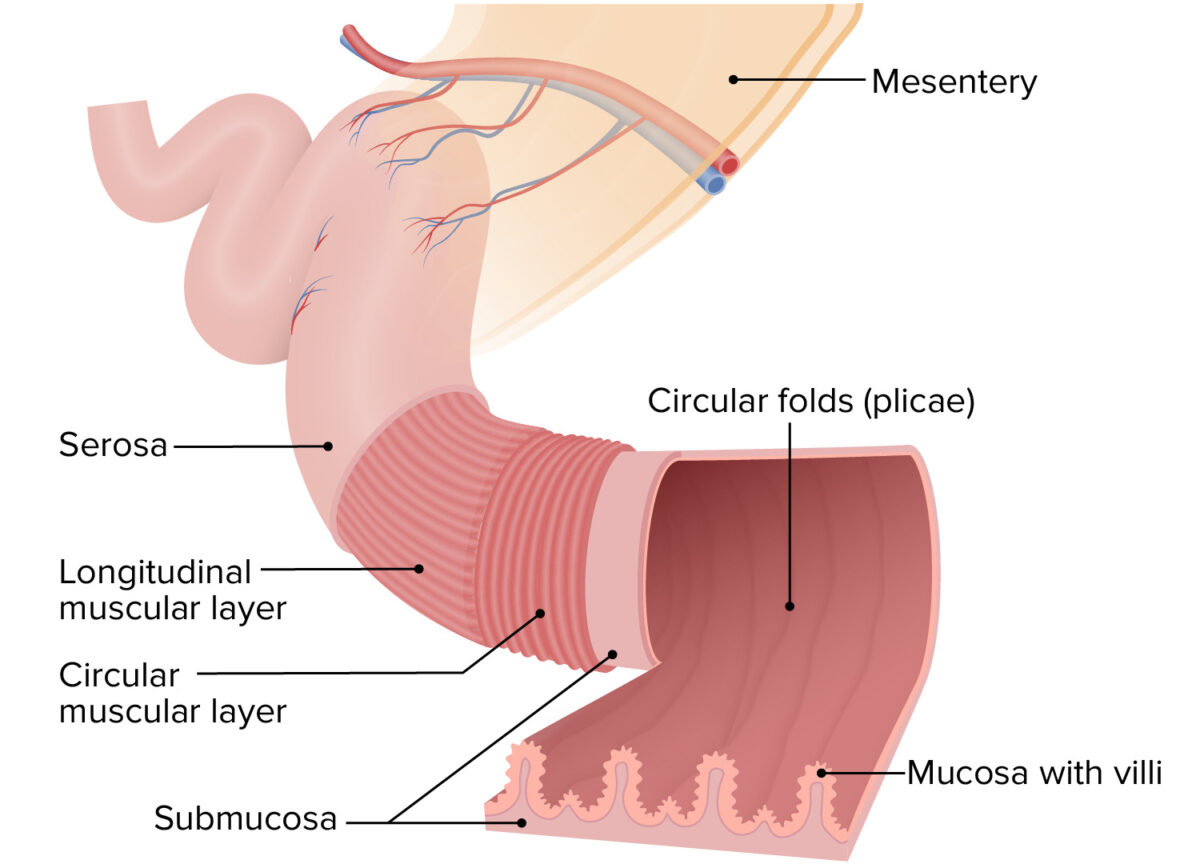
Similar to other segments of the GI tract, the layers of the small intestinal wall (from the inner lumen outward) are mucosa → submucosa → muscular layer → serosa. The walls have several different types of folds.

Folds of the intestinal walls

Circular folds (known as plicae):

  • Large folds of intestinal wall projecting into the lumen
  • Contain both mucosa and submucosa
  • Permanent folds (i.e., not obliterated when the intestine is distended, which is different from rugae in the stomach Stomach The stomach is a muscular sac in the upper left portion of the abdomen that plays a critical role in digestion. The stomach develops from the foregut and connects the esophagus with the duodenum. Structurally, the stomach is C-shaped and forms a greater and lesser curvature and is divided grossly into regions: the cardia, fundus, body, and pylorus. Stomach: Anatomy, which are obliterated as the stomach Stomach The stomach is a muscular sac in the upper left portion of the abdomen that plays a critical role in digestion. The stomach develops from the foregut and connects the esophagus with the duodenum. Structurally, the stomach is C-shaped and forms a greater and lesser curvature and is divided grossly into regions: the cardia, fundus, body, and pylorus. Stomach: Anatomy distends)
  • Locations:
    • Begin approximately 6 cm into the duodenum
    • Most prominent in the jejunum
    • Decrease as you move farther down the ileum
  • Function:
    • Mixes chyme 
    • ↑ Surface area required for:
      • Contact digestion Digestion Digestion refers to the process of the mechanical and chemical breakdown of food into smaller particles, which can then be absorbed and utilized by the body. Digestion and Absorption
      • Absorption Absorption Absorption involves the uptake of nutrient molecules and their transfer from the lumen of the GI tract across the enterocytes and into the interstitial space, where they can be taken up in the venous or lymphatic circulation. Digestion and Absorption

Intestinal villi: 

  • Finger-like projections of the mucosa only ( epithelium Epithelium The epithelium is a complex of specialized cellular organizations arranged into sheets and lining cavities and covering the surfaces of the body. The cells exhibit polarity, having an apical and a basal pole. Structures important for the epithelial integrity and function involve the basement membrane, the semipermeable sheet on which the cells rest, and interdigitations, as well as cellular junctions. Surface Epithelium: Histology and lamina propria Lamina propria Whipple’s Disease)
  • Location: off the entire lining of the small intestines, including off plicae folds
  • Function: significantly ↑ surface area
Layers and folds in the intestinal walls

Layers and folds in the intestinal walls

Image by Lecturio.

Layers of the intestinal walls

Mucosa:

  • Consists of 3 sublayers:
    1. Epithelium Epithelium The epithelium is a complex of specialized cellular organizations arranged into sheets and lining cavities and covering the surfaces of the body. The cells exhibit polarity, having an apical and a basal pole. Structures important for the epithelial integrity and function involve the basement membrane, the semipermeable sheet on which the cells rest, and interdigitations, as well as cellular junctions. Surface Epithelium: Histology (innermost lining):
      • Enterocytes (also called absorptive cells): simple columnar cells primarily responsible for contact digestion Digestion Digestion refers to the process of the mechanical and chemical breakdown of food into smaller particles, which can then be absorbed and utilized by the body. Digestion and Absorption and absorption Absorption Absorption involves the uptake of nutrient molecules and their transfer from the lumen of the GI tract across the enterocytes and into the interstitial space, where they can be taken up in the venous or lymphatic circulation. Digestion and Absorption
      • Goblet cells Goblet cells A glandular epithelial cell or a unicellular gland. Goblet cells secrete mucus. They are scattered in the epithelial linings of many organs, especially the small intestine and the respiratory tract. Glandular Epithelium: Histology: secrete mucus
      • Enteroendocrine cells Enteroendocrine cells Cells found throughout the lining of the gastrointestinal tract that contain and secrete regulatory peptide hormones and/or biogenic amines. Gastrointestinal Secretions: secrete hormones Hormones Hormones are messenger molecules that are synthesized in one part of the body and move through the bloodstream to exert specific regulatory effects on another part of the body. Hormones play critical roles in coordinating cellular activities throughout the body in response to the constant changes in both the internal and external environments. Hormones: Overview and Types into the blood
      • Cells are connected via tight junctions Tight junctions Cell-cell junctions that seal adjacent epithelial cells together, preventing the passage of most dissolved molecules from one side of the epithelial sheet to the other. The Cell: Cell Junctions
      • Have a brush border Brush border Tubular System: microvilli on the absorptive surface, which ↑ surface area and contain membrane-bound digestive enzymes Enzymes Enzymes are complex protein biocatalysts that accelerate chemical reactions without being consumed by them. Due to the body’s constant metabolic needs, the absence of enzymes would make life unsustainable, as reactions would occur too slowly without these molecules. Basics of Enzymes 
    2. Lamina propria Lamina propria Whipple’s Disease, which contains:
      • Vasculature: arterioles Arterioles The smallest divisions of the arteries located between the muscular arteries and the capillaries. Arteries: Histology, veins Veins Veins are tubular collections of cells, which transport deoxygenated blood and waste from the capillary beds back to the heart. Veins are classified into 3 types: small veins/venules, medium veins, and large veins. Each type contains 3 primary layers: tunica intima, tunica media, and tunica adventitia. Veins: Histology, and capillary networks, which absorb a majority of the nutrients except for fats Fats The glyceryl esters of a fatty acid, or of a mixture of fatty acids. They are generally odorless, colorless, and tasteless if pure, but they may be flavored according to origin. Fats are insoluble in water, soluble in most organic solvents. They occur in animal and vegetable tissue and are generally obtained by boiling or by extraction under pressure. They are important in the diet (dietary fats) as a source of energy. Energy Homeostasis
      • Lymphatic vessels Lymphatic Vessels Tubular vessels that are involved in the transport of lymph and lymphocytes. Lymphatic Drainage System: Anatomy known as lacteals: absorb a majority of the fat and fat-soluble vitamins
    3. Muscularis mucosa:
      • Thin layer of smooth muscle
      • Controls movement of the villi to ↑ mixing
  • Crypts of Lieberkuhn:
    • Pores at the base of the villi that open into tubular glands (similar to gastric glands Gastric glands Stomach: Anatomy)
    • Upper ½ contains enterocytes and goblet cells Goblet cells A glandular epithelial cell or a unicellular gland. Goblet cells secrete mucus. They are scattered in the epithelial linings of many organs, especially the small intestine and the respiratory tract. Glandular Epithelium: Histology
    • Lower ½ contains:
      • Stem cells: replace enterocytes and goblet cells Goblet cells A glandular epithelial cell or a unicellular gland. Goblet cells secrete mucus. They are scattered in the epithelial linings of many organs, especially the small intestine and the respiratory tract. Glandular Epithelium: Histology every 3–6 days 
      • Paneth cells: secrete defensins Defensins Family of antimicrobial peptides that have been identified in humans, animals, and plants. They are thought to play a role in host defenses against infections, inflammation, wound repair, and acquired immunity. Innate Immunity: Barriers, Complement, and Cytokines and lysozyme (which protect against bacterial infection)
    • Located throughout the small intestines
  • Peyer patches Patches Vitiligo:

Submucosa:

  • Loose connective tissue Loose connective tissue Connective Tissue: Histology
  • Contains larger vessels
  • Brunner glands:
    • Produce alkaline mucus that protects the mucosa from damage caused by stomach Stomach The stomach is a muscular sac in the upper left portion of the abdomen that plays a critical role in digestion. The stomach develops from the foregut and connects the esophagus with the duodenum. Structurally, the stomach is C-shaped and forms a greater and lesser curvature and is divided grossly into regions: the cardia, fundus, body, and pylorus. Stomach: Anatomy acid
    • Found only within the duodenum
  • Meissner plexus Meissner plexus One of two ganglionated neural networks which together form the enteric nervous system. The submucous (Meissner’s) plexus is in the connective tissue of the submucosa. Its neurons innervate the epithelium, blood vessels, endocrine cells, other submucosal ganglia, and myenteric ganglia, and play an important role in regulating ion and water transport. Gastrointestinal Neural and Hormonal Signaling:
    • Ganglia of the ANS ANS The ans is a component of the peripheral nervous system that uses both afferent (sensory) and efferent (effector) neurons, which control the functioning of the internal organs and involuntary processes via connections with the CNS. The ans consists of the sympathetic and parasympathetic nervous systems. Autonomic Nervous System: Anatomy
    • Control the muscularis mucosa (independently of the muscular layer of the intestines)

Muscular layer: 

The muscular layer is made up of 2 layers of smooth muscle that mix and move chyme along the tract.

  • Circular layer (inner layer)
  • Longitudinal layer (outer layer)
  • Auerbach (myenteric) plexus:
    • Ganglia of the autonomic nervous system Autonomic nervous system The ANS is a component of the peripheral nervous system that uses both afferent (sensory) and efferent (effector) neurons, which control the functioning of the internal organs and involuntary processes via connections with the CNS. The ANS consists of the sympathetic and parasympathetic nervous systems. Autonomic Nervous System: Anatomy, which controls the muscular layer
    • Located between the 2 layers of smooth muscle

Serosa: 

  • Made up of connective tissue Connective tissue Connective tissues originate from embryonic mesenchyme and are present throughout the body except inside the brain and spinal cord. The main function of connective tissues is to provide structural support to organs. Connective tissues consist of cells and an extracellular matrix. Connective Tissue: Histology
  • Joins with mesentery Mesentery A layer of the peritoneum which attaches the abdominal viscera to the abdominal wall and conveys their blood vessels and nerves. Peritoneum: Anatomy or peritoneum Peritoneum The peritoneum is a serous membrane lining the abdominopelvic cavity. This lining is formed by connective tissue and originates from the mesoderm. The membrane lines both the abdominal walls (as parietal peritoneum) and all of the visceral organs (as visceral peritoneum). Peritoneum: Anatomy
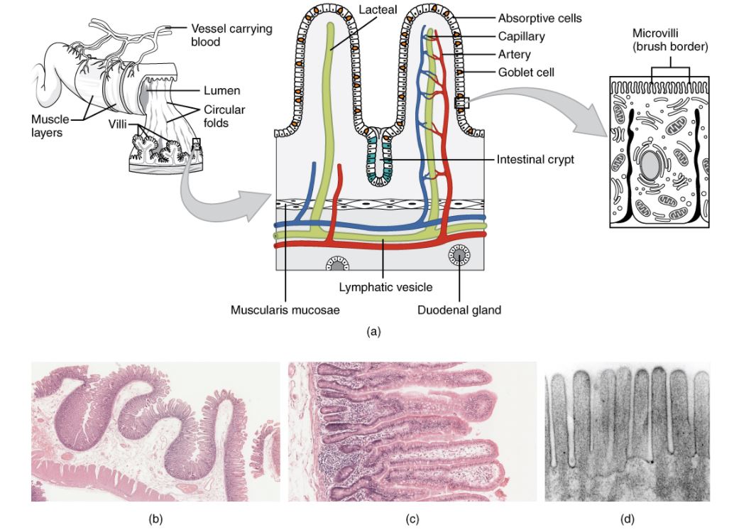
Histology of the small intestine

Histology of the small intestine:
(a): The absorptive surface of the small intestine is vastly enlarged by the presence of circular folds, villi, and microvilli.
(b): Micrograph of the circular folds: Note that the folds contain both the mucosa and the submucosa.
(c): Micrograph of the villi: Note that the villi contain only the epithelial and lamina propria layers of the mucosa; the muscularis mucosa is visible as a “pink line” along the left edge of the slide.
(d): Electron micrograph of the microvilli: from left to right—light microscope, ×56; light microscope, ×508; electron microscope, ×196,000

Image: “Histology of the Small Intestine” by Phil Schatz. License: CC BY 4.0

Anatomic Differences between the Jejunum and Ileum

Characteristic differences between the jejunum and ileum are summarized in the table.

Table: Anatomic differences between the jejunum and ileum
Jejunum Ileum
Diameter 4 cm (at the most proximal end) 2 cm (at the most distal end)
Wall thickness Thicker Thinner
Fat in mesentery Mesentery A layer of the peritoneum which attaches the abdominal viscera to the abdominal wall and conveys their blood vessels and nerves. Peritoneum: Anatomy Less More
Circular folds Many, best developed Some in the proximal ileum, very few distally
Lymphoid tissue/Peyer patches Patches Vitiligo Few Many

Neurovasculature

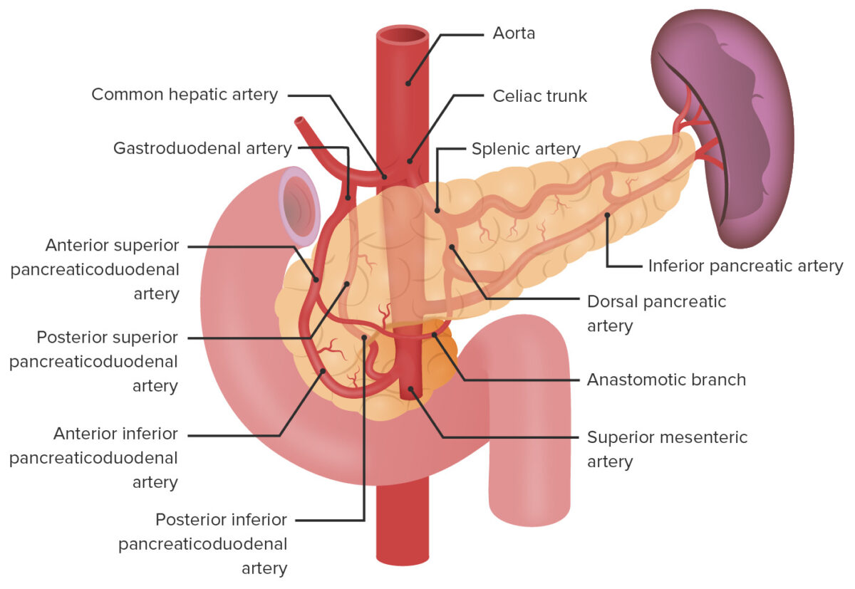
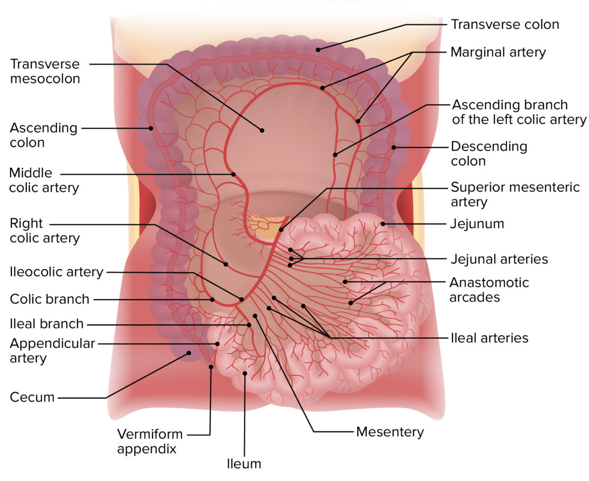
Arterial supply

  • Duodenum: superior and inferior pancreaticoduodenal arteries Arteries Arteries are tubular collections of cells that transport oxygenated blood and nutrients from the heart to the tissues of the body. The blood passes through the arteries in order of decreasing luminal diameter, starting in the largest artery (the aorta) and ending in the small arterioles. Arteries are classified into 3 types: large elastic arteries, medium muscular arteries, and small arteries and arterioles. Arteries: Histology
  • Jejunum and ileum: jejunal and ileal arteries Arteries Arteries are tubular collections of cells that transport oxygenated blood and nutrients from the heart to the tissues of the body. The blood passes through the arteries in order of decreasing luminal diameter, starting in the largest artery (the aorta) and ending in the small arterioles. Arteries are classified into 3 types: large elastic arteries, medium muscular arteries, and small arteries and arterioles. Arteries: Histology (branch off the SMA)
    • Run within the mesentery Mesentery A layer of the peritoneum which attaches the abdominal viscera to the abdominal wall and conveys their blood vessels and nerves. Peritoneum: Anatomy from the SMA toward the intestines
    • Form anastomotic loops with each other, known as arcades 
    • Vasa recta Vasa recta Glomerular Filtration: small straight arteries Arteries Arteries are tubular collections of cells that transport oxygenated blood and nutrients from the heart to the tissues of the body. The blood passes through the arteries in order of decreasing luminal diameter, starting in the largest artery (the aorta) and ending in the small arterioles. Arteries are classified into 3 types: large elastic arteries, medium muscular arteries, and small arteries and arterioles. Arteries: Histology off the arcades that supply the intestinal wall

Venous drainage

  • Veins Veins Veins are tubular collections of cells, which transport deoxygenated blood and waste from the capillary beds back to the heart. Veins are classified into 3 types: small veins/venules, medium veins, and large veins. Each type contains 3 primary layers: tunica intima, tunica media, and tunica adventitia. Veins: Histology running adjacent to the arterial supply
  • Drain into the superior mesenteric vein (SMV)
  • The SMV unites with the splenic vein to form the hepatic portal Hepatic portal Liver: Anatomy vein → brings nutrients absorbed through the gut to the liver Liver The liver is the largest gland in the human body. The liver is found in the superior right quadrant of the abdomen and weighs approximately 1.5 kilograms. Its main functions are detoxification, metabolism, nutrient storage (e.g., iron and vitamins), synthesis of coagulation factors, formation of bile, filtration, and storage of blood. Liver: Anatomy

Lymphatics

Lacteals within the intestinal villi drain into:

  • Pancreaticoduodenal nodes (duodenum only)
  • Superior mesenteric nodes (all 3 segments)

Innervation

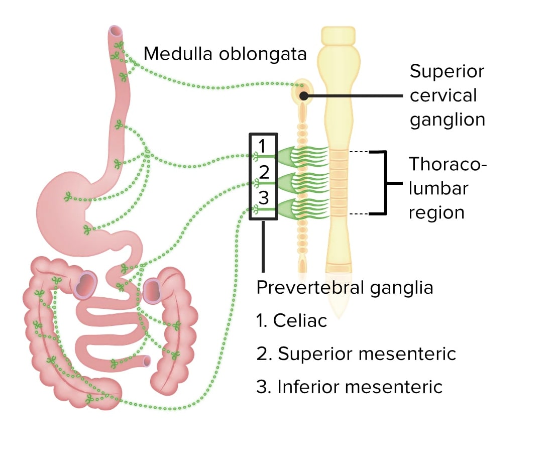
The small intestine is innervated by the ANS ANS The ans is a component of the peripheral nervous system that uses both afferent (sensory) and efferent (effector) neurons, which control the functioning of the internal organs and involuntary processes via connections with the CNS. The ans consists of the sympathetic and parasympathetic nervous systems. Autonomic Nervous System: Anatomy.

Parasympathetic innervation (stimulatory): 
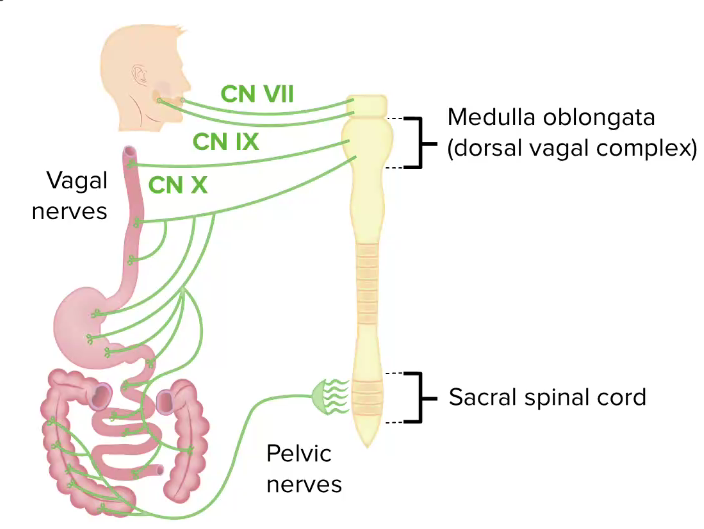
  • Posterior trunk of the vagus nerve Vagus nerve The 10th cranial nerve. The vagus is a mixed nerve which contains somatic afferents (from skin in back of the ear and the external auditory meatus), visceral afferents (from the pharynx, larynx, thorax, and abdomen), parasympathetic efferents (to the thorax and abdomen), and efferents to striated muscle (of the larynx and pharynx). Pharynx: Anatomy
  • ↑ Secretions, motility Motility The motor activity of the gastrointestinal tract. Gastrointestinal Motility, and blood flow Blood flow Blood flow refers to the movement of a certain volume of blood through the vasculature over a given unit of time (e.g., mL per minute). Vascular Resistance, Flow, and Mean Arterial Pressure
  • Relaxation of sphincters
Parasympathetic innervation of the gi tract

Parasympathetic innervation of the GI tract
CN: cranial nerve

Image by Lecturio.

Sympathetic innervation (inhibitory):

Sympathetic innervation of the gi tract

Sympathetic innervation of the GI tract

Image by Lecturio.

Function

The small intestines are the primary site of chemical digestion Digestion Digestion refers to the process of the mechanical and chemical breakdown of food into smaller particles, which can then be absorbed and utilized by the body. Digestion and Absorption and absorption Absorption Absorption involves the uptake of nutrient molecules and their transfer from the lumen of the GI tract across the enterocytes and into the interstitial space, where they can be taken up in the venous or lymphatic circulation. Digestion and Absorption of nutrients.

  • Receive:
    • Chyme from the stomach Stomach The stomach is a muscular sac in the upper left portion of the abdomen that plays a critical role in digestion. The stomach develops from the foregut and connects the esophagus with the duodenum. Structurally, the stomach is C-shaped and forms a greater and lesser curvature and is divided grossly into regions: the cardia, fundus, body, and pylorus. Stomach: Anatomy
    • Pancreatic juice Pancreatic Juice The fluid containing digestive enzymes secreted by the pancreas in response to food in the duodenum. Pancreas: Anatomy and enzymes Enzymes Enzymes are complex protein biocatalysts that accelerate chemical reactions without being consumed by them. Due to the body’s constant metabolic needs, the absence of enzymes would make life unsustainable, as reactions would occur too slowly without these molecules. Basics of Enzymes
    • Bile Bile An emulsifying agent produced in the liver and secreted into the duodenum. Its composition includes bile acids and salts; cholesterol; and electrolytes. It aids digestion of fats in the duodenum. Gallbladder and Biliary Tract: Anatomy
  • Secrete intestinal juice, which (along with the pancreatic juice Pancreatic Juice The fluid containing digestive enzymes secreted by the pancreas in response to food in the duodenum. Pancreas: Anatomy and bile Bile An emulsifying agent produced in the liver and secreted into the duodenum. Its composition includes bile acids and salts; cholesterol; and electrolytes. It aids digestion of fats in the duodenum. Gallbladder and Biliary Tract: Anatomy):
    • Neutralizes stomach Stomach The stomach is a muscular sac in the upper left portion of the abdomen that plays a critical role in digestion. The stomach develops from the foregut and connects the esophagus with the duodenum. Structurally, the stomach is C-shaped and forms a greater and lesser curvature and is divided grossly into regions: the cardia, fundus, body, and pylorus. Stomach: Anatomy acid
    • Digests nutrients ( carbohydrates Carbohydrates A class of organic compounds composed of carbon, hydrogen, and oxygen in a ratio of cn(H2O)n. The largest class of organic compounds, including starch; glycogen; cellulose; polysaccharides; and simple monosaccharides. Basics of Carbohydrates, fat, and proteins Proteins Linear polypeptides that are synthesized on ribosomes and may be further modified, crosslinked, cleaved, or assembled into complex proteins with several subunits. The specific sequence of amino acids determines the shape the polypeptide will take, during protein folding, and the function of the protein. Energy Homeostasis)
  • Primary site of nutrient absorption Absorption Absorption involves the uptake of nutrient molecules and their transfer from the lumen of the GI tract across the enterocytes and into the interstitial space, where they can be taken up in the venous or lymphatic circulation. Digestion and Absorption
  • Brush border Brush border Tubular System of small intestines:
    • Contains additional digestive enzymes Enzymes Enzymes are complex protein biocatalysts that accelerate chemical reactions without being consumed by them. Due to the body’s constant metabolic needs, the absence of enzymes would make life unsustainable, as reactions would occur too slowly without these molecules. Basics of Enzymes required for contact digestion Digestion Digestion refers to the process of the mechanical and chemical breakdown of food into smaller particles, which can then be absorbed and utilized by the body. Digestion and Absorption
    • ↑ Surface area for absorption Absorption Absorption involves the uptake of nutrient molecules and their transfer from the lumen of the GI tract across the enterocytes and into the interstitial space, where they can be taken up in the venous or lymphatic circulation. Digestion and Absorption of nutrients
  • Secrete hormones Hormones Hormones are messenger molecules that are synthesized in one part of the body and move through the bloodstream to exert specific regulatory effects on another part of the body. Hormones play critical roles in coordinating cellular activities throughout the body in response to the constant changes in both the internal and external environments. Hormones: Overview and Types 
  • Functions of intestinal motility Intestinal Motility Gastrointestinal Motility:
    • Mix chyme with bile Bile An emulsifying agent produced in the liver and secreted into the duodenum. Its composition includes bile acids and salts; cholesterol; and electrolytes. It aids digestion of fats in the duodenum. Gallbladder and Biliary Tract: Anatomy and digestive juices from the pancreas Pancreas The pancreas lies mostly posterior to the stomach and extends across the posterior abdominal wall from the duodenum on the right to the spleen on the left. This organ has both exocrine and endocrine tissue. Pancreas: Anatomy and intestines
    • Churn chyme, bringing it into contact with the mucosa for contact digestion Digestion Digestion refers to the process of the mechanical and chemical breakdown of food into smaller particles, which can then be absorbed and utilized by the body. Digestion and Absorption and absorption Absorption Absorption involves the uptake of nutrient molecules and their transfer from the lumen of the GI tract across the enterocytes and into the interstitial space, where they can be taken up in the venous or lymphatic circulation. Digestion and Absorption
    • Move contents toward the large intestine Large intestine The large intestines constitute the last portion of the digestive system. The large intestine consists of the cecum, appendix, colon (with ascending, transverse, descending, and sigmoid segments), rectum, and anal canal. The primary function of the colon is to remove water and compact the stool prior to expulsion from the body via the rectum and anal canal. Colon, Cecum, and Appendix: Anatomy

Clinical Relevance

  • Intestinal malrotation Malrotation Pediatric Gastrointestinal Abnormalities: congenital GI anomaly that results from failure of the normal primitive gut rotation Rotation Motion of an object in which either one or more points on a line are fixed. It is also the motion of a particle about a fixed point. X-rays around mesenteric vessels during embryologic development. Intestinal malrotation Malrotation Pediatric Gastrointestinal Abnormalities can result in a number of anatomic patterns characterized by abnormal location and attachments of the intestines within the abdominal cavity. These anomalies can be clinically silent or may present with a number of complications, the most catastrophic of which is midgut Midgut Development of the Abdominal Organs volvulus Volvulus A volvulus is the twisting or axial rotation of a portion of the bowel around its mesentery. The most common site of volvulus in adults is the colon; most frequently the sigmoid volvulus. Patients typically present with symptoms of bowel obstruction such as abdominal pain, distension, vomiting, and constipation/obstipation. Volvulus
  • Intussusception Intussusception Intussusception occurs when a part of the intestine (intussusceptum) telescopes into another part (intussuscipiens) of the intestine. The condition can cause obstruction and, if untreated, progress to bowel ischemia. Intussusception is most common in the pediatric population, but is occasionally encountered in adults. Intussusception: telescoping of the proximal part (intussusceptum) into a distal part (intussuscipiens) of the intestine. Intussusception Intussusception Intussusception occurs when a part of the intestine (intussusceptum) telescopes into another part (intussuscipiens) of the intestine. The condition can cause obstruction and, if untreated, progress to bowel ischemia. Intussusception is most common in the pediatric population, but is occasionally encountered in adults. Intussusception can cause obstruction and, if untreated, can progress to bowel ischemia Bowel ischemia Mesenteric ischemia is a rare, life-threatening condition caused by inadequate blood flow through the mesenteric vessels, which results in ischemia and necrosis of the intestinal wall. Mesenteric ischemia can be either acute or chronic. Mesenteric Ischemia. Intussusception Intussusception Intussusception occurs when a part of the intestine (intussusceptum) telescopes into another part (intussuscipiens) of the intestine. The condition can cause obstruction and, if untreated, progress to bowel ischemia. Intussusception is most common in the pediatric population, but is occasionally encountered in adults. Intussusception is most common in the pediatric population, but it is occasionally encountered in adults. The pediatric individual typically presents with acute cyclical abdominal pain Abdominal Pain Acute Abdomen and vomiting Vomiting The forcible expulsion of the contents of the stomach through the mouth. Hypokalemia, whereas adults present with symptoms of bowel obstruction Bowel obstruction Any impairment, arrest, or reversal of the normal flow of intestinal contents toward the anal canal. Ascaris/Ascariasis
  • Meckel diverticulum Diverticulum A pouch or sac opening from the colon. Diverticular Disease: persistent remnant of the omphalomesenteric (vitelline) duct. Meckel diverticulum Diverticulum A pouch or sac opening from the colon. Diverticular Disease is usually located in the antimesenteric border of the ileum. The mucosal lining of the diverticulum Diverticulum A pouch or sac opening from the colon. Diverticular Disease may contain heterotopic Heterotopic Transplantation of tissue typical of one area to a different recipient site. The tissue may be autologous, heterologous, or homologous. Organ Transplantation mucosa (most commonly gastric). Although it is frequently asymptomatic, Meckel diverticulum Diverticulum A pouch or sac opening from the colon. Diverticular Disease can cause ulceration Ulceration Corneal Abrasions, Erosion, and Ulcers and present with lower GI bleeding. Other complications include diverticulitis Diverticulitis Inflammation of a diverticulum or diverticula. Diverticular Disease and small bowel obstruction Bowel obstruction Any impairment, arrest, or reversal of the normal flow of intestinal contents toward the anal canal. Ascaris/Ascariasis
  • Small bowel obstruction Bowel obstruction Any impairment, arrest, or reversal of the normal flow of intestinal contents toward the anal canal. Ascaris/Ascariasis: The normal passage of bowel contents is interrupted either because of a functional decrease in peristalsis Peristalsis A movement, caused by sequential muscle contraction, that pushes the contents of the intestines or other tubular organs in one direction. Gastrointestinal Motility or mechanical obstruction Mechanical Obstruction Any impairment, arrest, or reversal of the normal flow of intestinal contents toward the anal canal. Imaging of the Intestines from intraluminal or extraluminal mechanical compression Compression Blunt Chest Trauma. The most common causes in developed countries include postsurgical adhesions, hernias, and malignancies. The most important clinical features include crampy abdominal pain Abdominal Pain Acute Abdomen, obstipation Obstipation Large Bowel Obstruction (i.e., inability to pass flatus or stool), nausea Nausea An unpleasant sensation in the stomach usually accompanied by the urge to vomit. Common causes are early pregnancy, sea and motion sickness, emotional stress, intense pain, food poisoning, and various enteroviruses. Antiemetics, and vomiting Vomiting The forcible expulsion of the contents of the stomach through the mouth. Hypokalemia.
  • Crohn’s disease: form of inflammatory bowel disease characterized by patchy transmural inflammation Transmural inflammation Crohn’s Disease; any portion of the luminal GI tract can be involved, though the terminal ileum and proximal colon Colon The large intestines constitute the last portion of the digestive system. The large intestine consists of the cecum, appendix, colon (with ascending, transverse, descending, and sigmoid segments), rectum, and anal canal. The primary function of the colon is to remove water and compact the stool prior to expulsion from the body via the rectum and anal canal. Colon, Cecum, and Appendix: Anatomy are most commonly affected. The most important clinical features include crampy abdominal pain Abdominal Pain Acute Abdomen, diarrhea Diarrhea Diarrhea is defined as ≥ 3 watery or loose stools in a 24-hour period. There are a multitude of etiologies, which can be classified based on the underlying mechanism of disease. The duration of symptoms (acute or chronic) and characteristics of the stools (e.g., watery, bloody, steatorrheic, mucoid) can help guide further diagnostic evaluation. Diarrhea, fatigue Fatigue The state of weariness following a period of exertion, mental or physical, characterized by a decreased capacity for work and reduced efficiency to respond to stimuli. Fibromyalgia, and weight loss Weight loss Decrease in existing body weight. Bariatric Surgery. Complications include fistulas, phlegmon/ abscess Abscess Accumulation of purulent material in tissues, organs, or circumscribed spaces, usually associated with signs of infection. Chronic Granulomatous Disease formation, and perianal disease.
  • Celiac disease Celiac disease Celiac disease (also known as celiac sprue or gluten enteropathy) is an autoimmune reaction to gliadin, which is a component of gluten. Celiac disease is closely associated with HLA-DQ2 and HLA-DQ8. The immune response is localized to the proximal small intestine and causes the characteristic histologic findings of villous atrophy, crypt hyperplasia, and intraepithelial lymphocytosis. Celiac Disease: also known as gluten-sensitive enteropathy Enteropathy IPEX Syndrome. Celiac disease Celiac disease Celiac disease (also known as celiac sprue or gluten enteropathy) is an autoimmune reaction to gliadin, which is a component of gluten. Celiac disease is closely associated with HLA-DQ2 and HLA-DQ8. The immune response is localized to the proximal small intestine and causes the characteristic histologic findings of villous atrophy, crypt hyperplasia, and intraepithelial lymphocytosis. Celiac Disease is a common immune-mediated inflammatory disease of the small intestine caused by sensitivity to gliadin Gliadin Simple protein, one of the prolamins, derived from the gluten of wheat, rye, etc. May be separated into 4 discrete electrophoretic fractions. It is the toxic factor associated with celiac disease. Celiac Disease, a component of gluten Gluten Prolamins in the endosperm of seeds from the triticeae tribe which includes species of wheat; barley; and rye. Celiac Disease. The most important clinical features include diarrhea Diarrhea Diarrhea is defined as ≥ 3 watery or loose stools in a 24-hour period. There are a multitude of etiologies, which can be classified based on the underlying mechanism of disease. The duration of symptoms (acute or chronic) and characteristics of the stools (e.g., watery, bloody, steatorrheic, mucoid) can help guide further diagnostic evaluation. Diarrhea with malabsorption Malabsorption General term for a group of malnutrition syndromes caused by failure of normal intestinal absorption of nutrients. Malabsorption and Maldigestion, crampy abdominal pain Abdominal Pain Acute Abdomen, and villous atrophy Villous Atrophy Giardia/Giardiasis on histology.

References

  1. Collins, J., Nguyen, A. (2021). Anatomy, abdomen and pelvis, small intestine. StatPearls. Retrieved August 23, 2021, from https://www.statpearls.com/articlelibrary/viewarticle/32127/ 
  2. Saladin, K.S., Miller, L. (2004). Anatomy and Physiology, 3rd ed., pp. 964–965. McGraw-Hill Education.
  3. Peppercorn, M.A., Kane, S.V. (2020). Clinical manifestations, diagnosis, and prognosis of Crohn’s disease in adults. UpToDate. Retrieved August 20, 2021, from https://www.uptodate.com/contents/clinical-manifestations-diagnosis-and-prognosis-of-crohn-disease-in-adults
  4. Peppercorn, M.A., Kane, S.V. (2020). Clinical manifestations, diagnosis, and prognosis of ulcerative colitis in adults. UpToDate. Retrieved August 20, 2021, from https://www.uptodate.com/contents/clinical-manifestations-diagnosis-and-prognosis-of-ulcerative-colitis-in-adults
  5. Drake, R.L., et al. (Ed.) (2020). Regional anatomy. In Gray’s Anatomy for Students, 4th ed. Churchill Livingstone/Elsevier, pp. 309–315.
  6. Schuppan, D., Dieterich, W. (2020). Epidemiology, pathogenesis, and clinical manifestations of celiac disease in adults. UpToDate. Retrieved August 20, 2021, from https://www.uptodate.com/contents/epidemiology-pathogenesis-and-clinical-manifestations-of-celiac-disease-in-adults

Create your free account or log in to continue reading!

Sign up now and get free access to Lecturio with concept pages, medical videos, and questions for your medical education.

User Reviews

Details