Advertisement

Advertisement

Advertisement

Advertisement

Skull: Anatomy

The skull (cranium) is the skeletal structure of the head supporting the face and forming a protective cavity for the brain Brain The part of central nervous system that is contained within the skull (cranium). Arising from the neural tube, the embryonic brain is comprised of three major parts including prosencephalon (the forebrain); mesencephalon (the midbrain); and rhombencephalon (the hindbrain). The developed brain consists of cerebrum; cerebellum; and other structures in the brain stem. Nervous System: Anatomy, Structure, and Classification. The skull consists of 22 bones divided into the viscerocranium (facial skeleton) and the neurocranium. The neurocranium is further subdivided into the calvarium (the skullcap) and the base of the skull (the cranial fossa).

Last updated: Nov 19, 2024

Editorial responsibility: Stanley Oiseth, Lindsay Jones, Evelin Maza

Advertisement

Advertisement

Advertisement

Advertisement

Advertisement

Advertisement

Viscerocranium

The viscerocranium is responsible for maintaining the structure for the muscles of the face Muscles of the Face The facial muscles (also called mimetic muscles) control facial expression and are supplied by the facial nerve. Most of them originate from the skull and attach to the skin around the facial openings, which serve as a method to group or classify them. Facial Muscles: Anatomy. The muscles are arranged in a complex anatomical pattern to allow for a variety of facial functions such as mastication Mastication The act and process of chewing and grinding food in the mouth. Jaw and Temporomandibular Joint: Anatomy, smiling, frowning, eye movement, and patency of the nasal passage.

Table: Bones of the viscerocranium
Viscerocranium bone Bone Bone is a compact type of hardened connective tissue composed of bone cells, membranes, an extracellular mineralized matrix, and central bone marrow. The 2 primary types of bone are compact and spongy. Bones: Structure and Types Number of bones Facial features
Zygomatic Paired Cheekbone
Lacrimal Paired Anterior medial wall of the orbit
Nasal Paired Bridge of the nose Nose The nose is the human body’s primary organ of smell and functions as part of the upper respiratory system. The nose may be best known for inhaling oxygen and exhaling carbon dioxide, but it also contributes to other important functions, such as tasting. The anatomy of the nose can be divided into the external nose and the nasal cavity. Nose Anatomy (External & Internal)
Palatine Paired Posterior quarter of the hard palate Hard palate The anteriorly located rigid section of the palate. Palate: Anatomy
Maxilla Paired Upper jaw Jaw The jaw is made up of the mandible, which comprises the lower jaw, and the maxilla, which comprises the upper jaw. The mandible articulates with the temporal bone via the temporomandibular joint (TMJ). The 4 muscles of mastication produce the movements of the TMJ to ensure the efficient chewing of food. Jaw and Temporomandibular Joint: Anatomy and ¾ of the hard palate Hard palate The anteriorly located rigid section of the palate. Palate: Anatomy (the palatine process of the maxilla)
Vomer Single Posterior portion of the nasal septum Nasal septum The partition separating the two nasal cavities in the midplane. It is formed by the septal nasal cartilage, parts of skull bones, and membranous parts. Nose Anatomy (External & Internal)
Mandible Mandible The largest and strongest bone of the face constituting the lower jaw. It supports the lower teeth. Jaw and Temporomandibular Joint: Anatomy Single Lower jaw Jaw The jaw is made up of the mandible, which comprises the lower jaw, and the maxilla, which comprises the upper jaw. The mandible articulates with the temporal bone via the temporomandibular joint (TMJ). The 4 muscles of mastication produce the movements of the TMJ to ensure the efficient chewing of food. Jaw and Temporomandibular Joint: Anatomy and chin Chin The anatomical frontal portion of the mandible, also known as the mentum, that contains the line of fusion of the two separate halves of the mandible (symphysis menti). This line of fusion divides inferiorly to enclose a triangular area called the mental protuberance. On each side, inferior to the second premolar tooth, is the mental foramen for the passage of blood vessels and a nerve. Melasma
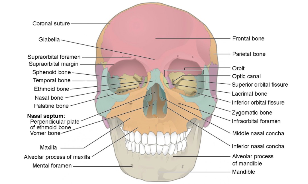
Anterior view of the skull

Anterior view of the skull featuring the bones of the viscerocranium:
Zygomatic (blue), lacrimal (green), nasal (blue/midline), inferior nasal conchae (blue inside the nasal cavity), palatine (dark orange), maxillary (light orange), vomer (blue/midline), mandible (beige), and ethmoid (gray within the nasal cavity)
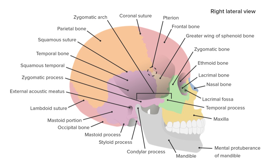
Image: “Anterior View of Skull” by Lumen Learning. License: CC BY 4.0
Right lateral view of the skull

Lateral view of the skull featuring the bones and sutures

Image by Lecturio.

Neurocranium

The neurocranium is the complex of bones encasing the brain Brain The part of central nervous system that is contained within the skull (cranium). Arising from the neural tube, the embryonic brain is comprised of three major parts including prosencephalon (the forebrain); mesencephalon (the midbrain); and rhombencephalon (the hindbrain). The developed brain consists of cerebrum; cerebellum; and other structures in the brain stem. Nervous System: Anatomy, Structure, and Classification. The calvarium is the upper portion of the neurocranium, and consists of the unpaired frontal bone Bone Bone is a compact type of hardened connective tissue composed of bone cells, membranes, an extracellular mineralized matrix, and central bone marrow. The 2 primary types of bone are compact and spongy. Bones: Structure and Types, unpaired occipital bone Bone Bone is a compact type of hardened connective tissue composed of bone cells, membranes, an extracellular mineralized matrix, and central bone marrow. The 2 primary types of bone are compact and spongy. Bones: Structure and Types, bilateral parietal bones, and bilateral temporal bones. The bones are initially separated (the fontanelles Fontanelles Physical Examination of the Newborn at birth) and fuse over childhood. The bones are then connected by specific sutures: coronal Coronal Computed Tomography (CT), squamous, lambdoid, and sagittal Sagittal Computed Tomography (CT).

Bones of the neurocranium:
  • Frontal bone Bone Bone is a compact type of hardened connective tissue composed of bone cells, membranes, an extracellular mineralized matrix, and central bone marrow. The 2 primary types of bone are compact and spongy. Bones: Structure and Types
  • Occipital bone Bone Bone is a compact type of hardened connective tissue composed of bone cells, membranes, an extracellular mineralized matrix, and central bone marrow. The 2 primary types of bone are compact and spongy. Bones: Structure and Types
  • Parietal bones (2)
  • Temporal bones (2)
  • Ethmoid bone Ethmoid bone A light and spongy (pneumatized) bone that lies between the orbital part of frontal bone and the anterior of sphenoid bone. Ethmoid bone separates the orbit from the ethmoid sinus. It consists of a horizontal plate, a perpendicular plate, and two lateral labyrinths. Orbit and Extraocular Muscles: Anatomy
  • Sphenoid bone Sphenoid bone An irregular unpaired bone situated at the skull base and wedged between the frontal, temporal, and occipital bones (frontal bone; temporal bone; occipital bone). Sphenoid bone consists of a median body and three pairs of processes resembling a bat with spread wings. The body is hollowed out in its inferior to form two large cavities (sphenoid sinus). Orbit and Extraocular Muscles: Anatomy
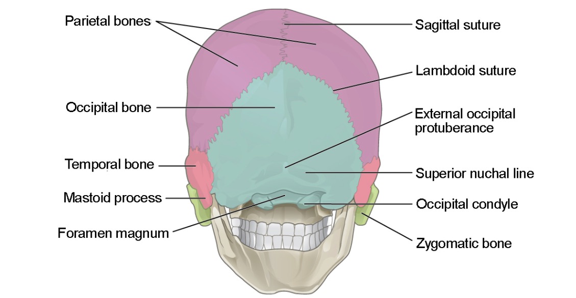
Posterior view of the skull

Posterior (slightly inferior) view of the skull featuring the bones of the calvarium:
the frontal bone (not pictured), occipital bone (blue), and parietal bone (purple)

Image: “Posterior View of Skull” by LumenLearning. License: CC BY 4.0

Main sutures are immobile joints between adjacent bones of the skull:

Skull sutures

Lateral view of the skull featuring the coronal, lambdoid, and squamous sutures

Image: “Skull sutures” by OpenStax College. License: CC BY 3.0
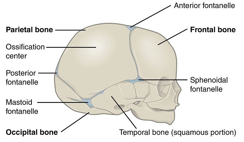
Major fontanelles Fontanelles Physical Examination of the Newborn are areas of connective tissue Connective tissue Connective tissues originate from embryonic mesenchyme and are present throughout the body except inside the brain and spinal cord. The main function of connective tissues is to provide structural support to organs. Connective tissues consist of cells and an extracellular matrix. Connective Tissue: Histology within the gaps/between the bone Bone Bone is a compact type of hardened connective tissue composed of bone cells, membranes, an extracellular mineralized matrix, and central bone marrow. The 2 primary types of bone are compact and spongy. Bones: Structure and Types plates, which are open during infancy:

  • Posterior, occipital, or lambdoid
  • Anterior, bregmatic, or frontal
  • Sphenoidal or anterolateral
  • Mastoid or posterolateral
Table: The major fontanelles Fontanelles Physical Examination of the Newborn
Fontanelle Shape Location Closure
Posterior, occipital, or lambdoid Triangle Junction of the sagittal Sagittal Computed Tomography (CT) and lambdoid sutures 3 months
Anterior, bregmatic, or frontal Diamond Junction of the sagittal Sagittal Computed Tomography (CT) and coronal Coronal Computed Tomography (CT) sutures 18–24 months
Sphenoidal or anterolateral Irregular and bilateral Between the frontal, parietal, and sphenoid bones 6 months
Mastoid or posterolateral Irregular and bilateral Between the parietal, temporal, and occipital bones 24 months
Lateral view of a newborn's skull

Lateral view of a newborn skull featuring the posterior, anterior, sphenoidal, and mastoid fontanelles

Image: “Lateral view of a newborn’s skull” by OpenStax College. License: CC BY 3.0

Base of the Skull

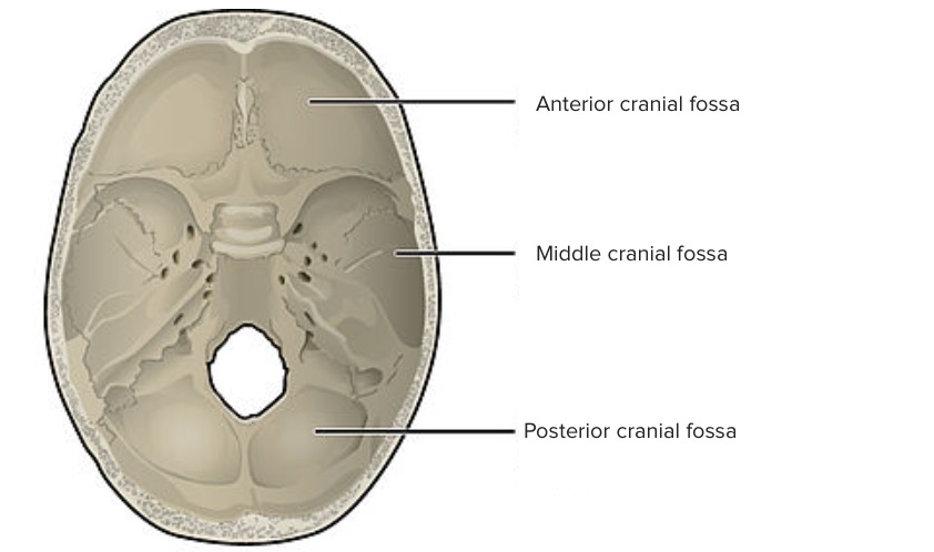
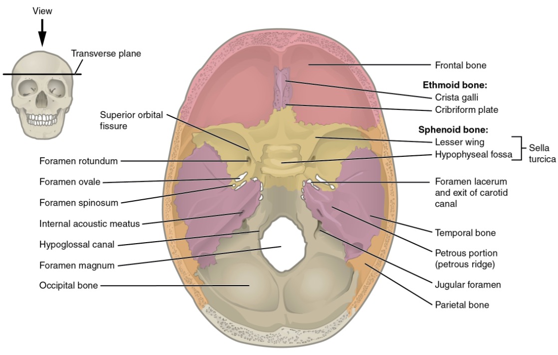
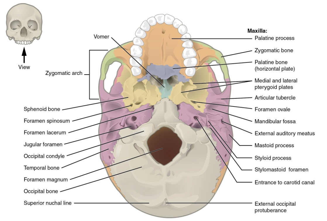
The skull base is a highly complex region of the skull with many important anatomical landmarks and foramen for cranial nerves Cranial nerves There are 12 pairs of cranial nerves (CNs), which run from the brain to various parts of the head, neck, and trunk. The CNs can be sensory or motor or both. The CNs are named and numbered in Roman numerals according to their location, from the front to the back of the brain. The 12 Cranial Nerves: Overview and Functions and vasculature to travel in and out of the skull. In-depth knowledge of the location of the foramen and the structures traversing the foramen are critically important.

Table: Regions of the base of the skull
Region Boundaries Bones Structures penetrating the foramina Content
Anterior cranial fossa
  • Anterior: frontal bones
  • Posterior: body and lesser wings of the sphenoid bone Sphenoid bone An irregular unpaired bone situated at the skull base and wedged between the frontal, temporal, and occipital bones (frontal bone; temporal bone; occipital bone). Sphenoid bone consists of a median body and three pairs of processes resembling a bat with spread wings. The body is hollowed out in its inferior to form two large cavities (sphenoid sinus). Orbit and Extraocular Muscles: Anatomy
Frontal (orbital plates) Foramen cecum Cecum The blind sac or outpouching area of the large intestine that is below the entrance of the small intestine. It has a worm-like extension, the vermiform appendix. Colon, Cecum, and Appendix: Anatomy: nasal emissary veins Veins Veins are tubular collections of cells, which transport deoxygenated blood and waste from the capillary beds back to the heart. Veins are classified into 3 types: small veins/venules, medium veins, and large veins. Each type contains 3 primary layers: tunica intima, tunica media, and tunica adventitia. Veins: Histology leading to the superior sagittal Sagittal Computed Tomography (CT) sinus
  • Frontal lobe Frontal lobe The part of the cerebral hemisphere anterior to the central sulcus, and anterior and superior to the lateral sulcus. Cerebral Cortex: Anatomy of the cerebral cortex Cerebral cortex The cerebral cortex is the largest and most developed part of the human brain and CNS. Occupying the upper part of the cranial cavity, the cerebral cortex has 4 lobes and is divided into 2 hemispheres that are joined centrally by the corpus callosum. Cerebral Cortex: Anatomy
  • Olfactory bulb Olfactory bulb Ovoid body resting on the cribriform plate of the ethmoid bone where the olfactory nerve terminates. The olfactory bulb contains several types of nerve cells including the mitral cells, on whose dendrites the olfactory nerve synapses, forming the olfactory glomeruli. The accessory olfactory bulb, which receives the projection from the vomeronasal organ via the vomeronasal nerve, is also included here. Olfaction: Anatomy
  • Olfactory tract Olfactory tract Olfaction: Anatomy
  • Orbital gyri
Ethmoid (cribriform plate and crista galli)
  • Foramina of the cribriform plate: olfactory nerve Olfactory nerve The 1st cranial nerve. The olfactory nerve conveys the sense of smell. It is formed by the axons of olfactory receptor neurons which project from the olfactory epithelium (in the nasal epithelium) to the olfactory bulb. Nose Anatomy (External & Internal) (I)
  • Anterior/posterior ethmoidal foramina: anterior/posterior ethmoidal nerves, arteries Arteries Arteries are tubular collections of cells that transport oxygenated blood and nutrients from the heart to the tissues of the body. The blood passes through the arteries in order of decreasing luminal diameter, starting in the largest artery (the aorta) and ending in the small arterioles. Arteries are classified into 3 types: large elastic arteries, medium muscular arteries, and small arteries and arterioles. Arteries: Histology, and veins Veins Veins are tubular collections of cells, which transport deoxygenated blood and waste from the capillary beds back to the heart. Veins are classified into 3 types: small veins/venules, medium veins, and large veins. Each type contains 3 primary layers: tunica intima, tunica media, and tunica adventitia. Veins: Histology
Middle cranial fossa
  • Anterior: lesser and greater wings of the sphenoid bone Sphenoid bone An irregular unpaired bone situated at the skull base and wedged between the frontal, temporal, and occipital bones (frontal bone; temporal bone; occipital bone). Sphenoid bone consists of a median body and three pairs of processes resembling a bat with spread wings. The body is hollowed out in its inferior to form two large cavities (sphenoid sinus). Orbit and Extraocular Muscles: Anatomy
  • Posterior: superior edge of the petrous portion of the temporal bones
  • Lateral: squamous part of the temporal bone Temporal bone Either of a pair of compound bones forming the lateral (left and right) surfaces and base of the skull which contains the organs of hearing. It is a large bone formed by the fusion of parts: the squamous (the flattened anterior-superior part), the tympanic (the curved anterior-inferior part), the mastoid (the irregular posterior portion), and the petrous (the part at the base of the skull). Jaw and Temporomandibular Joint: Anatomy and greater wings of the sphenoid
  • Medial: lateral aspect of the sphenoid (carotid sulcus, sella turcica Sella turcica A bony prominence situated on the upper surface of the body of the sphenoid bone. It houses the pituitary gland. Pituitary Gland: Anatomy, and dorsum sellae)
Sphenoid (lesser and greater wings)
  • Optic canal: optic nerve Optic nerve The 2nd cranial nerve which conveys visual information from the retina to the brain. The nerve carries the axons of the retinal ganglion cells which sort at the optic chiasm and continue via the optic tracts to the brain. The largest projection is to the lateral geniculate nuclei; other targets include the superior colliculi and the suprachiasmatic nuclei. Though known as the second cranial nerve, it is considered part of the central nervous system. The 12 Cranial Nerves: Overview and Functions (II) and ophthalmic artery Ophthalmic artery Artery originating from the internal carotid artery and distributing to the eye, orbit and adjacent facial structures. Eye: Anatomy
  • Superior orbital fissure Fissure A crack or split that extends into the dermis Generalized and Localized Rashes: oculomotor nerve Oculomotor nerve The 3D cranial nerve. The oculomotor nerve sends motor fibers to the levator muscles of the eyelid and to the superior rectus, inferior rectus, and inferior oblique muscles of the eye. It also sends parasympathetic efferents (via the ciliary ganglion) to the muscles controlling pupillary constriction and accommodation. The motor fibers originate in the oculomotor nuclei of the midbrain. The 12 Cranial Nerves: Overview and Functions (III), trochlear nerve Trochlear nerve The 4th cranial nerve. The trochlear nerve carries the motor innervation of the superior oblique muscles of the eye. The 12 Cranial Nerves: Overview and Functions (IV), 3 branches of the ophthalmic nerve (V1, lacrimal, frontal, and nasociliary nerves), abducent nerve (VI), and superior ophthalmic vein
  • Temporal lobes
  • Pituitary Pituitary A small, unpaired gland situated in the sella turcica. It is connected to the hypothalamus by a short stalk which is called the infundibulum. Hormones: Overview and Types gland
Sphenoid (greater wings)
  • Foramen rotundum: maxillary nerve (V2)
  • Foramen ovale Foramen ovale An opening in the wall between the right and the left upper chambers (heart atria) of a fetal heart. Oval foramen normally closes soon after birth; when it fails to close the condition is called patent oval foramen. Patent Foramen Ovale: mandibular nerve Mandibular nerve A branch of the trigeminal (5th cranial) nerve. The mandibular nerve carries motor fibers to the muscles of mastication and sensory fibers to the teeth and gingivae, the face in the region of the mandible, and parts of the dura. Jaw and Temporomandibular Joint: Anatomy (V3), accessory meningeal artery, and the vein connecting the cavernous sinus and pterygoid plexus
  • Foramen spinosum: meningeal branch of the mandibular nerve Mandibular nerve A branch of the trigeminal (5th cranial) nerve. The mandibular nerve carries motor fibers to the muscles of mastication and sensory fibers to the teeth and gingivae, the face in the region of the mandible, and parts of the dura. Jaw and Temporomandibular Joint: Anatomy and the middle meningeal artery Middle Meningeal Artery Epidural Hemorrhage/vein
Temporal
  • Foramen lacerum: nerve and artery of the pterygoid canal and meningeal branch of ascending pharyngeal artery and vein
  • Carotid canal: internal carotid plexus and artery
  • Hiatus for greater petrosal nerve: greater petrosal nerve
  • Hiatus for lesser petrosal nerve: lesser petrosal nerve and superior tympanic artery
Posterior cranial fossa Anterior: dorsum sellae of the sphenoid/superior border of the petrous part of the temporal bones Temporal
  • Internal acoustic meatus: facial nerve Facial nerve The 7th cranial nerve. The facial nerve has two parts, the larger motor root which may be called the facial nerve proper, and the smaller intermediate or sensory root. Together they provide efferent innervation to the muscles of facial expression and to the lacrimal and salivary glands, and convey afferent information for taste from the anterior two-thirds of the tongue and for touch from the external ear. The 12 Cranial Nerves: Overview and Functions (VII), vestibulocochlear nerve Vestibulocochlear nerve The 8th cranial nerve. The vestibulocochlear nerve has a cochlear part (cochlear nerve) which is concerned with hearing and a vestibular part (vestibular nerve) which mediates the sense of balance and head position. The fibers of the cochlear nerve originate from neurons of the spiral ganglion and project to the cochlear nuclei (cochlear nucleus). The fibers of the vestibular nerve arise from neurons of scarpa’s ganglion and project to the vestibular nuclei. The 12 Cranial Nerves: Overview and Functions (VIII), and labyrinthine artery/vein
  • Jugular foramen: glossopharyngeal nerve Glossopharyngeal nerve The 9th cranial nerve. The glossopharyngeal nerve is a mixed motor and sensory nerve; it conveys somatic and autonomic efferents as well as general, special, and visceral afferents. Among the connections are motor fibers to the stylopharyngeus muscle, parasympathetic fibers to the parotid glands, general and taste afferents from the posterior third of the tongue, the nasopharynx, and the palate, and afferents from baroreceptors and chemoreceptor cells of the carotid sinus. Pharynx: Anatomy (IX), vagus nerve Vagus nerve The 10th cranial nerve. The vagus is a mixed nerve which contains somatic afferents (from skin in back of the ear and the external auditory meatus), visceral afferents (from the pharynx, larynx, thorax, and abdomen), parasympathetic efferents (to the thorax and abdomen), and efferents to striated muscle (of the larynx and pharynx). Pharynx: Anatomy (X), accessory nerve (XI), internal jugular vein Internal jugular vein Parapharyngeal Abscess, inferior petrosal sinus, occipital artery, and posterior meningeal artery
  • Cerebellum Cerebellum The cerebellum, Latin for “little brain,” is located in the posterior cranial fossa, dorsal to the pons and midbrain, and its principal role is in the coordination of movements. The cerebellum consists of 3 lobes on either side of its 2 hemispheres and is connected in the middle by the vermis. Cerebellum: Anatomy
  • Brainstem
Occipital
  • Foramen magnum: medulla oblongata Medulla Oblongata The lower portion of the brain stem. It is inferior to the pons and anterior to the cerebellum. Medulla oblongata serves as a relay station between the brain and the spinal cord, and contains centers for regulating respiratory, vasomotor, cardiac, and reflex activities. Brain Stem: Anatomy, ascending fibers of accessory nerve (XI), vertebral arteries Arteries Arteries are tubular collections of cells that transport oxygenated blood and nutrients from the heart to the tissues of the body. The blood passes through the arteries in order of decreasing luminal diameter, starting in the largest artery (the aorta) and ending in the small arterioles. Arteries are classified into 3 types: large elastic arteries, medium muscular arteries, and small arteries and arterioles. Arteries: Histology, anterior spinal artery Anterior Spinal Artery Anterior Cord Syndrome, posterior spinal arteries Arteries Arteries are tubular collections of cells that transport oxygenated blood and nutrients from the heart to the tissues of the body. The blood passes through the arteries in order of decreasing luminal diameter, starting in the largest artery (the aorta) and ending in the small arterioles. Arteries are classified into 3 types: large elastic arteries, medium muscular arteries, and small arteries and arterioles. Arteries: Histology, and spinal veins Veins Veins are tubular collections of cells, which transport deoxygenated blood and waste from the capillary beds back to the heart. Veins are classified into 3 types: small veins/venules, medium veins, and large veins. Each type contains 3 primary layers: tunica intima, tunica media, and tunica adventitia. Veins: Histology
  • Hypoglossal canal: hypoglossal nerve Hypoglossal nerve The 12th cranial nerve. The hypoglossal nerve originates in the hypoglossal nucleus of the medulla and supplies motor innervation to all of the muscles of the tongue except the palatoglossus (which is supplied by the vagus). This nerve also contains proprioceptive afferents from the tongue muscles. Lips and Tongue: Anatomy (XII)

Related videos

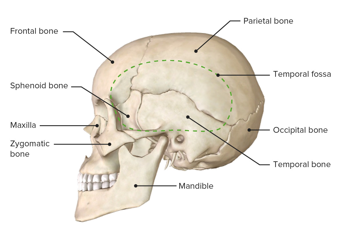
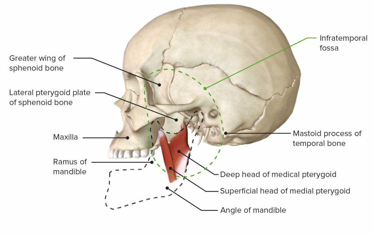
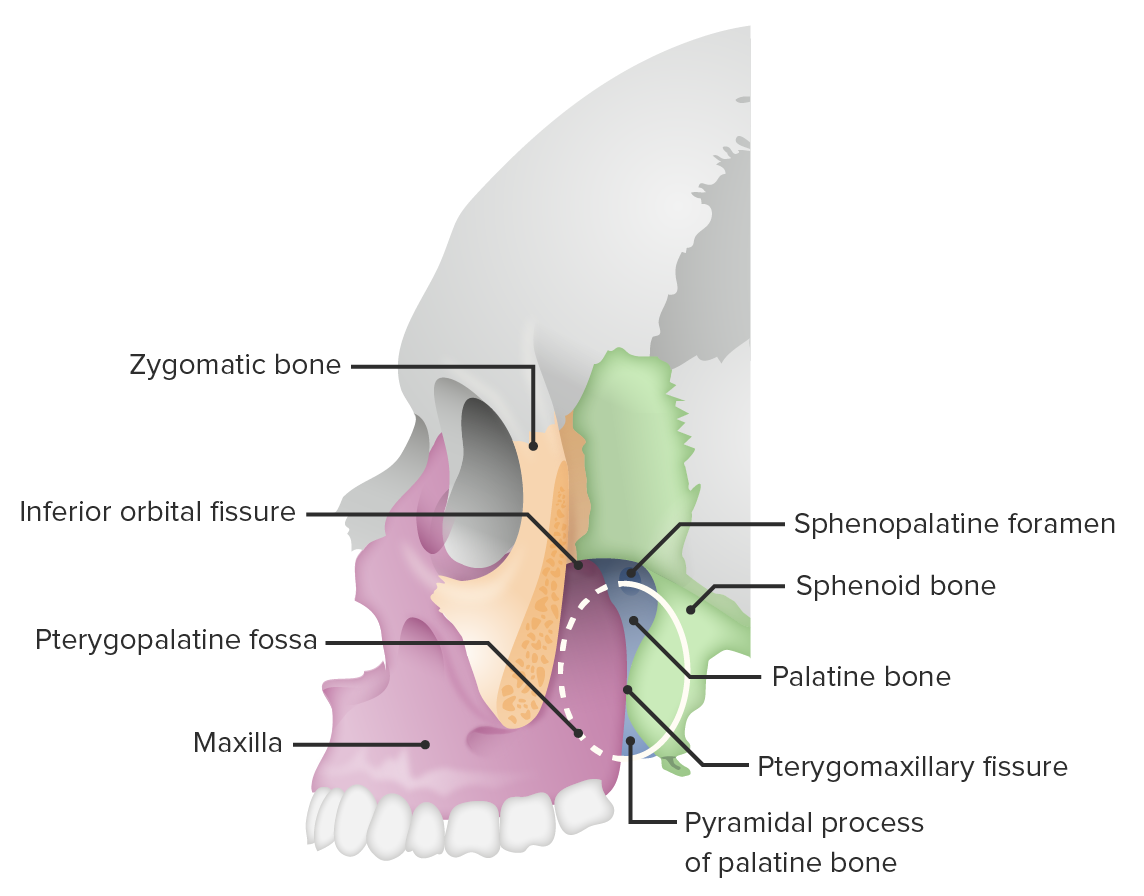
External Fossae

Table: External fossae
Temporal fossa* Infratemporal fossa Pterygopalatine fossa
Location Above the zygomatic arch of the temporal bone Temporal bone Either of a pair of compound bones forming the lateral (left and right) surfaces and base of the skull which contains the organs of hearing. It is a large bone formed by the fusion of parts: the squamous (the flattened anterior-superior part), the tympanic (the curved anterior-inferior part), the mastoid (the irregular posterior portion), and the petrous (the part at the base of the skull). Jaw and Temporomandibular Joint: Anatomy
  • Medial to the infratemporal fossa
  • Below the apex of the orbit
  • Between the sphenoid bone Sphenoid bone An irregular unpaired bone situated at the skull base and wedged between the frontal, temporal, and occipital bones (frontal bone; temporal bone; occipital bone). Sphenoid bone consists of a median body and three pairs of processes resembling a bat with spread wings. The body is hollowed out in its inferior to form two large cavities (sphenoid sinus). Orbit and Extraocular Muscles: Anatomy posteriorly, maxilla anteriorly, and palatine bone Palatine bone Orbit and Extraocular Muscles: Anatomy medially
Boundaries
  • Superior/posterior: superior and inferior temporal lines
  • Inferior: zygomatic arch
  • Anterior: frontal process of the zygomatic bone Zygomatic bone Either of a pair of bones that form the prominent part of the cheek and contribute to the orbit on each side of the skull. Orbit and Extraocular Muscles: Anatomy and the zygomatic process of the frontal bone Bone Bone is a compact type of hardened connective tissue composed of bone cells, membranes, an extracellular mineralized matrix, and central bone marrow. The 2 primary types of bone are compact and spongy. Bones: Structure and Types
  • Superior: greater wing of the sphenoid bone Sphenoid bone An irregular unpaired bone situated at the skull base and wedged between the frontal, temporal, and occipital bones (frontal bone; temporal bone; occipital bone). Sphenoid bone consists of a median body and three pairs of processes resembling a bat with spread wings. The body is hollowed out in its inferior to form two large cavities (sphenoid sinus). Orbit and Extraocular Muscles: Anatomy
  • Inferior: medial pterygoid Medial pterygoid Two of the masticatory muscles: the internal, or medial, pterygoid muscle and external, or lateral, pterygoid muscle. Action of the former is closing the jaws and that of the latter is opening the jaws, protruding the mandible, and moving the mandible from side to side. Jaw and Temporomandibular Joint: Anatomy muscle attachment near the mandibular angle
  • Anterior: posterior aspect of the maxilla
  • Posterior: mastoid process
  • Medial: lateral pterygoid Lateral pterygoid Two of the masticatory muscles: the internal, or medial, pterygoid muscle and external, or lateral, pterygoid muscle. Action of the former is closing the jaws and that of the latter is opening the jaws, protruding the mandible, and moving the mandible from side to side. Jaw and Temporomandibular Joint: Anatomy plate of the sphenoid bone Sphenoid bone An irregular unpaired bone situated at the skull base and wedged between the frontal, temporal, and occipital bones (frontal bone; temporal bone; occipital bone). Sphenoid bone consists of a median body and three pairs of processes resembling a bat with spread wings. The body is hollowed out in its inferior to form two large cavities (sphenoid sinus). Orbit and Extraocular Muscles: Anatomy
  • Lateral: ramus of the mandible Mandible The largest and strongest bone of the face constituting the lower jaw. It supports the lower teeth. Jaw and Temporomandibular Joint: Anatomy
Content
  • Inferior alveolar, lingual, and buccal nerves
  • Otic ganglion
  • Chorda tympani Chorda tympani A branch of the facial (7th cranial) nerve which passes through the middle ear and continues through the petrotympanic fissure. The chorda tympani nerve carries taste sensation from the anterior two-thirds of the tongue and conveys parasympathetic efferents to the salivary glands. Lips and Tongue: Anatomy
  • Maxillary artery
  • Pterygoid venous plexus
  • Medial/ lateral pterygoid Lateral pterygoid Two of the masticatory muscles: the internal, or medial, pterygoid muscle and external, or lateral, pterygoid muscle. Action of the former is closing the jaws and that of the latter is opening the jaws, protruding the mandible, and moving the mandible from side to side. Jaw and Temporomandibular Joint: Anatomy muscles
  • Tendon of the temporalis Temporalis A masticatory muscle whose action is closing the jaws; its posterior portion retracts the mandible. Jaw and Temporomandibular Joint: Anatomy muscle
  • Pterygopalatine ganglion
  • Greater/lesser palatine nerves
  • Maxillary nerve
  • Maxillary artery
Foramina (and the connecting structure) Inferior orbital fissure Fissure A crack or split that extends into the dermis Generalized and Localized Rashes: orbit
  • Inferior orbital fissure Fissure A crack or split that extends into the dermis Generalized and Localized Rashes: orbit
  • Foramen ovale Foramen ovale An opening in the wall between the right and the left upper chambers (heart atria) of a fetal heart. Oval foramen normally closes soon after birth; when it fails to close the condition is called patent oval foramen. Patent Foramen Ovale and foramen spinosum: middle cranial fossa
  • Pterygomaxillary fissure Fissure A crack or split that extends into the dermis Generalized and Localized Rashes: pterygopalatine fossa
  • Inferior orbital fissure Fissure A crack or split that extends into the dermis Generalized and Localized Rashes: orbit
  • Foramen rotundum: middle cranial fossa
  • Pterygoid canal: foramen lacerum
  • Sphenopalatine foramen: nasal cavity Nasal cavity The proximal portion of the respiratory passages on either side of the nasal septum. Nasal cavities, extending from the nares to the nasopharynx, are lined with ciliated nasal mucosa. Nose Anatomy (External & Internal)
  • Pterygomaxillary fissure Fissure A crack or split that extends into the dermis Generalized and Localized Rashes: infratemporal fossa
  • Greater/lesser palatine foramina: oral cavity
*The temporal fossa is also called the pterion:
  • Point of convergence for the temporal, parietal, sphenoid, and frontal bones
  • One of the weakest areas of the skull (susceptible to fracture)

Clinical Relevance

  • Le Fort fractures Le Fort fractures Le Fort fractures are a group midface fracture patterns classified into 3 types: Le Fort I, II, and III. Le Fort fractures represent 10%-20% of all facial fractures and can be caused by any significant blunt trauma to the face, most commonly from motor vehicle accidents. Le Fort Fractures: transverse fractures of the midface. Three types of Le Fort fractures Le Fort fractures Le Fort fractures are a group midface fracture patterns classified into 3 types: Le Fort I, II, and III. Le Fort fractures represent 10%-20% of all facial fractures and can be caused by any significant blunt trauma to the face, most commonly from motor vehicle accidents. Le Fort Fractures exist and involve the separation of all or a portion of the midface from the skull base. The fractures are repaired surgically by adding stabilizing plates.
  • Skull fractures: an interruption in the continuation of the bones comprising the calvarium. The fractures usually occur due to blunt force trauma and are often associated with a traumatic brain injury Traumatic brain injury A form of acquired brain injury which occurs when a sudden trauma causes damage to the brain. Le Fort Fractures. Fractures of the pterion may rupture the middle meningeal artery Middle Meningeal Artery Epidural Hemorrhage. Fractures of the petrous portion of the temporal bone Temporal bone Either of a pair of compound bones forming the lateral (left and right) surfaces and base of the skull which contains the organs of hearing. It is a large bone formed by the fusion of parts: the squamous (the flattened anterior-superior part), the tympanic (the curved anterior-inferior part), the mastoid (the irregular posterior portion), and the petrous (the part at the base of the skull). Jaw and Temporomandibular Joint: Anatomy may cause blood or cerebrospinal fluid Cerebrospinal Fluid A watery fluid that is continuously produced in the choroid plexus and circulates around the surface of the brain; spinal cord; and in the cerebral ventricles. Ventricular System: Anatomy to escape from the ear, which may result in hearing loss Hearing loss Hearing loss, also known as hearing impairment, is any degree of impairment in the ability to apprehend sound as determined by audiometry to be below normal hearing thresholds. Clinical presentation may occur at birth or as a gradual loss of hearing with age, including a short-term or sudden loss at any point. Hearing Loss
  • Orbital fractures Orbital fractures An orbital fracture is a break in the continuity of one or multiple bones of the eye socket, caused by direct or indirect trauma to the orbit. Patients frequently present with lacerations around the eye, orbital pain, edema, ecchymosis, diplopia on upward gaze, numbness around the eye, and signs of muscle entrapment. Orbital Fractures: fractures to the eye sockets. Orbital fractures Orbital fractures An orbital fracture is a break in the continuity of one or multiple bones of the eye socket, caused by direct or indirect trauma to the orbit. Patients frequently present with lacerations around the eye, orbital pain, edema, ecchymosis, diplopia on upward gaze, numbness around the eye, and signs of muscle entrapment. Orbital Fractures are classified as orbital rim Orbital Rim Orbital Fractures fractures, direct orbital floor Orbital Floor Orbital Fractures fractures, and blowout fractures. Individuals present with bruising around the eyes accompanied by blurry, decreased, or double vision Vision Ophthalmic Exam.
  • Craniosynostosis Craniosynostosis Craniosynostosis is the premature fusion of 1 or more cranial sutures during the 1st year of life. Craniosynostosis is classified as simple or complex, and can be caused by environmental factors or genetic syndromes. Craniosynostosis: the premature Premature Childbirth before 37 weeks of pregnancy (259 days from the first day of the mother’s last menstrual period, or 245 days after fertilization). Necrotizing Enterocolitis fusion of 1 or more cranial sutures. Craniosynostosis Craniosynostosis Craniosynostosis is the premature fusion of 1 or more cranial sutures during the 1st year of life. Craniosynostosis is classified as simple or complex, and can be caused by environmental factors or genetic syndromes. Craniosynostosis can be classified as simple or complex and typically results in an abnormally shaped head. Craniosynostosis Craniosynostosis Craniosynostosis is the premature fusion of 1 or more cranial sutures during the 1st year of life. Craniosynostosis is classified as simple or complex, and can be caused by environmental factors or genetic syndromes. Craniosynostosis occurs in approximately 4 per 10,000 children and can be improved with surgical intervention.
  • Acrania: a rare congenital disorder characterized by the partial or complete absence of the bones of the skull. The condition is commonly associated with anencephaly Anencephaly A malformation of the nervous system caused by failure of the anterior neuropore to close. Infants are born with intact spinal cords, cerebellums, and brainstems, but lack formation of neural structures above this level. The skull is only partially formed but the eyes are usually normal. This condition may be associated with folate deficiency. Affected infants are only capable of primitive (brain stem) reflexes and usually do not survive for more than two weeks. Neural Tube Defects
  • Cranium bifidum: a neural tube Neural tube A tube of ectodermal tissue in an embryo that will give rise to the central nervous system, including the spinal cord and the brain. Lumen within the neural tube is called neural canal which gives rise to the central canal of the spinal cord and the ventricles of the brain. Gastrulation and Neurulation defect characterized by the improper closure of the fetal skull during development. Cranium bifidum is usually associated with encephalocele Encephalocele Brain tissue herniation through a congenital or acquired defect in the skull. The majority of congenital encephaloceles occur in the occipital or frontal regions. Clinical features include a protuberant mass that may be pulsatile. The quantity and location of protruding neural tissue determines the type and degree of neurologic deficit. Visual defects, psychomotor developmental delay, and persistent motor deficits frequently occur. Neural Tube Defects or the protrusion of cerebral matter covered by meninges Meninges The brain and the spinal cord are enveloped by 3 overlapping layers of connective tissue called the meninges. The layers are, from the most external layer to the most internal layer, the dura mater, arachnoid mater, and pia mater. Between these layers are 3 potential spaces called the epidural, subdural, and subarachnoid spaces. Meninges: Anatomy through the bony defect. Presentation typically includes a groove down the midline of the skull between the forehead Forehead The part of the face above the eyes. Melasma, nose Nose The nose is the human body’s primary organ of smell and functions as part of the upper respiratory system. The nose may be best known for inhaling oxygen and exhaling carbon dioxide, but it also contributes to other important functions, such as tasting. The anatomy of the nose can be divided into the external nose and the nasal cavity. Nose Anatomy (External & Internal), or occipital region.

References

  1. Johnson, D., & Wilkie, A. O. (2011). Craniosynostosis. European journal of human genetics: EJHG, 19(4), 369–376. https://pubmed.ncbi.nlm.nih.gov/21248745/
  2. Okamoto, T., Nuri, T., Harada, A., Kyutoku, S., & Ueda, K. (2019). Cranial Suture Measurement by 2-point Method in Ultrasound Screening of Craniosynostosis. Plastic and reconstructive surgery. Global open, 7(5), e2225. https://pubmed.ncbi.nlm.nih.gov/31333954/
  3. Kliegman, R., Stanton, B., St. Geme, J. W., Schor, N. F., & Behrman, R. E. (2016). Nelson textbook of pediatrics (Edition 20.). Phialdelphia, PA: Elsevier.

Create your free account or log in to continue reading!

Sign up now and get free access to Lecturio with concept pages, medical videos, and questions for your medical education.

User Reviews

Details