Sideroblastic Anemia

Sideroblastic anemias are a heterogeneous group of bone marrow Bone marrow The soft tissue filling the cavities of bones. Bone marrow exists in two types, yellow and red. Yellow marrow is found in the large cavities of large bones and consists mostly of fat cells and a few primitive blood cells. Red marrow is a hematopoietic tissue and is the site of production of erythrocytes and granular leukocytes. Bone marrow is made up of a framework of connective tissue containing branching fibers with the frame being filled with marrow cells. Bone Marrow: Composition and Hematopoiesis disorders characterized by abnormal iron Iron A metallic element with atomic symbol fe, atomic number 26, and atomic weight 55. 85. It is an essential constituent of hemoglobins; cytochromes; and iron-binding proteins. It plays a role in cellular redox reactions and in the transport of oxygen. Trace Elements accumulation in the mitochondria Mitochondria Semiautonomous, self-reproducing organelles that occur in the cytoplasm of all cells of most, but not all, eukaryotes. Each mitochondrion is surrounded by a double limiting membrane. The inner membrane is highly invaginated, and its projections are called cristae. Mitochondria are the sites of the reactions of oxidative phosphorylation, which result in the formation of ATP. They contain distinctive ribosomes, transfer RNAs; amino Acyl tRNA synthetases; and elongation and termination factors. Mitochondria depend upon genes within the nucleus of the cells in which they reside for many essential messenger RNAs. Mitochondria are believed to have arisen from aerobic bacteria that established a symbiotic relationship with primitive protoeukaryotes. The Cell: Organelles of erythroid precursors. The accumulated iron Iron A metallic element with atomic symbol fe, atomic number 26, and atomic weight 55. 85. It is an essential constituent of hemoglobins; cytochromes; and iron-binding proteins. It plays a role in cellular redox reactions and in the transport of oxygen. Trace Elements appears as granules in a ringlike distribution around the nucleus Nucleus Within a eukaryotic cell, a membrane-limited body which contains chromosomes and one or more nucleoli (cell nucleolus). The nuclear membrane consists of a double unit-type membrane which is perforated by a number of pores; the outermost membrane is continuous with the endoplasmic reticulum. A cell may contain more than one nucleus. The Cell: Organelles, giving rise to the characteristic morphological feature of a ring sideroblast. Sideroblastic anemias may be due to inherited defects in heme synthesis Synthesis Polymerase Chain Reaction (PCR) or can be acquired through alcoholism Alcoholism A primary, chronic disease with genetic, psychosocial, and environmental factors influencing its development and manifestations. The disease is often progressive and fatal. It is characterized by impaired control over drinking, preoccupation with the drug alcohol, use of alcohol despite adverse consequences, and distortions in thinking, most notably denial. Each of these symptoms may be continuous or periodic. Wernicke Encephalopathy and Korsakoff Syndrome, lead poisoning Lead poisoning Poisoning that results from chronic or acute ingestion, injection, inhalation, or skin absorption of lead or lead compounds. Metal Poisoning (Lead, Arsenic, Iron), medications, or vitamin deficiencies. The anemia Anemia Anemia is a condition in which individuals have low Hb levels, which can arise from various causes. Anemia is accompanied by a reduced number of RBCs and may manifest with fatigue, shortness of breath, pallor, and weakness. Subtypes are classified by the size of RBCs, chronicity, and etiology. Anemia: Overview and Types is commonly microcytic with low to normal reticulocyte Reticulocyte Immature erythrocytes. In humans, these are erythroid cells that have just undergone extrusion of their cell nucleus. They still contain some organelles that gradually decrease in number as the cells mature. Ribosomes are last to disappear. Certain staining techniques cause components of the ribosomes to precipitate into characteristic 'reticulum' (not the same as the endoplasmic reticulum), hence the name reticulocytes. Erythrocytes: Histology count. Serum iron Iron A metallic element with atomic symbol fe, atomic number 26, and atomic weight 55. 85. It is an essential constituent of hemoglobins; cytochromes; and iron-binding proteins. It plays a role in cellular redox reactions and in the transport of oxygen. Trace Elements levels are typically elevated. A bone marrow Bone marrow The soft tissue filling the cavities of bones. Bone marrow exists in two types, yellow and red. Yellow marrow is found in the large cavities of large bones and consists mostly of fat cells and a few primitive blood cells. Red marrow is a hematopoietic tissue and is the site of production of erythrocytes and granular leukocytes. Bone marrow is made up of a framework of connective tissue containing branching fibers with the frame being filled with marrow cells. Bone Marrow: Composition and Hematopoiesis examination showing ring sideroblasts establishes the diagnosis. Management involves treating the underlying condition, avoiding causative medication and/or toxins, and phlebotomy Phlebotomy The techniques used to draw blood from a vein for diagnostic purposes or for treatment of certain blood disorders such as erythrocytosis, hemochromatosis, polycythemia vera, and porphyria cutanea tarda. Hereditary Hemochromatosis in cases of iron overload Iron overload An excessive accumulation of iron in the body due to a greater than normal absorption of iron from the gastrointestinal tract or from parenteral injection. This may arise from idiopathic hemochromatosis, excessive iron intake, chronic alcoholism, certain types of refractory anemia, or transfusional hemosiderosis. Hereditary Hemochromatosis.

Last updated: May 17, 2024

Editorial responsibility: Stanley Oiseth, Lindsay Jones, Evelin Maza

Overview

Definition

Sideroblastic anemias are a heterogeneous group of bone marrow Bone marrow The soft tissue filling the cavities of bones. Bone marrow exists in two types, yellow and red. Yellow marrow is found in the large cavities of large bones and consists mostly of fat cells and a few primitive blood cells. Red marrow is a hematopoietic tissue and is the site of production of erythrocytes and granular leukocytes. Bone marrow is made up of a framework of connective tissue containing branching fibers with the frame being filled with marrow cells. Bone Marrow: Composition and Hematopoiesis disorders characterized by abnormal iron Iron A metallic element with atomic symbol fe, atomic number 26, and atomic weight 55. 85. It is an essential constituent of hemoglobins; cytochromes; and iron-binding proteins. It plays a role in cellular redox reactions and in the transport of oxygen. Trace Elements accumulation in the mitochondria Mitochondria Semiautonomous, self-reproducing organelles that occur in the cytoplasm of all cells of most, but not all, eukaryotes. Each mitochondrion is surrounded by a double limiting membrane. The inner membrane is highly invaginated, and its projections are called cristae. Mitochondria are the sites of the reactions of oxidative phosphorylation, which result in the formation of ATP. They contain distinctive ribosomes, transfer RNAs; amino Acyl tRNA synthetases; and elongation and termination factors. Mitochondria depend upon genes within the nucleus of the cells in which they reside for many essential messenger RNAs. Mitochondria are believed to have arisen from aerobic bacteria that established a symbiotic relationship with primitive protoeukaryotes. The Cell: Organelles of erythroid precursors.

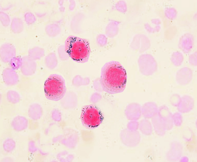
The distribution of the iron Iron A metallic element with atomic symbol fe, atomic number 26, and atomic weight 55. 85. It is an essential constituent of hemoglobins; cytochromes; and iron-binding proteins. It plays a role in cellular redox reactions and in the transport of oxygen. Trace Elements is ringlike around the nucleus Nucleus Within a eukaryotic cell, a membrane-limited body which contains chromosomes and one or more nucleoli (cell nucleolus). The nuclear membrane consists of a double unit-type membrane which is perforated by a number of pores; the outermost membrane is continuous with the endoplasmic reticulum. A cell may contain more than one nucleus. The Cell: Organelles, manifested by the precursors (ring sideroblasts) in the bone marrow Bone marrow The soft tissue filling the cavities of bones. Bone marrow exists in two types, yellow and red. Yellow marrow is found in the large cavities of large bones and consists mostly of fat cells and a few primitive blood cells. Red marrow is a hematopoietic tissue and is the site of production of erythrocytes and granular leukocytes. Bone marrow is made up of a framework of connective tissue containing branching fibers with the frame being filled with marrow cells. Bone Marrow: Composition and Hematopoiesis.

Epidemiology

  • A rare disease → incidence Incidence The number of new cases of a given disease during a given period in a specified population. It also is used for the rate at which new events occur in a defined population. It is differentiated from prevalence, which refers to all cases in the population at a given time. Measures of Disease Frequency and prevalence Prevalence The total number of cases of a given disease in a specified population at a designated time. It is differentiated from incidence, which refers to the number of new cases in the population at a given time. Measures of Disease Frequency not well characterized
  • Men > women
  • No racial predominance
  • Hereditary forms usually manifest before 30 years of age.
  • Acquired type is often seen at age 65 years or older.

Etiology

Congenital Congenital Chorioretinitis disorders:

  • Syndromic: with accompanying nonhematologic manifestations
  • Nonsyndromic: 
    • Present with anemia Anemia Anemia is a condition in which individuals have low Hb levels, which can arise from various causes. Anemia is accompanied by a reduced number of RBCs and may manifest with fatigue, shortness of breath, pallor, and weakness. Subtypes are classified by the size of RBCs, chronicity, and etiology. Anemia: Overview and Types, no other clinical manifestations
    • Majority of cases

Acquired:

  • Clonal hematopoietic disorders:
    • Myelodysplastic syndromes Myelodysplastic Syndromes Myelodysplastic syndromes (MDS) are a group of clonal neoplasms with maturation defects characterized by dysplasia, cytopenia, and immature bone marrow precursors. Myelodysplastic syndromes can be idiopathic, or secondary to various injurious exposures such as cytotoxic chemotherapy, ionizing radiation, or environmental toxins. Myelodysplastic Syndromes ( MDS MDS Myelodysplastic syndromes (MDS) are a group of clonal neoplasms with maturation defects characterized by dysplasia, cytopenia, and immature bone marrow precursors. Myelodysplastic syndromes can be idiopathic, or secondary to various injurious exposures such as cytotoxic chemotherapy, ionizing radiation, or environmental toxins. Myelodysplastic Syndromes)
    • Myeloproliferative neoplasms Neoplasms New abnormal growth of tissue. Malignant neoplasms show a greater degree of anaplasia and have the properties of invasion and metastasis, compared to benign neoplasms. Benign Bone Tumors (MPN)
  • Reversible causes:
    • Alcohol 
    • Vitamin deficiencies (vitamin B6, cofactor for protoporphyrin synthesis Synthesis Polymerase Chain Reaction (PCR))
    • Lead poisoning Lead poisoning Poisoning that results from chronic or acute ingestion, injection, inhalation, or skin absorption of lead or lead compounds. Metal Poisoning (Lead, Arsenic, Iron) (inhibits the conversion of protoporphyrins Protoporphyrins Porphyrins with four methyl, two vinyl, and two propionic acid side chains attached to the pyrrole rings. Protoporphyrin IX occurs in hemoglobin, myoglobin, and most of the cytochromes. Heme Metabolism)
    • Medications ( isoniazid Isoniazid Antibacterial agent used primarily as a tuberculostatic. It remains the treatment of choice for tuberculosis. Antimycobacterial Drugs, chloramphenicol Chloramphenicol Chloramphenicol, the only clinically relevant drug in the amphenicol class, is a potent inhibitor of bacterial protein synthesis by binding to the 50S ribosomal subunit and preventing peptide bond formation. Chloramphenicol is a broad-spectrum antibiotic with wide distribution; however, due to its toxicity, its use is limited to severe infections. Chloramphenicol, linezolid Linezolid An oxazolidinone and acetamide derived anti-bacterial agent and protein synthesis inhibitor that is used in the treatment of gram-positive bacterial infections of the skin and respiratory tract. Oxazolidinones)
    • Copper Copper A heavy metal trace element with the atomic symbol cu, atomic number 29, and atomic weight 63. 55. Trace Elements deficiency
    • Zinc Zinc A metallic element of atomic number 30 and atomic weight 65. 38. It is a necessary trace element in the diet, forming an essential part of many enzymes, and playing an important role in protein synthesis and in cell division. Zinc deficiency is associated with anemia, short stature, hypogonadism, impaired wound healing, and geophagia. It is known by the symbol zn. Trace Elements poisoning
    • Hypothermia Hypothermia Hypothermia can be defined as a drop in the core body temperature below 35°C (95°F) and is classified into mild, moderate, severe, and profound forms based on the degree of temperature decrease. Hypothermia

Pathophysiology

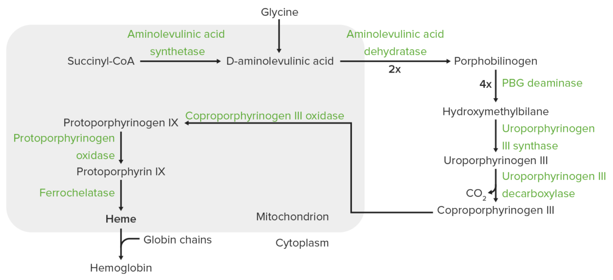
Heme synthesis Synthesis Polymerase Chain Reaction (PCR)

  • Heme:
    • A porphyrin-containing compound, synthesized through an enzymatic pathway that takes place in the mitochondria Mitochondria Semiautonomous, self-reproducing organelles that occur in the cytoplasm of all cells of most, but not all, eukaryotes. Each mitochondrion is surrounded by a double limiting membrane. The inner membrane is highly invaginated, and its projections are called cristae. Mitochondria are the sites of the reactions of oxidative phosphorylation, which result in the formation of ATP. They contain distinctive ribosomes, transfer RNAs; amino Acyl tRNA synthetases; and elongation and termination factors. Mitochondria depend upon genes within the nucleus of the cells in which they reside for many essential messenger RNAs. Mitochondria are believed to have arisen from aerobic bacteria that established a symbiotic relationship with primitive protoeukaryotes. The Cell: Organelles and cytoplasm
    • Most production occurs in the bone marrow Bone marrow The soft tissue filling the cavities of bones. Bone marrow exists in two types, yellow and red. Yellow marrow is found in the large cavities of large bones and consists mostly of fat cells and a few primitive blood cells. Red marrow is a hematopoietic tissue and is the site of production of erythrocytes and granular leukocytes. Bone marrow is made up of a framework of connective tissue containing branching fibers with the frame being filled with marrow cells. Bone Marrow: Composition and Hematopoiesis.
  • In the mitochondria Mitochondria Semiautonomous, self-reproducing organelles that occur in the cytoplasm of all cells of most, but not all, eukaryotes. Each mitochondrion is surrounded by a double limiting membrane. The inner membrane is highly invaginated, and its projections are called cristae. Mitochondria are the sites of the reactions of oxidative phosphorylation, which result in the formation of ATP. They contain distinctive ribosomes, transfer RNAs; amino Acyl tRNA synthetases; and elongation and termination factors. Mitochondria depend upon genes within the nucleus of the cells in which they reside for many essential messenger RNAs. Mitochondria are believed to have arisen from aerobic bacteria that established a symbiotic relationship with primitive protoeukaryotes. The Cell: Organelles:
    • Succinyl-CoA Succinyl-CoA Citric Acid Cycle + glycine Glycine A non-essential amino acid. It is found primarily in gelatin and silk fibroin and used therapeutically as a nutrient. It is also a fast inhibitory neurotransmitter. Synthesis of Nonessential Amino Acids aminolevulinic acid Aminolevulinic acid A compound produced from succinyl-CoA and glycine as an intermediate in heme synthesis. It is used as a photochemotherapy for actinic keratosis. Heme Metabolism (ALA) 
    • Reaction is catalyzed by aminolevulinic acid Aminolevulinic acid A compound produced from succinyl-CoA and glycine as an intermediate in heme synthesis. It is used as a photochemotherapy for actinic keratosis. Heme Metabolism synthase (ALAS2 in the erythrocytes Erythrocytes Erythrocytes, or red blood cells (RBCs), are the most abundant cells in the blood. While erythrocytes in the fetus are initially produced in the yolk sac then the liver, the bone marrow eventually becomes the main site of production. Erythrocytes: Histology).
    • ALA → exits to the cytoplasm
  • In the cytoplasm: 
    • 2 ALA molecules condense to produce porphobilinogen Porphobilinogen Heme Metabolism.
    • Porphobilinogen Porphobilinogen Heme Metabolism → uroporphyrinogen III → coproporphyrinogen III 
    • Coproporphyrinogen III is transported back to the mitochondria Mitochondria Semiautonomous, self-reproducing organelles that occur in the cytoplasm of all cells of most, but not all, eukaryotes. Each mitochondrion is surrounded by a double limiting membrane. The inner membrane is highly invaginated, and its projections are called cristae. Mitochondria are the sites of the reactions of oxidative phosphorylation, which result in the formation of ATP. They contain distinctive ribosomes, transfer RNAs; amino Acyl tRNA synthetases; and elongation and termination factors. Mitochondria depend upon genes within the nucleus of the cells in which they reside for many essential messenger RNAs. Mitochondria are believed to have arisen from aerobic bacteria that established a symbiotic relationship with primitive protoeukaryotes. The Cell: Organelles.
  • In the mitochondria Mitochondria Semiautonomous, self-reproducing organelles that occur in the cytoplasm of all cells of most, but not all, eukaryotes. Each mitochondrion is surrounded by a double limiting membrane. The inner membrane is highly invaginated, and its projections are called cristae. Mitochondria are the sites of the reactions of oxidative phosphorylation, which result in the formation of ATP. They contain distinctive ribosomes, transfer RNAs; amino Acyl tRNA synthetases; and elongation and termination factors. Mitochondria depend upon genes within the nucleus of the cells in which they reside for many essential messenger RNAs. Mitochondria are believed to have arisen from aerobic bacteria that established a symbiotic relationship with primitive protoeukaryotes. The Cell: Organelles:
    • Coproporphyrinogen III → protoporphyrinogen Protoporphyrinogen Heme Metabolism III, which then is converted to protoporphyrin IX
    • Ferrous iron Iron A metallic element with atomic symbol fe, atomic number 26, and atomic weight 55. 85. It is an essential constituent of hemoglobins; cytochromes; and iron-binding proteins. It plays a role in cellular redox reactions and in the transport of oxygen. Trace Elements is inserted into protoporphyrin IX (catalyzed by ferrochelatase Ferrochelatase A mitochondrial enzyme found in a wide variety of cells and tissues. It is the final enzyme in the 8-enzyme biosynthetic pathway of heme. Ferrochelatase catalyzes ferrous insertion into protoporphyrin IX to form protoheme or heme. Heme Metabolism), forming heme.
Heme synthesis

Heme synthesis:
The process of heme synthesis takes place in the mitochondria and cytoplasm.
In the mitochondria, succinyl coenzyme A (CoA) combines with glycine to form aminolevulinic acid.
This reaction is catalyzed by aminolevulinic acid synthase. The aminolevulinic acid exits to the cytoplasm, where 2 aminolevulinic acid molecules condense to produce porphobilinogen (PBG). The subsequent steps lead to the formation of coproporphyrinogen III, which is transported back to the mitochondria. Oxidase facilitates conversion of coproporphyrinogen III to protoporphyrinogen IX, which then is converted to protoporphyrin IX. Ferrous iron is inserted into protoporphyrin IX, forming heme (catalyzed by ferrochelatase).

Image by Lecturio.

Pathogenesis

Defects in heme synthesis Synthesis Polymerase Chain Reaction (PCR):

  • X-linked X-linked Genetic diseases that are linked to gene mutations on the X chromosome in humans or the X chromosome in other species. Included here are animal models of human X-linked diseases. Common Variable Immunodeficiency (CVID) sideroblastic anemia Anemia Anemia is a condition in which individuals have low Hb levels, which can arise from various causes. Anemia is accompanied by a reduced number of RBCs and may manifest with fatigue, shortness of breath, pallor, and weakness. Subtypes are classified by the size of RBCs, chronicity, and etiology. Anemia: Overview and Types (XLSA):
    • Mutations in ALAS2 gene Gene A category of nucleic acid sequences that function as units of heredity and which code for the basic instructions for the development, reproduction, and maintenance of organisms. Basic Terms of Genetics, which encodes aminolevulinic acid Aminolevulinic acid A compound produced from succinyl-CoA and glycine as an intermediate in heme synthesis. It is used as a photochemotherapy for actinic keratosis. Heme Metabolism synthase, the rate-limiting enzyme in heme synthesis Synthesis Polymerase Chain Reaction (PCR)
    • Frequently (> 50% of cases) responsive to B6 supplementation
  • Autosomal recessive Autosomal recessive Autosomal inheritance, both dominant and recessive, refers to the transmission of genes from the 22 autosomal chromosomes. Autosomal recessive diseases are only expressed when 2 copies of the recessive allele are inherited. Autosomal Recessive and Autosomal Dominant Inheritance sideroblastic anemia Anemia Anemia is a condition in which individuals have low Hb levels, which can arise from various causes. Anemia is accompanied by a reduced number of RBCs and may manifest with fatigue, shortness of breath, pallor, and weakness. Subtypes are classified by the size of RBCs, chronicity, and etiology. Anemia: Overview and Types:
    • SLC25A38 mutation Mutation Genetic mutations are errors in DNA that can cause protein misfolding and dysfunction. There are various types of mutations, including chromosomal, point, frameshift, and expansion mutations. Types of Mutations: impaired glycine Glycine A non-essential amino acid. It is found primarily in gelatin and silk fibroin and used therapeutically as a nutrient. It is also a fast inhibitory neurotransmitter. Synthesis of Nonessential Amino Acids transporter into mitochondria Mitochondria Semiautonomous, self-reproducing organelles that occur in the cytoplasm of all cells of most, but not all, eukaryotes. Each mitochondrion is surrounded by a double limiting membrane. The inner membrane is highly invaginated, and its projections are called cristae. Mitochondria are the sites of the reactions of oxidative phosphorylation, which result in the formation of ATP. They contain distinctive ribosomes, transfer RNAs; amino Acyl tRNA synthetases; and elongation and termination factors. Mitochondria depend upon genes within the nucleus of the cells in which they reside for many essential messenger RNAs. Mitochondria are believed to have arisen from aerobic bacteria that established a symbiotic relationship with primitive protoeukaryotes. The Cell: Organelles 
    • FECH mutation Mutation Genetic mutations are errors in DNA that can cause protein misfolding and dysfunction. There are various types of mutations, including chromosomal, point, frameshift, and expansion mutations. Types of Mutations: impaired ferrochelatase Ferrochelatase A mitochondrial enzyme found in a wide variety of cells and tissues. It is the final enzyme in the 8-enzyme biosynthetic pathway of heme. Ferrochelatase catalyzes ferrous insertion into protoporphyrin IX to form protoheme or heme. Heme Metabolism
  • Acquired:
    • Isoniazid Isoniazid Antibacterial agent used primarily as a tuberculostatic. It remains the treatment of choice for tuberculosis. Antimycobacterial Drugs (INH): causes ALAS2 impairment
    • Alcohol

Defects in iron-sulfur cluster (ISC) biogenesis:

  • ISCs are involved in multiple biological functions including electron transport, iron Iron A metallic element with atomic symbol fe, atomic number 26, and atomic weight 55. 85. It is an essential constituent of hemoglobins; cytochromes; and iron-binding proteins. It plays a role in cellular redox reactions and in the transport of oxygen. Trace Elements regulation, and DNA DNA A deoxyribonucleotide polymer that is the primary genetic material of all cells. Eukaryotic and prokaryotic organisms normally contain DNA in a double-stranded state, yet several important biological processes transiently involve single-stranded regions. DNA, which consists of a polysugar-phosphate backbone possessing projections of purines (adenine and guanine) and pyrimidines (thymine and cytosine), forms a double helix that is held together by hydrogen bonds between these purines and pyrimidines (adenine to thymine and guanine to cytosine). DNA Types and Structure repair.
  • Dysfunction in ISC transfer in and out of mitochondria Mitochondria Semiautonomous, self-reproducing organelles that occur in the cytoplasm of all cells of most, but not all, eukaryotes. Each mitochondrion is surrounded by a double limiting membrane. The inner membrane is highly invaginated, and its projections are called cristae. Mitochondria are the sites of the reactions of oxidative phosphorylation, which result in the formation of ATP. They contain distinctive ribosomes, transfer RNAs; amino Acyl tRNA synthetases; and elongation and termination factors. Mitochondria depend upon genes within the nucleus of the cells in which they reside for many essential messenger RNAs. Mitochondria are believed to have arisen from aerobic bacteria that established a symbiotic relationship with primitive protoeukaryotes. The Cell: Organelles disables erythropoiesis Erythropoiesis The production of red blood cells (erythrocytes). In humans, erythrocytes are produced by the yolk sac in the first trimester; by the liver in the second trimester; by the bone marrow in the third trimester and after birth. In normal individuals, the erythrocyte count in the peripheral blood remains relatively constant implying a balance between the rate of erythrocyte production and rate of destruction. Erythrocytes: Histology.
  • Seen in mutations such as:
    • ABCB7: associated with ataxia Ataxia Impairment of the ability to perform smoothly coordinated voluntary movements. This condition may affect the limbs, trunk, eyes, pharynx, larynx, and other structures. Ataxia may result from impaired sensory or motor function. Sensory ataxia may result from posterior column injury or peripheral nerve diseases. Motor ataxia may be associated with cerebellar diseases; cerebral cortex diseases; thalamic diseases; basal ganglia diseases; injury to the red nucleus; and other conditions. Ataxia-telangiectasia (syndromic)
    • HSPA9
    • GLRX5

Defects in mitochondrial protein synthesis Synthesis Polymerase Chain Reaction (PCR):

  • Congenital Congenital Chorioretinitis etiologies affect multifunctional proteins Proteins Linear polypeptides that are synthesized on ribosomes and may be further modified, crosslinked, cleaved, or assembled into complex proteins with several subunits. The specific sequence of amino acids determines the shape the polypeptide will take, during protein folding, and the function of the protein. Energy Homeostasis, so these are associated with systemic signs (syndromic) such as:
    • Pearson marrow-pancreas syndrome
    • Myopathy Myopathy Dermatomyositis, lactic acidosis Lactic Acidosis Oxazolidinones, and sideroblastic anemia Anemia Anemia is a condition in which individuals have low Hb levels, which can arise from various causes. Anemia is accompanied by a reduced number of RBCs and may manifest with fatigue, shortness of breath, pallor, and weakness. Subtypes are classified by the size of RBCs, chronicity, and etiology. Anemia: Overview and Types (MLASA)
    • TRNT1 mutation Mutation Genetic mutations are errors in DNA that can cause protein misfolding and dysfunction. There are various types of mutations, including chromosomal, point, frameshift, and expansion mutations. Types of Mutations ( immunodeficiency Immunodeficiency Chédiak-Higashi Syndrome, recurrent fevers, developmental delay) 
  • Acquired:
    • Chloramphenicol Chloramphenicol Chloramphenicol, the only clinically relevant drug in the amphenicol class, is a potent inhibitor of bacterial protein synthesis by binding to the 50S ribosomal subunit and preventing peptide bond formation. Chloramphenicol is a broad-spectrum antibiotic with wide distribution; however, due to its toxicity, its use is limited to severe infections. Chloramphenicol
    • Linezolid Linezolid An oxazolidinone and acetamide derived anti-bacterial agent and protein synthesis inhibitor that is used in the treatment of gram-positive bacterial infections of the skin and respiratory tract. Oxazolidinones

Dysfunction in erythropoiesis Erythropoiesis The production of red blood cells (erythrocytes). In humans, erythrocytes are produced by the yolk sac in the first trimester; by the liver in the second trimester; by the bone marrow in the third trimester and after birth. In normal individuals, the erythrocyte count in the peripheral blood remains relatively constant implying a balance between the rate of erythrocyte production and rate of destruction. Erythrocytes: Histology:

  • RBCs RBCs Erythrocytes, or red blood cells (RBCs), are the most abundant cells in the blood. While erythrocytes in the fetus are initially produced in the yolk sac then the liver, the bone marrow eventually becomes the main site of production. Erythrocytes: Histology unable to mature
  • Leads to cell death Cell death Injurious stimuli trigger the process of cellular adaptation, whereby cells respond to withstand the harmful changes in their environment. Overwhelmed adaptive mechanisms lead to cell injury. Mild stimuli produce reversible injury. If the stimulus is severe or persistent, injury becomes irreversible. Apoptosis is programmed cell death, a mechanism with both physiologic and pathologic effects. Cell Injury and Death in the marrow (intramedullary hemolysis)
  • ↑ Inappropriate iron absorption Iron absorption Digestion and Absorption iron overload Iron overload An excessive accumulation of iron in the body due to a greater than normal absorption of iron from the gastrointestinal tract or from parenteral injection. This may arise from idiopathic hemochromatosis, excessive iron intake, chronic alcoholism, certain types of refractory anemia, or transfusional hemosiderosis. Hereditary Hemochromatosis
  • Seen in:
    • XLSA
    • SLC25A38 mutation Mutation Genetic mutations are errors in DNA that can cause protein misfolding and dysfunction. There are various types of mutations, including chromosomal, point, frameshift, and expansion mutations. Types of Mutations
    • MDS MDS Myelodysplastic syndromes (MDS) are a group of clonal neoplasms with maturation defects characterized by dysplasia, cytopenia, and immature bone marrow precursors. Myelodysplastic syndromes can be idiopathic, or secondary to various injurious exposures such as cytotoxic chemotherapy, ionizing radiation, or environmental toxins. Myelodysplastic Syndromes/MPN

Clinical Presentation

Clinical presentation varies depending on the underlying disease.

General features of anemia Anemia Anemia is a condition in which individuals have low Hb levels, which can arise from various causes. Anemia is accompanied by a reduced number of RBCs and may manifest with fatigue, shortness of breath, pallor, and weakness. Subtypes are classified by the size of RBCs, chronicity, and etiology. Anemia: Overview and Types:

  • Growth retardation Growth Retardation Failure of a fetus to attain expected growth. Fetal Alcohol Spectrum Disorder in children 
  • Fatigue Fatigue The state of weariness following a period of exertion, mental or physical, characterized by a decreased capacity for work and reduced efficiency to respond to stimuli. Fibromyalgia and muscle weakness
  • Hypothermia Hypothermia Hypothermia can be defined as a drop in the core body temperature below 35°C (95°F) and is classified into mild, moderate, severe, and profound forms based on the degree of temperature decrease. Hypothermia
  • Photosensitivity Photosensitivity Tetracyclines

Patients Patients Individuals participating in the health care system for the purpose of receiving therapeutic, diagnostic, or preventive procedures. Clinician–Patient Relationship with vitamin B6 deficiency may have:

  • Peripheral neuropathy Neuropathy Leprosy
  • Ataxia Ataxia Impairment of the ability to perform smoothly coordinated voluntary movements. This condition may affect the limbs, trunk, eyes, pharynx, larynx, and other structures. Ataxia may result from impaired sensory or motor function. Sensory ataxia may result from posterior column injury or peripheral nerve diseases. Motor ataxia may be associated with cerebellar diseases; cerebral cortex diseases; thalamic diseases; basal ganglia diseases; injury to the red nucleus; and other conditions. Ataxia-telangiectasia
  • Dermatitis Dermatitis Any inflammation of the skin. Atopic Dermatitis (Eczema)
  • Glossitis

Patients Patients Individuals participating in the health care system for the purpose of receiving therapeutic, diagnostic, or preventive procedures. Clinician–Patient Relationship with lead poisoning Lead poisoning Poisoning that results from chronic or acute ingestion, injection, inhalation, or skin absorption of lead or lead compounds. Metal Poisoning (Lead, Arsenic, Iron) may have:

Others:
  • Pink-stained urine from porphyrins
  • Those with syndromic sideroblastic anemias will have other nonhematologic manifestations.

Diagnosis

Medical history

  • Family history Family History Adult Health Maintenance of anemia Anemia Anemia is a condition in which individuals have low Hb levels, which can arise from various causes. Anemia is accompanied by a reduced number of RBCs and may manifest with fatigue, shortness of breath, pallor, and weakness. Subtypes are classified by the size of RBCs, chronicity, and etiology. Anemia: Overview and Types
  • Presence of concurrent clinical features (in syndromic forms)
  • Possibilities of copper Copper A heavy metal trace element with the atomic symbol cu, atomic number 29, and atomic weight 63. 55. Trace Elements deficiency:
    • History of gastrointestinal surgery 
    • Enteral or parenteral nutrition Parenteral nutrition The administering of nutrients for assimilation and utilization by a patient who cannot maintain adequate nutrition by enteral feeding alone. Nutrients are administered by a route other than the alimentary canal (e.g., intravenously, subcutaneously). Central Venous Catheter without copper Copper A heavy metal trace element with the atomic symbol cu, atomic number 29, and atomic weight 63. 55. Trace Elements 
    • Excessive zinc Zinc A metallic element of atomic number 30 and atomic weight 65. 38. It is a necessary trace element in the diet, forming an essential part of many enzymes, and playing an important role in protein synthesis and in cell division. Zinc deficiency is associated with anemia, short stature, hypogonadism, impaired wound healing, and geophagia. It is known by the symbol zn. Trace Elements intake 
  • Alcohol use
  • History of exposure to lead
  • Medications 
  • Long-term dialysis Dialysis Renal replacement therapy refers to dialysis and/or kidney transplantation. Dialysis is a procedure by which toxins and excess water are removed from the circulation. Hemodialysis and peritoneal dialysis (PD) are the two types of dialysis, and their primary difference is the location of the filtration process (external to the body in hemodialysis versus inside the body for PD). Peritoneal Dialysis and Hemodialysis

Laboratory findings

  • Complete blood count:
    • Microcytosis (MCV < 80)
    • ↑ RBC distribution width (RDW)
    • Clinical significance: In children, neither microcytic anemia Microcytic anemia Conditions in which there is a generalized increase in the iron stores of body tissues, particularly of liver and the mononuclear phagocyte system, without demonstrable tissue damage. The name refers to the presence of stainable iron in the tissue in the form of hemosiderin. Anemia: Overview and Types with no iron Iron A metallic element with atomic symbol fe, atomic number 26, and atomic weight 55. 85. It is an essential constituent of hemoglobins; cytochromes; and iron-binding proteins. It plays a role in cellular redox reactions and in the transport of oxygen. Trace Elements deficiency nor thalassemia Thalassemia Thalassemia is a hereditary cause of microcytic hypochromic anemia and results from a deficiency in either the α or β globin chains, resulting in hemoglobinopathy. The presentation of thalassemia depends on the number of defective chains present and can range from being asymptomatic to rendering the more severely affected patients to be transfusion dependent. Thalassemia is suggestive of XLSA.
  • Normal to low reticulocyte Reticulocyte Immature erythrocytes. In humans, these are erythroid cells that have just undergone extrusion of their cell nucleus. They still contain some organelles that gradually decrease in number as the cells mature. Ribosomes are last to disappear. Certain staining techniques cause components of the ribosomes to precipitate into characteristic ‘reticulum’ (not the same as the endoplasmic reticulum), hence the name reticulocytes. Erythrocytes: Histology count
  • Iron studies Iron Studies Iron Deficiency Anemia:
    • ↑ Serum iron Iron A metallic element with atomic symbol fe, atomic number 26, and atomic weight 55. 85. It is an essential constituent of hemoglobins; cytochromes; and iron-binding proteins. It plays a role in cellular redox reactions and in the transport of oxygen. Trace Elements 
    • Ferritin Ferritin Iron-containing proteins that are widely distributed in animals, plants, and microorganisms. Their major function is to store iron in a nontoxic bioavailable form. Each ferritin molecule consists of ferric iron in a hollow protein shell (apoferritins) made of 24 subunits of various sequences depending on the species and tissue types. Hereditary Hemochromatosis levels
    • Normal or ↓ total iron-binding capacity (TIBC)
    • Transferrin Transferrin An iron-binding beta1-globulin that is synthesized in the liver and secreted into the blood. It plays a central role in the transport of iron throughout the circulation. Heme Metabolism saturation
  • Abnormal peripheral blood smear Peripheral Blood Smear Anemia: Overview and Types:
    • Anisocytosis, poikilocytosis
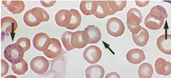
    • Siderocytes: hypochromic erythrocytes Erythrocytes Erythrocytes, or red blood cells (RBCs), are the most abundant cells in the blood. While erythrocytes in the fetus are initially produced in the yolk sac then the liver, the bone marrow eventually becomes the main site of production. Erythrocytes: Histology with basophilic granules staining positive for iron Iron A metallic element with atomic symbol fe, atomic number 26, and atomic weight 55. 85. It is an essential constituent of hemoglobins; cytochromes; and iron-binding proteins. It plays a role in cellular redox reactions and in the transport of oxygen. Trace Elements
  • Diagnosis is based on bone marrow Bone marrow The soft tissue filling the cavities of bones. Bone marrow exists in two types, yellow and red. Yellow marrow is found in the large cavities of large bones and consists mostly of fat cells and a few primitive blood cells. Red marrow is a hematopoietic tissue and is the site of production of erythrocytes and granular leukocytes. Bone marrow is made up of a framework of connective tissue containing branching fibers with the frame being filled with marrow cells. Bone Marrow: Composition and Hematopoiesis aspirate (in Prussian blue-stained smear): ring sideroblasts

Differentiating sideroblastic anemia Anemia Anemia is a condition in which individuals have low Hb levels, which can arise from various causes. Anemia is accompanied by a reduced number of RBCs and may manifest with fatigue, shortness of breath, pallor, and weakness. Subtypes are classified by the size of RBCs, chronicity, and etiology. Anemia: Overview and Types from other microcytic anemias

Table: Differentiating sideroblastic anemia Anemia Anemia is a condition in which individuals have low Hb levels, which can arise from various causes. Anemia is accompanied by a reduced number of RBCs and may manifest with fatigue, shortness of breath, pallor, and weakness. Subtypes are classified by the size of RBCs, chronicity, and etiology. Anemia: Overview and Types from other microcytic anemias (MCV < 80)
CBC finding Diagnosis Iron Iron A metallic element with atomic symbol fe, atomic number 26, and atomic weight 55. 85. It is an essential constituent of hemoglobins; cytochromes; and iron-binding proteins. It plays a role in cellular redox reactions and in the transport of oxygen. Trace Elements level Features or historical setting
Anemia Anemia Anemia is a condition in which individuals have low Hb levels, which can arise from various causes. Anemia is accompanied by a reduced number of RBCs and may manifest with fatigue, shortness of breath, pallor, and weakness. Subtypes are classified by the size of RBCs, chronicity, and etiology. Anemia: Overview and Types with MCV < 80 Sideroblastic anemia Anemia Anemia is a condition in which individuals have low Hb levels, which can arise from various causes. Anemia is accompanied by a reduced number of RBCs and may manifest with fatigue, shortness of breath, pallor, and weakness. Subtypes are classified by the size of RBCs, chronicity, and etiology. Anemia: Overview and Types Iron Iron A metallic element with atomic symbol fe, atomic number 26, and atomic weight 55. 85. It is an essential constituent of hemoglobins; cytochromes; and iron-binding proteins. It plays a role in cellular redox reactions and in the transport of oxygen. Trace Elements level Alcoholic Alcoholic Persons who have a history of physical or psychological dependence on ethanol. Mallory-Weiss Syndrome (Mallory-Weiss Tear), MDS MDS Myelodysplastic syndromes (MDS) are a group of clonal neoplasms with maturation defects characterized by dysplasia, cytopenia, and immature bone marrow precursors. Myelodysplastic syndromes can be idiopathic, or secondary to various injurious exposures such as cytotoxic chemotherapy, ionizing radiation, or environmental toxins. Myelodysplastic Syndromes
Iron Iron A metallic element with atomic symbol fe, atomic number 26, and atomic weight 55. 85. It is an essential constituent of hemoglobins; cytochromes; and iron-binding proteins. It plays a role in cellular redox reactions and in the transport of oxygen. Trace Elements deficiency anemia Anemia Anemia is a condition in which individuals have low Hb levels, which can arise from various causes. Anemia is accompanied by a reduced number of RBCs and may manifest with fatigue, shortness of breath, pallor, and weakness. Subtypes are classified by the size of RBCs, chronicity, and etiology. Anemia: Overview and Types Iron Iron A metallic element with atomic symbol fe, atomic number 26, and atomic weight 55. 85. It is an essential constituent of hemoglobins; cytochromes; and iron-binding proteins. It plays a role in cellular redox reactions and in the transport of oxygen. Trace Elements level Blood loss
Thalassemia Thalassemia Thalassemia is a hereditary cause of microcytic hypochromic anemia and results from a deficiency in either the α or β globin chains, resulting in hemoglobinopathy. The presentation of thalassemia depends on the number of defective chains present and can range from being asymptomatic to rendering the more severely affected patients to be transfusion dependent. Thalassemia Normal iron Iron A metallic element with atomic symbol fe, atomic number 26, and atomic weight 55. 85. It is an essential constituent of hemoglobins; cytochromes; and iron-binding proteins. It plays a role in cellular redox reactions and in the transport of oxygen. Trace Elements level Variant dependent (asymptomatic to severe anemia Anemia Anemia is a condition in which individuals have low Hb levels, which can arise from various causes. Anemia is accompanied by a reduced number of RBCs and may manifest with fatigue, shortness of breath, pallor, and weakness. Subtypes are classified by the size of RBCs, chronicity, and etiology. Anemia: Overview and Types)
MDS: myelodysplastic syndromes

Additional tests

  • If indicated based on history:
    • Copper Copper A heavy metal trace element with the atomic symbol cu, atomic number 29, and atomic weight 63. 55. Trace Elements and ceruloplasmin Ceruloplasmin A multi-copper blood ferroxidase involved in iron and copper homeostasis and inflammation. Wilson Disease levels
    • Lead level
  • Genetic testing Genetic Testing Detection of a mutation; genotype; karyotype; or specific alleles associated with genetic traits, heritable diseases, or predisposition to a disease, or that may lead to the disease in descendants. It includes prenatal genetic testing. Myotonic Dystrophies:
    • Necessary to establish a specific diagnosis
    • Performed on leukocytes Leukocytes White blood cells. These include granular leukocytes (basophils; eosinophils; and neutrophils) as well as non-granular leukocytes (lymphocytes and monocytes). White Myeloid Cells: Histology from the peripheral blood

Management

Goals

  • Prevent organ damage from subsequent iron overload Iron overload An excessive accumulation of iron in the body due to a greater than normal absorption of iron from the gastrointestinal tract or from parenteral injection. This may arise from idiopathic hemochromatosis, excessive iron intake, chronic alcoholism, certain types of refractory anemia, or transfusional hemosiderosis. Hereditary Hemochromatosis
  • Control symptoms associated with anemia Anemia Anemia is a condition in which individuals have low Hb levels, which can arise from various causes. Anemia is accompanied by a reduced number of RBCs and may manifest with fatigue, shortness of breath, pallor, and weakness. Subtypes are classified by the size of RBCs, chronicity, and etiology. Anemia: Overview and Types.

Treatment options

Treatment options depend on the etiology and include:

  • Drug induced: withdrawal of the medication
  • Alcohol or toxin induced: alcohol abstinence, removal of the toxic agents
  • Treatment of underlying condition, i.e., MDS MDS Myelodysplastic syndromes (MDS) are a group of clonal neoplasms with maturation defects characterized by dysplasia, cytopenia, and immature bone marrow precursors. Myelodysplastic syndromes can be idiopathic, or secondary to various injurious exposures such as cytotoxic chemotherapy, ionizing radiation, or environmental toxins. Myelodysplastic Syndromes/MPN 
  • In the case of INH intake: Co-administration of B6 is required.
  • For XLSA: vitamin B6 (pyridoxine) supplements 
  • For thiamine-responsive megaloblastic anemia Megaloblastic anemia Megaloblastic anemia is a subset of macrocytic anemias that arises because of impaired nucleic acid synthesis in erythroid precursors. This impairment leads to ineffective RBC production and intramedullary hemolysis that is characterized by large cells with arrested nuclear maturation. The most common causes are vitamin B12 and folic acid deficiencies. Megaloblastic Anemia: vitamin B1 
  • For symptomatic anemia Anemia Anemia is a condition in which individuals have low Hb levels, which can arise from various causes. Anemia is accompanied by a reduced number of RBCs and may manifest with fatigue, shortness of breath, pallor, and weakness. Subtypes are classified by the size of RBCs, chronicity, and etiology. Anemia: Overview and Types: transfusion if indicated
  • Iron overload Iron overload An excessive accumulation of iron in the body due to a greater than normal absorption of iron from the gastrointestinal tract or from parenteral injection. This may arise from idiopathic hemochromatosis, excessive iron intake, chronic alcoholism, certain types of refractory anemia, or transfusional hemosiderosis. Hereditary Hemochromatosis: phlebotomy Phlebotomy The techniques used to draw blood from a vein for diagnostic purposes or for treatment of certain blood disorders such as erythrocytosis, hemochromatosis, polycythemia vera, and porphyria cutanea tarda. Hereditary Hemochromatosis, iron Iron A metallic element with atomic symbol fe, atomic number 26, and atomic weight 55. 85. It is an essential constituent of hemoglobins; cytochromes; and iron-binding proteins. It plays a role in cellular redox reactions and in the transport of oxygen. Trace Elements chelation
  • For some inherited sideroblastic anemias: possible allogeneic hematopoietic stem cell transplantation Hematopoietic stem cell transplantation Transfer of hematopoietic stem cells from bone marrow or blood between individuals within the same species (homologous transplantation) or transfer within the same individual (autologous transplantation). Hematopoietic stem cell transplantation has been used as an alternative to bone marrow transplantation in the treatment of a variety of neoplasms. Organ Transplantation

Clinical Relevance

  • Lead poisoning Lead poisoning Poisoning that results from chronic or acute ingestion, injection, inhalation, or skin absorption of lead or lead compounds. Metal Poisoning (Lead, Arsenic, Iron): lead inhibits ferrochelatase Ferrochelatase A mitochondrial enzyme found in a wide variety of cells and tissues. It is the final enzyme in the 8-enzyme biosynthetic pathway of heme. Ferrochelatase catalyzes ferrous insertion into protoporphyrin IX to form protoheme or heme. Heme Metabolism and ALA dehydratase ( enzymes Enzymes Enzymes are complex protein biocatalysts that accelerate chemical reactions without being consumed by them. Due to the body’s constant metabolic needs, the absence of enzymes would make life unsustainable, as reactions would occur too slowly without these molecules. Basics of Enzymes in the heme synthesis Synthesis Polymerase Chain Reaction (PCR) pathway), resulting in decreased heme synthesis Synthesis Polymerase Chain Reaction (PCR). Features include gastrointestinal symptoms and neurological manifestations. Laboratory findings include anemia Anemia Anemia is a condition in which individuals have low Hb levels, which can arise from various causes. Anemia is accompanied by a reduced number of RBCs and may manifest with fatigue, shortness of breath, pallor, and weakness. Subtypes are classified by the size of RBCs, chronicity, and etiology. Anemia: Overview and Types, erythrocyte basophilic stippling in peripheral smear, and ringed sideroblasts in bone marrow Bone marrow The soft tissue filling the cavities of bones. Bone marrow exists in two types, yellow and red. Yellow marrow is found in the large cavities of large bones and consists mostly of fat cells and a few primitive blood cells. Red marrow is a hematopoietic tissue and is the site of production of erythrocytes and granular leukocytes. Bone marrow is made up of a framework of connective tissue containing branching fibers with the frame being filled with marrow cells. Bone Marrow: Composition and Hematopoiesis. Diagnosis is by lead level measurement.
  • Myelodysplastic syndrome: a group of malignant myeloid stem cell disorders characterized by dysplastic and ineffective bone marrow Bone marrow The soft tissue filling the cavities of bones. Bone marrow exists in two types, yellow and red. Yellow marrow is found in the large cavities of large bones and consists mostly of fat cells and a few primitive blood cells. Red marrow is a hematopoietic tissue and is the site of production of erythrocytes and granular leukocytes. Bone marrow is made up of a framework of connective tissue containing branching fibers with the frame being filled with marrow cells. Bone Marrow: Composition and Hematopoiesis myeloid line cell production and an increased risk of transformation Transformation Change brought about to an organism’s genetic composition by unidirectional transfer (transfection; transduction, genetic; conjugation, genetic, etc.) and incorporation of foreign DNA into prokaryotic or eukaryotic cells by recombination of part or all of that DNA into the cell’s genome. Bacteriology to acute leukemia. Myelodysplastic syndrome usually presents in older patients Patients Individuals participating in the health care system for the purpose of receiving therapeutic, diagnostic, or preventive procedures. Clinician–Patient Relationship, greater than 60 years of age. The syndrome manifests as thrombocytopenia Thrombocytopenia Thrombocytopenia occurs when the platelet count is < 150,000 per microliter. The normal range for platelets is usually 150,000-450,000/µL of whole blood. Thrombocytopenia can be a result of decreased production, increased destruction, or splenic sequestration of platelets. Patients are often asymptomatic until platelet counts are < 50,000/µL. Thrombocytopenia, anemia Anemia Anemia is a condition in which individuals have low Hb levels, which can arise from various causes. Anemia is accompanied by a reduced number of RBCs and may manifest with fatigue, shortness of breath, pallor, and weakness. Subtypes are classified by the size of RBCs, chronicity, and etiology. Anemia: Overview and Types, and neutropenia Neutropenia Neutrophils are an important component of the immune system and play a significant role in the eradication of infections. Low numbers of circulating neutrophils, referred to as neutropenia, predispose the body to recurrent infections or sepsis, though patients can also be asymptomatic. Neutropenia with dysfunctional granulocytes Granulocytes Leukocytes with abundant granules in the cytoplasm. They are divided into three groups according to the staining properties of the granules: neutrophilic, eosinophilic, and basophilic. Mature granulocytes are the neutrophils; eosinophils; and basophils. White Myeloid Cells: Histology, despite a hypercellular bone marrow Bone marrow The soft tissue filling the cavities of bones. Bone marrow exists in two types, yellow and red. Yellow marrow is found in the large cavities of large bones and consists mostly of fat cells and a few primitive blood cells. Red marrow is a hematopoietic tissue and is the site of production of erythrocytes and granular leukocytes. Bone marrow is made up of a framework of connective tissue containing branching fibers with the frame being filled with marrow cells. Bone Marrow: Composition and Hematopoiesis.
  • Vitamin B6 deficiency: caused by pyridoxine-inactivating drugs (e.g., isoniazid Isoniazid Antibacterial agent used primarily as a tuberculostatic. It remains the treatment of choice for tuberculosis. Antimycobacterial Drugs), protein-energy undernutrition, malabsorption Malabsorption General term for a group of malnutrition syndromes caused by failure of normal intestinal absorption of nutrients. Malabsorption and Maldigestion, alcoholism Alcoholism A primary, chronic disease with genetic, psychosocial, and environmental factors influencing its development and manifestations. The disease is often progressive and fatal. It is characterized by impaired control over drinking, preoccupation with the drug alcohol, use of alcohol despite adverse consequences, and distortions in thinking, most notably denial. Each of these symptoms may be continuous or periodic. Wernicke Encephalopathy and Korsakoff Syndrome, or excessive loss due to hemodialysis Hemodialysis Procedures which temporarily or permanently remedy insufficient cleansing of body fluids by the kidneys. Crush Syndrome. Deficiency can cause peripheral neuropathy Neuropathy Leprosy, seizures Seizures A seizure is abnormal electrical activity of the neurons in the cerebral cortex that can manifest in numerous ways depending on the region of the brain affected. Seizures consist of a sudden imbalance that occurs between the excitatory and inhibitory signals in cortical neurons, creating a net excitation. The 2 major classes of seizures are focal and generalized. Seizures, seborrheic dermatitis Dermatitis Any inflammation of the skin. Atopic Dermatitis (Eczema), glossitis, cheilosis, and sideroblastic anemia Anemia Anemia is a condition in which individuals have low Hb levels, which can arise from various causes. Anemia is accompanied by a reduced number of RBCs and may manifest with fatigue, shortness of breath, pallor, and weakness. Subtypes are classified by the size of RBCs, chronicity, and etiology. Anemia: Overview and Types.

References

  1. Ashorobi D, Chhabra A. (2020). Sideroblastic Anemia. StatPearls: StatPearls Publishing. Retrieved April 6, 2021, from https://www.ncbi.nlm.nih.gov/books/NBK538287/
  2. Bottomley SS. (2021). Sideroblastic anemias: Diagnosis and management. UpToDate. Retrieved April 6, 2021, from https://www.uptodate.com/contents/sideroblastic-anemias-diagnosis-and-management
  3. Bottomley SS. (2021). Causes and pathophysiology of the sideroblastic anemias. UpToDate. Retrieved April 6, 2021, from https://www.uptodate.com/contents/causes-and-pathophysiology-of-the-sideroblastic-anemias
  4. Ducamp S, Fleming MD. (2019). The molecular genetics of sideroblastic anemia. https://pubmed.ncbi.nlm.nih.gov/30401706/
  5. Sticco KL, Yarrarapu, SNS, Al Obaidi NM. (2021) Refractory Anemia With Ring Sideroblasts. StatPearls [Internet]. Treasure Island (FL): StatPearls Publishing. Retrieved April 6, 2021, from https://www.ncbi.nlm.nih.gov/books/NBK537073/

Create your free account or log in to continue reading!

Sign up now and get free access to Lecturio with concept pages, medical videos, and questions for your medical education.

User Reviews

Details