Advertisement

Advertisement

Advertisement

Advertisement

Sex-Linked Inheritance

Sex-linked inheritance is a form of mendelian inheritance. The term describes traits that are inherited via either the X or the Y chromosome Y chromosome The male sex chromosome, being the differential sex chromosome carried by half the male gametes and none of the female gametes in humans and in some other male-heterogametic species in which the homologue of the X chromosome has been retained. Basic Terms of Genetics. For X-linked recessive X-Linked Recessive Duchenne Muscular Dystrophy transmission, the allele Allele Variant forms of the same gene, occupying the same locus on homologous chromosomes, and governing the variants in production of the same gene product. Basic Terms of Genetics is recessive and carried on the X chromosome X chromosome The female sex chromosome, being the differential sex chromosome carried by half the male gametes and all female gametes in human and other male-heterogametic species. Basic Terms of Genetics. Males are more likely to express X-linked recessive X-Linked Recessive Duchenne Muscular Dystrophy disorders because they possess only 1 X chromosome X chromosome The female sex chromosome, being the differential sex chromosome carried by half the male gametes and all female gametes in human and other male-heterogametic species. Basic Terms of Genetics. For X-linked X-linked Genetic diseases that are linked to gene mutations on the X chromosome in humans or the X chromosome in other species. Included here are animal models of human X-linked diseases. Common Variable Immunodeficiency (CVID) dominant transmission, the allele Allele Variant forms of the same gene, occupying the same locus on homologous chromosomes, and governing the variants in production of the same gene product. Basic Terms of Genetics is dominant and carried on the X chromosome X chromosome The female sex chromosome, being the differential sex chromosome carried by half the male gametes and all female gametes in human and other male-heterogametic species. Basic Terms of Genetics. Because of this, only 1 copy of the disease allele Allele Variant forms of the same gene, occupying the same locus on homologous chromosomes, and governing the variants in production of the same gene product. Basic Terms of Genetics is required for phenotypic expression. X-linked X-linked Genetic diseases that are linked to gene mutations on the X chromosome in humans or the X chromosome in other species. Included here are animal models of human X-linked diseases. Common Variable Immunodeficiency (CVID) dominant disorders are less common than X-linked recessive X-Linked Recessive Duchenne Muscular Dystrophy disorders.

Last updated: Mar 21, 2024

Editorial responsibility: Stanley Oiseth, Lindsay Jones, Evelin Maza

Advertisement

Advertisement

Advertisement

Advertisement

Advertisement

Advertisement

Definition

Sex-linked inheritance occurs when genetic traits are inherited on either the X or the Y chromosome Y chromosome The male sex chromosome, being the differential sex chromosome carried by half the male gametes and none of the female gametes in humans and in some other male-heterogametic species in which the homologue of the X chromosome has been retained. Basic Terms of Genetics. There are 2 major sex-linked modes of inheritance: X-linked recessive X-Linked Recessive Duchenne Muscular Dystrophy and X-linked X-linked Genetic diseases that are linked to gene mutations on the X chromosome in humans or the X chromosome in other species. Included here are animal models of human X-linked diseases. Common Variable Immunodeficiency (CVID) dominant.

X-linked recessive X-Linked Recessive Duchenne Muscular Dystrophy inheritance:

  • Females:
  • Males:
    • More frequently affected than females (only 1 X chromosome X chromosome The female sex chromosome, being the differential sex chromosome carried by half the male gametes and all female gametes in human and other male-heterogametic species. Basic Terms of Genetics)
    • Male-to-male inheritance is impossible (father transmits only Y chromosome Y chromosome The male sex chromosome, being the differential sex chromosome carried by half the male gametes and none of the female gametes in humans and in some other male-heterogametic species in which the homologue of the X chromosome has been retained. Basic Terms of Genetics to son).

X-linked X-linked Genetic diseases that are linked to gene mutations on the X chromosome in humans or the X chromosome in other species. Included here are animal models of human X-linked diseases. Common Variable Immunodeficiency (CVID) dominant inheritance:

  • Very rare
  • Males and females have an equal probability Probability Probability is a mathematical tool used to study randomness and provide predictions about the likelihood of something happening. There are several basic rules of probability that can be used to help determine the probability of multiple events happening together, separately, or sequentially. Basics of Probability of expressing the trait. 
  • Females:
    • Inheritance can be from either the maternal or the paternal X chromosome X chromosome The female sex chromosome, being the differential sex chromosome carried by half the male gametes and all female gametes in human and other male-heterogametic species. Basic Terms of Genetics.
    • Usually less severe phenotypical presentation in females because of 2nd X chromosome X chromosome The female sex chromosome, being the differential sex chromosome carried by half the male gametes and all female gametes in human and other male-heterogametic species. Basic Terms of Genetics

X-Linked Recessive Inheritance and Diseases

X-linked recessive X-Linked Recessive Duchenne Muscular Dystrophy inheritance

Children of an unaffected father and a heterozygous ( carrier Carrier Vaccination) mother have the following risks:

  • Male offspring: 50% risk of disease
  • Female offspring: 0% risk of disease and 50% risk of being a carrier Carrier Vaccination
Table: Risk of children of an unaffected father and a heterozygous ( carrier Carrier Vaccination) mother
Unaffected father
x y
Heterozygous ( carrier Carrier Vaccination) mother x xx (unaffected female) xy (unaffected male)
x xx ( carrier Carrier Vaccination female) xy (affected male)
x: affected X chromosome; x: unaffected x chromosome; y: unaffected Y chromosome

Children of an affected father and a heterozygous ( carrier Carrier Vaccination) mother have the following risks:

  • Male offspring: 50% risk of disease
  • Female offspring: affected chromosome Chromosome In a prokaryotic cell or in the nucleus of a eukaryotic cell, a structure consisting of or containing DNA which carries the genetic information essential to the cell. Basic Terms of Genetics always transmitted → 50% risk of disease and 50% risk of being a carrier Carrier Vaccination
Table: Risk of children of an affected father and a heterozygous ( carrier Carrier Vaccination) mother
Hemizygously affected father
x y
Heterozygous ( carrier Carrier Vaccination) mother x xx ( carrier Carrier Vaccination female) xy (unaffected male)
x xx (affected female) xy (affected male)
x: affected X chromosome; x: unaffected x chromosome; y: unaffected Y chromosome

Children of an affected father and an unaffected mother have the following risks:

  • Male offspring: 0% risk of disease
  • Female offspring: affected chromosome Chromosome In a prokaryotic cell or in the nucleus of a eukaryotic cell, a structure consisting of or containing DNA which carries the genetic information essential to the cell. Basic Terms of Genetics from father always transmitted → 100% risk of being a carrier Carrier Vaccination, but 0% risk of disease
Table: Risk of children of an affected father and an unaffected mother
Hemizygously affected father
x y
Unaffected mother x xx ( carrier Carrier Vaccination female) xy (unaffected male)
x xx ( carrier Carrier Vaccination female) xy (unaffected male)
x: affected X chromosome; x: unaffected x chromosome; y: unaffected Y chromosome

Children of a hemizygous affected father and a homozygous (affected) mother have the following risks:

  • Male offspring: 100% affected
  • Female offspring: 100% affected
Table: Risk of children of a hemizygous affected father and a homozygous (affected) mother
Hemizygously affected father
x y
Homozygously affected mother x xx (affected female) xy (affected male)
x xx (affected female) xy (affected male)
x: affected X chromosome; x: unaffected x chromosome; y: unaffected Y chromosome

X-linked recessive X-Linked Recessive Duchenne Muscular Dystrophy diseases

  • Red-green color blindness Blindness The inability to see or the loss or absence of perception of visual stimuli. This condition may be the result of eye diseases; optic nerve diseases; optic chiasm diseases; or brain diseases affecting the visual pathways or occipital lobe. Retinopathy of Prematurity: also known as daltonism. Red-green color blindness Blindness The inability to see or the loss or absence of perception of visual stimuli. This condition may be the result of eye diseases; optic nerve diseases; optic chiasm diseases; or brain diseases affecting the visual pathways or occipital lobe. Retinopathy of Prematurity is a common trait in humans that results in confusion of green and red. About 56 different genes Genes A category of nucleic acid sequences that function as units of heredity and which code for the basic instructions for the development, reproduction, and maintenance of organisms. DNA Types and Structure have been associated with causing color blindness Blindness The inability to see or the loss or absence of perception of visual stimuli. This condition may be the result of eye diseases; optic nerve diseases; optic chiasm diseases; or brain diseases affecting the visual pathways or occipital lobe. Retinopathy of Prematurity when mutated, not all of which are located on the X chromosome X chromosome The female sex chromosome, being the differential sex chromosome carried by half the male gametes and all female gametes in human and other male-heterogametic species. Basic Terms of Genetics. Red-green color blindness Blindness The inability to see or the loss or absence of perception of visual stimuli. This condition may be the result of eye diseases; optic nerve diseases; optic chiasm diseases; or brain diseases affecting the visual pathways or occipital lobe. Retinopathy of Prematurity can be present from birth or develop later in life. About 8% of males, but only 0.4% of females, have some form of color blindness Blindness The inability to see or the loss or absence of perception of visual stimuli. This condition may be the result of eye diseases; optic nerve diseases; optic chiasm diseases; or brain diseases affecting the visual pathways or occipital lobe. Retinopathy of Prematurity.
  • Hemophilia A Hemophilia A The classic hemophilia resulting from a deficiency of factor VIII. It is an inherited disorder of blood coagulation characterized by a permanent tendency to hemorrhage. Hemophilia: blood clotting disorder caused by mutations in the F8 gene Gene A category of nucleic acid sequences that function as units of heredity and which code for the basic instructions for the development, reproduction, and maintenance of organisms. Basic Terms of Genetics, resulting in factor VIII Factor VIII Factor VIII of blood coagulation. Antihemophilic factor that is part of the factor viii/von Willebrand factor complex. Factor VIII is produced in the liver and acts in the intrinsic pathway of blood coagulation. It serves as a cofactor in factor X activation and this action is markedly enhanced by small amounts of thrombin. Hemostasis deficiency. Even heterozygous females can have mild symptoms of hemophilia Hemophilia The hemophilias are a group of inherited, or sometimes acquired, disorders of secondary hemostasis due to deficiency of specific clotting factors. Hemophilia A is a deficiency of factor VIII, hemophilia B a deficiency of factor IX, and hemophilia C a deficiency of factor XI. Patients present with bleeding events that may be spontaneous or associated with minor or major trauma. Hemophilia. Management includes factor VIII Factor VIII Factor VIII of blood coagulation. Antihemophilic factor that is part of the factor viii/von Willebrand factor complex. Factor VIII is produced in the liver and acts in the intrinsic pathway of blood coagulation. It serves as a cofactor in factor X activation and this action is markedly enhanced by small amounts of thrombin. Hemostasis replacement therapy and desmopressin Desmopressin Hemophilia.
  • Hemophilia B Hemophilia B A deficiency of blood coagulation factor IX inherited as an X-linked disorder. (also known as Christmas disease, after the first patient studied in detail, not the holiday.) historical and clinical features resemble those in classic hemophilia, but patients present with fewer symptoms. Severity of bleeding is usually similar in members of a single family. Many patients are asymptomatic until the hemostatic system is stressed by surgery or trauma. Treatment is similar to that for hemophilia A. Hemophilia: blood clotting disorder caused by mutations in the F9 gene Gene A category of nucleic acid sequences that function as units of heredity and which code for the basic instructions for the development, reproduction, and maintenance of organisms. Basic Terms of Genetics, resulting in factor IX Factor IX Storage-stable blood coagulation factor acting in the intrinsic pathway of blood coagulation. Its activated form, ixa, forms a complex with factor VIII and calcium on platelet factor 3 to activate factor X to Xa. Hemostasis deficiency. Management is mostly by factor IX Factor IX Storage-stable blood coagulation factor acting in the intrinsic pathway of blood coagulation. Its activated form, ixa, forms a complex with factor VIII and calcium on platelet factor 3 to activate factor X to Xa. Hemostasis replacement therapy. 
  • Duchenne muscular dystrophy Muscular Dystrophy Becker Muscular Dystrophy ( DMD DMD Duchenne muscular dystrophy (DMD) is an X-linked recessive genetic disorder that is caused by a mutation in the dmd gene. The mutation leads to the production of abnormal dystrophin, resulting in muscle-fiber destruction and replacement with fatty or fibrous tissue. Duchenne Muscular Dystrophy): caused by mutations in the DMD DMD Duchenne muscular dystrophy (DMD) is an X-linked recessive genetic disorder that is caused by a mutation in the dmd gene. The mutation leads to the production of abnormal dystrophin, resulting in muscle-fiber destruction and replacement with fatty or fibrous tissue. Duchenne Muscular Dystrophy gene Gene A category of nucleic acid sequences that function as units of heredity and which code for the basic instructions for the development, reproduction, and maintenance of organisms. Basic Terms of Genetics. Duchenne muscular dystrophy Muscular Dystrophy Becker Muscular Dystrophy is characterized by rapidly progressive muscle weakness and degeneration, starting with proximal muscle groups in early infancy. Management is symptomatic, including physical therapy Physical Therapy Becker Muscular Dystrophy, steroid injections to improve muscle strength Muscle strength The amount of force generated by muscle contraction. Muscle strength can be measured during isometric, isotonic, or isokinetic contraction, either manually or using a device such as a muscle strength dynamometer. Neurological Examination, assistive breathing devices, and heart transplantation Heart Transplantation The transference of a heart from one human or animal to another. Organ Transplantation.
  • Becker muscular dystrophy Muscular Dystrophy Becker Muscular Dystrophy (BMD): also caused by mutations in the DMD DMD Duchenne muscular dystrophy (DMD) is an X-linked recessive genetic disorder that is caused by a mutation in the dmd gene. The mutation leads to the production of abnormal dystrophin, resulting in muscle-fiber destruction and replacement with fatty or fibrous tissue. Duchenne Muscular Dystrophy gene Gene A category of nucleic acid sequences that function as units of heredity and which code for the basic instructions for the development, reproduction, and maintenance of organisms. Basic Terms of Genetics. Becker muscular dystrophy Muscular Dystrophy Becker Muscular Dystrophy presents similarly to DMD DMD Duchenne muscular dystrophy (DMD) is an X-linked recessive genetic disorder that is caused by a mutation in the dmd gene. The mutation leads to the production of abnormal dystrophin, resulting in muscle-fiber destruction and replacement with fatty or fibrous tissue. Duchenne Muscular Dystrophy, but it has a later onset and a milder clinical course.
  • X-linked ichthyosis X-linked ichthyosis Chronic form of ichthyosis that is inherited as a seX-linked recessive trait carried on the x-chromosome and transmitted to the male offspring. It is characterized by severe scaling, especially on the extremities, and is associated with steroid sulfatase deficiency. Ichthyosis Vulgaris: skin Skin The skin, also referred to as the integumentary system, is the largest organ of the body. The skin is primarily composed of the epidermis (outer layer) and dermis (deep layer). The epidermis is primarily composed of keratinocytes that undergo rapid turnover, while the dermis contains dense layers of connective tissue. Skin: Structure and Functions condition caused by a mutation Mutation Genetic mutations are errors in DNA that can cause protein misfolding and dysfunction. There are various types of mutations, including chromosomal, point, frameshift, and expansion mutations. Types of Mutations or deletion in the STS gene Gene A category of nucleic acid sequences that function as units of heredity and which code for the basic instructions for the development, reproduction, and maintenance of organisms. Basic Terms of Genetics. This mutation Mutation Genetic mutations are errors in DNA that can cause protein misfolding and dysfunction. There are various types of mutations, including chromosomal, point, frameshift, and expansion mutations. Types of Mutations or deletion results in diffuse skin Skin The skin, also referred to as the integumentary system, is the largest organ of the body. The skin is primarily composed of the epidermis (outer layer) and dermis (deep layer). The epidermis is primarily composed of keratinocytes that undergo rapid turnover, while the dermis contains dense layers of connective tissue. Skin: Structure and Functions scaling due to a slow rate of shedding of skin Skin The skin, also referred to as the integumentary system, is the largest organ of the body. The skin is primarily composed of the epidermis (outer layer) and dermis (deep layer). The epidermis is primarily composed of keratinocytes that undergo rapid turnover, while the dermis contains dense layers of connective tissue. Skin: Structure and Functions cells developing in the 1st few weeks of life. Management is symptomatic with alpha-hydroxy acids Acids Chemical compounds which yield hydrogen ions or protons when dissolved in water, whose hydrogen can be replaced by metals or basic radicals, or which react with bases to form salts and water (neutralization). An extension of the term includes substances dissolved in media other than water. Acid-Base Balance, lubricating bath oils, and emollients Emollients Oleaginous substances used topically to soothe, soften or protect skin or mucous membranes. They are used also as vehicles for other dermatologic agents. Pityriasis Rosea.
  • X-linked X-linked Genetic diseases that are linked to gene mutations on the X chromosome in humans or the X chromosome in other species. Included here are animal models of human X-linked diseases. Common Variable Immunodeficiency (CVID) agammaglobulinemia (XLA): type of primary immunodeficiency Immunodeficiency Chédiak-Higashi Syndrome caused by mutations in the BTK gene Gene A category of nucleic acid sequences that function as units of heredity and which code for the basic instructions for the development, reproduction, and maintenance of organisms. Basic Terms of Genetics. Affected individuals develop recurrent bacterial infections Infections Invasion of the host organism by microorganisms or their toxins or by parasites that can cause pathological conditions or diseases. Chronic Granulomatous Disease early in life owing to absence of mature B cells B cells Lymphoid cells concerned with humoral immunity. They are short-lived cells resembling bursa-derived lymphocytes of birds in their production of immunoglobulin upon appropriate stimulation. B cells: Types and Functions. Also, these individuals may have no tonsils Tonsils Tonsillitis or adenoids. 
  • Glucose-6-phosphate dehydrogenase Glucose-6-phosphate dehydrogenase Pentose Phosphate Pathway deficiency ( G6PD G6PD Pentose Phosphate Pathway): nonimmune hemolytic anemia Hemolytic Anemia Hemolytic anemia (HA) is the term given to a large group of anemias that are caused by the premature destruction/hemolysis of circulating red blood cells (RBCs). Hemolysis can occur within (intravascular hemolysis) or outside the blood vessels (extravascular hemolysis). Hemolytic Anemia due to mutations in the G6PD G6PD Pentose Phosphate Pathway gene Gene A category of nucleic acid sequences that function as units of heredity and which code for the basic instructions for the development, reproduction, and maintenance of organisms. Basic Terms of Genetics. Hemolysis tends to occur secondary to infection or exposure to certain medications, chemicals, or foods. The disorder is associated with neonatal jaundice Neonatal jaundice Yellow discoloration of the skin; mucous membrane; and sclera in the newborn. It is a sign of neonatal hyperbilirubinemia. Most cases are transient self-limiting (physiological neonatal jaundice) occurring in the first week of life, but some can be a sign of pathological disorders, particularly liver diseases. Jaundice, Heinz bodies Heinz bodies Abnormal intracellular inclusions, composed of denatured hemoglobin, found on the membrane of red blood cells. They are seen in thalassemias, enzymopathies, hemoglobinopathies, and after splenectomy. Asplenia on peripheral smear, and increased susceptibility to infections Infections Invasion of the host organism by microorganisms or their toxins or by parasites that can cause pathological conditions or diseases. Chronic Granulomatous Disease due to NADPH NADPH Nicotinamide adenine dinucleotide phosphate. A coenzyme composed of ribosylnicotinamide 5′-phosphate (nmn) coupled by pyrophosphate linkage to the 5′-phosphate adenosine 2. Pentose Phosphate Pathway deficiency in phagocytes.
Patterns of x-linked recessive inheritance

Patterns of X-linked recessive inheritance:
Both maternal and paternal X chromosomes must be affected for females to express the trait. Females are usually carriers of the X-linked recessive trait. With only 1 X chromosome, males are more frequently affected than females. Male-to-male inheritance is impossible because a father can only transmit a Y chromosome to a son.
Note: A few carriers may be mildly affected due to skewed X-chromosome inactivation.

Image by Lecturio.

X-Linked Dominant Inheritance and Diseases

X-linked X-linked Genetic diseases that are linked to gene mutations on the X chromosome in humans or the X chromosome in other species. Included here are animal models of human X-linked diseases. Common Variable Immunodeficiency (CVID) dominant inheritance

Children of a hemizygous (unaffected) father and a heterozygous (affected) mother have the following risks:

  • Male offspring: 50% risk of disease
  • Female offspring: 50% risk of disease
Table: Risk of children of a hemizygous (unaffected) father and a heterozygous (affected) mother
Unaffected father
x y
Heterozygously affected mother x xx (unaffected female) xy (unaffected male)
X Xx (affected female) Xy (affected male)
X: affected X chromosome; x: unaffected x chromosome; y: unaffected Y chromosome

Children of a hemizygous (unaffected) father and a homozygous (affected) mother have the following risks:

  • Male offspring: 100% risk of disease
  • Female offspring: 100% risk of disease
Table: Risk of children of a hemizygous (unaffected) father and a homozygous (affected) mother
Unaffected father
x y
Homozygously affected mother X Xx (affected female) Xy (affected male)
X Xx (affected female) Xy (affected male)
X: affected X chromosome; x: unaffected x chromosome; y: unaffected Y chromosome

Children of a hemizygous (affected) father and a heterozygous (affected) mother have the following risks:

  • Male offspring: 50% risk of disease
  • Female offspring: 100% risk of disease
Table: Risk of children of a hemizygous (affected) father and a heterozygous (affected) mother
Hemizygously affected father
X y
Heterozygously affected mother X XX (affected female) Xy (affected male)
x Xx (affected female) xy (unaffected male)
X: affected X chromosome; x: unaffected x chromosome; y: unaffected Y chromosome

Children of a hemizygous (affected) father and a homozygous (unaffected) mother have the following risks:

  • Male offspring: 0% risk of disease
  • Female offspring: 100% risk of disease
Table: Risk of children of a hemizygous (affected) father and a homozygous (unaffected) mother
Hemizygously affected father
X y
Unaffected mother x Xx (affected female) xy (unaffected male)
x Xx (affected female) xy (unaffected male)
X: affected X chromosome; x: unaffected x chromosome; y: unaffected Y chromosome

X-linked X-linked Genetic diseases that are linked to gene mutations on the X chromosome in humans or the X chromosome in other species. Included here are animal models of human X-linked diseases. Common Variable Immunodeficiency (CVID) dominant diseases

  • Hypophosphatemic rickets Hypophosphatemic rickets A disorder characterized by hypophosphatemia; rickets; osteomalacia; resulting from lack of phosphate reabsorption by the kidneys and possible defects in vitamin D metabolism. Osteomalacia and Rickets: condition characterized by reduced phosphate Phosphate Inorganic salts of phosphoric acid. Electrolytes reabsorption, most commonly caused by mutations in the PHEX PHEX A membrane-bound metalloendopeptidase that may play a role in the degradation or activation of a variety of peptide hormones and intercellular signaling peptides and proteins. Genetic mutations that result in loss of function of this protein are a cause of hypophosphatemic rickets, X-linked dominant. X-linked Hypophosphatemic Rickets gene Gene A category of nucleic acid sequences that function as units of heredity and which code for the basic instructions for the development, reproduction, and maintenance of organisms. Basic Terms of Genetics. Symptoms tend to vary significantly among affected individuals. Severe forms may cause bowing of the legs and other bone Bone Bone is a compact type of hardened connective tissue composed of bone cells, membranes, an extracellular mineralized matrix, and central bone marrow. The 2 primary types of bone are compact and spongy. Bones: Structure and Types deformities, bone Bone Bone is a compact type of hardened connective tissue composed of bone cells, membranes, an extracellular mineralized matrix, and central bone marrow. The 2 primary types of bone are compact and spongy. Bones: Structure and Types and joint pain Pain An unpleasant sensation induced by noxious stimuli which are detected by nerve endings of nociceptive neurons. Pain: Types and Pathways, and poor bone Bone Bone is a compact type of hardened connective tissue composed of bone cells, membranes, an extracellular mineralized matrix, and central bone marrow. The 2 primary types of bone are compact and spongy. Bones: Structure and Types growth.
  • Rett syndrome Rett syndrome Rett syndrome is a rare genetic neurological and developmental disorder that affects the development of the brain. There are various stages of Rett syndrome characterized by different symptoms and clinical signs, including increasing problems with the use of muscles that control movement, coordination, and communication. Rett Syndrome: neurodevelopmental disorder caused by mutations in the MECP2 gene Gene A category of nucleic acid sequences that function as units of heredity and which code for the basic instructions for the development, reproduction, and maintenance of organisms. Basic Terms of Genetics and almost exclusively affecting females. The majority of cases of Rett syndrome Rett syndrome Rett syndrome is a rare genetic neurological and developmental disorder that affects the development of the brain. There are various stages of Rett syndrome characterized by different symptoms and clinical signs, including increasing problems with the use of muscles that control movement, coordination, and communication. Rett Syndrome are caused by de novo mutations, and affected males tend to die shortly after birth. Rett syndrome Rett syndrome Rett syndrome is a rare genetic neurological and developmental disorder that affects the development of the brain. There are various stages of Rett syndrome characterized by different symptoms and clinical signs, including increasing problems with the use of muscles that control movement, coordination, and communication. Rett Syndrome is characterized by normal initial development followed by loss of developmental milestones Developmental milestones Developmental milestones are the skills or abilities that most children are able to perform when they reach a certain age. Understanding the appropriate milestones and at what age they are reached helps clinicians identify symptoms of delayed development. Developmental milestones are divided into 5 important domains: gross motor, fine motor, language, social, and cognitive. Developmental Milestones and Normal Growth, stereotypic hand Hand The hand constitutes the distal part of the upper limb and provides the fine, precise movements needed in activities of daily living. It consists of 5 metacarpal bones and 14 phalanges, as well as numerous muscles innervated by the median and ulnar nerves. Hand: Anatomy movements, seizures Seizures A seizure is abnormal electrical activity of the neurons in the cerebral cortex that can manifest in numerous ways depending on the region of the brain affected. Seizures consist of a sudden imbalance that occurs between the excitatory and inhibitory signals in cortical neurons, creating a net excitation. The 2 major classes of seizures are focal and generalized. Seizures, and gait Gait Manner or style of walking. Neurological Examination abnormalities. Treatment is purely symptomatic and includes physiotherapy Physiotherapy Spinal Stenosis and anticonvulsants. 
  • Alport syndrome Alport Syndrome Alport syndrome, also called hereditary nephritis, is a genetic disorder caused by a mutation in the genes encoding for the alpha chains of type IV collagen, resulting in the production of abnormal type IV collagen strands. Patients present with glomerulonephritis, hypertension, edema, hematuria, and proteinuria, as well as with ocular and auditory findings. Alport Syndrome: common genetic kidney disorder caused by mutations in COL4A5, COL4A4, and COL4A3. These mutations result in defective type IV collagen Type IV collagen A non-fibrillar collagen found in the structure of basement membrane. Collagen type IV molecules assemble to form a sheet-like network which is involved in maintaining the structural integrity of basement membranes. The predominant form of the protein is comprised of two alpha1(IV) subunits and one alpha2(IV) subunit, however, at least six different alpha subunits can be incorporated into the heterotrimer. Alport Syndrome leading to glomerular disease, ocular abnormalities, and sensorineural hearing loss Sensorineural hearing loss Hearing loss resulting from damage to the cochlea and the sensorineural elements which lie internally beyond the oval and round windows. These elements include the auditory nerve and its connections in the brainstem. Hearing Loss. Management includes strict blood pressure control, hearing aids AIDS Chronic HIV infection and depletion of CD4 cells eventually results in acquired immunodeficiency syndrome (AIDS), which can be diagnosed by the presence of certain opportunistic diseases called AIDS-defining conditions. These conditions include a wide spectrum of bacterial, viral, fungal, and parasitic infections as well as several malignancies and generalized conditions. HIV Infection and AIDS, and kidney transplantation Kidney Transplantation The transference of a kidney from one human or animal to another. Organ Transplantation.
  • Fragile X syndrome Fragile X syndrome Fragile X syndrome (FXS), also known as Martin-Bell syndrome, is a genetic condition with X-linked inheritance. Both boys and girls may be affected, but the severity is much worse in boys. Characteristic features include a long face, prominent forehead and chin, large ears, flat feet, and large testes post-puberty for boys. Fragile X Syndrome: condition caused by mutations in the FMR1 gene Gene A category of nucleic acid sequences that function as units of heredity and which code for the basic instructions for the development, reproduction, and maintenance of organisms. Basic Terms of Genetics leading to mild-to-moderate intellectual disability Disability Determination of the degree of a physical, mental, or emotional handicap. The diagnosis is applied to legal qualification for benefits and income under disability insurance and to eligibility for social security and workman’s compensation benefits. ABCDE Assessment. Typical physical features include a long, narrow face, large ears, flexible fingers, and large testicles Testicles The testicles, also known as the testes or the male gonads, are a pair of egg-shaped glands suspended within the scrotum. The testicles have multiple layers: an outer tunica vaginalis, an intermediate tunica albuginea, and an innermost tunica vasculosa. The testicles are composed of testicular lobules and seminiferous tubules. Testicles: Anatomy. Other symptoms are problems with social interactions, delayed speech development, hyperactivity Hyperactivity Attention Deficit Hyperactivity Disorder, and seizures Seizures A seizure is abnormal electrical activity of the neurons in the cerebral cortex that can manifest in numerous ways depending on the region of the brain affected. Seizures consist of a sudden imbalance that occurs between the excitatory and inhibitory signals in cortical neurons, creating a net excitation. The 2 major classes of seizures are focal and generalized. Seizures. Management is purely symptomatic.
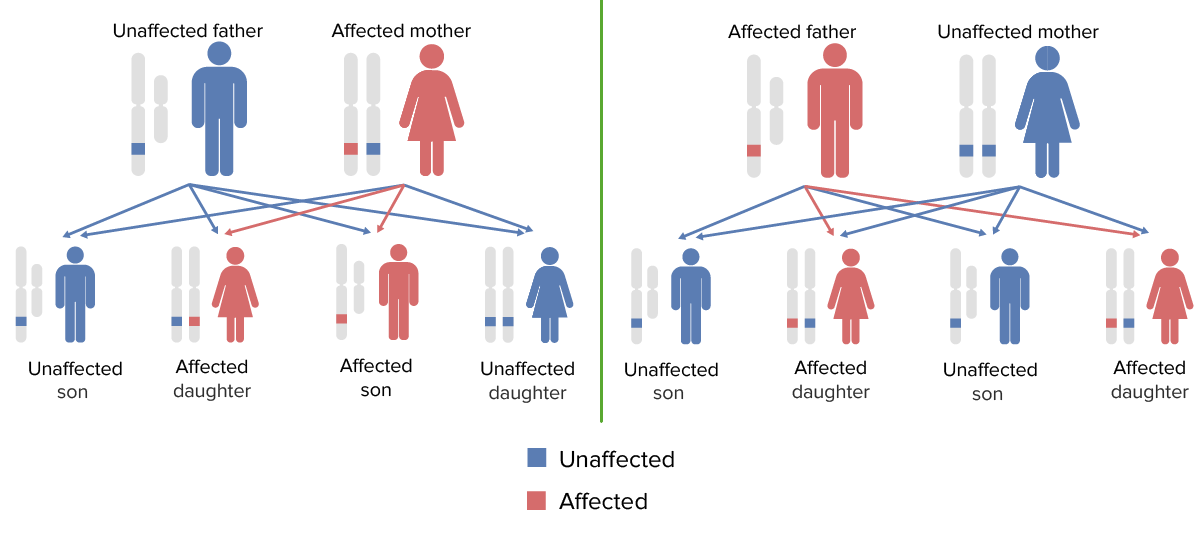
Patterns of x-linked dominant inheritance

Patterns of X-linked dominant inheritance (a very rare mode of inheritance):
Males and females have an equal probability of expressing the trait.
Note: Some X-linked dominant disorders are embryonic lethal in males; most affect females less severely.

Image by Lecturio.

References

  1. Raby, B. (2021). Inheritance patterns of monogenic disorders (Mendelian and non-Mendelian). UpToDate. Retrieved June 11, 2021, from https://www.uptodate.com/contents/inheritance-patterns-of-monogenic-disorders-mendelian-and-non-mendelian
  2. Van Esch, H. (2021). Fragile X syndrome: clinical features and diagnosis in children and adolescents. UpToDate. Retrieved June 26, 2021, from https://www.uptodate.com/contents/fragile-x-syndrome-clinical-features-and-diagnosis-in-children-and-adolescents
  3. Kashtan, C. (2021). Clinical manifestation, diagnosis, and treatment of Alport syndrome (hereditary nephritis). UpToDate. Retrieved June 26, 2021, from https://www.uptodate.com/contents/clinical-manifestations-diagnosis-and-treatment-of-alport-syndrome-hereditary-nephritis
  4. Schultz, R., Glaze, D. (2021). Rett syndrome: genetics, clinical features, and diagnosis. UpToDate. Retrieved June 26, 2021, from https://www.uptodate.com/contents/rett-syndrome-genetics-clinical-features-and-diagnosis
  5. Scheinman, S., Carpenter, T., Drezner, M. (2021). Hereditary hypophosphatemic rickets and tumor-induced osteomalacia. UpToDate. Retrieved June 26, 2021, from https://www.uptodate.com/contents/hereditary-hypophosphatemic-rickets-and-tumor-induced-osteomalacia
  6. Darras, B. (2021). Duchenne and Becker muscular dystrophy: clinical features and diagnosis. UpToDate. Retrieved June 26, 2021, from https://www.uptodate.com/contents/duchenne-and-becker-muscular-dystrophy-clinical-features-and-diagnosis
  7. Hand, J. (2021). Recessive X-linked ichthyosis. UpToDate. Retrieved June 26, 2021, from https://www.uptodate.com/contents/recessive-x-linked-ichthyosis
  8. Hernandex-Trujilo, V. (2021). Agammaglobulinemia. UpToDate. Retrieved June 26, 2021, from https://www.uptodate.com/contents/agammaglobulinemia
  9. Glader, B. (2021). Genetics and pathophysiology of glucose-6-phosphate dehydrogenase (G6PD) deficiency. UpToDate. Retrieved June 26, 2021, from https://www.uptodate.com/contents/genetics-and-pathophysiology-of-glucose-6-phosphate-dehydrogenase-g6pd-deficiency
  10. NIH, Genetic and Rare Diseases Information Center. (n.a.) Hemophilia A. Retrieved September 5, 2021, from https://rarediseases.info.nih.gov/diseases/6591/hemophilia-a

User Reviews

It's Deal Week! 💸 Save 30% on all plans!

Details