Retropharyngeal Abscess

Retropharyngeal abscesses occur in the retropharyngeal space, which extends from the base of the skull Base of the skull The inferior region of the skull consisting of an internal (cerebral), and an external (basilar) surface. Skull: Anatomy to the posterior mediastinum Posterior mediastinum Mediastinum and Great Vessels: Anatomy. The abscesses occur due to extension Extension Examination of the Upper Limbs of local infections Infections Invasion of the host organism by microorganisms or their toxins or by parasites that can cause pathological conditions or diseases. Chronic Granulomatous Disease, including upper respiratory infections Infections Invasion of the host organism by microorganisms or their toxins or by parasites that can cause pathological conditions or diseases. Chronic Granulomatous Disease or localized infections Infections Invasion of the host organism by microorganisms or their toxins or by parasites that can cause pathological conditions or diseases. Chronic Granulomatous Disease from trauma such as dental procedures. Infections Infections Invasion of the host organism by microorganisms or their toxins or by parasites that can cause pathological conditions or diseases. Chronic Granulomatous Disease most commonly occur in children. Key clinical features include trismus Trismus Spasmodic contraction of the masseter muscle resulting in forceful jaw closure. This may be seen with a variety of diseases, including tetanus, as a complication of radiation therapy, trauma, or in association with neoplastic conditions. Tetanus, dysphagia Dysphagia Dysphagia is the subjective sensation of difficulty swallowing. Symptoms can range from a complete inability to swallow, to the sensation of solids or liquids becoming "stuck." Dysphagia is classified as either oropharyngeal or esophageal, with esophageal dysphagia having 2 sub-types: functional and mechanical. Dysphagia, and an inability to extend the neck Neck The part of a human or animal body connecting the head to the rest of the body. Peritonsillar Abscess. Diagnosis is confirmed by computed tomography of the neck Neck The part of a human or animal body connecting the head to the rest of the body. Peritonsillar Abscess. Management is primarily through antibiotics and surgical drainage. Complications include airway Airway ABCDE Assessment compromise, mediastinitis Mediastinitis Mediastinitis refers to an infection or inflammation involving the mediastinum (a region in the thoracic cavity containing the heart, thymus gland, portions of the esophagus, and trachea). Acute mediastinitis can be caused by bacterial infection due to direct contamination, hematogenous or lymphatic spread, or extension of infection from nearby structures. Mediastinitis, and internal jugular vein Internal jugular vein Parapharyngeal Abscess thrombosis Thrombosis Formation and development of a thrombus or blood clot in the blood vessel. Epidemic Typhus.

Last updated: Jan 15, 2024

Editorial responsibility: Stanley Oiseth, Lindsay Jones, Evelin Maza

Epidemiology and Etiology

Epidemiology

Etiology

  • Abscesses occur due to infections Infections Invasion of the host organism by microorganisms or their toxins or by parasites that can cause pathological conditions or diseases. Chronic Granulomatous Disease that drain to lateral retropharyngeal lymph nodes Lymph Nodes They are oval or bean shaped bodies (1 – 30 mm in diameter) located along the lymphatic system. Lymphatic Drainage System: Anatomy.
  • May be secondary to:
    • Upper respiratory tract infections Infections Invasion of the host organism by microorganisms or their toxins or by parasites that can cause pathological conditions or diseases. Chronic Granulomatous Disease (more common in children) 
    • Otitis media
    • Sinusitis Sinusitis Sinusitis refers to inflammation of the mucosal lining of the paranasal sinuses. The condition usually occurs concurrently with inflammation of the nasal mucosa (rhinitis), a condition known as rhinosinusitis. Acute sinusitis is due to an upper respiratory infection caused by a viral, bacterial, or fungal agent. Sinusitis
    • Pharyngeal trauma (more common in adults)
      • Foreign body Foreign Body Foreign Body Aspiration (e.g., fishbone)
      • Endoscopy Endoscopy Procedures of applying endoscopes for disease diagnosis and treatment. Endoscopy involves passing an optical instrument through a small incision in the skin i.e., percutaneous; or through a natural orifice and along natural body pathways such as the digestive tract; and/or through an incision in the wall of a tubular structure or organ, i.e. Transluminal, to examine or perform surgery on the interior parts of the body. Gastroesophageal Reflux Disease (GERD)
      • Dental procedures
  • Infections Infections Invasion of the host organism by microorganisms or their toxins or by parasites that can cause pathological conditions or diseases. Chronic Granulomatous Disease are often polymicrobial and may include:
    • Aerobes
      • Staphylococcus aureus Staphylococcus aureus Potentially pathogenic bacteria found in nasal membranes, skin, hair follicles, and perineum of warm-blooded animals. They may cause a wide range of infections and intoxications. Brain Abscess 
      • Streptococcus Streptococcus Streptococcus is one of the two medically important genera of gram-positive cocci, the other being Staphylococcus. Streptococci are identified as different species on blood agar on the basis of their hemolytic pattern and sensitivity to optochin and bacitracin. There are many pathogenic species of streptococci, including S. pyogenes, S. agalactiae, S. pneumoniae, and the viridans streptococci. Streptococcus pyogenes
    • Anaerobes Anaerobes Lincosamides
      • Bacteroides Bacteroides Bacteroides is a genus of opportunistic, anaerobic, gram-negative bacilli. Bacteroides fragilis is the most common species involved in human disease and is part of the normal flora of the large intestine. Bacteroides 
      • Fusobacteria Fusobacteria A genus of gram-negative, anaerobic, rod-shaped bacteria found in cavities of humans and other animals. No endospores are formed. Some species are pathogenic and occur in various purulent or gangrenous infections. Perianal and Perirectal Abscess
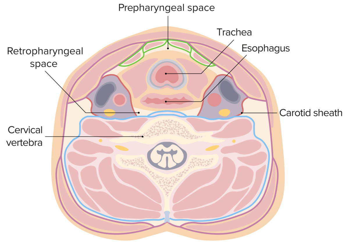
Retropharyngeal space

The retropharyngeal space in this figure is located between the yellow (buccopharyngeal fascia) and blue (prevertebral fascia) lines. The lateral borders are formed by the carotid sheath (red lines).

Image by Lecturio.
Clinical Presentation

Early symptoms

  • May mimic pharyngitis Pharyngitis Pharyngitis is an inflammation of the back of the throat (pharynx). Pharyngitis is usually caused by an upper respiratory tract infection, which is viral in most cases. It typically results in a sore throat and fever. Other symptoms may include a runny nose, cough, headache, and hoarseness. Pharyngitis
    • Fever Fever Fever is defined as a measured body temperature of at least 38°C (100.4°F). Fever is caused by circulating endogenous and/or exogenous pyrogens that increase levels of prostaglandin E2 in the hypothalamus. Fever is commonly associated with chills, rigors, sweating, and flushing of the skin. Fever
    • Sore throat Sore throat Pharyngitis is an inflammation of the back of the throat (pharynx). Pharyngitis is usually caused by an upper respiratory tract infection, which is viral in most cases. It typically results in a sore throat and fever. Other symptoms may include a runny nose, cough, headache, and hoarseness. Pharyngitis
    • Pharyngeal erythema Erythema Redness of the skin produced by congestion of the capillaries. This condition may result from a variety of disease processes. Chalazion
  • Dysphagia Dysphagia Dysphagia is the subjective sensation of difficulty swallowing. Symptoms can range from a complete inability to swallow, to the sensation of solids or liquids becoming “stuck.” Dysphagia is classified as either oropharyngeal or esophageal, with esophageal dysphagia having 2 sub-types: functional and mechanical. Dysphagia
  • Odynophagia Odynophagia Epiglottitis
  • Neck stiffness Neck Stiffness Meningitis/ asymmetry Asymmetry Examination of the Upper Limbs 
  • Trismus Trismus Spasmodic contraction of the masseter muscle resulting in forceful jaw closure. This may be seen with a variety of diseases, including tetanus, as a complication of radiation therapy, trauma, or in association with neoplastic conditions. Tetanus ( lockjaw Lockjaw Spasmodic contraction of the masseter muscle resulting in forceful jaw closure. This may be seen with a variety of diseases, including tetanus, as a complication of radiation therapy, trauma, or in association with neoplastic conditions. Tetanus)

Later symptoms

Diagnosis and Management

Diagnosis

  • Initial work-up includes:
    • CBC
    • Blood culture
  • Diagnosis is confirmed by imaging.
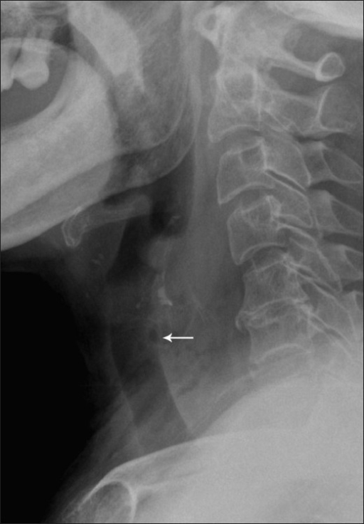
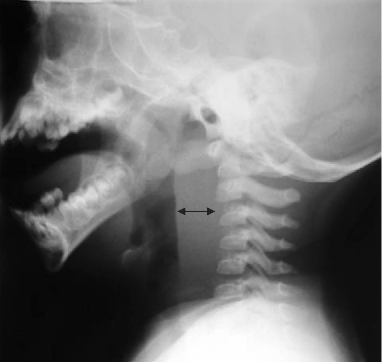
    • Lateral neck Neck The part of a human or animal body connecting the head to the rest of the body. Peritonsillar Abscess X-ray X-ray Penetrating electromagnetic radiation emitted when the inner orbital electrons of an atom are excited and release radiant energy. X-ray wavelengths range from 1 pm to 10 nm. Hard x-rays are the higher energy, shorter wavelength x-rays. Soft x-rays or grenz rays are less energetic and longer in wavelength. The short wavelength end of the x-ray spectrum overlaps the gamma rays wavelength range. The distinction between gamma rays and x-rays is based on their radiation source. Pulmonary Function Tests:
      • Widened retropharyngeal space 
      • Gas in the retropharyngeal space
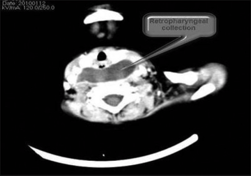
    • Computed tomography (CT) of the neck Neck The part of a human or animal body connecting the head to the rest of the body. Peritonsillar Abscess with contrast (gold standard):
      • Rim-enhancing hypodense collection in retropharyngeal space
      • Thickening of the prevertebral space 
      • Presence of air or air-fluid levels

Management

  • The 1st and most important step is protecting the airway Airway ABCDE Assessment intubation Intubation Peritonsillar Abscess if needed! 
  • Surgical drainage
  • Antibiotics
    • Ampicillin Ampicillin Semi-synthetic derivative of penicillin that functions as an orally active broad-spectrum antibiotic. Penicillins/ sulbactam Sulbactam A beta-lactamase inhibitor with very weak antibacterial action. The compound prevents antibiotic destruction of beta-lactam antibiotics by inhibiting beta-lactamases, thus extending their spectrum activity. Combinations of sulbactam with beta-lactam antibiotics have been used successfully for the therapy of infections caused by organisms resistant to the antibiotic alone. Cephalosporins or clindamycin Clindamycin An antibacterial agent that is a semisynthetic analog of lincomycin. Lincosamides
    • If no response, vancomycin Vancomycin Antibacterial obtained from streptomyces orientalis. It is a glycopeptide related to ristocetin that inhibits bacterial cell wall assembly and is toxic to kidneys and the inner ear. Glycopeptides or linezolid Linezolid An oxazolidinone and acetamide derived anti-bacterial agent and protein synthesis inhibitor that is used in the treatment of gram-positive bacterial infections of the skin and respiratory tract. Oxazolidinones
    • When clinically improved, may switch to amoxicillin Amoxicillin A broad-spectrum semisynthetic antibiotic similar to ampicillin except that its resistance to gastric acid permits higher serum levels with oral administration. Penicillins/clavulanate or clindamycin Clindamycin An antibacterial agent that is a semisynthetic analog of lincomycin. Lincosamides (with oral linezolid Linezolid An oxazolidinone and acetamide derived anti-bacterial agent and protein synthesis inhibitor that is used in the treatment of gram-positive bacterial infections of the skin and respiratory tract. Oxazolidinones for MRSA MRSA A strain of Staphylococcus aureus that is non-susceptible to the action of methicillin. The mechanism of resistance usually involves modification of normal or the presence of acquired penicillin binding proteins. Staphylococcus coverage).

Complications

Retropharyngeal abscesses are considered the most deadly deep neck Neck The part of a human or animal body connecting the head to the rest of the body. Peritonsillar Abscess infection. 

The most common complications include:

Differential Diagnosis

  • Epiglottitis Epiglottitis Epiglottitis (or “supraglottitis”) is an inflammation of the epiglottis and adjacent supraglottic structures. The majority of cases are caused by bacterial infection. Symptoms are rapid in onset and severe. Epiglottitis: an inflammation Inflammation Inflammation is a complex set of responses to infection and injury involving leukocytes as the principal cellular mediators in the body’s defense against pathogenic organisms. Inflammation is also seen as a response to tissue injury in the process of wound healing. The 5 cardinal signs of inflammation are pain, heat, redness, swelling, and loss of function. Inflammation of the epiglottis Epiglottis A thin leaf-shaped cartilage that is covered with laryngeal mucosa and situated posterior to the root of the tongue and hyoid bone. During swallowing, the epiglottis folds back over the larynx inlet thus prevents foods from entering the airway. Larynx: Anatomy most commonly caused by infection with Haemophilus Haemophilus Haemophilus is a genus of Gram-negative coccobacilli, all of whose strains require at least 1 of 2 factors for growth (factor V [NAD] and factor X [heme]); therefore, it is most often isolated on chocolate agar, which can supply both factors. The pathogenic species are H. influenzae and H. ducreyi. Haemophilus influenzae. Epiglottitis Epiglottitis Epiglottitis (or “supraglottitis”) is an inflammation of the epiglottis and adjacent supraglottic structures. The majority of cases are caused by bacterial infection. Symptoms are rapid in onset and severe. Epiglottitis presents with “cherry-red” epiglottis Epiglottis A thin leaf-shaped cartilage that is covered with laryngeal mucosa and situated posterior to the root of the tongue and hyoid bone. During swallowing, the epiglottis folds back over the larynx inlet thus prevents foods from entering the airway. Larynx: Anatomy, fever Fever Fever is defined as a measured body temperature of at least 38°C (100.4°F). Fever is caused by circulating endogenous and/or exogenous pyrogens that increase levels of prostaglandin E2 in the hypothalamus. Fever is commonly associated with chills, rigors, sweating, and flushing of the skin. Fever, dysphagia Dysphagia Dysphagia is the subjective sensation of difficulty swallowing. Symptoms can range from a complete inability to swallow, to the sensation of solids or liquids becoming “stuck.” Dysphagia is classified as either oropharyngeal or esophageal, with esophageal dysphagia having 2 sub-types: functional and mechanical. Dysphagia, drooling Drooling Peritonsillar Abscess, and difficulty breathing. The condition is commonly seen in unvaccinated children. Treatment is with rocephin and steroids Steroids A group of polycyclic compounds closely related biochemically to terpenes. They include cholesterol, numerous hormones, precursors of certain vitamins, bile acids, alcohols (sterols), and certain natural drugs and poisons. Steroids have a common nucleus, a fused, reduced 17-carbon atom ring system, cyclopentanoperhydrophenanthrene. Most steroids also have two methyl groups and an aliphatic side-chain attached to the nucleus. Benign Liver Tumors.
  • Peritonsillar abscess: a bacterial infection (most commonly S. pyogenes) that causes an abscess next to the palatine tonsils. The condition is more common in children and young adults and presents with trismus, fever, and sore throat, with a “hot potato” voice. Physical exam shows an abscess Abscess Accumulation of purulent material in tissues, organs, or circumscribed spaces, usually associated with signs of infection. Chronic Granulomatous Disease near the pharyngeal tonsil causing uvula Uvula A fleshy extension at the back of the soft palate that hangs above the opening of the throat. Peritonsillar Abscess deviation. Treatment is with antibiotics and surgical drainage. 
  • Parapharyngeal abscess Abscess Accumulation of purulent material in tissues, organs, or circumscribed spaces, usually associated with signs of infection. Chronic Granulomatous Disease or prevertebral space infection: very similar clinical presentation to retropharyngeal abscesses but most often caused by dental infections Infections Invasion of the host organism by microorganisms or their toxins or by parasites that can cause pathological conditions or diseases. Chronic Granulomatous Disease that extend into the lateral or posterior pharyngeal space. The best way to differentiate pharyngeal from retropharyngeal abscess Abscess Accumulation of purulent material in tissues, organs, or circumscribed spaces, usually associated with signs of infection. Chronic Granulomatous Disease is by CT of the neck Neck The part of a human or animal body connecting the head to the rest of the body. Peritonsillar Abscess. Treatment is with antibiotics and surgical drainage. 
  • Meningitis Meningitis Meningitis is inflammation of the meninges, the protective membranes of the brain, and spinal cord. The causes of meningitis are varied, with the most common being bacterial or viral infection. The classic presentation of meningitis is a triad of fever, altered mental status, and nuchal rigidity. Meningitis: an infection of the meninges Meninges The brain and the spinal cord are enveloped by 3 overlapping layers of connective tissue called the meninges. The layers are, from the most external layer to the most internal layer, the dura mater, arachnoid mater, and pia mater. Between these layers are 3 potential spaces called the epidural, subdural, and subarachnoid spaces. Meninges: Anatomy, the protective membranes around the brain Brain The part of central nervous system that is contained within the skull (cranium). Arising from the neural tube, the embryonic brain is comprised of three major parts including prosencephalon (the forebrain); mesencephalon (the midbrain); and rhombencephalon (the hindbrain). The developed brain consists of cerebrum; cerebellum; and other structures in the brain stem. Nervous System: Anatomy, Structure, and Classification, most often caused by Streptococcus Streptococcus Streptococcus is one of the two medically important genera of gram-positive cocci, the other being Staphylococcus. Streptococci are identified as different species on blood agar on the basis of their hemolytic pattern and sensitivity to optochin and bacitracin. There are many pathogenic species of streptococci, including S. pyogenes, S. agalactiae, S. pneumoniae, and the viridans streptococci. Streptococcus pneumoniae or H. influenzae H. influenzae A species of Haemophilus found on the mucous membranes of humans and a variety of animals. The species is further divided into biotypes I through VIII. Haemophilus. Meningitis Meningitis Meningitis is inflammation of the meninges, the protective membranes of the brain, and spinal cord. The causes of meningitis are varied, with the most common being bacterial or viral infection. The classic presentation of meningitis is a triad of fever, altered mental status, and nuchal rigidity. Meningitis presents with fever Fever Fever is defined as a measured body temperature of at least 38°C (100.4°F). Fever is caused by circulating endogenous and/or exogenous pyrogens that increase levels of prostaglandin E2 in the hypothalamus. Fever is commonly associated with chills, rigors, sweating, and flushing of the skin. Fever, stiff neck Neck The part of a human or animal body connecting the head to the rest of the body. Peritonsillar Abscess, and headache Headache The symptom of pain in the cranial region. It may be an isolated benign occurrence or manifestation of a wide variety of headache disorders. Brain Abscess. The condition is differentiated from retropharyngeal abscess Abscess Accumulation of purulent material in tissues, organs, or circumscribed spaces, usually associated with signs of infection. Chronic Granulomatous Disease by the presence of light sensitivity, headache Headache The symptom of pain in the cranial region. It may be an isolated benign occurrence or manifestation of a wide variety of headache disorders. Brain Abscess, and nuchal rigidity Nuchal Rigidity Meningitis. Diagnosis is by lumbar puncture Lumbar Puncture Febrile Infant for cerebrospinal (CSF) evaluation. Treatment consists of the rapid administration of antibiotics.

References

  1. Jain H, Knorr TL, Sinha V. Retropharyngeal Abscess. StatPearls. Retrieved Oct 5, 2020, from https://www.statpearls.com/TodaysPearl/3-9-2018
  2. Wald, E. R. (2019). Retropharyngeal Infections in Children. UpToDate. Retrieved Sept 23, 2020, https://www.uptodate.com/contents/retropharyngeal-infections-in-children

Create your free account or log in to continue reading!

Sign up now and get free access to Lecturio with concept pages, medical videos, and questions for your medical education.

User Reviews

Details