Human cells are reliant on aerobic metabolism. Obtaining O2 from the environment and transporting it to the tissues while excreting the byproduct of cellular respiration Respiration The act of breathing with the lungs, consisting of inhalation, or the taking into the lungs of the ambient air, and of exhalation, or the expelling of the modified air which contains more carbon dioxide than the air taken in. Nose Anatomy (External & Internal) (CO2) are processes key to survival and must be closely regulated. Chemoreceptors in the lungs Lungs Lungs are the main organs of the respiratory system. Lungs are paired viscera located in the thoracic cavity and are composed of spongy tissue. The primary function of the lungs is to oxygenate blood and eliminate CO2. Lungs: Anatomy and tissues sense changes in the concentration of respiratory gasses and send messages to the CNS, which, in turn, modifies breathing parameters such as the respiratory rate Respiratory rate The number of times an organism breathes with the lungs (respiration) per unit time, usually per minute. Pulmonary Examination or tidal volume Tidal volume The volume of air inspired or expired during each normal, quiet respiratory cycle. Common abbreviations are tv or V with subscript t. Ventilation: Mechanics of Breathing to compensate for any imbalance. Disruption of this control mechanism can be caused by severe disease and also result in severe disease.
Last updated: Dec 15, 2025
Respiratory centers are specialized neuron clusters located in the medulla oblongata Medulla Oblongata The lower portion of the brain stem. It is inferior to the pons and anterior to the cerebellum. Medulla oblongata serves as a relay station between the brain and the spinal cord, and contains centers for regulating respiratory, vasomotor, cardiac, and reflex activities. Brain Stem: Anatomy. They regulate 2 respiratory parameters in response to changing demands:
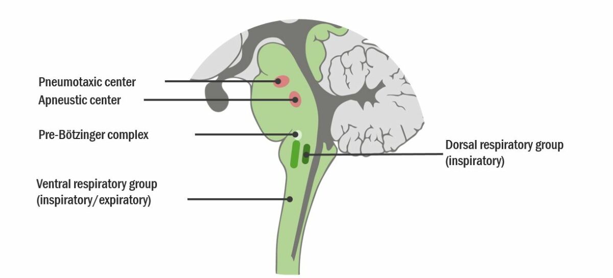
Respiratory control centers
Image by Lecturio. License: CC BY-NC-SA 4.0Respiratory centers set the rate and depth of breathing. Changes in O₂, CO₂, and H+ concentrations are sensed by central and peripheral chemoreceptors, which regulate the activity of the respiratory centers to match metabolic and situational needs.
Central chemoreceptors are located in the medulla oblongata Medulla Oblongata The lower portion of the brain stem. It is inferior to the pons and anterior to the cerebellum. Medulla oblongata serves as a relay station between the brain and the spinal cord, and contains centers for regulating respiratory, vasomotor, cardiac, and reflex activities. Brain Stem: Anatomy and regulate respiratory center activity based on changes in blood gases.
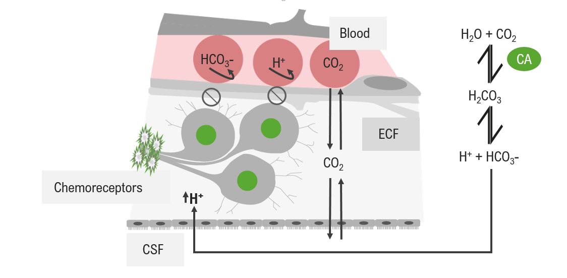
Diagram of the stimulation of central chemoreceptors by H+:
since H+ cannot enter the CNS, they are produced as a result of the carbonic anhydrase reaction using CO2. H+ is an indirect indicator for the central chemoreceptors that depicts the amount of CO2 in the systemic circulation.
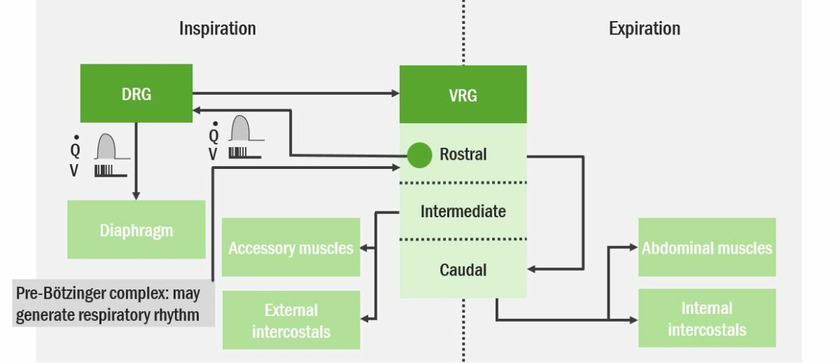
Integration of the ventral respiratory group neurons (VRG) and the dorsal respiratory group neurons (DRG) for respiration control
Image by Lecturio. License: CC BY-NC-SA 4.0Peripheral chemoreceptors are located in the carotid bodies and aortic bodies and are more responsive to blood gases than central chemoreceptors.
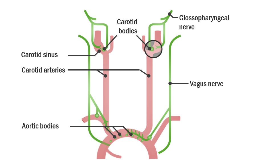
Anatomical location of peripheral chemoreceptors
Image by Lecturio. License: CC BY-NC-SA 4.0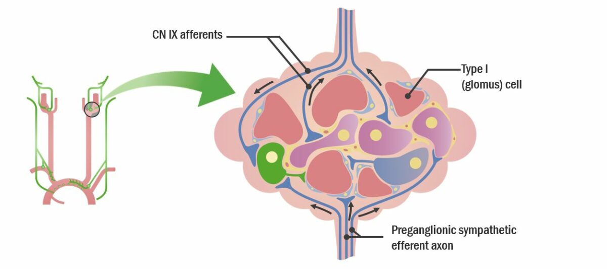
Anatomical location of peripheral chemoreceptors
CN: cranial nerve
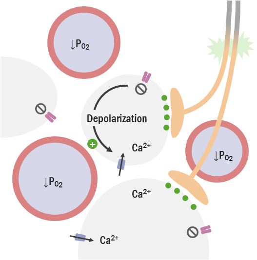
Diagram of the effect of low partial pressure of O2 (PO2) in the type I (glomus) cells of peripheral chemoreceptors
Image by Lecturio. License: CC BY-NC-SA 4.0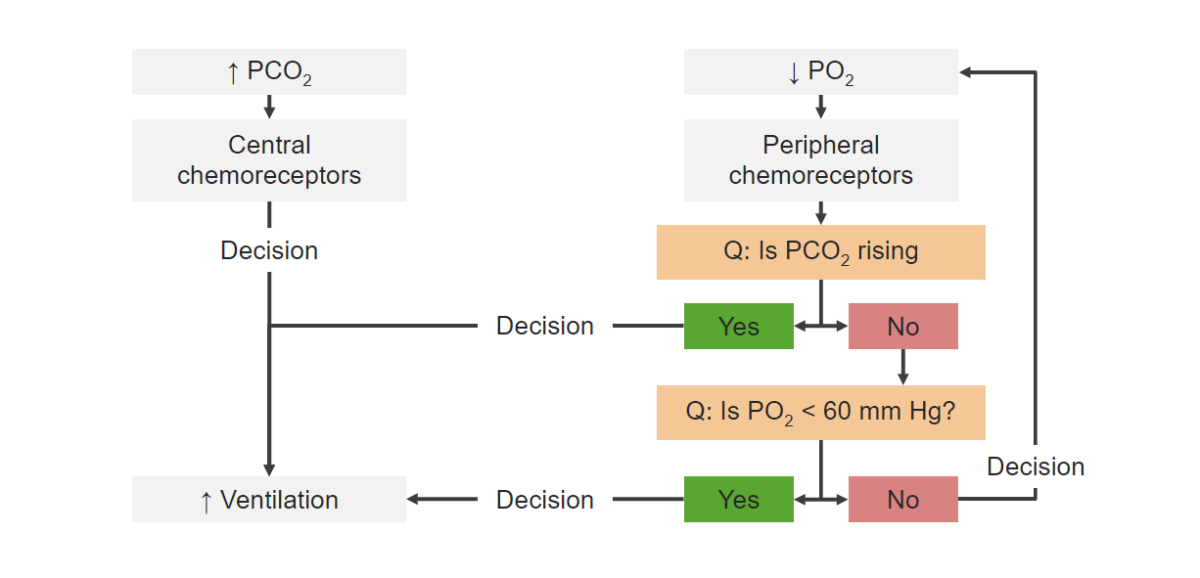
Flowchart of the effects of blood gases on chemoreceptors and, ultimately, ventilation
Image by Lecturio. License: CC BY-NC-SA 4.0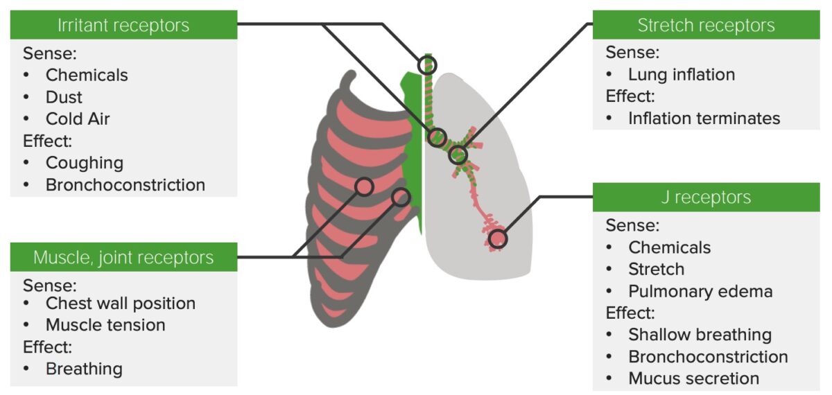
Receptors along the respiratory tract, and in muscles and joints of the thorax
Image by Lecturio. License: CC BY-NC-SA 4.0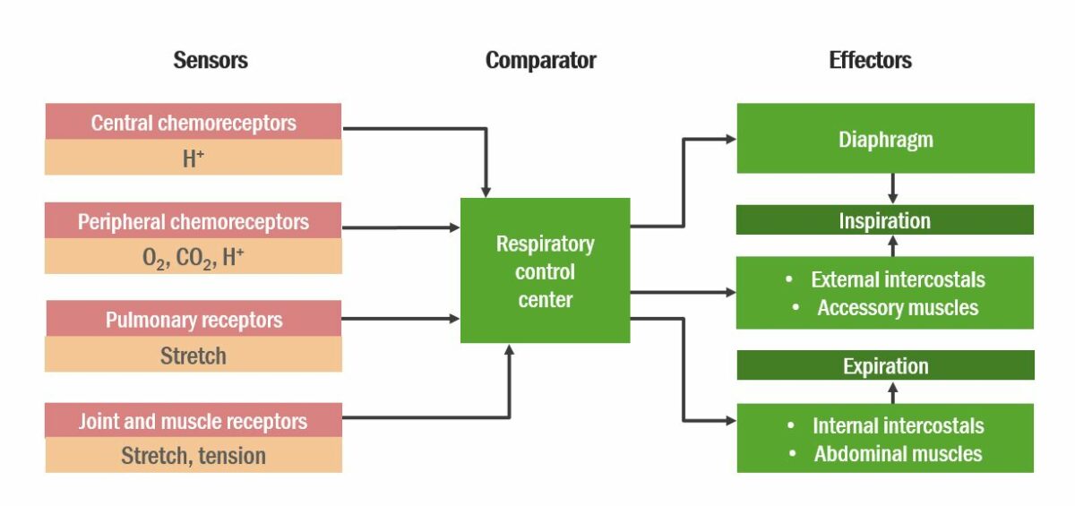
Diagram of integration of stimuli by the respiratory control center
Image by Lecturio. License: CC BY-NC-SA 4.0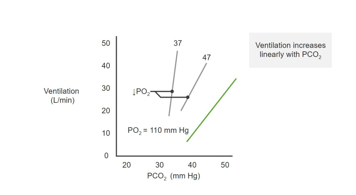
Relationship between ventilation and partial pressure of O₂ (PO₂), and PCO₂:
See how either an increase in PCO₂ or a decrease in PO₂ can increase the amount of air drawn per minute (ventilation).
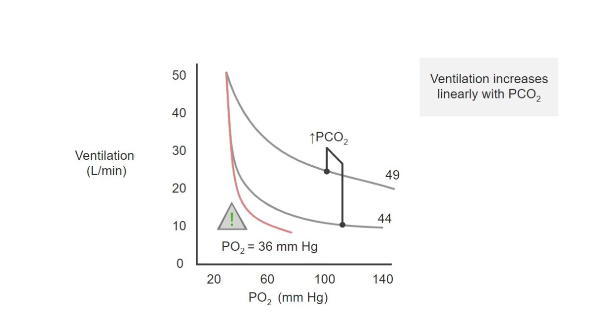
Chart of PO2 (x-axis) and ventilation (y-axis):
See the inverse relationship between the 2 components.
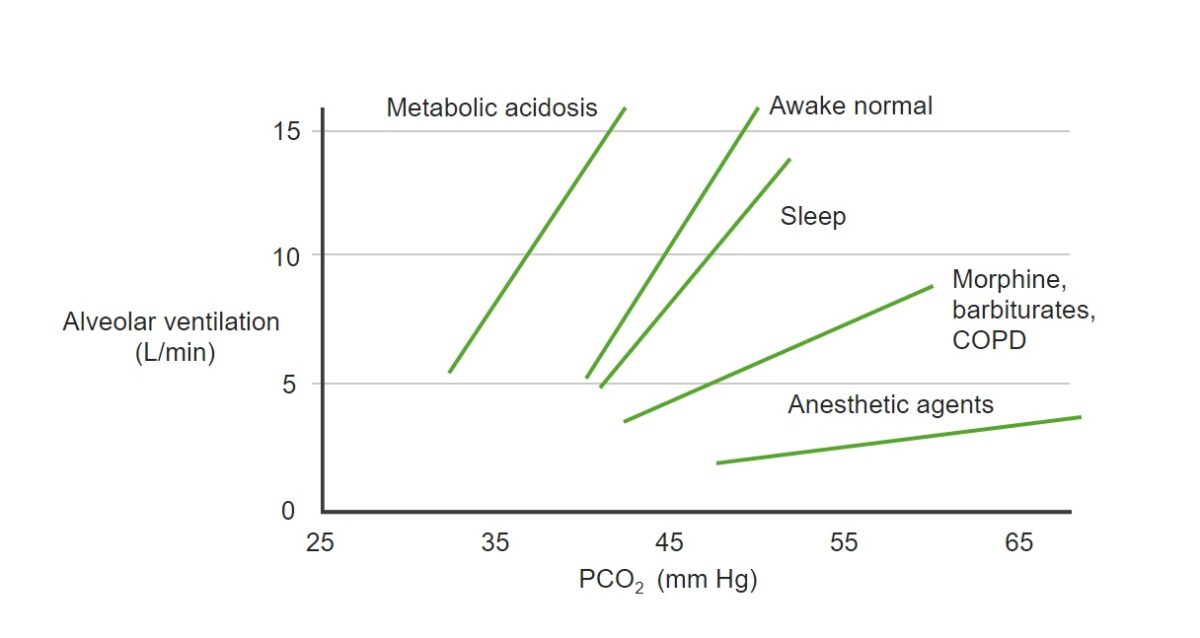
Changes of PCO2 and alveolar ventilation relationship under various conditions:
Notice how pharmacological agents blunt the ventilatory response to high PCO2.
COPD: chronic obstructive pulmonary disease