Diagnostic modalities such as chest X-rays X-rays X-rays are high-energy particles of electromagnetic radiation used in the medical field for the generation of anatomical images. X-rays are projected through the body of a patient and onto a film, and this technique is called conventional or projectional radiography. X-rays provide static images Static Images Ultrasound (Sonography) of the thoracic cavity, including the lungs Lungs Lungs are the main organs of the respiratory system. Lungs are paired viscera located in the thoracic cavity and are composed of spongy tissue. The primary function of the lungs is to oxygenate blood and eliminate CO2. Lungs: Anatomy and airways. While providing a wealth of anatomical information necessary for the diagnosis of pulmonary disease Pulmonary disease Diseases involving the respiratory system. Blastomyces/Blastomycosis, chest X-rays X-rays X-rays are high-energy particles of electromagnetic radiation used in the medical field for the generation of anatomical images. X-rays are projected through the body of a patient and onto a film, and this technique is called conventional or projectional radiography. X-rays do not give much information about the individual’s respiratory function. Pulmonary function tests are a group of diagnostic procedures yielding useful, quantifiable information about the rate of the flow Flow Blood flows through the heart, arteries, capillaries, and veins in a closed, continuous circuit. Flow is the movement of volume per unit of time. Flow is affected by the pressure gradient and the resistance fluid encounters between 2 points. Vascular resistance is the opposition to flow, which is caused primarily by blood friction against vessel walls. Vascular Resistance, Flow, and Mean Arterial Pressure of air through the individual’s airways, lung capacity, and the efficiency of gas exchange Gas exchange Human cells are primarily reliant on aerobic metabolism. The respiratory system is involved in pulmonary ventilation and external respiration, while the circulatory system is responsible for transport and internal respiration. Pulmonary ventilation (breathing) represents movement of air into and out of the lungs. External respiration, or gas exchange, is represented by the O2 and CO2 exchange between the lungs and the blood. Gas Exchange in relation to time. The most commonly utilized tests include spirometry (before and after bronchodilator use), lung volumes, and quantitation of diffusing capacity for carbon monoxide Carbon monoxide Carbon monoxide (CO). A poisonous colorless, odorless, tasteless gas. It combines with hemoglobin to form carboxyhemoglobin, which has no oxygen carrying capacity. The resultant oxygen deprivation causes headache, dizziness, decreased pulse and respiratory rates, unconsciousness, and death. Carbon Monoxide Poisoning (CO). The tests can be influenced by the individual's effort/ fatigue Fatigue The state of weariness following a period of exertion, mental or physical, characterized by a decreased capacity for work and reduced efficiency to respond to stimuli. Fibromyalgia, disease state, or anatomical malformation.
Last updated: Dec 15, 2025
Lung volumes are measured by:

Graphical representation of lung volumes as measured by body plethysmography:
Lung volumes can be measured and calculated by placing an individual in a box of known volume and performing specific breathing maneuvers, allowing the physician to diagnose pathology that causes changes in lung volumes.
Lung volumes:
Lung capacities:
Spirometry records the volume of air moving in and out of an individual’s lungs Lungs Lungs are the main organs of the respiratory system. Lungs are paired viscera located in the thoracic cavity and are composed of spongy tissue. The primary function of the lungs is to oxygenate blood and eliminate CO2. Lungs: Anatomy.
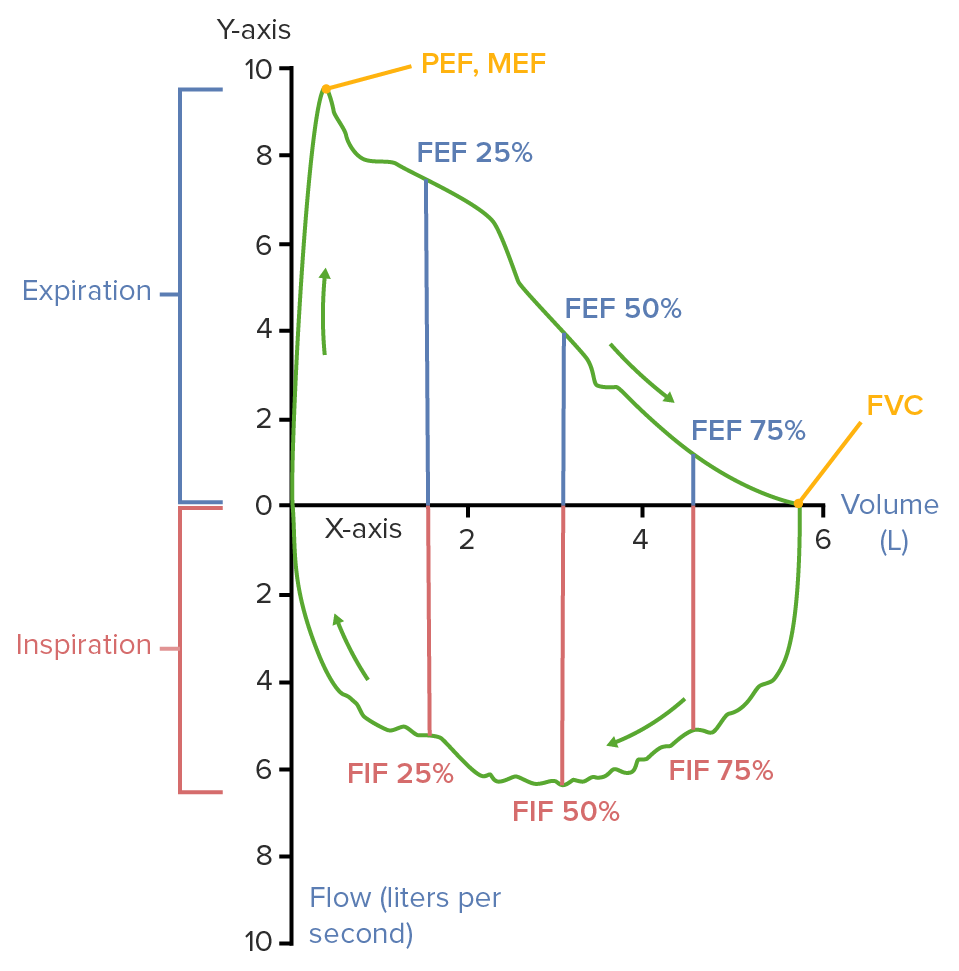
Flow volume loops:
Spirometry testing results are often represented as standardized graphs called flow volume loops. The graphs usually depict volume on the x-axis and flow on the y-axis (flow out of the lungs is positive, flow into the lungs is negative). A flow volume loop represents a respiratory cycle captured through spirometry.
PEF: peak expiratory flow
MEF: maximal expiratory flow
FEF: forced expiratory flow
FVC: forced vital capacity
FIF: forced inspiratory flow
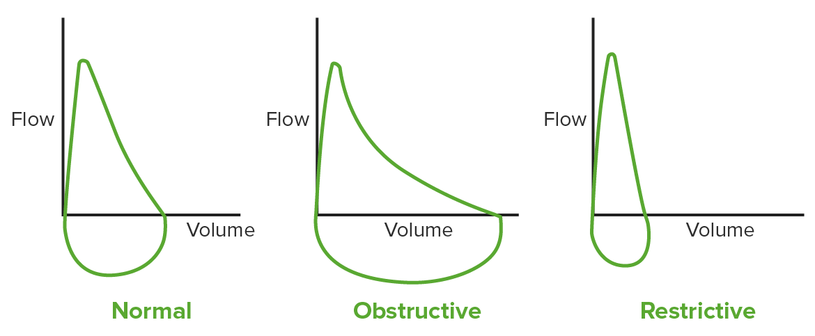
Left: a normal flow volume loop
Middle: obstructive loop spirometry (the expiratory flow is decreased)
Right: restrictive loop spirometry (the volumes are decreased, but the flow remains relatively the same)
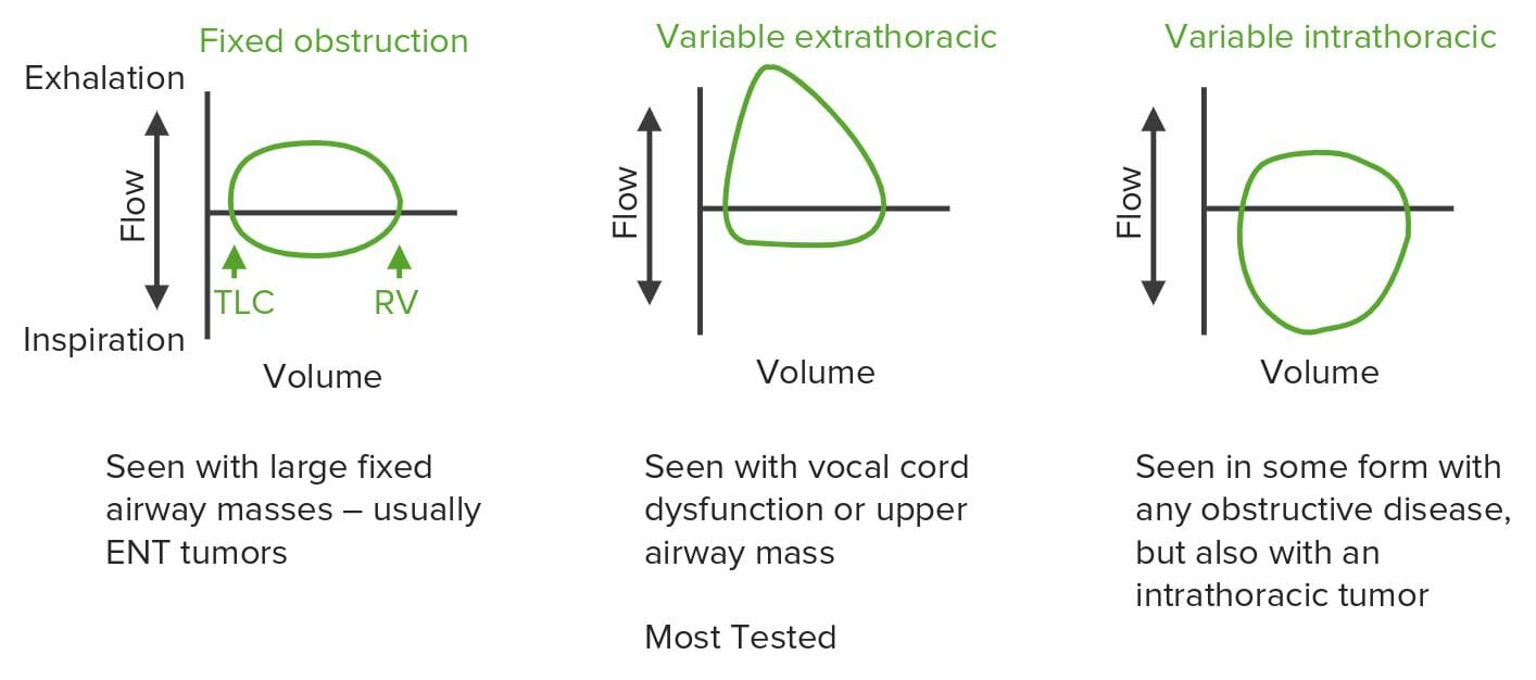
Fixed obstruction flow volume loop: The diameter of the airway is fixed (airway tumors), so expiratory and inspiratory flow cannot increase past a set point determined by the physical diameter of the airway, leading to plateauing of both the expiratory and inspiratory portion of the loop.
Variable extrathoracic flow volume loop: Variable extrathoracic airway obstruction (vocal cord dysfunction) worsens on inspiration when negative pressure in the extrathoracic portion of the airway is the highest, leading to flattening of the inspiratory portion of the loop.
Variable intrathoracic flow volume loop: Variable intrathoracic obstruction (asthma) is highest on expiration when the diameter of the intrathoracic airways is the smallest (shown as flattening of the expiratory phase of the loop).
| Parameter | Obstructive lung disease | Restrictive lung disease |
|---|---|---|
| TLC | High/normal | Very low |
| VC or FVC | Low/normal | Very low |
| RV | High | Low |
| FRC | High | Low |
| FEV1 | Very Low | Normal/low |
| FEV1/FVC | Low | Normal/high |
The following conditions are diagnosed by pulmonary function tests: