Advertisement

Advertisement

Advertisement

Advertisement

Primary Ciliary Dyskinesia

Primary ciliary dyskinesia (PCD), also known as immotile-cilia syndrome, is an autosomal recessive Autosomal recessive Autosomal inheritance, both dominant and recessive, refers to the transmission of genes from the 22 autosomal chromosomes. Autosomal recessive diseases are only expressed when 2 copies of the recessive allele are inherited. Autosomal Recessive and Autosomal Dominant Inheritance disorder leading to an impairment that affects mucociliary clearance Mucociliary clearance A nonspecific host defense mechanism that removes mucus and other material from the lungs by ciliary and secretory activity of the tracheobronchial submucosal glands. It is measured in vivo as mucus transfer, ciliary beat frequency, and clearance of radioactive tracers. Acute Bronchitis. Primary ciliary dyskinesia is caused by defective ciliary function in the airways and is characterized by the loss of oscillation (immotility), abnormal oscillation (dyskinesia), or absence of cilia ( aplasia Aplasia Cranial Nerve Palsies). In most cases, PCD commonly presents with recurrent infections Recurrent infections Common Variable Immunodeficiency (CVID) of the upper and lower respiratory tract. Clinical features include bronchiectasis Bronchiectasis Bronchiectasis is a chronic disease of the airways that results from permanent bronchial distortion. This results from a continuous cycle of inflammation, bronchial damage and dilation, impaired clearance of secretions, and recurrent infections. Bronchiectasis, chronic rhinosinusitis, and situs inversus. There is no gold standard diagnostic test for PCD, as several tests are used in the diagnosis. Treatment is individualized, and the primary goal is to remove trapped mucus and treat infections Infections Invasion of the host organism by microorganisms or their toxins or by parasites that can cause pathological conditions or diseases. Chronic Granulomatous Disease. Patients Patients Individuals participating in the health care system for the purpose of receiving therapeutic, diagnostic, or preventive procedures. Clinician–Patient Relationship with PCD usually have a regular Regular Insulin lifespan.

Last updated: Jan 13, 2023

Editorial responsibility: Stanley Oiseth, Lindsay Jones, Evelin Maza

Advertisement

Advertisement

Advertisement

Advertisement

Advertisement

Advertisement

Overview

Definition

Primary ciliary dyskinesia (PCD) is a genetic condition in which cilia in the respiratory tract have defective functionality that leads to ineffective mucociliary clearance Mucociliary clearance A nonspecific host defense mechanism that removes mucus and other material from the lungs by ciliary and secretory activity of the tracheobronchial submucosal glands. It is measured in vivo as mucus transfer, ciliary beat frequency, and clearance of radioactive tracers. Acute Bronchitis.
Epidemiology

  • Affects 1 in 16,000–20,000 live births 
  • Equal prevalence Prevalence The total number of cases of a given disease in a specified population at a designated time. It is differentiated from incidence, which refers to the number of new cases in the population at a given time. Measures of Disease Frequency in men and women
  • Affects all racial and ethnic groups

Etiology

Pathophysiology

Normal physiology

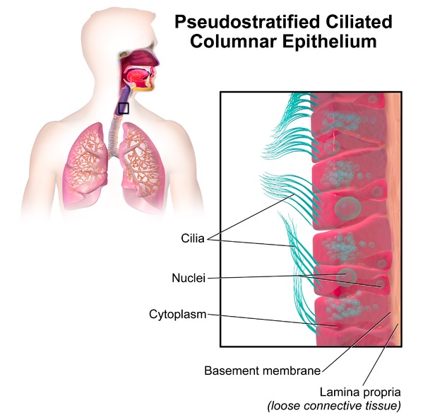
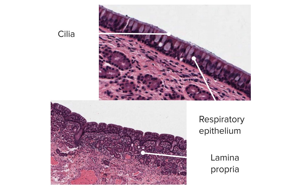
  • Cilia are microscopic, finger-like structures projecting out of the cell surface.
  • Cilia line the airways, reproductive tissues, and other organs.
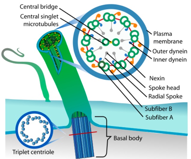
  • Normal, motile cilia in the upper and lower respiratory tract are composed of:
    • Alpha- and beta-tubulin monomers
    • A complex central core (axonemal) structure including:
      • Dynein Dynein A family of multisubunit cytoskeletal motor proteins that use the energy of ATP hydrolysis, generated by a ring of aaa ATPases in the dynein heavy chain, to power a variety of cellular functions. Dyneins fall into two major classes based upon structural and functional criteria. The Cell: Cytosol and Cytoskeleton arms
      • Radial spokes
      • Nexin links
  • Cilia usually beat in a coordinated fashion to move respiratory secretions.

Ciliary dysfunction

  • PCD is a highly heterogeneous syndrome that can be caused by a defect in:
    • 1 of the many polypeptide species in the central core of the cilia 
    • Proteins Proteins Linear polypeptides that are synthesized on ribosomes and may be further modified, crosslinked, cleaved, or assembled into complex proteins with several subunits. The specific sequence of amino acids determines the shape the polypeptide will take, during protein folding, and the function of the protein. Energy Homeostasis needed for the proper assembly of cilia
  • Mutations involving dynein Dynein A family of multisubunit cytoskeletal motor proteins that use the energy of ATP hydrolysis, generated by a ring of aaa ATPases in the dynein heavy chain, to power a variety of cellular functions. Dyneins fall into two major classes based upon structural and functional criteria. The Cell: Cytosol and Cytoskeleton are commonly implicated in PCD.
  • Effects:
    • Abnormal ciliary movement (ciliary dyskinesia)
    • Inability to move (ciliary immobility)
    • Absent cilia (ciliary aplasia Aplasia Cranial Nerve Palsies)
  • Proper ciliary function is necessary for the rotation Rotation Motion of an object in which either one or more points on a line are fixed. It is also the motion of a particle about a fixed point. X-rays of organs in the abdomen and chest.
  • With dysfunction, situs inversus and/or abnormal organ positioning can occur.

Clinical Presentation

Onset

  • Commonly presents in children, but can also present in adults
  • Median age of diagnosis:
    • In children, 5–5.5 years of age
    • In adults, 22 years of age

Signs and symptoms

  • Recurrent infections Recurrent infections Common Variable Immunodeficiency (CVID) of the upper and lower respiratory tract:
    • Most common symptom
    • Common causative bacteria Bacteria Bacteria are prokaryotic single-celled microorganisms that are metabolically active and divide by binary fission. Some of these organisms play a significant role in the pathogenesis of diseases. Bacteriology include:
      • Haemophilus influenzae Haemophilus Influenzae A species of Haemophilus found on the mucous membranes of humans and a variety of animals. The species is further divided into biotypes I through viii. Haemophilus
      • Streptococcus Streptococcus Streptococcus is one of the two medically important genera of gram-positive cocci, the other being Staphylococcus. Streptococci are identified as different species on blood agar on the basis of their hemolytic pattern and sensitivity to optochin and bacitracin. There are many pathogenic species of streptococci, including S. pyogenes, S. agalactiae, S. pneumoniae, and the viridans streptococci. Streptococcus pneumoniae
      • Staphylococcus aureus Staphylococcus aureus Potentially pathogenic bacteria found in nasal membranes, skin, hair follicles, and perineum of warm-blooded animals. They may cause a wide range of infections and intoxications. Brain Abscess
      • Pseudomonas aeruginosa Pseudomonas aeruginosa A species of gram-negative, aerobic, rod-shaped bacteria commonly isolated from clinical specimens (wound, burn, and urinary tract infections). It is also found widely distributed in soil and water. P. Aeruginosa is a major agent of nosocomial infection. Pseudomonas
      • Non-tuberculosis mycobacteria Mycobacteria Mycobacterium is a genus of the family Mycobacteriaceae in the phylum Actinobacteria. Mycobacteria comprise more than 150 species of facultative intracellular bacilli that are mostly obligate aerobes. Mycobacteria are responsible for multiple human infections including serious diseases, such as tuberculosis (M. tuberculosis), leprosy (M. leprae), and M. avium complex infections. Mycobacterium
  • Chronic rhinosinusitis:
    • Occurs in almost 100% of patients Patients Individuals participating in the health care system for the purpose of receiving therapeutic, diagnostic, or preventive procedures. Clinician–Patient Relationship
    • Year-round runny nose Nose The nose is the human body’s primary organ of smell and functions as part of the upper respiratory system. The nose may be best known for inhaling oxygen and exhaling carbon dioxide, but it also contributes to other important functions, such as tasting. The anatomy of the nose can be divided into the external nose and the nasal cavity. Nose Anatomy (External & Internal) and nasal congestion
    • Often begins in early childhood
    • Usually involves the maxillary and ethmoid sinuses
    • Can be associated with headaches
    • Can lead to nasal polyposis
  • Mild respiratory distress (in newborns):
  • Bronchiectasis Bronchiectasis Bronchiectasis is a chronic disease of the airways that results from permanent bronchial distortion. This results from a continuous cycle of inflammation, bronchial damage and dilation, impaired clearance of secretions, and recurrent infections. Bronchiectasis
    • Auscultatory crackles
    • Wheezing Wheezing Wheezing is an abnormal breath sound characterized by a whistling noise that can be relatively high-pitched and shrill (more common) or coarse. Wheezing is produced by the movement of air through narrowed or compressed small (intrathoracic) airways. Wheezing that mimics asthma Asthma Asthma is a chronic inflammatory respiratory condition characterized by bronchial hyperresponsiveness and airflow obstruction. The disease is believed to result from the complex interaction of host and environmental factors that increase disease predisposition, with inflammation causing symptoms and structural changes. Patients typically present with wheezing, cough, and dyspnea. Asthma
  • Chronic secretory otitis media with recurrent bouts of acute otitis media Acute Otitis Media Acute otitis media is an infection in the middle ear characterized by mucosal inflammation and retention of fluid. The most common pathogens are Streptococcus pneumoniae, Haemophilus influenzae, and Moraxella catarrhalis. The condition can present with fever, otalgia, and diminished hearing. Acute Otitis Media (↓ frequency after puberty Puberty Puberty is a complex series of physical, psychosocial, and cognitive transitions usually experienced by adolescents (11-19 years of age). Puberty is marked by a growth in stature and the development of secondary sexual characteristics, achievement of fertility, and changes in most body systems. Puberty)

Other related findings

  • Situs inversus:
    • Present in about 50% of patients Patients Individuals participating in the health care system for the purpose of receiving therapeutic, diagnostic, or preventive procedures. Clinician–Patient Relationship with PCD
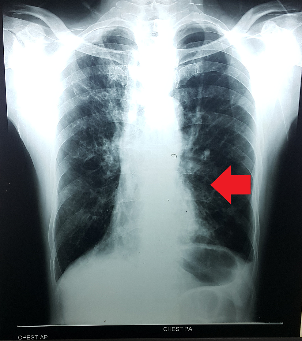
    • Situs inversus presents as situs inversus totalis, which is a complete mirror-image reversal of the circulatory system and viscera, including the heart (dextrocardia).
    • Causes no apparent health problems and often remains undiagnosed until a chest radiograph is ordered
    • Can be part of Kartagener syndrome: a triad of situs inversus, chronic sinusitis Sinusitis Sinusitis refers to inflammation of the mucosal lining of the paranasal sinuses. The condition usually occurs concurrently with inflammation of the nasal mucosa (rhinitis), a condition known as rhinosinusitis. Acute sinusitis is due to an upper respiratory infection caused by a viral, bacterial, or fungal agent. Sinusitis, and bronchiectasis Bronchiectasis Bronchiectasis is a chronic disease of the airways that results from permanent bronchial distortion. This results from a continuous cycle of inflammation, bronchial damage and dilation, impaired clearance of secretions, and recurrent infections. Bronchiectasis
  • Heterotaxy syndrome Heterotaxy syndrome Abnormal thoracoabdominal viscera arrangement (visceral heterotaxy) or malformation that involves additional congenital heart defects (e.g., heart isomerism; dextrocardia) and/or abnormal spleen (e.g., asplenia and polysplenia). Irregularities with the central nervous system, the skeleton and urinary tract are often associated with the syndrome. Asplenia (situs ambiguus):
    • Defect in right-left laterality, which results in abnormalities in arrangements of the heart, liver Liver The liver is the largest gland in the human body. The liver is found in the superior right quadrant of the abdomen and weighs approximately 1.5 kilograms. Its main functions are detoxification, metabolism, nutrient storage (e.g., iron and vitamins), synthesis of coagulation factors, formation of bile, filtration, and storage of blood. Liver: Anatomy, intestines, or spleen Spleen The spleen is the largest lymphoid organ in the body, located in the LUQ of the abdomen, superior to the left kidney and posterior to the stomach at the level of the 9th-11th ribs just below the diaphragm. The spleen is highly vascular and acts as an important blood filter, cleansing the blood of pathogens and damaged erythrocytes. Spleen: Anatomy
    • Organs may be structurally abnormal or abnormally positioned.
    • Patients Patients Individuals participating in the health care system for the purpose of receiving therapeutic, diagnostic, or preventive procedures. Clinician–Patient Relationship may exhibit asplenia Asplenia Asplenia is the absence of splenic tissue or function and can stem from several factors ranging from congenital to iatrogenic. There is a distinction between anatomic asplenia, which is due to the surgical removal of the spleen, and functional asplenia, which is due to a condition that leads to splenic atrophy, infarct, congestion, or infiltrative disease. Asplenia or polysplenia.
  • Transposition of the great arteries Arteries Arteries are tubular collections of cells that transport oxygenated blood and nutrients from the heart to the tissues of the body. The blood passes through the arteries in order of decreasing luminal diameter, starting in the largest artery (the aorta) and ending in the small arterioles. Arteries are classified into 3 types: large elastic arteries, medium muscular arteries, and small arteries and arterioles. Arteries: Histology
  • Hydrocephalus Hydrocephalus Excessive accumulation of cerebrospinal fluid within the cranium which may be associated with dilation of cerebral ventricles, intracranial. Subarachnoid Hemorrhage
  • Fertility issues:
    • Infertility Infertility Infertility is the inability to conceive in the context of regular intercourse. The most common causes of infertility in women are related to ovulatory dysfunction or tubal obstruction, whereas, in men, abnormal sperm is a common cause. Infertility in men due to live, but immotile Immotile Shigella sperm
    • Impaired fertility in women due to impaired ciliary function in the fallopian tubes Fallopian tubes The uterus, cervix, and fallopian tubes are part of the internal female reproductive system. The fallopian tubes receive an ovum after ovulation and help move it and/or a fertilized embryo toward the uterus via ciliated cells lining the tubes and peristaltic movements of its smooth muscle. Uterus, Cervix, and Fallopian Tubes: Anatomy
  • Epispadias Epispadias A birth defect due to malformation of the urethra in which the urethral opening is above its normal location. In the male, the malformed urethra generally opens on the top or the side of the penis, but the urethra can also be open the entire length of the penis. In the female, the malformed urethral opening is often between the clitoris and the labia, or in the abdomen. Penile Anomalies and Conditions 
  • Pyloric stenosis Stenosis Hypoplastic Left Heart Syndrome (HLHS)

Diagnosis

Evaluation of ciliary function

There is no gold standard diagnostic test for PCD, but the following evaluations aid in the diagnosis: 

  • Nasal nitric oxide Nitric Oxide A free radical gas produced endogenously by a variety of mammalian cells, synthesized from arginine by nitric oxide synthase. Nitric oxide is one of the endothelium-dependent relaxing factors released by the vascular endothelium and mediates vasodilation. It also inhibits platelet aggregation, induces disaggregation of aggregated platelets, and inhibits platelet adhesion to the vascular endothelium. Nitric oxide activates cytosolic guanylate cyclase and thus elevates intracellular levels of cyclic gmp. Pulmonary Hypertension Drugs (nNO) determination:
  • Assessment of ciliary motion and ultrastructure using high-speed video microscopy analysis (HSVA) and transmission electron microscopy:
    • Ciliated epithelium Epithelium The epithelium is a complex of specialized cellular organizations arranged into sheets and lining cavities and covering the surfaces of the body. The cells exhibit polarity, having an apical and a basal pole. Structures important for the epithelial integrity and function involve the basement membrane, the semipermeable sheet on which the cells rest, and interdigitations, as well as cellular junctions. Surface Epithelium: Histology obtained via nasal brushing using bronchoscopy Bronchoscopy Endoscopic examination, therapy or surgery of the bronchi. Laryngomalacia and Tracheomalacia (at least 4–6 weeks after a respiratory infection)
    • Ciliary beat frequency and pattern observed on video
    • Structural defects of cilia determined using electron microscopy
  • Cell cultures of ciliated cells:
    • Epithelia obtained using a cytology brush
    • Cilia are regrown and their ability to rotate cells is observed.
    • PCD: cells do not rotate
  • Genetic testing Genetic Testing Detection of a mutation; genotype; karyotype; or specific alleles associated with genetic traits, heritable diseases, or predisposition to a disease, or that may lead to the disease in descendants. It includes prenatal genetic testing. Myotonic Dystrophies is useful in individuals exhibiting characteristic clinical features.
  • Measures of mucociliary transport:
    • Low availability and low sensitivity
    • Rarely used
Electron microscope photograph of the ciliary structure in the bronchiole annotated

Electron microscope photograph of the ciliary structure in a bronchiole:
Left: diagram of the ciliary internal structures
Middle: cross-section image obtained using electron microscopy
Right: The microtubules are labeled.
Left image by Lecturio.
Middle and right images: “Bronchiolar area cilia cross-sections 1” by Louisa Howard, Michael Binder. License: Public Domain, edited by Lecturio.

Additional tests

  • Spirometry Spirometry Measurement of volume of air inhaled or exhaled by the lung. Pulmonary Function Tests: reveals mild-to-moderate obstructive disease with variable Variable Variables represent information about something that can change. The design of the measurement scales, or of the methods for obtaining information, will determine the data gathered and the characteristics of that data. As a result, a variable can be qualitative or quantitative, and may be further classified into subgroups. Types of Variables response to bronchodilators Bronchodilators Asthma Drugs
  • Chest X-ray X-ray Penetrating electromagnetic radiation emitted when the inner orbital electrons of an atom are excited and release radiant energy. X-ray wavelengths range from 1 pm to 10 nm. Hard x-rays are the higher energy, shorter wavelength x-rays. Soft x-rays or grenz rays are less energetic and longer in wavelength. The short wavelength end of the x-ray spectrum overlaps the gamma rays wavelength range. The distinction between gamma rays and x-rays is based on their radiation source. Pulmonary Function Tests and high-resolution CT High-resolution CT Imaging of the Lungs and Pleura ( HRCT HRCT Pulmonary Function Tests) used to evaluate worsening lung function can usually reveal:
    • Hyperinflation Hyperinflation Imaging of the Lungs and Pleura
    • Peribronchial thickening
    • Atelectasis Atelectasis Atelectasis is the partial or complete collapse of a part of the lung. Atelectasis is almost always a secondary phenomenon from conditions causing bronchial obstruction, external compression, surfactant deficiency, or scarring. Atelectasis
    • Bronchiectasis Bronchiectasis Bronchiectasis is a chronic disease of the airways that results from permanent bronchial distortion. This results from a continuous cycle of inflammation, bronchial damage and dilation, impaired clearance of secretions, and recurrent infections. Bronchiectasis (present on HRCT HRCT Pulmonary Function Tests in all adults and about 50% of children with PCD)
  • Sinus CT scan is obtained in patients Patients Individuals participating in the health care system for the purpose of receiving therapeutic, diagnostic, or preventive procedures. Clinician–Patient Relationship with chronic sinusitis Sinusitis Sinusitis refers to inflammation of the mucosal lining of the paranasal sinuses. The condition usually occurs concurrently with inflammation of the nasal mucosa (rhinitis), a condition known as rhinosinusitis. Acute sinusitis is due to an upper respiratory infection caused by a viral, bacterial, or fungal agent. Sinusitis who present with the following signs and symptoms:
    • Persistent mucociliary drainage
    • Nasal obstruction or blockage
    • Facial pain Facial pain Pain in the facial region including orofacial pain and craniofacial pain. Associated conditions include local inflammatory and neoplastic disorders and neuralgic syndromes involving the trigeminal, facial, and glossopharyngeal nerves. Conditions which feature recurrent or persistent facial pain as the primary manifestation of disease are referred to as facial pain syndromes. Trigeminal Neuralgia or pressure
Kartagener syndrome

Chest X-ray diagnosing Kartagener syndrome:
Mild cardiomegaly with pulmonary congestion and dextrocardia (arrow) noted in a patient with a form of primary ciliary dyskinesia. Kartagener syndrome (primary ciliary dyskinesia: situs inversus, chronic sinusitis, and bronchiectasis)

Image: “Chest X-ray” by Madeeha Subhan, Waleed Sadiq. License: CC BY 3.0

Management and Prognosis

Management

Treatment is individualized and guided by the clinical presentation of the patient.

  • Main goals of management:
    • Control and treat all infections Infections Invasion of the host organism by microorganisms or their toxins or by parasites that can cause pathological conditions or diseases. Chronic Granulomatous Disease.
    • Remove trapped mucus from the lungs Lungs Lungs are the main organs of the respiratory system. Lungs are paired viscera located in the thoracic cavity and are composed of spongy tissue. The primary function of the lungs is to oxygenate blood and eliminate CO2. Lungs: Anatomy and airways.
    • Treatment techniques are based on the experience of treating cystic Cystic Fibrocystic Change fibrosis Fibrosis Any pathological condition where fibrous connective tissue invades any organ, usually as a consequence of inflammation or other injury. Bronchiolitis Obliterans and other forms of bronchiectasis Bronchiectasis Bronchiectasis is a chronic disease of the airways that results from permanent bronchial distortion. This results from a continuous cycle of inflammation, bronchial damage and dilation, impaired clearance of secretions, and recurrent infections. Bronchiectasis.
  • Monitoring:
    • Close and continuous follow-up 
    • Spirometry Spirometry Measurement of volume of air inhaled or exhaled by the lung. Pulmonary Function Tests at every visit (average 1–3 times annually) starting at 6 years of age
  • Management depends on the clinical course of the patient:
    • Bronchiectasis Bronchiectasis Bronchiectasis is a chronic disease of the airways that results from permanent bronchial distortion. This results from a continuous cycle of inflammation, bronchial damage and dilation, impaired clearance of secretions, and recurrent infections. Bronchiectasis:
      • Daily chest physiotherapy Physiotherapy Spinal Stenosis
      • Use of selective antibiotics (guided by sputum cultures) to help clear secretions and decrease microbial load
      • For acute exacerbation of bronchiectasis Bronchiectasis Bronchiectasis is a chronic disease of the airways that results from permanent bronchial distortion. This results from a continuous cycle of inflammation, bronchial damage and dilation, impaired clearance of secretions, and recurrent infections. Bronchiectasis: oral antibiotics
      • For P. aeruginosa P. aeruginosa A species of gram-negative, aerobic, rod-shaped bacteria commonly isolated from clinical specimens (wound, burn, and urinary tract infections). It is also found widely distributed in soil and water. P. Aeruginosa is a major agent of nosocomial infection. Pseudomonas infections Infections Invasion of the host organism by microorganisms or their toxins or by parasites that can cause pathological conditions or diseases. Chronic Granulomatous Disease or severe pneumonia Pneumonia Pneumonia or pulmonary inflammation is an acute or chronic inflammation of lung tissue. Causes include infection with bacteria, viruses, or fungi. In more rare cases, pneumonia can also be caused through toxic triggers through inhalation of toxic substances, immunological processes, or in the course of radiotherapy. Pneumonia: IV antibiotics
      • Preventive antibiotic therapy for recurrent exacerbations in patients Patients Individuals participating in the health care system for the purpose of receiving therapeutic, diagnostic, or preventive procedures. Clinician–Patient Relationship ≥ 7 years of age is usually using macrolides Macrolides Macrolides and ketolides are antibiotics that inhibit bacterial protein synthesis by binding to the 50S ribosomal subunit and blocking transpeptidation. These antibiotics have a broad spectrum of antimicrobial activity but are best known for their coverage of atypical microorganisms. Macrolides and Ketolides
      • Vaccination Vaccination Vaccination is the administration of a substance to induce the immune system to develop protection against a disease. Unlike passive immunization, which involves the administration of pre-performed antibodies, active immunization constitutes the administration of a vaccine to stimulate the body to produce its own antibodies. Vaccination against influenza Influenza Influenza viruses are members of the Orthomyxoviridae family and the causative organisms of influenza, a highly contagious febrile respiratory disease. There are 3 primary influenza viruses (A, B, and C) and various subtypes, which are classified based on their virulent surface antigens, hemagglutinin (HA) and neuraminidase (NA). Influenza typically presents with a fever, myalgia, headache, and symptoms of an upper respiratory infection. Influenza Viruses/Influenza and pneumococcus
      • Smoking Smoking Willful or deliberate act of inhaling and exhaling smoke from burning substances or agents held by hand. Interstitial Lung Diseases cessation
    • Chronic rhinosinusitis and nasal polyposis:
      • Nasal saline lavage
      • Intranasal glucocorticoids Glucocorticoids Glucocorticoids are a class within the corticosteroid family. Glucocorticoids are chemically and functionally similar to endogenous cortisol. There are a wide array of indications, which primarily benefit from the antiinflammatory and immunosuppressive effects of this class of drugs. Glucocorticoids for nasal polyposis
      • Antibiotics for exacerbations
    • Chronic otitis media with effusion:
      • Can lead to hearing loss Hearing loss Hearing loss, also known as hearing impairment, is any degree of impairment in the ability to apprehend sound as determined by audiometry to be below normal hearing thresholds. Clinical presentation may occur at birth or as a gradual loss of hearing with age, including a short-term or sudden loss at any point. Hearing Loss
      • Consider the use of a tympanostomy tube.
    • Male infertility Infertility Infertility is the inability to conceive in the context of regular intercourse. The most common causes of infertility in women are related to ovulatory dysfunction or tubal obstruction, whereas, in men, abnormal sperm is a common cause. Infertility: Consider in vitro fertilization Fertilization To undergo fertilization, the sperm enters the uterus, travels towards the ampulla of the fallopian tube, and encounters the oocyte. The zona pellucida (the outer layer of the oocyte) deteriorates along with the zygote, which travels towards the uterus and eventually forms a blastocyst, allowing for implantation to occur. Fertilization and First Week techniques.
  • End-stage respiratory insufficiency:
    • Bilateral lung transplantation Lung transplantation The transference of either one or both of the lungs from one human or animal to another. Organ Transplantation
    • In case of situs inversus: heart-lung transplantation

Prognosis Prognosis A prediction of the probable outcome of a disease based on a individual’s condition and the usual course of the disease as seen in similar situations. Non-Hodgkin Lymphomas

  • Patients Patients Individuals participating in the health care system for the purpose of receiving therapeutic, diagnostic, or preventive procedures. Clinician–Patient Relationship generally have a normal lifespan.
  • Slower rate of reduction in lung function than cystic Cystic Fibrocystic Change fibrosis Fibrosis Any pathological condition where fibrous connective tissue invades any organ, usually as a consequence of inflammation or other injury. Bronchiolitis Obliterans
  • Repeated and chronic infections Infections Invasion of the host organism by microorganisms or their toxins or by parasites that can cause pathological conditions or diseases. Chronic Granulomatous Disease may impact the ability to work full-time.

Differential Diagnosis

  • Cystic Cystic Fibrocystic Change fibrosis Fibrosis Any pathological condition where fibrous connective tissue invades any organ, usually as a consequence of inflammation or other injury. Bronchiolitis Obliterans: an autosomal recessive Autosomal recessive Autosomal inheritance, both dominant and recessive, refers to the transmission of genes from the 22 autosomal chromosomes. Autosomal recessive diseases are only expressed when 2 copies of the recessive allele are inherited. Autosomal Recessive and Autosomal Dominant Inheritance disorder caused by mutations in the CFTR gene Gene A category of nucleic acid sequences that function as units of heredity and which code for the basic instructions for the development, reproduction, and maintenance of organisms. Basic Terms of Genetics leading to dysfunctional chloride Chloride Inorganic compounds derived from hydrochloric acid that contain the Cl- ion. Electrolytes channels Channels The Cell: Cell Membrane. The defective chloride Chloride Inorganic compounds derived from hydrochloric acid that contain the Cl- ion. Electrolytes channels Channels The Cell: Cell Membrane lead to hyperviscous mucus and the accumulation of secretions. Cystic Cystic Fibrocystic Change fibrosis Fibrosis Any pathological condition where fibrous connective tissue invades any organ, usually as a consequence of inflammation or other injury. Bronchiolitis Obliterans commonly presents with recurrent respiratory infections Infections Invasion of the host organism by microorganisms or their toxins or by parasites that can cause pathological conditions or diseases. Chronic Granulomatous Disease, pancreatic insufficiency, and failure to thrive Failure to Thrive Failure to thrive (FTT), or faltering growth, describes suboptimal weight gain and growth in children. The majority of cases are due to inadequate caloric intake; however, genetic, infectious, and oncological etiologies are also common. Failure to Thrive. Diagnosis is using a sweat chloride Chloride Inorganic compounds derived from hydrochloric acid that contain the Cl- ion. Electrolytes test followed by genetic testing Genetic Testing Detection of a mutation; genotype; karyotype; or specific alleles associated with genetic traits, heritable diseases, or predisposition to a disease, or that may lead to the disease in descendants. It includes prenatal genetic testing. Myotonic Dystrophies. Cystic Cystic Fibrocystic Change fibrosis Fibrosis Any pathological condition where fibrous connective tissue invades any organ, usually as a consequence of inflammation or other injury. Bronchiolitis Obliterans has a worse prognosis Prognosis A prediction of the probable outcome of a disease based on a individual’s condition and the usual course of the disease as seen in similar situations. Non-Hodgkin Lymphomas than PCD. 
  • IgG IgG The major immunoglobulin isotype class in normal human serum. There are several isotype subclasses of igg, for example, igg1, igg2a, and igg2b. Hypersensitivity Pneumonitis subclass deficiency: a decrease in 1 or more subclasses of IgG IgG The major immunoglobulin isotype class in normal human serum. There are several isotype subclasses of igg, for example, igg1, igg2a, and igg2b. Hypersensitivity Pneumonitis antibodies Antibodies Immunoglobulins (Igs), also known as antibodies, are glycoprotein molecules produced by plasma cells that act in immune responses by recognizing and binding particular antigens. The various Ig classes are IgG (the most abundant), IgM, IgE, IgD, and IgA, which differ in their biologic features, structure, target specificity, and distribution. Immunoglobulins: Types and Functions with normal levels of other immunoglobulins Immunoglobulins Immunoglobulins (Igs), also known as antibodies, are glycoprotein molecules produced by plasma cells that act in immune responses by recognizing and binding particular antigens. The various Ig classes are IgG (the most abundant), IgM, IgE, IgD, and IgA, which differ in their biologic features, structure, target specificity, and distribution. Immunoglobulins: Types and Functions. Patients Patients Individuals participating in the health care system for the purpose of receiving therapeutic, diagnostic, or preventive procedures. Clinician–Patient Relationship are often asymptomatic but may have recurrent infections Recurrent infections Common Variable Immunodeficiency (CVID) of the ears, bronchi Bronchi The larger air passages of the lungs arising from the terminal bifurcation of the trachea. They include the largest two primary bronchi which branch out into secondary bronchi, and tertiary bronchi which extend into bronchioles and pulmonary alveoli. Bronchial Tree: Anatomy, sinuses, and lungs Lungs Lungs are the main organs of the respiratory system. Lungs are paired viscera located in the thoracic cavity and are composed of spongy tissue. The primary function of the lungs is to oxygenate blood and eliminate CO2. Lungs: Anatomy. Updated vaccinations and antibiotics are part of the management. Immunoglobulin therapy is an option if initial management is not successful in controlling infections Infections Invasion of the host organism by microorganisms or their toxins or by parasites that can cause pathological conditions or diseases. Chronic Granulomatous Disease.
  • Granulomatosis with polyangiitis Granulomatosis with Polyangiitis A multisystemic disease of a complex genetic background. It is characterized by inflammation of the blood vessels (vasculitis) leading to damage in any number of organs. The common features include granulomatous inflammation of the respiratory tract and kidneys. Most patients have measurable autoantibodies (antineutrophil cytoplasmic antibodies) against myeloblastin. Granulomatosis with Polyangiitis: a type of vasculitis Vasculitis Inflammation of any one of the blood vessels, including the arteries; veins; and rest of the vasculature system in the body. Systemic Lupus Erythematosus that results in damage to various organ systems, particularly the kidneys Kidneys The kidneys are a pair of bean-shaped organs located retroperitoneally against the posterior wall of the abdomen on either side of the spine. As part of the urinary tract, the kidneys are responsible for blood filtration and excretion of water-soluble waste in the urine. Kidneys: Anatomy and respiratory tract. Granulomatosis with polyangiitis Granulomatosis with Polyangiitis A multisystemic disease of a complex genetic background. It is characterized by inflammation of the blood vessels (vasculitis) leading to damage in any number of organs. The common features include granulomatous inflammation of the respiratory tract and kidneys. Most patients have measurable autoantibodies (antineutrophil cytoplasmic antibodies) against myeloblastin. Granulomatosis with Polyangiitis can lead to constitutional symptoms Constitutional Symptoms Antineutrophil Cytoplasmic Antibody (ANCA)-Associated Vasculitis, chronic upper respiratory infections Infections Invasion of the host organism by microorganisms or their toxins or by parasites that can cause pathological conditions or diseases. Chronic Granulomatous Disease, nosebleeds Nosebleeds Bleeding from the nose. Granulomatosis with Polyangiitis, ear pain Ear Pain Acute Otitis Media, hemoptysis Hemoptysis Hemoptysis is defined as the expectoration of blood originating in the lower respiratory tract. Hemoptysis is a consequence of another disease process and can be classified as either life threatening or non-life threatening. Hemoptysis can result in significant morbidity and mortality due to both drowning (reduced gas exchange as the lungs fill with blood) and hemorrhagic shock. Hemoptysis, and/or hematuria Hematuria Presence of blood in the urine. Renal Cell Carcinoma. Patients Patients Individuals participating in the health care system for the purpose of receiving therapeutic, diagnostic, or preventive procedures. Clinician–Patient Relationship can progress to kidney failure. Granulomatosis with polyangiitis Granulomatosis with Polyangiitis A multisystemic disease of a complex genetic background. It is characterized by inflammation of the blood vessels (vasculitis) leading to damage in any number of organs. The common features include granulomatous inflammation of the respiratory tract and kidneys. Most patients have measurable autoantibodies (antineutrophil cytoplasmic antibodies) against myeloblastin. Granulomatosis with Polyangiitis demonstrates c-ANCA c-ANCA Antineutrophil Cytoplasmic Antibody (ANCA)-Associated Vasculitis positivity and is treated with corticosteroids Corticosteroids Chorioretinitis along with an immunosuppressant.

References

  1. Bergström, S. (2021). Primary ciliary dyskinesia (immotile-cilia syndrome). UpToDate. Retrieved March 2, 2020, from https://www.uptodate.com/contents/primary-ciliary-dyskinesia-immotile-cilia-syndrome
  2. Husain, A. (2021). The Lung. In Kumar V, Abbas AK, Aster JC. Robbins & Cotran Pathologic Basis of Disease. Philadelphia, PA: Elsevier Saunders.
  3. MedlinePlus (2020). Primary ciliary dyskinesia. Retrieved March 2, 2020, from https://medlineplus.gov/genetics/condition/primary-ciliary-dyskinesia/
  4. National Heart, Lung, and Blood Institute. Primary Ciliary Dyskinesia. Retrieved March 2, 2020, from https://www.nhlbi.nih.gov/health-topics/primary-ciliary-dyskinesia.
  5. National Organization for Rare Disorders (2020). Primary Ciliary Dyskinesia. Retrieved March 2, 2020, from https://rarediseases.org/rare-diseases/primary-ciliary-dyskinesia/

Create your free account or log in to continue reading!

Sign up now and get free access to Lecturio with concept pages, medical videos, and questions for your medical education.

User Reviews

Details