Postpartum Hemorrhage

Postpartum hemorrhage is one of the most common and deadly obstetric complications. Since 2017, postpartum hemorrhage has been defined as blood loss greater than 1,000 mL for both cesarean and vaginal deliveries, or excessive blood loss with signs of hemodynamic instability. Blood loss of more than 500 mL following a vaginal delivery is considered abnormal, though no longer classified as postpartum hemorrhage. The most common cause of postpartum hemorrhage is uterine atony. Retained products of conception, trauma, and coagulopathies Coagulopathies Hemothorax may also cause postpartum hemorrhage. Appropriate management requires rapid identification Identification Defense Mechanisms of the cause of the hemorrhage, medical and/or surgical correction, and patient resuscitation Resuscitation The restoration to life or consciousness of one apparently dead. . Neonatal Respiratory Distress Syndrome.

Last updated: Sep 29, 2022

Editorial responsibility: Stanley Oiseth, Lindsay Jones, Evelin Maza

Definition and Epidemiology

Definition

Postpartum hemorrhage is defined as excessive blood loss following the delivery of an infant > 20 weeks gestational age Gestational age The age of the conceptus, beginning from the time of fertilization. In clinical obstetrics, the gestational age is often estimated as the time from the last day of the last menstruation which is about 2 weeks before ovulation and fertilization. Pregnancy: Diagnosis, Physiology, and Care.

  • The classic definition of postpartum hemorrhage was based on mode of delivery:
    • Blood loss > 500 mL following a vaginal delivery 
    • Blood loss > 1,000 mL following a cesarean section
  • The newer meaning of postpartum hemorrhage was defined by the American College of Obstetricians and Gynecologists in 2017:
    • Postpartum hemorrhage:
      • Cumulative blood loss ≥ 1,000 mL regardless of delivery route, OR
      • Bleeding associated with signs/symptoms of hypovolemia Hypovolemia Sepsis in Children within 24 hours of delivery
    • While reducing cases labeled as postpartum hemorrhage, blood loss of > 500 mL following a vaginal delivery is still considered abnormal and should prompt investigation.
  • Postpartum hemorrhage can be primary or secondary:
    • Primary postpartum hemorrhage: occurs within 24 hours of delivery
    • Secondary postpartum hemorrhage: occurs between 24 hours and 12 weeks after delivery

Epidemiology

  • Incidence Incidence The number of new cases of a given disease during a given period in a specified population. It also is used for the rate at which new events occur in a defined population. It is differentiated from prevalence, which refers to all cases in the population at a given time. Measures of Disease Frequency
    • 1%–10% of deliveries
    • Higher in sites using quantitative (compared to estimated) blood loss measurements
  • Leading cause of maternal mortality Mortality All deaths reported in a given population. Measures of Health Status:
    • Approximately 11% of pregnancy-related mortality Mortality All deaths reported in a given population. Measures of Health Status in developed countries
    • Up to 60% of maternal deaths in developing countries
  • Uterine atony accounts for 70%–80% of all postpartum hemorrhage

Etiology

Primary postpartum hemorrhage

The primary etiologies can be remembered as “the 4 Ts” (tone, tissue, trauma, and thrombin Thrombin An enzyme formed from prothrombin that converts fibrinogen to fibrin. Hemostasis).
  • Tone (uterine atony):
    • Inability of the uterus Uterus The uterus, cervix, and fallopian tubes are part of the internal female reproductive system. The uterus has a thick wall made of smooth muscle (the myometrium) and an inner mucosal layer (the endometrium). The most inferior portion of the uterus is the cervix, which connects the uterine cavity to the vagina. Uterus, Cervix, and Fallopian Tubes: Anatomy to contract effectively following delivery
    • The most common cause of postpartum hemorrhage
    • Risk factors:
      • Prolonged labor (uterine muscle fatigue Fatigue The state of weariness following a period of exertion, mental or physical, characterized by a decreased capacity for work and reduced efficiency to respond to stimuli. Fibromyalgia)
      • Intraamniotic infections Infections Invasion of the host organism by microorganisms or their toxins or by parasites that can cause pathological conditions or diseases. Chronic Granulomatous Disease (e.g., chorioamnionitis Chorioamnionitis Chorioamnionitis, commonly referred to as intraamniotic infection (IAI), is a common obstetric complication involving infection and inflammation of the fetal membranes, amniotic fluid, placenta, or the fetus itself. Chorioamnionitis is typically caused by a polymicrobial infection that ascends from the lower genitourinary tract. Chorioamnionitis)
      • Uterine overdistension: a multiple pregnancy Pregnancy The status during which female mammals carry their developing young (embryos or fetuses) in utero before birth, beginning from fertilization to birth. Pregnancy: Diagnosis, Physiology, and Care (e.g., twins), polyhydramnios Polyhydramnios Polyhydramnios is a pathological excess of amniotic fluid. Common causes of polyhydramnios include fetal anomalies, gestational diabetes, multiple gestations, and congenital infections. Patients are often asymptomatic but may present with dyspnea, extremity swelling, or abdominal distention. Polyhydramnios, large for gestational age Gestational age The age of the conceptus, beginning from the time of fertilization. In clinical obstetrics, the gestational age is often estimated as the time from the last day of the last menstruation which is about 2 weeks before ovulation and fertilization. Pregnancy: Diagnosis, Physiology, and Care infants
      • Grand multiparity
      • Uterine inversion
      • General anesthesia General anesthesia Procedure in which patients are induced into an unconscious state through use of various medications so that they do not feel pain during surgery. Anesthesiology: History and Basic Concepts
  • Tissue (retained products of conception):
    • Placenta Placenta A highly vascularized mammalian fetal-maternal organ and major site of transport of oxygen, nutrients, and fetal waste products. It includes a fetal portion (chorionic villi) derived from trophoblasts and a maternal portion (decidua) derived from the uterine endometrium. The placenta produces an array of steroid, protein and peptide hormones (placental hormones). Placenta, Umbilical Cord, and Amniotic Cavity tear during the 3rd stage of labor 3rd stage of labor The final period of obstetric labor that is from the expulsion of the fetus to the expulsion of the placenta. Normal and Abnormal Labor
    • Abnormal placentation:
      • Placenta accreta Placenta Accreta Abnormal placentation in which all or parts of the placenta are attached directly to the myometrium due to a complete or partial absence of decidua. It is associated with postpartum hemorrhage because of the failure of placental separation. Placental Abnormalities
      • Placenta previa Placenta Previa Abnormal placentation in which the placenta implants in the lower segment of the uterus (the zone of dilation) and may cover part or all of the opening of the cervix. It is often associated with serious antepartum bleeding and premature labor. Placental Abnormalities
  • Trauma: 
    • Lacerations:
      • Vagina Vagina The vagina is the female genital canal, extending from the vulva externally to the cervix uteri internally. The structures have sexual, reproductive, and urinary functions and a rich blood supply, mainly arising from the internal iliac artery. Vagina, Vulva, and Pelvic Floor: Anatomy
      • Perineum Perineum The body region lying between the genital area and the anus on the surface of the trunk, and to the shallow compartment lying deep to this area that is inferior to the pelvic diaphragm. The surface area is between the vulva and the anus in the female, and between the scrotum and the anus in the male. Vagina, Vulva, and Pelvic Floor: Anatomy
      • Cervix Cervix The uterus, cervix, and fallopian tubes are part of the internal female reproductive system. The most inferior portion of the uterus is the cervix, which connects the uterine cavity to the vagina. Externally, the cervix is lined by stratified squamous cells; however, the cervical canal is lined by columnar epithelium. Uterus, Cervix, and Fallopian Tubes: Anatomy
    • Lateral extension Extension Examination of the Upper Limbs of the uterine incision into the uterine arteries Arteries Arteries are tubular collections of cells that transport oxygenated blood and nutrients from the heart to the tissues of the body. The blood passes through the arteries in order of decreasing luminal diameter, starting in the largest artery (the aorta) and ending in the small arterioles. Arteries are classified into 3 types: large elastic arteries, medium muscular arteries, and small arteries and arterioles. Arteries: Histology during cesarean section
    • Uterine rupture Uterine Rupture A complete separation or tear in the wall of the uterus with or without expulsion of the fetus. It may be due to injuries, multiple pregnancies, large fetus, previous scarring, or obstruction. Antepartum Hemorrhage (during a vaginal birth after cesarean delivery Cesarean Delivery Cesarean delivery (CD) is the operative delivery of ≥ 1 infants through a surgical incision in the maternal abdomen and uterus. Cesarean deliveries may be indicated for a number of either maternal or fetal reasons, most commonly including fetal intolerance to labor, arrest of labor, a history of prior uterine surgery, fetal malpresentation, and placental abnormalities. Cesarean Delivery through a prior uterine incision)
    • Hematomas
    • Risk factors:
      • Operative vaginal delivery Operative Vaginal Delivery Operative vaginal delivery is the use of obstetric forceps or a vacuum extractor to effect delivery of a fetus. Complications during Childbirth
      • Precipitous delivery
      • Emergent cesarean delivery Cesarean Delivery Cesarean delivery (CD) is the operative delivery of ≥ 1 infants through a surgical incision in the maternal abdomen and uterus. Cesarean deliveries may be indicated for a number of either maternal or fetal reasons, most commonly including fetal intolerance to labor, arrest of labor, a history of prior uterine surgery, fetal malpresentation, and placental abnormalities. Cesarean Delivery
  • Thrombin Thrombin An enzyme formed from prothrombin that converts fibrinogen to fibrin. Hemostasis disorders ( coagulopathies Coagulopathies Hemothorax):
    • DIC DIC Disseminated intravascular coagulation (DIC) is a condition characterized by systemic bodywide activation of the coagulation cascade. This cascade results in both widespread microvascular thrombi contributing to multiple organ dysfunction and consumption of clotting factors and platelets, leading to hemorrhage. Disseminated Intravascular Coagulation, which is more likely to occur with:
      • Intrauterine fetal demise (IUFD)
      • Placental abruption Placental Abruption Premature separation of the normally implanted placenta from the uterus. Signs of varying degree of severity include uterine bleeding, uterine muscle hypertonia, and fetal distress or fetal death. Antepartum Hemorrhage
      • Amniotic fluid embolism Amniotic Fluid Embolism Blocking of maternal circulation by amniotic fluid that is forced into uterine veins by strong uterine contraction near the end of pregnancy. It is characterized by the sudden onset of severe respiratory distress and hypotension that can lead to maternal death. Complications during Childbirth
      • Sepsis Sepsis Systemic inflammatory response syndrome with a proven or suspected infectious etiology. When sepsis is associated with organ dysfunction distant from the site of infection, it is called severe sepsis. When sepsis is accompanied by hypotension despite adequate fluid infusion, it is called septic shock. Sepsis and Septic Shock
    • Hemolysis, elevated liver Liver The liver is the largest gland in the human body. The liver is found in the superior right quadrant of the abdomen and weighs approximately 1.5 kilograms. Its main functions are detoxification, metabolism, nutrient storage (e.g., iron and vitamins), synthesis of coagulation factors, formation of bile, filtration, and storage of blood. Liver: Anatomy enzymes Enzymes Enzymes are complex protein biocatalysts that accelerate chemical reactions without being consumed by them. Due to the body’s constant metabolic needs, the absence of enzymes would make life unsustainable, as reactions would occur too slowly without these molecules. Basics of Enzymes, and low platelet count (HELLP) syndrome
    • Gestational thrombocytopenia Gestational Thrombocytopenia Hypocoagulable Conditions
    • Idiopathic Idiopathic Dermatomyositis thrombocytopenic purpura
    • Von Willebrand disease Von Willebrand disease Von Willebrand disease (vWD) is a bleeding disorder characterized by a qualitative or quantitative deficiency of von Willebrand factor (vWF). Von Willebrand factor is a multimeric protein involved in the plate adhesion phase of hemostasis by forming a bridge between platelets and damaged portions of the vessel wall. Von Willebrand Disease ( von Willebrand factor von Willebrand factor A high-molecular-weight plasma protein, produced by endothelial cells and megakaryocytes, that is part of the factor VIII/von Willebrand factor complex. The von Willebrand factor has receptors for collagen, platelets, and ristocetin activity as well as the immunologically distinct antigenic determinants. It functions in adhesion of platelets to collagen and hemostatic plug formation. The prolonged bleeding time in von Willebrand diseases is due to the deficiency of this factor. Hemostasis rapidly decreases in delivery)
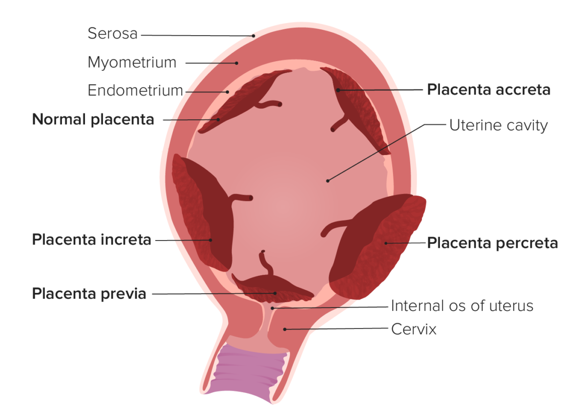
Types of abnormal placentation

Types of abnormal placentation.
In placenta accreta, the placenta attaches (at least in part) to the underlying myometrium. In placenta increta, the placental villi actually invade into the myometrium. In placenta percreta, the placental villi invade through the entire uterine wall and may invade surrounding structures (e.g., the bladder). Placenta previa occurs when the placenta covers the internal cervical os.

Image by Lecturio. License: CC BY-NC-SA 4.0

Secondary postpartum hemorrhage

  • Uterine infection ( postpartum endometritis Postpartum Endometritis Inflammation of the endometrium, usually caused by intrauterine infections. Endometritis is the most common cause of postpartum fever. Postpartum Endometritis)
  • Retained products of conception
  • Subinvolution of the uterus Uterus The uterus, cervix, and fallopian tubes are part of the internal female reproductive system. The uterus has a thick wall made of smooth muscle (the myometrium) and an inner mucosal layer (the endometrium). The most inferior portion of the uterus is the cervix, which connects the uterine cavity to the vagina. Uterus, Cervix, and Fallopian Tubes: Anatomy
  • Inherited coagulation defects

Pathophysiology

Postpartum hemorrhage due to a nontraumatic etiology results when 1 or both of the primary mechanisms required for normal postpartum hemostasis Hemostasis Hemostasis refers to the innate, stepwise body processes that occur following vessel injury, resulting in clot formation and cessation of bleeding. Hemostasis occurs in 2 phases, namely, primary and secondary. Primary hemostasis involves forming a plug that stops the bleeding temporarily. Secondary hemostasis involves the activation of the coagulation cascade. Hemostasis are disrupted. The 2 primary mechanisms are mechanical hemostasis Hemostasis Hemostasis refers to the innate, stepwise body processes that occur following vessel injury, resulting in clot formation and cessation of bleeding. Hemostasis occurs in 2 phases, namely, primary and secondary. Primary hemostasis involves forming a plug that stops the bleeding temporarily. Secondary hemostasis involves the activation of the coagulation cascade. Hemostasis and hemostatic factors.

Mechanical hemostasis Hemostasis Hemostasis refers to the innate, stepwise body processes that occur following vessel injury, resulting in clot formation and cessation of bleeding. Hemostasis occurs in 2 phases, namely, primary and secondary. Primary hemostasis involves forming a plug that stops the bleeding temporarily. Secondary hemostasis involves the activation of the coagulation cascade. Hemostasis

  • Normal contraction of the myometrium → compression Compression Blunt Chest Trauma of myometrial vasculature in the crisscross latticework of the smooth muscle fibers of the uterus Uterus The uterus, cervix, and fallopian tubes are part of the internal female reproductive system. The uterus has a thick wall made of smooth muscle (the myometrium) and an inner mucosal layer (the endometrium). The most inferior portion of the uterus is the cervix, which connects the uterine cavity to the vagina. Uterus, Cervix, and Fallopian Tubes: Anatomy
  • Disrupted in:
    • Uterine atony:
      • Failure of myometrial contraction → continued uterine blood flow Blood flow Blood flow refers to the movement of a certain volume of blood through the vasculature over a given unit of time (e.g., mL per minute). Vascular Resistance, Flow, and Mean Arterial Pressure → hemorrhage from the exposed vessels supplying the placental bed
      • Anything impairing postpartum contraction of the uterus Uterus The uterus, cervix, and fallopian tubes are part of the internal female reproductive system. The uterus has a thick wall made of smooth muscle (the myometrium) and an inner mucosal layer (the endometrium). The most inferior portion of the uterus is the cervix, which connects the uterine cavity to the vagina. Uterus, Cervix, and Fallopian Tubes: Anatomy leads to postpartum hemorrhage.
    • Retained products of conception:
      • Blood flows into the maternal side of the placenta Placenta A highly vascularized mammalian fetal-maternal organ and major site of transport of oxygen, nutrients, and fetal waste products. It includes a fetal portion (chorionic villi) derived from trophoblasts and a maternal portion (decidua) derived from the uterine endometrium. The placenta produces an array of steroid, protein and peptide hormones (placental hormones). Placenta, Umbilical Cord, and Amniotic Cavity and out through the placental fragment
      • Retained products prevent the uterus Uterus The uterus, cervix, and fallopian tubes are part of the internal female reproductive system. The uterus has a thick wall made of smooth muscle (the myometrium) and an inner mucosal layer (the endometrium). The most inferior portion of the uterus is the cervix, which connects the uterine cavity to the vagina. Uterus, Cervix, and Fallopian Tubes: Anatomy from fully contracting → uterus Uterus The uterus, cervix, and fallopian tubes are part of the internal female reproductive system. The uterus has a thick wall made of smooth muscle (the myometrium) and an inner mucosal layer (the endometrium). The most inferior portion of the uterus is the cervix, which connects the uterine cavity to the vagina. Uterus, Cervix, and Fallopian Tubes: Anatomy fills up with blood → uterine atony

Hemostatic factors

  • Clotting is facilitated by:
    • Factors released from the uterine decidua (i.e., tissue factor)
    • Systemic, circulating coagulation factors Coagulation factors Endogenous substances, usually proteins, that are involved in the blood coagulation process. Hemostasis and platelets Platelets Platelets are small cell fragments involved in hemostasis. Thrombopoiesis takes place primarily in the bone marrow through a series of cell differentiation and is influenced by several cytokines. Platelets are formed after fragmentation of the megakaryocyte cytoplasm. Platelets: Histology
  • Disrupted in coagulopathies Coagulopathies Hemothorax (inadequate hemostatic factors to stop the bleeding of the placental bed)

Clinical Presentation and Diagnosis

Most patients Patients Individuals participating in the health care system for the purpose of receiving therapeutic, diagnostic, or preventive procedures. Clinician–Patient Relationship present with heavy bleeding and/or signs of hemodynamic instability. Diagnosis is usually clinical.

Clinical presentation

  • Heavy vaginal bleeding (often dramatic)
  • Hemodynamic instability:
    • Tachycardia Tachycardia Abnormally rapid heartbeat, usually with a heart rate above 100 beats per minute for adults. Tachycardia accompanied by disturbance in the cardiac depolarization (cardiac arrhythmia) is called tachyarrhythmia. Sepsis in Children
    • Tachypnea Tachypnea Increased respiratory rate. Pulmonary Examination
    • Hypotension Hypotension Hypotension is defined as low blood pressure, specifically < 90/60 mm Hg, and is most commonly a physiologic response. Hypotension may be mild, serious, or life threatening, depending on the cause. Hypotension
    • ↓ Urine output 
    • Skin Skin The skin, also referred to as the integumentary system, is the largest organ of the body. The skin is primarily composed of the epidermis (outer layer) and dermis (deep layer). The epidermis is primarily composed of keratinocytes that undergo rapid turnover, while the dermis contains dense layers of connective tissue. Skin: Structure and Functions findings: 
      • Cool and clammy
      • Delayed capillary refill
      • Pale
    • Mental status changes

Diagnosis

  • On exam look for:
    • Uterine atony: soft, boggy uterus Uterus The uterus, cervix, and fallopian tubes are part of the internal female reproductive system. The uterus has a thick wall made of smooth muscle (the myometrium) and an inner mucosal layer (the endometrium). The most inferior portion of the uterus is the cervix, which connects the uterine cavity to the vagina. Uterus, Cervix, and Fallopian Tubes: Anatomy
    • Uterine inversion: firm, round mass Mass Three-dimensional lesion that occupies a space within the breast Imaging of the Breast in the vagina Vagina The vagina is the female genital canal, extending from the vulva externally to the cervix uteri internally. The structures have sexual, reproductive, and urinary functions and a rich blood supply, mainly arising from the internal iliac artery. Vagina, Vulva, and Pelvic Floor: Anatomy
    • Retained products of conception: 
      • Visible trailing membranes
      • Palpable irregularities within the uterus Uterus The uterus, cervix, and fallopian tubes are part of the internal female reproductive system. The uterus has a thick wall made of smooth muscle (the myometrium) and an inner mucosal layer (the endometrium). The most inferior portion of the uterus is the cervix, which connects the uterine cavity to the vagina. Uterus, Cervix, and Fallopian Tubes: Anatomy
    • Lacerations/trauma
  • Monitor vital signs for signs of hemodynamic instability.
  • Measure blood loss:
    • Clinical estimate of blood loss by visual assessment 
    • Quantitative blood loss measurements:
      • Volumetric assessment of blood is collected via suction or in graduated drapes.
      • Weight assessment of blood is collected on surgical sponges.
      • Efforts are made to collect and quantify all blood lost.
  • Imaging:
    • Ultrasound is the modality of choice.
    • May be help identify retained products of conception (especially in secondary postpartum hemorrhage)
  • Laboratory tests:
    • Useful for management, but not diagnosis
    • The following should be ordered once postpartum hemorrhage is diagnosed:
      • Type and crossmatch to prepare for a blood transfusion
      • CBC
      • Coagulation panel: PT, PTT, fibrinogen Fibrinogen Plasma glycoprotein clotted by thrombin, composed of a dimer of three non-identical pairs of polypeptide chains (alpha, beta, gamma) held together by disulfide bonds. Fibrinogen clotting is a sol-gel change involving complex molecular arrangements: whereas fibrinogen is cleaved by thrombin to form polypeptides a and b, the proteolytic action of other enzymes yields different fibrinogen degradation products. Hemostasis

Classification of hemorrhage

The 4 classes of hemorrhage are described by the Advanced Trauma Life Support manual, which can also classify postpartum hemorrhage.

Table: 4 classes of hemorrhage (Advanced Trauma Life Support manual)
Class Percentage of blood volume lost Clinical findings
Class I Up to 15% Mild tachycardia Tachycardia Abnormally rapid heartbeat, usually with a heart rate above 100 beats per minute for adults. Tachycardia accompanied by disturbance in the cardiac depolarization (cardiac arrhythmia) is called tachyarrhythmia. Sepsis in Children
Class II 15%–30%
  • Tachycardia Tachycardia Abnormally rapid heartbeat, usually with a heart rate above 100 beats per minute for adults. Tachycardia accompanied by disturbance in the cardiac depolarization (cardiac arrhythmia) is called tachyarrhythmia. Sepsis in Children
  • Tachypnea Tachypnea Increased respiratory rate. Pulmonary Examination
  • Cool and clammy skin Skin The skin, also referred to as the integumentary system, is the largest organ of the body. The skin is primarily composed of the epidermis (outer layer) and dermis (deep layer). The epidermis is primarily composed of keratinocytes that undergo rapid turnover, while the dermis contains dense layers of connective tissue. Skin: Structure and Functions
Class III 30%–40%
  • Marked tachycardia Tachycardia Abnormally rapid heartbeat, usually with a heart rate above 100 beats per minute for adults. Tachycardia accompanied by disturbance in the cardiac depolarization (cardiac arrhythmia) is called tachyarrhythmia. Sepsis in Children
  • Marked tachypnea Tachypnea Increased respiratory rate. Pulmonary Examination
  • Hypotension Hypotension Hypotension is defined as low blood pressure, specifically < 90/60 mm Hg, and is most commonly a physiologic response. Hypotension may be mild, serious, or life threatening, depending on the cause. Hypotension
  • Mental status changes
  • Diminished urine output
Class IV > 40%
  • Marked tachycardia Tachycardia Abnormally rapid heartbeat, usually with a heart rate above 100 beats per minute for adults. Tachycardia accompanied by disturbance in the cardiac depolarization (cardiac arrhythmia) is called tachyarrhythmia. Sepsis in Children
  • Marked tachypnea Tachypnea Increased respiratory rate. Pulmonary Examination
  • Hypotension Hypotension Hypotension is defined as low blood pressure, specifically < 90/60 mm Hg, and is most commonly a physiologic response. Hypotension may be mild, serious, or life threatening, depending on the cause. Hypotension
  • Mental status changes
  • Minimal or absent urine output
  • Cold, pale skin Skin The skin, also referred to as the integumentary system, is the largest organ of the body. The skin is primarily composed of the epidermis (outer layer) and dermis (deep layer). The epidermis is primarily composed of keratinocytes that undergo rapid turnover, while the dermis contains dense layers of connective tissue. Skin: Structure and Functions

Management

Prevention and preparation

  • Active management of the 3rd stage of labor 3rd stage of labor The final period of obstetric labor that is from the expulsion of the fetus to the expulsion of the placenta. Normal and Abnormal Labor:
    • Give oxytocin immediately after delivery of the infant.
    • Continuous uterine massage with gentle traction on the umbilical cord Umbilical cord The flexible rope-like structure that connects a developing fetus to the placenta in mammals. The cord contains blood vessels which carry oxygen and nutrients from the mother to the fetus and waste products away from the fetus. Placenta, Umbilical Cord, and Amniotic Cavity until placental delivery
    • Always inspect placenta Placenta A highly vascularized mammalian fetal-maternal organ and major site of transport of oxygen, nutrients, and fetal waste products. It includes a fetal portion (chorionic villi) derived from trophoblasts and a maternal portion (decidua) derived from the uterine endometrium. The placenta produces an array of steroid, protein and peptide hormones (placental hormones). Placenta, Umbilical Cord, and Amniotic Cavity after delivery for missing cotyledons.
  • Quantitative measurement of blood loss:
    • Better assessment of blood loss
    • Providers consistently underestimate visible blood loss.
  • Be prepared for postpartum hemorrhage in high-risk patients Patients Individuals participating in the health care system for the purpose of receiving therapeutic, diagnostic, or preventive procedures. Clinician–Patient Relationship:
    • Appropriate personnel are nearby during delivery.
    • Ensure adequate IV access.
    • Uterotonic agents are immediately available.
    • Ensure the patient has a current type and screen (consider converting it to type and crossmatch if blood transfusions Blood transfusions The introduction of whole blood or blood component directly into the bloodstream. Transfusion Products are likely).
    • Considered high risk:
      • Low-lying placenta Placenta A highly vascularized mammalian fetal-maternal organ and major site of transport of oxygen, nutrients, and fetal waste products. It includes a fetal portion (chorionic villi) derived from trophoblasts and a maternal portion (decidua) derived from the uterine endometrium. The placenta produces an array of steroid, protein and peptide hormones (placental hormones). Placenta, Umbilical Cord, and Amniotic Cavity or placenta previa Placenta Previa Abnormal placentation in which the placenta implants in the lower segment of the uterus (the zone of dilation) and may cover part or all of the opening of the cervix. It is often associated with serious antepartum bleeding and premature labor. Placental Abnormalities
      • Active bleeding
      • Hematocrit Hematocrit The volume of packed red blood cells in a blood specimen. The volume is measured by centrifugation in a tube with graduated markings, or with automated blood cell counters. It is an indicator of erythrocyte status in disease. For example, anemia shows a low value; polycythemia, a high value. Neonatal Polycythemia < 30%
      • Known coagulopathy
      • Platelet count < 100,000
    • Important note: Postpartum hemorrhage can occur in anyone (e.g., a postterm, pregnant patient with prolonged induction of labor may develop an intraamniotic infection).

Emergency stabilization

  • Call for help
    • Other colleagues (e.g., obstetrician, nurse, anesthesiologist)
    • Alert the blood bank.
    • Consider holding an OR on standby if equipment and personnel would not otherwise be immediately available.
  • Call for blood.
  • IV access:
    • Start 2, large-bore IV needles. 
    • Give IV fluids IV fluids Intravenous fluids are one of the most common interventions administered in medicine to approximate physiologic bodily fluids. Intravenous fluids are divided into 2 categories: crystalloid and colloid solutions. Intravenous fluids have a wide variety of indications, including intravascular volume expansion, electrolyte manipulation, and maintenance fluids. Intravenous Fluids
    • IV access becomes increasingly difficult as a patient continues to bleed.
  • Tranexamic acid Tranexamic acid Antifibrinolytic hemostatic used in severe hemorrhage. Hemophilia, an antifibrinolytic agent, is given to control bleeding; however, the benefit is reduced if postpartum hemorrhage is diagnosed more than 3 hours postdelivery.
  • Get labs to assist in resuscitation Resuscitation The restoration to life or consciousness of one apparently dead. . Neonatal Respiratory Distress Syndrome
    • CBC
    • Coagulation studies Coagulation studies Coagulation studies are a group of hematologic laboratory studies that reflect the function of blood vessels, platelets, and coagulation factors, which all interact with one another to achieve hemostasis. Coagulation studies are usually ordered to evaluate patients with bleeding or hypercoagulation disorders. Coagulation Studies: PT, PTT, INR, fibrinogen Fibrinogen Plasma glycoprotein clotted by thrombin, composed of a dimer of three non-identical pairs of polypeptide chains (alpha, beta, gamma) held together by disulfide bonds. Fibrinogen clotting is a sol-gel change involving complex molecular arrangements: whereas fibrinogen is cleaved by thrombin to form polypeptides a and b, the proteolytic action of other enzymes yields different fibrinogen degradation products. Hemostasis
  • Red top tube test (if a concern for DIC DIC Disseminated intravascular coagulation (DIC) is a condition characterized by systemic bodywide activation of the coagulation cascade. This cascade results in both widespread microvascular thrombi contributing to multiple organ dysfunction and consumption of clotting factors and platelets, leading to hemorrhage. Disseminated Intravascular Coagulation): 
    • Place 2–3 mL of the patient’s blood in a test tube and let the blood sit for 6 minutes. 
    • If the blood does not clot within 6–10 minutes, suspect DIC DIC Disseminated intravascular coagulation (DIC) is a condition characterized by systemic bodywide activation of the coagulation cascade. This cascade results in both widespread microvascular thrombi contributing to multiple organ dysfunction and consumption of clotting factors and platelets, leading to hemorrhage. Disseminated Intravascular Coagulation.
  • Consider the etiology (therapy is directed at the underlying cause).

Treatment for uterine atony

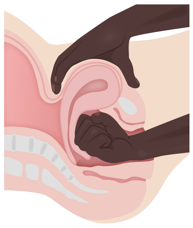
  • Step 1: uterine massage:
    • Instruct patient to empty the bladder Bladder A musculomembranous sac along the urinary tract. Urine flows from the kidneys into the bladder via the ureters, and is held there until urination. Pyelonephritis and Perinephric Abscess to increase effectiveness.
  • Step 2: Give additional oxytocin.
  • Step 3: If uterine atony persists, give other uterotonic agents:
    • Methylergonovine
    • PGF 
    • Misoprostol Misoprostol A synthetic analog of natural prostaglandin e1. It produces a dose-related inhibition of gastric acid and pepsin secretion, and enhances mucosal resistance to injury. It is an effective anti-ulcer agent and also has oxytocic properties. Eicosanoids
  • All 4 medications may be given if the patient has no contraindication.
  • Step 4: uterine packing:
    • Place balloons, sponges, etc ETC The electron transport chain (ETC) sends electrons through a series of proteins, which generate an electrochemical proton gradient that produces energy in the form of adenosine triphosphate (ATP). Electron Transport Chain (ETC). in the uterus Uterus The uterus, cervix, and fallopian tubes are part of the internal female reproductive system. The uterus has a thick wall made of smooth muscle (the myometrium) and an inner mucosal layer (the endometrium). The most inferior portion of the uterus is the cervix, which connects the uterine cavity to the vagina. Uterus, Cervix, and Fallopian Tubes: Anatomy to provide compression Compression Blunt Chest Trauma.
    • Carefully count all foreign bodies placed inside the uterus Uterus The uterus, cervix, and fallopian tubes are part of the internal female reproductive system. The uterus has a thick wall made of smooth muscle (the myometrium) and an inner mucosal layer (the endometrium). The most inferior portion of the uterus is the cervix, which connects the uterine cavity to the vagina. Uterus, Cervix, and Fallopian Tubes: Anatomy.
  • Step 5: surgery:
  • Alternative option: uterine artery Uterine Artery A branch arising from the internal iliac artery in females, that supplies blood to the uterus. Uterus, Cervix, and Fallopian Tubes: Anatomy embolization Embolization A method of hemostasis utilizing various agents such as gelfoam, silastic, metal, glass, or plastic pellets, autologous clot, fat, and muscle as emboli. It has been used in the treatment of spinal cord and intracranial arteriovenous malformations, renal arteriovenous fistulas, gastrointestinal bleeding, epistaxis, hypersplenism, certain highly vascular tumors, traumatic rupture of blood vessels, and control of operative hemorrhage. Gastrointestinal Bleeding:
    • An interventional radiologist identifies the uterine artery Uterine Artery A branch arising from the internal iliac artery in females, that supplies blood to the uterus. Uterus, Cervix, and Fallopian Tubes: Anatomy via fluoroscopy Fluoroscopy Production of an image when x-rays strike a fluorescent screen. X-rays and performs an embolization Embolization A method of hemostasis utilizing various agents such as gelfoam, silastic, metal, glass, or plastic pellets, autologous clot, fat, and muscle as emboli. It has been used in the treatment of spinal cord and intracranial arteriovenous malformations, renal arteriovenous fistulas, gastrointestinal bleeding, epistaxis, hypersplenism, certain highly vascular tumors, traumatic rupture of blood vessels, and control of operative hemorrhage. Gastrointestinal Bleeding.
    • Requires the patient to be stable enough to undergo the procedure (uncommon)
Table: Uterotonic agents used in postpartum hemorrhage
Medication (generic) Class Route Contraindications Contraindications A condition or factor associated with a recipient that makes the use of a drug, procedure, or physical agent improper or inadvisable. Contraindications may be absolute (life threatening) or relative (higher risk of complications in which benefits may outweigh risks). Noninvasive Ventilation
Oxytocin Natural hormone IV, IM None
Methylergonovine Ergot alkaloid IM Hypertension Hypertension Hypertension, or high blood pressure, is a common disease that manifests as elevated systemic arterial pressures. Hypertension is most often asymptomatic and is found incidentally as part of a routine physical examination or during triage for an unrelated medical encounter. Hypertension
Carboprost Carboprost A nonsteroidal abortifacient agent that is effective in both the first and second trimesters of pregnancy. Eicosanoids (PGF) Prostaglandin analogue IM Severe hepatic, renal, and cardiovascular disease, and asthma Asthma Asthma is a chronic inflammatory respiratory condition characterized by bronchial hyperresponsiveness and airflow obstruction. The disease is believed to result from the complex interaction of host and environmental factors that increase disease predisposition, with inflammation causing symptoms and structural changes. Patients typically present with wheezing, cough, and dyspnea. Asthma
Misoprostol Misoprostol A synthetic analog of natural prostaglandin e1. It produces a dose-related inhibition of gastric acid and pepsin secretion, and enhances mucosal resistance to injury. It is an effective anti-ulcer agent and also has oxytocic properties. Eicosanoids Prostaglandin analogue Oral, rectal None
Uterine massage

Uterine massage/manual compression for uterine atony

Image by Lecturio.

Treatment for retained products of conception

  • Suspect retained products of conception if the placenta Placenta A highly vascularized mammalian fetal-maternal organ and major site of transport of oxygen, nutrients, and fetal waste products. It includes a fetal portion (chorionic villi) derived from trophoblasts and a maternal portion (decidua) derived from the uterine endometrium. The placenta produces an array of steroid, protein and peptide hormones (placental hormones). Placenta, Umbilical Cord, and Amniotic Cavity is not delivered intact.
  • Management:
    • Manual exploration of the uterus Uterus The uterus, cervix, and fallopian tubes are part of the internal female reproductive system. The uterus has a thick wall made of smooth muscle (the myometrium) and an inner mucosal layer (the endometrium). The most inferior portion of the uterus is the cervix, which connects the uterine cavity to the vagina. Uterus, Cervix, and Fallopian Tubes: Anatomy to remove any retained pieces of membrane or placenta Placenta A highly vascularized mammalian fetal-maternal organ and major site of transport of oxygen, nutrients, and fetal waste products. It includes a fetal portion (chorionic villi) derived from trophoblasts and a maternal portion (decidua) derived from the uterine endometrium. The placenta produces an array of steroid, protein and peptide hormones (placental hormones). Placenta, Umbilical Cord, and Amniotic Cavity
    • Dilation and curettage Curettage A scraping, usually of the interior of a cavity or tract, for removal of new growth or other abnormal tissue, or to obtain material for tissue diagnosis. It is performed with a curet (curette), a spoon-shaped instrument designed for that purpose. Benign Bone Tumors
    • Uterine packing may be helpful

Treatment of trauma

  • Careful assessment for lacerations: 
    • Adequate lighting and equipment for pelvic exam
    • Inspect the entire cervix Cervix The uterus, cervix, and fallopian tubes are part of the internal female reproductive system. The most inferior portion of the uterus is the cervix, which connects the uterine cavity to the vagina. Externally, the cervix is lined by stratified squamous cells; however, the cervical canal is lined by columnar epithelium. Uterus, Cervix, and Fallopian Tubes: Anatomy (often difficult due to active bleeding and full dilation).
  • Management:
    • Suture repair of any bleeding lacerations
    • Surgical repair of uterine rupture Uterine Rupture A complete separation or tear in the wall of the uterus with or without expulsion of the fetus. It may be due to injuries, multiple pregnancies, large fetus, previous scarring, or obstruction. Antepartum Hemorrhage
    • Apply compression Compression Blunt Chest Trauma to hematomas to prevent expansion of bleeding.

Treatment for thrombin Thrombin An enzyme formed from prothrombin that converts fibrinogen to fibrin. Hemostasis disorders

Activate the hospital’s massive transfusion protocol and administer blood products:
  • Packed RBCs Packed RBCs Transfusion Products: 1 unit should ↑ hemoglobin by 1 g/dL and hematocrit Hematocrit The volume of packed red blood cells in a blood specimen. The volume is measured by centrifugation in a tube with graduated markings, or with automated blood cell counters. It is an indicator of erythrocyte status in disease. For example, anemia shows a low value; polycythemia, a high value. Neonatal Polycythemia by 3%
  • Platelets Platelets Platelets are small cell fragments involved in hemostasis. Thrombopoiesis takes place primarily in the bone marrow through a series of cell differentiation and is influenced by several cytokines. Platelets are formed after fragmentation of the megakaryocyte cytoplasm. Platelets: Histology: 1 unit should ↑ platelet count 5,000–10,000
  • FFP FFP Transfusion Products
    • Contains all coagulation factors Coagulation factors Endogenous substances, usually proteins, that are involved in the blood coagulation process. Hemostasis and plasma Plasma The residual portion of blood that is left after removal of blood cells by centrifugation without prior blood coagulation. Transfusion Products proteins Proteins Linear polypeptides that are synthesized on ribosomes and may be further modified, crosslinked, cleaved, or assembled into complex proteins with several subunits. The specific sequence of amino acids determines the shape the polypeptide will take, during protein folding, and the function of the protein. Energy Homeostasis 
    • 1 unit should ↑ fibrinogen Fibrinogen Plasma glycoprotein clotted by thrombin, composed of a dimer of three non-identical pairs of polypeptide chains (alpha, beta, gamma) held together by disulfide bonds. Fibrinogen clotting is a sol-gel change involving complex molecular arrangements: whereas fibrinogen is cleaved by thrombin to form polypeptides a and b, the proteolytic action of other enzymes yields different fibrinogen degradation products. Hemostasis 10 mg/dL
  • Cryoprecipitate Cryoprecipitate Transfusion Products
    • Contains fibrinogen Fibrinogen Plasma glycoprotein clotted by thrombin, composed of a dimer of three non-identical pairs of polypeptide chains (alpha, beta, gamma) held together by disulfide bonds. Fibrinogen clotting is a sol-gel change involving complex molecular arrangements: whereas fibrinogen is cleaved by thrombin to form polypeptides a and b, the proteolytic action of other enzymes yields different fibrinogen degradation products. Hemostasis (factor I), factor VIII Factor VIII Factor VIII of blood coagulation. Antihemophilic factor that is part of the factor viii/von Willebrand factor complex. Factor VIII is produced in the liver and acts in the intrinsic pathway of blood coagulation. It serves as a cofactor in factor X activation and this action is markedly enhanced by small amounts of thrombin. Hemostasis, factor XIII, and von Willebrand factor von Willebrand factor A high-molecular-weight plasma protein, produced by endothelial cells and megakaryocytes, that is part of the factor VIII/von Willebrand factor complex. The von Willebrand factor has receptors for collagen, platelets, and ristocetin activity as well as the immunologically distinct antigenic determinants. It functions in adhesion of platelets to collagen and hemostatic plug formation. The prolonged bleeding time in von Willebrand diseases is due to the deficiency of this factor. Hemostasis
    • 1 unit should ↑ fibrinogen Fibrinogen Plasma glycoprotein clotted by thrombin, composed of a dimer of three non-identical pairs of polypeptide chains (alpha, beta, gamma) held together by disulfide bonds. Fibrinogen clotting is a sol-gel change involving complex molecular arrangements: whereas fibrinogen is cleaved by thrombin to form polypeptides a and b, the proteolytic action of other enzymes yields different fibrinogen degradation products. Hemostasis 10 mg/dL
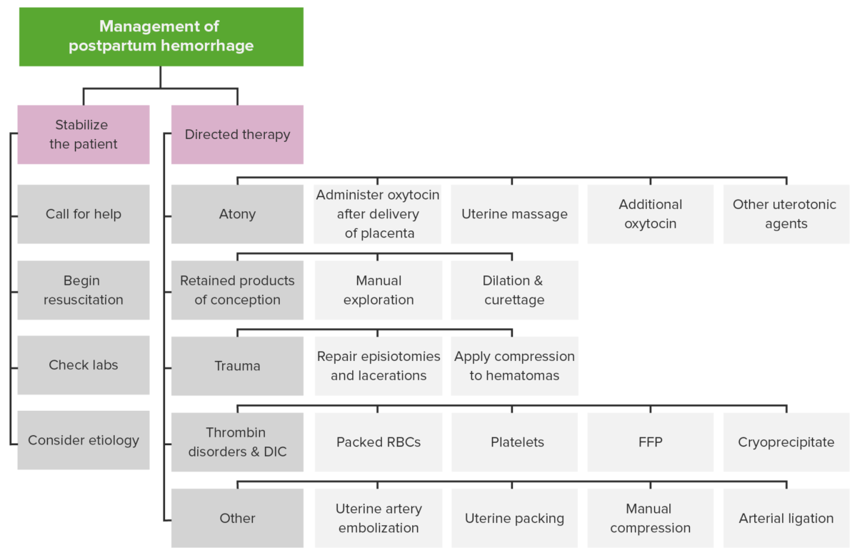
Management of postpartum hemorrhage

Management of postpartum hemorrhage

Image by Lecturio.

Complications

  • DIC DIC Disseminated intravascular coagulation (DIC) is a condition characterized by systemic bodywide activation of the coagulation cascade. This cascade results in both widespread microvascular thrombi contributing to multiple organ dysfunction and consumption of clotting factors and platelets, leading to hemorrhage. Disseminated Intravascular Coagulation
  • Acute renal failure Renal failure Conditions in which the kidneys perform below the normal level in the ability to remove wastes, concentrate urine, and maintain electrolyte balance; blood pressure; and calcium metabolism. Renal insufficiency can be classified by the degree of kidney damage (as measured by the level of proteinuria) and reduction in glomerular filtration rate. Crush Syndrome
  • Adult respiratory distress syndrome
  • Loss of fertility (hysterectomy)
  • Pituitary Pituitary A small, unpaired gland situated in the sella turcica. It is connected to the hypothalamus by a short stalk which is called the infundibulum. Hormones: Overview and Types necrosis Necrosis The death of cells in an organ or tissue due to disease, injury or failure of the blood supply. Ischemic Cell Damage (Sheehan syndrome)

Clinical Relevance

  • Cesarean section: The operative delivery of 1 or more infants through surgical incision Surgical Incision Surgical Site Infections into the maternal abdomen, surgical opening of the uterus Uterus The uterus, cervix, and fallopian tubes are part of the internal female reproductive system. The uterus has a thick wall made of smooth muscle (the myometrium) and an inner mucosal layer (the endometrium). The most inferior portion of the uterus is the cervix, which connects the uterine cavity to the vagina. Uterus, Cervix, and Fallopian Tubes: Anatomy, and delivery of the infant(s). 
  • Prolonged labor: longer-than-normal rate of labor progression. Prolonged labor increases the risk for obstetric complications including postpartum hemorrhage, intraamniotic infection, and fetal distress. 
  • Rapid labor: shorter-than-normal rate of labor progression. The normal duration of labor is usually at least 3 hours. Rapid labor increases the risk for postpartum hemorrhage.
  • Chorioamnionitis Chorioamnionitis Chorioamnionitis, commonly referred to as intraamniotic infection (IAI), is a common obstetric complication involving infection and inflammation of the fetal membranes, amniotic fluid, placenta, or the fetus itself. Chorioamnionitis is typically caused by a polymicrobial infection that ascends from the lower genitourinary tract. Chorioamnionitis: an infection of the chorion Chorion The outermost extraembryonic membrane surrounding the developing embryo. In reptiles and birds, it adheres to the shell and allows exchange of gases between the egg and its environment. In mammals, the chorion evolves into the fetal contribution of the placenta. Placenta, Umbilical Cord, and Amniotic Cavity and amnion Amnion The innermost membranous sac that surrounds and protects the developing embryo which is bathed in the amniotic fluid. Amnion cells are secretory epithelial cells and contribute to the amniotic fluid. Placenta, Umbilical Cord, and Amniotic Cavity. An infected uterus Uterus The uterus, cervix, and fallopian tubes are part of the internal female reproductive system. The uterus has a thick wall made of smooth muscle (the myometrium) and an inner mucosal layer (the endometrium). The most inferior portion of the uterus is the cervix, which connects the uterine cavity to the vagina. Uterus, Cervix, and Fallopian Tubes: Anatomy does not contract well, which leads to prolonged labor and significantly increases a patient’s risk for postpartum hemorrhage. Chorioamnionitis Chorioamnionitis Chorioamnionitis, commonly referred to as intraamniotic infection (IAI), is a common obstetric complication involving infection and inflammation of the fetal membranes, amniotic fluid, placenta, or the fetus itself. Chorioamnionitis is typically caused by a polymicrobial infection that ascends from the lower genitourinary tract. Chorioamnionitis is a common complication of prolonged rupture of membranes. Chorioamnionitis Chorioamnionitis Chorioamnionitis, commonly referred to as intraamniotic infection (IAI), is a common obstetric complication involving infection and inflammation of the fetal membranes, amniotic fluid, placenta, or the fetus itself. Chorioamnionitis is typically caused by a polymicrobial infection that ascends from the lower genitourinary tract. Chorioamnionitis may lead to maternal sepsis Sepsis Systemic inflammatory response syndrome with a proven or suspected infectious etiology. When sepsis is associated with organ dysfunction distant from the site of infection, it is called severe sepsis. When sepsis is accompanied by hypotension despite adequate fluid infusion, it is called septic shock. Sepsis and Septic Shock and/or postpartum endometritis Postpartum Endometritis Inflammation of the endometrium, usually caused by intrauterine infections. Endometritis is the most common cause of postpartum fever. Postpartum Endometritis.
  • Multiple pregnancy Pregnancy The status during which female mammals carry their developing young (embryos or fetuses) in utero before birth, beginning from fertilization to birth. Pregnancy: Diagnosis, Physiology, and Care: a pregnancy Pregnancy The status during which female mammals carry their developing young (embryos or fetuses) in utero before birth, beginning from fertilization to birth. Pregnancy: Diagnosis, Physiology, and Care with more than 1 fetus (e.g., twins, triplets, quadruplets). Multiple pregnancy Pregnancy The status during which female mammals carry their developing young (embryos or fetuses) in utero before birth, beginning from fertilization to birth. Pregnancy: Diagnosis, Physiology, and Care leads to overdistension of the uterus Uterus The uterus, cervix, and fallopian tubes are part of the internal female reproductive system. The uterus has a thick wall made of smooth muscle (the myometrium) and an inner mucosal layer (the endometrium). The most inferior portion of the uterus is the cervix, which connects the uterine cavity to the vagina. Uterus, Cervix, and Fallopian Tubes: Anatomy, which is a risk factor for several obstetric complications, including postpartum hemorrhage and prelabor rupture of membranes Prelabor Rupture of Membranes Prelabor rupture of membranes (PROM), previously known as premature rupture of membranes, refers to the rupture of the amniotic sac before the onset of labor. Prelabor rupture of membranes may occur in term or preterm pregnancies. Prelabor Rupture of Membranes.
  • Abnormal placentation: placental implantation Implantation Endometrial implantation of embryo, mammalian at the blastocyst stage. Fertilization and First Week occurring in an abnormal site within the uterus Uterus The uterus, cervix, and fallopian tubes are part of the internal female reproductive system. The uterus has a thick wall made of smooth muscle (the myometrium) and an inner mucosal layer (the endometrium). The most inferior portion of the uterus is the cervix, which connects the uterine cavity to the vagina. Uterus, Cervix, and Fallopian Tubes: Anatomy. Abnormal placentation also includes placenta accreta Placenta Accreta Abnormal placentation in which all or parts of the placenta are attached directly to the myometrium due to a complete or partial absence of decidua. It is associated with postpartum hemorrhage because of the failure of placental separation. Placental Abnormalities, which is an invasion of the placenta Placenta A highly vascularized mammalian fetal-maternal organ and major site of transport of oxygen, nutrients, and fetal waste products. It includes a fetal portion (chorionic villi) derived from trophoblasts and a maternal portion (decidua) derived from the uterine endometrium. The placenta produces an array of steroid, protein and peptide hormones (placental hormones). Placenta, Umbilical Cord, and Amniotic Cavity into the myometrium. Abnormal placentation increases the risk of postpartum hemorrhage due to retained products of conception.
  • Antepartum hemorrhage Antepartum hemorrhage Antepartum hemorrhage is defined as vaginal bleeding that occurs after 20 weeks of gestation and is unrelated to labor. The most important causes are placental abruption (most common), placenta previa (2nd most common), vasa previa, and uterine rupture. Antepartum Hemorrhage: obstetric bleeding during the 3rd trimester. Common causes include placenta previa Placenta Previa Abnormal placentation in which the placenta implants in the lower segment of the uterus (the zone of dilation) and may cover part or all of the opening of the cervix. It is often associated with serious antepartum bleeding and premature labor. Placental Abnormalities, placental abruption Placental Abruption Premature separation of the normally implanted placenta from the uterus. Signs of varying degree of severity include uterine bleeding, uterine muscle hypertonia, and fetal distress or fetal death. Antepartum Hemorrhage, and preterm labor Preterm labor Preterm labor refers to regular uterine contractions leading to cervical change prior to 37 weeks of gestation; preterm birth refers to birth prior to 37 weeks of gestation. Preterm birth may be spontaneous due to preterm labor, preterm prelabor rupture of membranes (PPROM), or cervical insufficiency. Preterm Labor and Birth. Bleeding due to abnormal placentation may increase the risk of postpartum hemorrhage due to retained products of conception.
  • Disseminated intravascular coagulation Disseminated intravascular coagulation Disseminated intravascular coagulation (DIC) is a condition characterized by systemic bodywide activation of the coagulation cascade. This cascade results in both widespread microvascular thrombi contributing to multiple organ dysfunction and consumption of clotting factors and platelets, leading to hemorrhage. Disseminated Intravascular Coagulation ( DIC DIC Disseminated intravascular coagulation (DIC) is a condition characterized by systemic bodywide activation of the coagulation cascade. This cascade results in both widespread microvascular thrombi contributing to multiple organ dysfunction and consumption of clotting factors and platelets, leading to hemorrhage. Disseminated Intravascular Coagulation): a serious medical disorder in which multiple clots form and can lead to permanent organ damage. Systemic activation of blood coagulation occurs and results in the generation and deposition of excess fibrin Fibrin A protein derived from fibrinogen in the presence of thrombin, which forms part of the blood clot. Rapidly Progressive Glomerulonephritis. The systemic activation of the coagulation cascade Coagulation cascade The coagulation cascade is a series of reactions that ultimately generates a strong, cross-linked fibrin clot. Hemostasis uses many coagulation factors Coagulation factors Endogenous substances, usually proteins, that are involved in the blood coagulation process. Hemostasis, which result in uncontrolled bleeding.
  • Hemolysis, elevated liver Liver The liver is the largest gland in the human body. The liver is found in the superior right quadrant of the abdomen and weighs approximately 1.5 kilograms. Its main functions are detoxification, metabolism, nutrient storage (e.g., iron and vitamins), synthesis of coagulation factors, formation of bile, filtration, and storage of blood. Liver: Anatomy enzymes Enzymes Enzymes are complex protein biocatalysts that accelerate chemical reactions without being consumed by them. Due to the body’s constant metabolic needs, the absence of enzymes would make life unsustainable, as reactions would occur too slowly without these molecules. Basics of Enzymes, and low platelet count (HELLP) syndrome: a preeclampsia Preeclampsia A complication of pregnancy, characterized by a complex of symptoms including maternal hypertension and proteinuria with or without pathological edema. Symptoms may range between mild and severe. Pre-eclampsia usually occurs after the 20th week of gestation, but may develop before this time in the presence of trophoblastic disease. Hypertensive Pregnancy Disorders complication involving a triad of hemolysis, elevated liver Liver The liver is the largest gland in the human body. The liver is found in the superior right quadrant of the abdomen and weighs approximately 1.5 kilograms. Its main functions are detoxification, metabolism, nutrient storage (e.g., iron and vitamins), synthesis of coagulation factors, formation of bile, filtration, and storage of blood. Liver: Anatomy enzymes Enzymes Enzymes are complex protein biocatalysts that accelerate chemical reactions without being consumed by them. Due to the body’s constant metabolic needs, the absence of enzymes would make life unsustainable, as reactions would occur too slowly without these molecules. Basics of Enzymes, and low platelets Platelets Platelets are small cell fragments involved in hemostasis. Thrombopoiesis takes place primarily in the bone marrow through a series of cell differentiation and is influenced by several cytokines. Platelets are formed after fragmentation of the megakaryocyte cytoplasm. Platelets: Histology ( thrombocytopenia Thrombocytopenia Thrombocytopenia occurs when the platelet count is < 150,000 per microliter. The normal range for platelets is usually 150,000-450,000/µL of whole blood. Thrombocytopenia can be a result of decreased production, increased destruction, or splenic sequestration of platelets. Patients are often asymptomatic until platelet counts are < 50,000/µL. Thrombocytopenia). Thrombocytopenia Thrombocytopenia Thrombocytopenia occurs when the platelet count is < 150,000 per microliter. The normal range for platelets is usually 150,000-450,000/µL of whole blood. Thrombocytopenia can be a result of decreased production, increased destruction, or splenic sequestration of platelets. Patients are often asymptomatic until platelet counts are < 50,000/µL. Thrombocytopenia represents a hemostasis Hemostasis Hemostasis refers to the innate, stepwise body processes that occur following vessel injury, resulting in clot formation and cessation of bleeding. Hemostasis occurs in 2 phases, namely, primary and secondary. Primary hemostasis involves forming a plug that stops the bleeding temporarily. Secondary hemostasis involves the activation of the coagulation cascade. Hemostasis abnormality and increases the risk of postpartum hemorrhage.

References

  1. Belfort, M. A. (2021). Overview of postpartum hemorrhage. In Barss, V.A. (Ed.), UpToDate. Retrieved March 11, 2021, from https://www.uptodate.com/contents/overview-of-postpartum-hemorrhage
  2. Wormer, K. (2020). Acute postpartum hemorrhage. In Bryant, S. (Ed.), StatPearls. Retrieved March 11, 2021, from https://www.statpearls.com/articlelibrary/viewarticle/27638/ 
  3. The American College of Obstetrics and Gynecology Committee on Obstetrics. (2017). Practice Bulletin No. 183: Postpartum hemorrhage. Retrieved March 11, 2021, from https://www.acog.org/clinical/clinical-guidance/practice-bulletin/articles/2017/10/postpartum-hemorrhage 
  4. The American College of Obstetrics and Gynecology Committee on Obstetrics. (2019). Committee Opinion No. 794: Quantitative blood loss in obstetric hemorrhage. Retrieved March 11, 2021, from https://www.acog.org/clinical/clinical-guidance/committee-opinion/articles/2019/12/quantitative-blood-loss-in-obstetric-hemorrhage 
  5. Tobian, A. (2021). Clinical use of Cryoprecipitate. UpToDate. Retrieved May 15, 2021, from https://www.uptodate.com/contents/clinical-use-of-cryoprecipitate

Create your free account or log in to continue reading!

Sign up now and get free access to Lecturio with concept pages, medical videos, and questions for your medical education.

User Reviews

Details