Postoperative Care

After any procedure performed in the operating room, all patients Patients Individuals participating in the health care system for the purpose of receiving therapeutic, diagnostic, or preventive procedures. Clinician–Patient Relationship must undergo close observation at least in the recovery room. After larger procedures and for patients Patients Individuals participating in the health care system for the purpose of receiving therapeutic, diagnostic, or preventive procedures. Clinician–Patient Relationship who require hospitalization Hospitalization The confinement of a patient in a hospital. Delirium, observation must continue on the surgical ward. The primary intent of this practice is the early detection of postoperative complications. The entire medical team must be vigilant and aware of the patient’s history, the procedure performed, and risk factors for potential complications. Some of the most important things to attend to include monitoring vital signs (which may indicate severe complications such as bleeding), wound and drain care, postoperative pain Pain An unpleasant sensation induced by noxious stimuli which are detected by nerve endings of nociceptive neurons. Pain: Types and Pathways control, nausea Nausea An unpleasant sensation in the stomach usually accompanied by the urge to vomit. Common causes are early pregnancy, sea and motion sickness, emotional stress, intense pain, food poisoning, and various enteroviruses. Antiemetics and vomiting Vomiting The forcible expulsion of the contents of the stomach through the mouth. Hypokalemia, pulmonary hygiene to prevent atelectasis Atelectasis Atelectasis is the partial or complete collapse of a part of the lung. Atelectasis is almost always a secondary phenomenon from conditions causing bronchial obstruction, external compression, surfactant deficiency, or scarring. Atelectasis, deep vein thrombosis Thrombosis Formation and development of a thrombus or blood clot in the blood vessel. Epidemic Typhus prophylaxis Prophylaxis Cephalosporins, and fluid management.

Last updated: Mar 27, 2024

Editorial responsibility: Stanley Oiseth, Lindsay Jones, Evelin Maza

Recovery Room

The recovery room is reserved for patients Patients Individuals participating in the health care system for the purpose of receiving therapeutic, diagnostic, or preventive procedures. Clinician–Patient Relationship in the immediate postoperative period who have received general anesthesia General anesthesia Procedure in which patients are induced into an unconscious state through use of various medications so that they do not feel pain during surgery. Anesthesiology: History and Basic Concepts. Observation usually lasts 6 hours (until they are conscious and their vital signs are stable).
Monitoring airway Airway ABCDE Assessment, breathing, and circulation Circulation The movement of the blood as it is pumped through the cardiovascular system. ABCDE Assessment (the ABCs) and vital signs

  • Monitoring of the ABCs is the priority.
  • The patient may initially remain intubated, even in the recovery room.
  • Following extubation Extubation Removal of an endotracheal tube from the patient. Invasive Mechanical Ventilation:
  • Assess the patient’s vital signs:
    • ↑ Temperature, consider:
      • Atelectasis Atelectasis Atelectasis is the partial or complete collapse of a part of the lung. Atelectasis is almost always a secondary phenomenon from conditions causing bronchial obstruction, external compression, surfactant deficiency, or scarring. Atelectasis
      • Infection  
    • ↑ Pulse rate (PR), consider:
      • Pain Pain An unpleasant sensation induced by noxious stimuli which are detected by nerve endings of nociceptive neurons. Pain: Types and Pathways (most common)
      • Hemorrhage
      • Pulmonary embolism Pulmonary Embolism Pulmonary embolism (PE) is a potentially fatal condition that occurs as a result of intraluminal obstruction of the main pulmonary artery or its branches. The causative factors include thrombi, air, amniotic fluid, and fat. In PE, gas exchange is impaired due to the decreased return of deoxygenated blood to the lungs. Pulmonary Embolism (PE)
      • MI MI MI is ischemia and death of an area of myocardial tissue due to insufficient blood flow and oxygenation, usually from thrombus formation on a ruptured atherosclerotic plaque in the epicardial arteries. Clinical presentation is most commonly with chest pain, but women and patients with diabetes may have atypical symptoms. Myocardial Infarction
      • Infection  
    • ↓ BP, consider:
      • Hemorrhage
      • Cardiac dysfunction
      • Infection
    • RR RR Relative risk (RR) is the risk of a disease or condition occurring in a group or population with a particular exposure relative to a control (unexposed) group. Measures of Risk, consider:
      • Pulmonary embolism Pulmonary Embolism Pulmonary embolism (PE) is a potentially fatal condition that occurs as a result of intraluminal obstruction of the main pulmonary artery or its branches. The causative factors include thrombi, air, amniotic fluid, and fat. In PE, gas exchange is impaired due to the decreased return of deoxygenated blood to the lungs. Pulmonary Embolism
      • Atelectasis Atelectasis Atelectasis is the partial or complete collapse of a part of the lung. Atelectasis is almost always a secondary phenomenon from conditions causing bronchial obstruction, external compression, surfactant deficiency, or scarring. Atelectasis
      • Infection   
    • Oxygen saturation Oxygen Saturation Basic Procedures, consider:
      • PE
      • MI MI MI is ischemia and death of an area of myocardial tissue due to insufficient blood flow and oxygenation, usually from thrombus formation on a ruptured atherosclerotic plaque in the epicardial arteries. Clinical presentation is most commonly with chest pain, but women and patients with diabetes may have atypical symptoms. Myocardial Infarction
      • Airway Airway ABCDE Assessment obstruction
      • Atelectasis Atelectasis Atelectasis is the partial or complete collapse of a part of the lung. Atelectasis is almost always a secondary phenomenon from conditions causing bronchial obstruction, external compression, surfactant deficiency, or scarring. Atelectasis
    • ↓ Level of consciousness, consider:
      • Stroke
      • Cerebral hemorrhage
      • Lingering anesthetic effects
  • Major complications:
    • Airway Airway ABCDE Assessment obstruction
    • MI MI MI is ischemia and death of an area of myocardial tissue due to insufficient blood flow and oxygenation, usually from thrombus formation on a ruptured atherosclerotic plaque in the epicardial arteries. Clinical presentation is most commonly with chest pain, but women and patients with diabetes may have atypical symptoms. Myocardial Infarction
    • Hemorrhage 
    • Respiratory failure Respiratory failure Respiratory failure is a syndrome that develops when the respiratory system is unable to maintain oxygenation and/or ventilation. Respiratory failure may be acute or chronic and is classified as hypoxemic, hypercapnic, or a combination of the two. Respiratory Failure
    • Acute kidney injury Acute Kidney Injury Acute kidney injury refers to sudden and often reversible loss of renal function, which develops over days or weeks. Azotemia refers to elevated levels of nitrogen-containing substances in the blood that accompany AKI, which include BUN and creatinine. Acute Kidney Injury
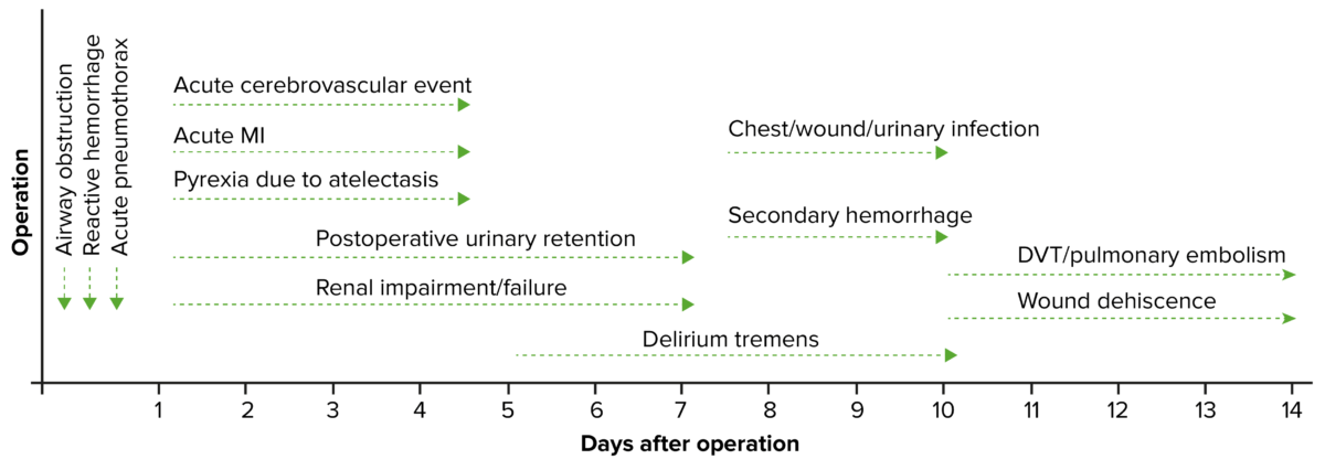
Timeline of postoperative complications

Timeline of postoperative complications
DVT: deep vein thrombosis
MI: myocardial infarction

Image by Lecturio.

Other activities in the recovery room

  • The patient’s symptoms (e.g., dyspnea Dyspnea Dyspnea is the subjective sensation of breathing discomfort. Dyspnea is a normal manifestation of heavy physical or psychological exertion, but also may be caused by underlying conditions (both pulmonary and extrapulmonary). Dyspnea) and concerns (e.g., pain Pain An unpleasant sensation induced by noxious stimuli which are detected by nerve endings of nociceptive neurons. Pain: Types and Pathways or nausea Nausea An unpleasant sensation in the stomach usually accompanied by the urge to vomit. Common causes are early pregnancy, sea and motion sickness, emotional stress, intense pain, food poisoning, and various enteroviruses. Antiemetics) should be addressed.
  • Evaluate symptoms that could be caused by complications:
    • Chest pain Pain An unpleasant sensation induced by noxious stimuli which are detected by nerve endings of nociceptive neurons. Pain: Types and Pathways ECG ECG An electrocardiogram (ECG) is a graphic representation of the electrical activity of the heart plotted against time. Adhesive electrodes are affixed to the skin surface allowing measurement of cardiac impulses from many angles. The ECG provides 3-dimensional information about the conduction system of the heart, the myocardium, and other cardiac structures. Electrocardiogram (ECG) to assess for possible MI MI MI is ischemia and death of an area of myocardial tissue due to insufficient blood flow and oxygenation, usually from thrombus formation on a ruptured atherosclerotic plaque in the epicardial arteries. Clinical presentation is most commonly with chest pain, but women and patients with diabetes may have atypical symptoms. Myocardial Infarction
    • Uncontrolled pain Pain An unpleasant sensation induced by noxious stimuli which are detected by nerve endings of nociceptive neurons. Pain: Types and Pathways → evaluate for internal surgical bleeding
  • Serial physical examinations are performed with a focus on ruling out complications:
    • Heart and lung exam
    • Surgical wound exam
  • Based on the procedure performed, the patient’s risks, and the patient’s current status, the surgeon will determine the disposition of the case, which may be:
    • Discharge to home:
      • For healthy individuals undergoing minor and/or minimally invasive procedures
      • Without worrisome signs/symptoms in recovery
    • Transfer the individual to the inpatient ward for continued, routine postoperative care.
    • Transfer the individual to the ICU ICU Hospital units providing continuous surveillance and care to acutely ill patients. West Nile Virus for more complex care.

Surgical Ward

The patient is moved to the surgical ward for continued observation.

Basics of routine surgical care on the ward

All patients Patients Individuals participating in the health care system for the purpose of receiving therapeutic, diagnostic, or preventive procedures. Clinician–Patient Relationship should be physically evaluated by a member of the surgical team at least once daily (if not more often).

  • Monitor vital signs, urinary output, and mental status.
  • Monitor the state and output of drains and tubes (e.g., Foley catheter, nasogastric tube Nasogastric tube Malnutrition in children in resource-limited countries, chest tube).
  • Manage fluids.
  • Check the surgical site for signs of:
    • Dehiscence
    • Infection
    • Bleeding
  • Help control common postoperative symptoms:
    • Pain Pain An unpleasant sensation induced by noxious stimuli which are detected by nerve endings of nociceptive neurons. Pain: Types and Pathways
    • Nausea Nausea An unpleasant sensation in the stomach usually accompanied by the urge to vomit. Common causes are early pregnancy, sea and motion sickness, emotional stress, intense pain, food poisoning, and various enteroviruses. Antiemetics and vomiting Vomiting The forcible expulsion of the contents of the stomach through the mouth. Hypokalemia
  • Prophylaxis Prophylaxis Cephalosporins against:
    • Atelectasis Atelectasis Atelectasis is the partial or complete collapse of a part of the lung. Atelectasis is almost always a secondary phenomenon from conditions causing bronchial obstruction, external compression, surfactant deficiency, or scarring. Atelectasis
    • Deep vein thrombosis Thrombosis Formation and development of a thrombus or blood clot in the blood vessel. Epidemic Typhus ( DVT DVT Deep vein thrombosis (DVT) usually occurs in the deep veins of the lower extremities. The affected veins include the femoral, popliteal, iliofemoral, and pelvic veins. Proximal DVT is more likely to cause a pulmonary embolism (PE) and is generally considered more serious. Deep Vein Thrombosis)
  • For patients Patients Individuals participating in the health care system for the purpose of receiving therapeutic, diagnostic, or preventive procedures. Clinician–Patient Relationship at risk of internal bleeding and/or metabolic issues, order and follow up on basic lab work:
    • CBC (typically within the first 24 hours after surgery); consider transfusion based on Hb Hb The oxygen-carrying proteins of erythrocytes. They are found in all vertebrates and some invertebrates. The number of globin subunits in the hemoglobin quaternary structure differs between species. Structures range from monomeric to a variety of multimeric arrangements. Gas Exchange levels and blood loss during surgery
    • Consider a basic metabolic panel Basic Metabolic Panel Primary vs Secondary Headaches ( BMP BMP Primary vs Secondary Headaches).
    • Labs unique to the patient and/or procedure
  • Work toward discharge:
    • Ambulation
    • Tolerating nutrition
    • Normal voiding and appropriate bowel activity
    • Symptoms controlled without IV or IM medications

Drains

  • Drains should be checked and emptied regularly:
    • Lost fluid should be replaced with a fluid of similar characteristics.
    • Continuous blood loss through a drain should be investigated. 
    • Remove as soon as clinically appropriate in order to minimize complication risks.
  • Urinary catheter: for most procedures, typically removed once the patient can ambulate

Wound care

Wound infections Infections Invasion of the host organism by microorganisms or their toxins or by parasites that can cause pathological conditions or diseases. Chronic Granulomatous Disease can affect either the superficial or the deep incisional spaces. Pressure sores may also develop in immobile patients Patients Individuals participating in the health care system for the purpose of receiving therapeutic, diagnostic, or preventive procedures. Clinician–Patient Relationship.

  • Wounds should be examined daily for signs of infection:
  • Wounds should be covered with a dry dressing, which should be removed when clinically appropriate, often within 48 hours.
  • Superficial infections Infections Invasion of the host organism by microorganisms or their toxins or by parasites that can cause pathological conditions or diseases. Chronic Granulomatous Disease involve only the skin Skin The skin, also referred to as the integumentary system, is the largest organ of the body. The skin is primarily composed of the epidermis (outer layer) and dermis (deep layer). The epidermis is primarily composed of keratinocytes that undergo rapid turnover, while the dermis contains dense layers of connective tissue. Skin: Structure and Functions and subcutaneous tissue Subcutaneous tissue Loose connective tissue lying under the dermis, which binds skin loosely to subjacent tissues. It may contain a pad of adipocytes, which vary in number according to the area of the body and vary in size according to the nutritional state. Soft Tissue Abscess near the incision and can present with:
    • Cellulitis Cellulitis Cellulitis is a common infection caused by bacteria that affects the dermis and subcutaneous tissue of the skin. It is frequently caused by Staphylococcus aureus and Streptococcus pyogenes. The skin infection presents as an erythematous and edematous area with warmth and tenderness. Cellulitis
    • Purulent drainage
    • Superficial abscess Abscess Accumulation of purulent material in tissues, organs, or circumscribed spaces, usually associated with signs of infection. Chronic Granulomatous Disease
  • Deep infections Infections Invasion of the host organism by microorganisms or their toxins or by parasites that can cause pathological conditions or diseases. Chronic Granulomatous Disease may involve the peritoneal or organ spaces and present with:
    • Pain Pain An unpleasant sensation induced by noxious stimuli which are detected by nerve endings of nociceptive neurons. Pain: Types and Pathways
    • Fever Fever Fever is defined as a measured body temperature of at least 38°C (100.4°F). Fever is caused by circulating endogenous and/or exogenous pyrogens that increase levels of prostaglandin E2 in the hypothalamus. Fever is commonly associated with chills, rigors, sweating, and flushing of the skin. Fever
    • Ileus Ileus A condition caused by the lack of intestinal peristalsis or intestinal motility without any mechanical obstruction. This interference of the flow of intestinal contents often leads to intestinal obstruction. Ileus may be classified into postoperative, inflammatory, metabolic, neurogenic, and drug-induced. Small Bowel Obstruction (in GI surgery)
  • Signs of infection should be managed:
    • Wound swab for Gram staining Gram staining Bacteriology and culture
    • Antibiotics if clinical infection is apparent on physical exam
    • +/– surgical debridement Debridement The removal of foreign material and devitalized or contaminated tissue from or adjacent to a traumatic or infected lesion until surrounding healthy tissue is exposed. Stevens-Johnson Syndrome
  • Pressure sores:
    • A result of friction/persistent pressure over soft tissue Soft Tissue Soft Tissue Abscess affecting pressure points (e.g., sacrum Sacrum Five fused vertebrae forming a triangle-shaped structure at the back of the pelvis. It articulates superiorly with the lumbar vertebrae, inferiorly with the coccyx, and anteriorly with the ilium of the pelvis. The sacrum strengthens and stabilizes the pelvis. Vertebral Column: Anatomy)
    • Risk factors:
      • Immobility
      • Malnutrition Malnutrition Malnutrition is a clinical state caused by an imbalance or deficiency of calories and/or micronutrients and macronutrients. The 2 main manifestations of acute severe malnutrition are marasmus (total caloric insufficiency) and kwashiorkor (protein malnutrition with characteristic edema). Malnutrition in children in resource-limited countries
      • Poor peripheral circulation Circulation The movement of the blood as it is pumped through the cardiovascular system. ABCDE Assessment 
      • Diabetes Diabetes Diabetes mellitus (DM) is a metabolic disease characterized by hyperglycemia and dysfunction of the regulation of glucose metabolism by insulin. Type 1 DM is diagnosed mostly in children and young adults as the result of autoimmune destruction of β cells in the pancreas and the resulting lack of insulin. Type 2 DM has a significant association with obesity and is characterized by insulin resistance. Diabetes Mellitus mellitus 
      • Edema Edema Edema is a condition in which excess serous fluid accumulates in the body cavity or interstitial space of connective tissues. Edema is a symptom observed in several medical conditions. It can be categorized into 2 types, namely, peripheral (in the extremities) and internal (in an organ or body cavity). Edema
    • Prevention:
      • Early mobilization
      • Adequate nutrition 
      • Regular Regular Insulin turning by staff, if immobile 
      • Air filter mattress for high-risk patients Patients Individuals participating in the health care system for the purpose of receiving therapeutic, diagnostic, or preventive procedures. Clinician–Patient Relationship 
  • Risk factors for impaired wound healing Wound healing Wound healing is a physiological process involving tissue repair in response to injury. It involves a complex interaction of various cell types, cytokines, and inflammatory mediators. Wound healing stages include hemostasis, inflammation, granulation, and remodeling. Wound Healing:
    • Obesity Obesity Obesity is a condition associated with excess body weight, specifically with the deposition of excessive adipose tissue. Obesity is considered a global epidemic. Major influences come from the western diet and sedentary lifestyles, but the exact mechanisms likely include a mixture of genetic and environmental factors. Obesity
    • Smoking Smoking Willful or deliberate act of inhaling and exhaling smoke from burning substances or agents held by hand. Interstitial Lung Diseases
    • Diabetes Diabetes Diabetes mellitus (DM) is a metabolic disease characterized by hyperglycemia and dysfunction of the regulation of glucose metabolism by insulin. Type 1 DM is diagnosed mostly in children and young adults as the result of autoimmune destruction of β cells in the pancreas and the resulting lack of insulin. Type 2 DM has a significant association with obesity and is characterized by insulin resistance. Diabetes Mellitus mellitus
    • Peripheral vascular disease

Fluid replacement and oral intake

  • Fluid input (fluid intake) and output (sensible and insensible losses Insensible losses Loss of water by diffusion through the skin and by evaporation from the respiratory tract. Volume Depletion and Dehydration) should be balanced.
    • Fluid input sources:
      • Exogenous sources: oral and IV fluid
      • Endogenous sources: water produced by metabolism
    • Fluid output sources:
      • Sensible losses: urine and drains
      • Insensible losses Insensible losses Loss of water by diffusion through the skin and by evaporation from the respiratory tract. Volume Depletion and Dehydration: respiration Respiration The act of breathing with the lungs, consisting of inhalation, or the taking into the lungs of the ambient air, and of exhalation, or the expelling of the modified air which contains more carbon dioxide than the air taken in. Nose Anatomy (External & Internal), feces, sweat
    • Extra losses, such as increased insensible losses Insensible losses Loss of water by diffusion through the skin and by evaporation from the respiratory tract. Volume Depletion and Dehydration due to temperature in tropical climates, must be kept in mind.
    • Maintenance fluid requirements for adults are approximately 1.5–2.5 L daily.
  • Goals of fluid management:
    • Guard against ongoing physiologic fluid loss in patients Patients Individuals participating in the health care system for the purpose of receiving therapeutic, diagnostic, or preventive procedures. Clinician–Patient Relationship who cannot or should not eat/drink; these losses may be increased due to:
      • Drains
      • GI losses: vomiting Vomiting The forcible expulsion of the contents of the stomach through the mouth. Hypokalemia, diarrhea Diarrhea Diarrhea is defined as ≥ 3 watery or loose stools in a 24-hour period. There are a multitude of etiologies, which can be classified based on the underlying mechanism of disease. The duration of symptoms (acute or chronic) and characteristics of the stools (e.g., watery, bloody, steatorrheic, mucoid) can help guide further diagnostic evaluation. Diarrhea
      • Insensible losses Insensible losses Loss of water by diffusion through the skin and by evaporation from the respiratory tract. Volume Depletion and Dehydration: tachypnea Tachypnea Increased respiratory rate. Pulmonary Examination, perspiration
    • Support cardiac preload Preload Cardiac Mechanics
    • Maintain intravascular volume 
    • Electrolyte balance
  • Provide maintenance fluid:
    • 4 ml/kg/hour (or 100 ml/kg/24 hours) for the 1st 10 kg of body weight
    • 2 ml/kg/hour (or 50 ml/kg/24 hours) for the 2nd 10 kg 
    • 1 ml/kg/hour (or 20 ml/kg/24 hours) for each kilogram after the 20th
  • Nutrition:
    • Enteral nutrition Enteral nutrition Nutritional support given via the alimentary canal or any route connected to the gastrointestinal system (i.e., the enteral route). This includes oral feeding, sip feeding, and tube feeding using nasogastric, gastrostomy, and jejunostomy tubes. Short Bowel Syndrome (e.g., oral intake) is preferred and should start as soon as possible as long as there are no contraindications Contraindications A condition or factor associated with a recipient that makes the use of a drug, procedure, or physical agent improper or inadvisable. Contraindications may be absolute (life threatening) or relative (higher risk of complications in which benefits may outweigh risks). Noninvasive Ventilation (e.g., intestinal obstruction Intestinal obstruction Any impairment, arrest, or reversal of the normal flow of intestinal contents toward the anal canal. Ascaris/Ascariasis).
    • Total parenteral nutrition Parenteral nutrition The administering of nutrients for assimilation and utilization by a patient who cannot maintain adequate nutrition by enteral feeding alone. Nutrients are administered by a route other than the alimentary canal (e.g., intravenously, subcutaneously). Central Venous Catheter (TPN) may be initiated if prolonged nil per os (NPO; nothing by mouth) state is anticipated.
  • It is not necessary to replace potassium Potassium An element in the alkali group of metals with an atomic symbol k, atomic number 19, and atomic weight 39. 10. It is the chief cation in the intracellular fluid of muscle and other cells. Potassium ion is a strong electrolyte that plays a significant role in the regulation of fluid volume and maintenance of the water-electrolyte balance. Hyperkalemia within the first 24–48 hours after surgery because potassium Potassium An element in the alkali group of metals with an atomic symbol k, atomic number 19, and atomic weight 39. 10. It is the chief cation in the intracellular fluid of muscle and other cells. Potassium ion is a strong electrolyte that plays a significant role in the regulation of fluid volume and maintenance of the water-electrolyte balance. Hyperkalemia is released from injured cells.

Pain Pain An unpleasant sensation induced by noxious stimuli which are detected by nerve endings of nociceptive neurons. Pain: Types and Pathways management

  • Pain Pain An unpleasant sensation induced by noxious stimuli which are detected by nerve endings of nociceptive neurons. Pain: Types and Pathways should be regularly assessed and addressed.
  • Therapeutic options:
    • Parenteral analgesics:
      • Opioids Opioids Opiates are drugs that are derived from the sap of the opium poppy. Opiates have been used since antiquity for the relief of acute severe pain. Opioids are synthetic opiates with properties that are substantially similar to those of opiates. Opioid Analgesics: morphine Morphine The principal alkaloid in opium and the prototype opiate analgesic and narcotic. Morphine has widespread effects in the central nervous system and on smooth muscle. Opioid Analgesics, hydromorphone Hydromorphone An opioid analgesic made from morphine and used mainly as an analgesic. It has a shorter duration of action than morphine. Opioid Analgesics, fentanyl Fentanyl A potent narcotic analgesic, abuse of which leads to habituation or addiction. It is primarily a mu-opioid agonist. Fentanyl is also used as an adjunct to general anesthetics, and as an anesthetic for induction and maintenance. Opioid Analgesics
      • Patient-controlled analgesia Analgesia Methods of pain relief that may be used with or in place of analgesics. Anesthesiology: History and Basic Concepts: opioids Opioids Opiates are drugs that are derived from the sap of the opium poppy. Opiates have been used since antiquity for the relief of acute severe pain. Opioids are synthetic opiates with properties that are substantially similar to those of opiates. Opioid Analgesics
      • IV NSAIDs NSAIDS Primary vs Secondary Headaches
      • IV acetaminophen Acetaminophen Acetaminophen is an over-the-counter nonopioid analgesic and antipyretic medication and the most commonly used analgesic worldwide. Despite the widespread use of acetaminophen, its mechanism of action is not entirely understood. Acetaminophen
      • Ketamine Ketamine A cyclohexanone derivative used for induction of anesthesia. Its mechanism of action is not well understood, but ketamine can block NMDA receptors (n-methyl-d-aspartate receptors) and may interact with sigma receptors. Intravenous Anesthetics
    • Oral analgesia Analgesia Methods of pain relief that may be used with or in place of analgesics. Anesthesiology: History and Basic Concepts:
      • Opioids Opioids Opiates are drugs that are derived from the sap of the opium poppy. Opiates have been used since antiquity for the relief of acute severe pain. Opioids are synthetic opiates with properties that are substantially similar to those of opiates. Opioid Analgesics
      • NSAIDs NSAIDS Primary vs Secondary Headaches
      • Acetaminophen Acetaminophen Acetaminophen is an over-the-counter nonopioid analgesic and antipyretic medication and the most commonly used analgesic worldwide. Despite the widespread use of acetaminophen, its mechanism of action is not entirely understood. Acetaminophen
      • Gabapentin Gabapentin A cyclohexane-gamma-aminobutyric acid derivative that is used for the treatment of partial seizures; neuralgia; and restless legs syndrome. Second-Generation Anticonvulsant Drugs
    • Regional analgesia Analgesia Methods of pain relief that may be used with or in place of analgesics. Anesthesiology: History and Basic Concepts:
      • Intrathecal opioids Opioids Opiates are drugs that are derived from the sap of the opium poppy. Opiates have been used since antiquity for the relief of acute severe pain. Opioids are synthetic opiates with properties that are substantially similar to those of opiates. Opioid Analgesics
      • Epidural block
      • Peripheral nerve block
  • Use a multimodal approach.
  • Maximize the use of nonopioid analgesics before administering opioids Opioids Opiates are drugs that are derived from the sap of the opium poppy. Opiates have been used since antiquity for the relief of acute severe pain. Opioids are synthetic opiates with properties that are substantially similar to those of opiates. Opioid Analgesics
  • Typical example regimen:
    • NSAID NSAID Nonsteroidal antiinflammatory drugs (NSAIDs) are a class of medications consisting of aspirin, reversible NSAIDs, and selective NSAIDs. NSAIDs are used as antiplatelet, analgesic, antipyretic, and antiinflammatory agents. Nonsteroidal Antiinflammatory Drugs (NSAIDs) + acetaminophen Acetaminophen Acetaminophen is an over-the-counter nonopioid analgesic and antipyretic medication and the most commonly used analgesic worldwide. Despite the widespread use of acetaminophen, its mechanism of action is not entirely understood. Acetaminophen (scheduled)
    • Opioid Opioid Compounds with activity like opiate alkaloids, acting at opioid receptors. Properties include induction of analgesia or narcosis. Constipation as needed or opioid Opioid Compounds with activity like opiate alkaloids, acting at opioid receptors. Properties include induction of analgesia or narcosis. Constipation by patient-controlled analgesia Analgesia Methods of pain relief that may be used with or in place of analgesics. Anesthesiology: History and Basic Concepts
    • Additional measures:
      • Consider adjuvant Adjuvant Substances that augment, stimulate, activate, potentiate, or modulate the immune response at either the cellular or humoral level. The classical agents (freund’s adjuvant, bcg, corynebacterium parvum, et al.) contain bacterial antigens. Some are endogenous (e.g., histamine, interferon, transfer factor, tuftsin, interleukin-1). Their mode of action is either non-specific, resulting in increased immune responsiveness to a wide variety of antigens, or antigen-specific, i.e., affecting a restricted type of immune response to a narrow group of antigens. The therapeutic efficacy of many biological response modifiers is related to their antigen-specific immunoadjuvanticity. Vaccination analgesics (e.g., gabapentin Gabapentin A cyclohexane-gamma-aminobutyric acid derivative that is used for the treatment of partial seizures; neuralgia; and restless legs syndrome. Second-Generation Anticonvulsant Drugs) and/or regional anesthesia Anesthesia A state characterized by loss of feeling or sensation. This depression of nerve function is usually the result of pharmacologic action and is induced to allow performance of surgery or other painful procedures. Anesthesiology: History and Basic Concepts
  • Start with IV forms and transition to oral as tolerated.
  • Stay alert: Uncontrolled pain Pain An unpleasant sensation induced by noxious stimuli which are detected by nerve endings of nociceptive neurons. Pain: Types and Pathways may indicate a complication, such as internal hemorrhage or infection.

Postoperative nausea Nausea An unpleasant sensation in the stomach usually accompanied by the urge to vomit. Common causes are early pregnancy, sea and motion sickness, emotional stress, intense pain, food poisoning, and various enteroviruses. Antiemetics and vomiting Vomiting The forcible expulsion of the contents of the stomach through the mouth. Hypokalemia

  • Risk factors:
    • Patient-specific:
      • Women
      • Nonsmokers 
      • Previous history of postoperative nausea Nausea An unpleasant sensation in the stomach usually accompanied by the urge to vomit. Common causes are early pregnancy, sea and motion sickness, emotional stress, intense pain, food poisoning, and various enteroviruses. Antiemetics and vomiting Vomiting The forcible expulsion of the contents of the stomach through the mouth. Hypokalemia
      • Motion sickness  
    • Procedure-specific:
      • Volatile anesthetic agents (e.g., nitrous oxide Nitrous oxide Nitrogen oxide (N2O). A colorless, odorless gas that is used as an anesthetic and analgesic. High concentrations cause a narcotic effect and may replace oxygen, causing death by asphyxia. Inhaled Anesthetics)
      • Postoperative opioids Opioids Opiates are drugs that are derived from the sap of the opium poppy. Opiates have been used since antiquity for the relief of acute severe pain. Opioids are synthetic opiates with properties that are substantially similar to those of opiates. Opioid Analgesics 
      • Type of surgery (e.g., cholecystectomy Cholecystectomy Cholecystectomy is a surgical procedure performed with the goal of resecting and extracting the gallbladder. It is one of the most common abdominal surgeries performed in the Western world. Cholecystectomy is performed for symptomatic cholelithiasis, cholecystitis, gallbladder polyps > 0.5 cm, porcelain gallbladder, choledocholithiasis and gallstone pancreatitis, and rarely, for gallbladder cancer. Cholecystectomy
  • Major complications may result from postoperative nausea Nausea An unpleasant sensation in the stomach usually accompanied by the urge to vomit. Common causes are early pregnancy, sea and motion sickness, emotional stress, intense pain, food poisoning, and various enteroviruses. Antiemetics and vomiting Vomiting The forcible expulsion of the contents of the stomach through the mouth. Hypokalemia, including:
  • Postoperative nausea Nausea An unpleasant sensation in the stomach usually accompanied by the urge to vomit. Common causes are early pregnancy, sea and motion sickness, emotional stress, intense pain, food poisoning, and various enteroviruses. Antiemetics and vomiting Vomiting The forcible expulsion of the contents of the stomach through the mouth. Hypokalemia treatment:
    • Antiemetic agents:
      • Serotonin Serotonin A biochemical messenger and regulator, synthesized from the essential amino acid l-tryptophan. In humans it is found primarily in the central nervous system, gastrointestinal tract, and blood platelets. Serotonin mediates several important physiological functions including neurotransmission, gastrointestinal motility, hemostasis, and cardiovascular integrity. Receptors and Neurotransmitters of the CNS 5-hydroxytryptamine-3 receptor Receptor Receptors are proteins located either on the surface of or within a cell that can bind to signaling molecules known as ligands (e.g., hormones) and cause some type of response within the cell. Receptors antagonists: ondansetron Ondansetron A competitive serotonin type 3 receptor antagonist. It is effective in the treatment of nausea and vomiting caused by cytotoxic chemotherapy drugs, including cisplatin, and has reported anxiolytic and neuroleptic properties. Antiemetics
      • Dopamine Dopamine One of the catecholamine neurotransmitters in the brain. It is derived from tyrosine and is the precursor to norepinephrine and epinephrine. Dopamine is a major transmitter in the extrapyramidal system of the brain, and important in regulating movement. Receptors and Neurotransmitters of the CNS antagonists: metoclopramide Metoclopramide A dopamine d2 antagonist that is used as an antiemetic. Antiemetics, prochlorperazine Prochlorperazine A phenothiazine antipsychotic used principally in the treatment of nausea; vomiting; and vertigo. It is more likely than chlorpromazine to cause extrapyramidal disorders. Antiemetics
    • May combine agents from different classes

Mental status changes

  • Delirium Delirium Delirium is a medical condition characterized by acute disturbances in attention and awareness. Symptoms may fluctuate during the course of a day and involve memory deficits and disorientation. Delirium or confusion may occur postoperatively.
  • Rule out stroke with:
    • History
    • Exam
    • Potential brain Brain The part of central nervous system that is contained within the skull (cranium). Arising from the neural tube, the embryonic brain is comprised of three major parts including prosencephalon (the forebrain); mesencephalon (the midbrain); and rhombencephalon (the hindbrain). The developed brain consists of cerebrum; cerebellum; and other structures in the brain stem. Nervous System: Anatomy, Structure, and Classification imaging
  • Presentation:
    • Anxiety Anxiety Feelings or emotions of dread, apprehension, and impending disaster but not disabling as with anxiety disorders. Generalized Anxiety Disorder 
    • Incoherent speech
    • Decreased level of consciousness
    • Destructive behavior (e.g., pulling out drains)
  • Risk factors:
    • Older age
    • Preexisting dementia Dementia Major neurocognitive disorders (NCD), also known as dementia, are a group of diseases characterized by decline in a person’s memory and executive function. These disorders are progressive and persistent diseases that are the leading cause of disability among elderly people worldwide. Major Neurocognitive Disorders
    • Alcohol withdrawal Alcohol withdrawal With chronic alcohol use, body increases activity of excitatory receptors (e.g., glutamate) while decreasing activation of inhibitory receptors (e.g., GABA). Cessation of alcohol use leads to unchecked glutamate activation, presenting as withdrawal. Alcohol Use Disorder 
    • Benzodiazepine/ opioid Opioid Compounds with activity like opiate alkaloids, acting at opioid receptors. Properties include induction of analgesia or narcosis. Constipation use 
    • Electrolyte disturbances 
    • Infection
  • Treatment:
    • Treat the underlying cause. 
    • Haloperidol Haloperidol A phenyl-piperidinyl-butyrophenone that is used primarily to treat schizophrenia and other psychoses. It is also used in schizoaffective disorder, delusional disorders, ballism, and tourette syndrome (a drug of choice) and occasionally as adjunctive therapy in intellectual disability and the chorea of huntington disease. It is a potent antiemetic and is used in the treatment of intractable hiccups. First-Generation Antipsychotics in severe agitation Agitation A feeling of restlessness associated with increased motor activity. This may occur as a manifestation of nervous system drug toxicity or other conditions. St. Louis Encephalitis Virus
  • Anxiety Anxiety Feelings or emotions of dread, apprehension, and impending disaster but not disabling as with anxiety disorders. Generalized Anxiety Disorder, disorientation Disorientation St. Louis Encephalitis Virus, and minor changes in behavior or appearance can be early manifestations of complications.

Prophylaxis Prophylaxis Cephalosporins

  • DVT DVT Deep vein thrombosis (DVT) usually occurs in the deep veins of the lower extremities. The affected veins include the femoral, popliteal, iliofemoral, and pelvic veins. Proximal DVT is more likely to cause a pulmonary embolism (PE) and is generally considered more serious. Deep Vein Thrombosis prophylaxis Prophylaxis Cephalosporins:
    • Mechanical methods, which are used in almost all patients Patients Individuals participating in the health care system for the purpose of receiving therapeutic, diagnostic, or preventive procedures. Clinician–Patient Relationship:
    • Pharmacologic methods:
      • Used for moderate- to high-risk patients Patients Individuals participating in the health care system for the purpose of receiving therapeutic, diagnostic, or preventive procedures. Clinician–Patient Relationship (e.g., bariatric, orthopedic, or cancer patients Patients Individuals participating in the health care system for the purpose of receiving therapeutic, diagnostic, or preventive procedures. Clinician–Patient Relationship
      • Typically use low-molecular-weight heparin or unfractionated heparin Unfractionated heparin A highly acidic mucopolysaccharide formed of equal parts of sulfated d-glucosamine and d-glucuronic acid with sulfaminic bridges. The molecular weight ranges from six to twenty thousand. Heparin occurs in and is obtained from liver, lung, mast cells, etc. , of vertebrates. Its function is unknown, but it is used to prevent blood clotting in vivo and vitro, in the form of many different salts. Anticoagulants
  • Atelectasis Atelectasis Atelectasis is the partial or complete collapse of a part of the lung. Atelectasis is almost always a secondary phenomenon from conditions causing bronchial obstruction, external compression, surfactant deficiency, or scarring. Atelectasis prophylaxis Prophylaxis Cephalosporins:
    • Referred to as “pulmonary hygiene”
    • Atelectasis Atelectasis Atelectasis is the partial or complete collapse of a part of the lung. Atelectasis is almost always a secondary phenomenon from conditions causing bronchial obstruction, external compression, surfactant deficiency, or scarring. Atelectasis can result in decreased oxygenation, poorer wound healing Wound healing Wound healing is a physiological process involving tissue repair in response to injury. It involves a complex interaction of various cell types, cytokines, and inflammatory mediators. Wound healing stages include hemostasis, inflammation, granulation, and remodeling. Wound Healing, and respiratory infections Infections Invasion of the host organism by microorganisms or their toxins or by parasites that can cause pathological conditions or diseases. Chronic Granulomatous Disease.
    • Techniques used:
      • Early mobilization
      • Deep breathing and/or incentive spirometry Spirometry Measurement of volume of air inhaled or exhaled by the lung. Pulmonary Function Tests
      • Adequate analgesia Analgesia Methods of pain relief that may be used with or in place of analgesics. Anesthesiology: History and Basic Concepts (prevents shallow breathing due to pain Pain An unpleasant sensation induced by noxious stimuli which are detected by nerve endings of nociceptive neurons. Pain: Types and Pathways)
      • Continuous positive airway Airway ABCDE Assessment pressure ( CPAP CPAP A technique of respiratory therapy, in either spontaneously breathing or mechanically ventilated patients, in which airway pressure is maintained above atmospheric pressure throughout the respiratory cycle by pressurization of the ventilatory circuit. Noninvasive Ventilation)

Outpatient Care after Discharge

Patients Patients Individuals participating in the health care system for the purpose of receiving therapeutic, diagnostic, or preventive procedures. Clinician–Patient Relationship are typically ready for discharge when they meet the following criteria:

  • Stable vital signs and exam
  • Able to ambulate and provide basic self-care (or have help in doing so) 
  • Able to tolerate nutrition and hydration
  • Pain Pain An unpleasant sensation induced by noxious stimuli which are detected by nerve endings of nociceptive neurons. Pain: Types and Pathways controlled on oral medications
  • Voiding and defecating appropriately for their clinical situation

Review all restrictions and instructions with patients Patients Individuals participating in the health care system for the purpose of receiving therapeutic, diagnostic, or preventive procedures. Clinician–Patient Relationship prior to discharge:

  • Include any limitations Limitations Conflict of Interest to work/school (e.g., no heavy lifting for 6 weeks).
  • Teach patient how to care for their incisions.
  • Explain what to expect in terms of recovery.
  • Explain what requires more immediate evaluation.
  • Provide phone numbers and resources so patients Patients Individuals participating in the health care system for the purpose of receiving therapeutic, diagnostic, or preventive procedures. Clinician–Patient Relationship know who to call or where to go in case of concerns.
  • Provide documentation Documentation Systematic organization, storage, retrieval, and dissemination of specialized information, especially of a scientific or technical nature. It often involves authenticating or validating information. Advance Directives (“doctor’s note”) for work/school as needed. 

Ensure patient is set up with:

Most patients Patients Individuals participating in the health care system for the purpose of receiving therapeutic, diagnostic, or preventive procedures. Clinician–Patient Relationship should have a postoperative appointment around 7–14 days after discharge:

  • Assess surgical wound healing Wound healing Wound healing is a physiological process involving tissue repair in response to injury. It involves a complex interaction of various cell types, cytokines, and inflammatory mediators. Wound healing stages include hemostasis, inflammation, granulation, and remodeling. Wound Healing.
  • Assess functional status, pain Pain An unpleasant sensation induced by noxious stimuli which are detected by nerve endings of nociceptive neurons. Pain: Types and Pathways, and symptoms.
  • Review pathology/findings and next steps in the plan of care. 

Clinical Relevance: General Surgical Complications

The following conditions include common complications that can occur in any patient undergoing a surgical procedure that requires general anesthesia General anesthesia Procedure in which patients are induced into an unconscious state through use of various medications so that they do not feel pain during surgery. Anesthesiology: History and Basic Concepts.

Respiratory complications

  • DVT DVT Deep vein thrombosis (DVT) usually occurs in the deep veins of the lower extremities. The affected veins include the femoral, popliteal, iliofemoral, and pelvic veins. Proximal DVT is more likely to cause a pulmonary embolism (PE) and is generally considered more serious. Deep Vein Thrombosis: formation of a clinically important intravascular thrombus in the deep venous plexus of the lower limbs.
  • PE: when a clot becomes lodged within the pulmonary circulation Circulation The movement of the blood as it is pumped through the cardiovascular system. ABCDE Assessment. A pulmonary embolism Pulmonary Embolism Pulmonary embolism (PE) is a potentially fatal condition that occurs as a result of intraluminal obstruction of the main pulmonary artery or its branches. The causative factors include thrombi, air, amniotic fluid, and fat. In PE, gas exchange is impaired due to the decreased return of deoxygenated blood to the lungs. Pulmonary Embolism obstructs gas exchange Gas exchange Human cells are primarily reliant on aerobic metabolism. The respiratory system is involved in pulmonary ventilation and external respiration, while the circulatory system is responsible for transport and internal respiration. Pulmonary ventilation (breathing) represents movement of air into and out of the lungs. External respiration, or gas exchange, is represented by the O2 and CO2 exchange between the lungs and the blood. Gas Exchange and may be life-threatening.
  • Pneumonia Pneumonia Pneumonia or pulmonary inflammation is an acute or chronic inflammation of lung tissue. Causes include infection with bacteria, viruses, or fungi. In more rare cases, pneumonia can also be caused through toxic triggers through inhalation of toxic substances, immunological processes, or in the course of radiotherapy. Pneumonia: infection of the pulmonary parenchyma, most commonly by bacterial pathogens. 
  • Aspiration pneumonitis Pneumonitis Human Herpesvirus 6 and 7: Patients Patients Individuals participating in the health care system for the purpose of receiving therapeutic, diagnostic, or preventive procedures. Clinician–Patient Relationship who vomit while intubated may aspirate the vomitus and develop inflammation Inflammation Inflammation is a complex set of responses to infection and injury involving leukocytes as the principal cellular mediators in the body’s defense against pathogenic organisms. Inflammation is also seen as a response to tissue injury in the process of wound healing. The 5 cardinal signs of inflammation are pain, heat, redness, swelling, and loss of function. Inflammation of the pulmonary parenchyma due to the gastric contents.
  • Respiratory failure Respiratory failure Respiratory failure is a syndrome that develops when the respiratory system is unable to maintain oxygenation and/or ventilation. Respiratory failure may be acute or chronic and is classified as hypoxemic, hypercapnic, or a combination of the two. Respiratory Failure: inability to maintain normal PO2 and PCO2 in arterial blood, rendering the body unable to support systemic metabolic requirements. Respiratory failure Respiratory failure Respiratory failure is a syndrome that develops when the respiratory system is unable to maintain oxygenation and/or ventilation. Respiratory failure may be acute or chronic and is classified as hypoxemic, hypercapnic, or a combination of the two. Respiratory Failure can be due to inadequate oxygenation of blood or inadequate ventilation Ventilation The total volume of gas inspired or expired per unit of time, usually measured in liters per minute. Ventilation: Mechanics of Breathing.
  • Acute respiratory distress syndrome Acute Respiratory Distress Syndrome Acute respiratory distress syndrome is characterized by the sudden onset of hypoxemia and bilateral pulmonary edema without cardiac failure. Sepsis is the most common cause of ARDS. The underlying mechanism and histologic correlate is diffuse alveolar damage (DAD). Acute Respiratory Distress Syndrome (ARDS): severe inflammatory reaction of the lung that is characterized by the presence of pulmonary infiltrates due to alveolar fluid accumulation, without evidence suggestive of a cardiogenic etiology.
  • Pleural effusion Pleural Effusion Pleural effusion refers to the accumulation of fluid between the layers of the parietal and visceral pleura. Common causes of this condition include infection, malignancy, autoimmune disorders, or volume overload. Clinical manifestations include chest pain, cough, and dyspnea. Pleural Effusion: accumulation of fluid within the pleural space Pleural space The thin serous membrane enveloping the lungs (lung) and lining the thoracic cavity. Pleura consist of two layers, the inner visceral pleura lying next to the pulmonary parenchyma and the outer parietal pleura. Between the two layers is the pleural cavity which contains a thin film of liquid. Pleuritis, which can be due to a multitude of causes.
  • Pneumothorax Pneumothorax A pneumothorax is a life-threatening condition in which air collects in the pleural space, causing partial or full collapse of the lung. A pneumothorax can be traumatic or spontaneous. Patients present with a sudden onset of sharp chest pain, dyspnea, and diminished breath sounds on exam. Pneumothorax: potentially life-threatening condition in which air is trapped in the pleural space Pleural space The thin serous membrane enveloping the lungs (lung) and lining the thoracic cavity. Pleura consist of two layers, the inner visceral pleura lying next to the pulmonary parenchyma and the outer parietal pleura. Between the two layers is the pleural cavity which contains a thin film of liquid. Pleuritis and there is complete or partial collapse of the lung. Pneumothorax Pneumothorax A pneumothorax is a life-threatening condition in which air collects in the pleural space, causing partial or full collapse of the lung. A pneumothorax can be traumatic or spontaneous. Patients present with a sudden onset of sharp chest pain, dyspnea, and diminished breath sounds on exam. Pneumothorax may arise because of procedural complications or increased pulmonary pressures during mechanical ventilation Ventilation The total volume of gas inspired or expired per unit of time, usually measured in liters per minute. Ventilation: Mechanics of Breathing.

Cardiovascular complications

  • MI MI MI is ischemia and death of an area of myocardial tissue due to insufficient blood flow and oxygenation, usually from thrombus formation on a ruptured atherosclerotic plaque in the epicardial arteries. Clinical presentation is most commonly with chest pain, but women and patients with diabetes may have atypical symptoms. Myocardial Infarction: ischemia Ischemia A hypoperfusion of the blood through an organ or tissue caused by a pathologic constriction or obstruction of its blood vessels, or an absence of blood circulation. Ischemic Cell Damage and subsequent injury of the myocardial tissue due to severely reduced blood flow Blood flow Blood flow refers to the movement of a certain volume of blood through the vasculature over a given unit of time (e.g., mL per minute). Vascular Resistance, Flow, and Mean Arterial Pressure to a coronary artery Coronary Artery Truncus Arteriosus. MI MI MI is ischemia and death of an area of myocardial tissue due to insufficient blood flow and oxygenation, usually from thrombus formation on a ruptured atherosclerotic plaque in the epicardial arteries. Clinical presentation is most commonly with chest pain, but women and patients with diabetes may have atypical symptoms. Myocardial Infarction is diagnosed on the basis of an increase in cardiac enzymes Enzymes Enzymes are complex protein biocatalysts that accelerate chemical reactions without being consumed by them. Due to the body’s constant metabolic needs, the absence of enzymes would make life unsustainable, as reactions would occur too slowly without these molecules. Basics of Enzymes and ECG ECG An electrocardiogram (ECG) is a graphic representation of the electrical activity of the heart plotted against time. Adhesive electrodes are affixed to the skin surface allowing measurement of cardiac impulses from many angles. The ECG provides 3-dimensional information about the conduction system of the heart, the myocardium, and other cardiac structures. Electrocardiogram (ECG) changes suggestive of ischemia Ischemia A hypoperfusion of the blood through an organ or tissue caused by a pathologic constriction or obstruction of its blood vessels, or an absence of blood circulation. Ischemic Cell Damage
  • Heart failure Heart Failure A heterogeneous condition in which the heart is unable to pump out sufficient blood to meet the metabolic need of the body. Heart failure can be caused by structural defects, functional abnormalities (ventricular dysfunction), or a sudden overload beyond its capacity. Chronic heart failure is more common than acute heart failure which results from sudden insult to cardiac function, such as myocardial infarction. Total Anomalous Pulmonary Venous Return (TAPVR): inability of the heart to supply the body with a normal cardiac output Cardiac output The volume of blood passing through the heart per unit of time. It is usually expressed as liters (volume) per minute so as not to be confused with stroke volume (volume per beat). Cardiac Mechanics sufficient to meet metabolic demands.
  • Atrial fibrillation Atrial fibrillation Atrial fibrillation (AF or Afib) is a supraventricular tachyarrhythmia and the most common kind of arrhythmia. It is caused by rapid, uncontrolled atrial contractions and uncoordinated ventricular responses. Atrial Fibrillation: supraventricular arrhythmia produced by chaotic atrial depolarization Depolarization Membrane Potential, characterized on ECG ECG An electrocardiogram (ECG) is a graphic representation of the electrical activity of the heart plotted against time. Adhesive electrodes are affixed to the skin surface allowing measurement of cardiac impulses from many angles. The ECG provides 3-dimensional information about the conduction system of the heart, the myocardium, and other cardiac structures. Electrocardiogram (ECG) by absent P waves and an “irregularly irregular” ventricular rhythm.

Urinary complications

  • Urinary tract Urinary tract The urinary tract is located in the abdomen and pelvis and consists of the kidneys, ureters, urinary bladder, and urethra. The structures permit the excretion of urine from the body. Urine flows from the kidneys through the ureters to the urinary bladder and out through the urethra. Urinary Tract: Anatomy infection ( UTI UTI Urinary tract infections (UTIs) represent a wide spectrum of diseases, from self-limiting simple cystitis to severe pyelonephritis that can result in sepsis and death. Urinary tract infections are most commonly caused by Escherichia coli, but may also be caused by other bacteria and fungi. Urinary Tract Infections (UTIs)): infection of the urinary system most commonly caused by Escherichia coli Escherichia coli The gram-negative bacterium Escherichia coli is a key component of the human gut microbiota. Most strains of E. coli are avirulent, but occasionally they escape the GI tract, infecting the urinary tract and other sites. Less common strains of E. coli are able to cause disease within the GI tract, most commonly presenting as abdominal pain and diarrhea. Escherichia coli. In the postoperative patient, UTI UTI Urinary tract infections (UTIs) represent a wide spectrum of diseases, from self-limiting simple cystitis to severe pyelonephritis that can result in sepsis and death. Urinary tract infections are most commonly caused by Escherichia coli, but may also be caused by other bacteria and fungi. Urinary Tract Infections (UTIs) is most commonly due to use of an indwelling Foley catheter.
  • Acute kidney injury Acute Kidney Injury Acute kidney injury refers to sudden and often reversible loss of renal function, which develops over days or weeks. Azotemia refers to elevated levels of nitrogen-containing substances in the blood that accompany AKI, which include BUN and creatinine. Acute Kidney Injury ( AKI AKI Acute kidney injury refers to sudden and often reversible loss of renal function, which develops over days or weeks. Azotemia refers to elevated levels of nitrogen-containing substances in the blood that accompany AKI, which include BUN and creatinine. Acute Kidney Injury, also known as acute renal failure Renal failure Conditions in which the kidneys perform below the normal level in the ability to remove wastes, concentrate urine, and maintain electrolyte balance; blood pressure; and calcium metabolism. Renal insufficiency can be classified by the degree of kidney damage (as measured by the level of proteinuria) and reduction in glomerular filtration rate. Crush Syndrome): characterized by increases in serum creatinine and reductions in urinary output and glomerular filtration Glomerular filtration The kidneys are primarily in charge of the maintenance of water and solute homeostasis through the processes of filtration, reabsorption, secretion, and excretion. Glomerular filtration is the process of converting the systemic blood supply into a filtrate, which will ultimately become the urine. Glomerular Filtration. This condition may be due to periods of intraoperative hypotension Hypotension Hypotension is defined as low blood pressure, specifically < 90/60 mm Hg, and is most commonly a physiologic response. Hypotension may be mild, serious, or life threatening, depending on the cause. Hypotension leading to ischemia Ischemia A hypoperfusion of the blood through an organ or tissue caused by a pathologic constriction or obstruction of its blood vessels, or an absence of blood circulation. Ischemic Cell Damage either caused by blood loss and/or effects of anesthesia Anesthesia A state characterized by loss of feeling or sensation. This depression of nerve function is usually the result of pharmacologic action and is induced to allow performance of surgery or other painful procedures. Anesthesiology: History and Basic Concepts, use of nephrotoxic drugs, or embolic events.

Infectious complications

  • Sepsis Sepsis Systemic inflammatory response syndrome with a proven or suspected infectious etiology. When sepsis is associated with organ dysfunction distant from the site of infection, it is called severe sepsis. When sepsis is accompanied by hypotension despite adequate fluid infusion, it is called septic shock. Sepsis and Septic Shock: life-threatening syndrome resulting in multiorgan dysfunction caused by an unregulated and disproportionate host response to infection.

References

  1. Lal, P. (2018). Postoperative care and complications. In: Garden, O. James et al. Principles and practice of surgery (pp. 128–136). Retrieved June 8, 2021, from https://www.clinicalkey.es/#!/content/3-s2.0-B9780702068591000091
  2. James, Mollie M.,D.O., M.P.H., & Beilman, G. J., M.D. (2012). Mechanical ventilation. Surgical Clinics of North America, The, 92(6), 1463-1474. http://dx.doi.org/10.1016/j.suc.2012.08.003
  3. Ferri, Fred F. ,M.D., F.A.C.P. (2014). Pulmonary and critical care. In Ferri, Fred F., MD, FACP (Ed.), Ferri’s Practical Guide: Fast facts for patient care. Elseverier, pp. 340–371. Retrieved June 8, 2021, from https://www.clinicalkey.es/#!/content/3-s2.0-B9781455744596000118
  4. Thompson, B. T., Chambers, R. C., & Liu, K. D. (2017). Acute respiratory distress syndrome. N Engl J Med 377:562–572. doi:10.1056/NEJMra1608077
  5. Quick, Clive R.G. et al. (2020). Complications of surgery. In Quick, Clive R.G. et al., Essential Surgery: Problems, Diagnosis, and Management. Elsevier, pp. 170–184. Retrieved June 8, 2021, from https://www.clinicalkey.es/#!/content/3-s2.0-B9780702076312000122
  6. Goldstein, L. B. (2020). Ischemic cerebrovascular disease. In L. Goldman MD, & A. I. Schafer MD (Eds.), Goldman-Cecil medicine (pp. 2396-2407.e3). Retrieved June 8, 2021, from https://www.clinicalkey.es/#!/content/3-s2.0-B9780323532662003799
  7. Ko S. B. (2018). Perioperative stroke: pathophysiology and management. Korean Journal of Anesthesiology 71:3–11. https://doi.org/10.4097/kjae.2018.71.1.3
  8. Alexander JW, Solomkin JS, Edwards MJ. (2011). Updated recommendations for control of surgical site infections. Ann Surg 253:1082–1093. 
  9. Barbul A, Efron DT, Kavalukas SL. (2014). Wound healing. In: Brunicardi F, Andersen DK, Billiar TR, Dunn DL, Hunter JG, Matthews JB, Pollock RE. eds. Schwartz’s Principles of Surgery, 10th ed. New York: McGraw-Hill. 
  10. Beilman GJ, Dunn DL. (2014). Surgical infections. In: Brunicardi F, Andersen DK, Billiar TR, Dunn DL, Hunter JG, Matthews JB, Pollock RE. eds. Schwartz’s Principles of Surgery, 10th ed. New York: McGraw-Hill. 
  11. Culver DH, Horan TC, Gaynes RP, et al. Surgical wound infection rates by wound class, operative procedure, and patient risk index. National Nosocomial.

Create your free account or log in to continue reading!

Sign up now and get free access to Lecturio with concept pages, medical videos, and questions for your medical education.

User Reviews

Details