The posterior abdominal wall Abdominal wall The outer margins of the abdomen, extending from the osteocartilaginous thoracic cage to the pelvis. Though its major part is muscular, the abdominal wall consists of at least seven layers: the skin, subcutaneous fat, deep fascia; abdominal muscles, transversalis fascia, extraperitoneal fat, and the parietal peritoneum. Surgical Anatomy of the Abdomen is a complex musculoskeletal structure that houses the abdominal aorta Aorta The main trunk of the systemic arteries. Mediastinum and Great Vessels: Anatomy, the inferior vena cava Inferior vena cava The venous trunk which receives blood from the lower extremities and from the pelvic and abdominal organs. Mediastinum and Great Vessels: Anatomy, as well as important retroperitoneal Retroperitoneal Peritoneum: Anatomy organs, like the kidneys Kidneys The kidneys are a pair of bean-shaped organs located retroperitoneally against the posterior wall of the abdomen on either side of the spine. As part of the urinary tract, the kidneys are responsible for blood filtration and excretion of water-soluble waste in the urine. Kidneys: Anatomy, renal glands, pancreas Pancreas The pancreas lies mostly posterior to the stomach and extends across the posterior abdominal wall from the duodenum on the right to the spleen on the left. This organ has both exocrine and endocrine tissue. Pancreas: Anatomy, and duodenum Duodenum The shortest and widest portion of the small intestine adjacent to the pylorus of the stomach. It is named for having the length equal to about the width of 12 fingers. Small Intestine: Anatomy. This vital anatomical structure consists of the posterior abdominal muscles, their respective fascia Fascia Layers of connective tissue of variable thickness. The superficial fascia is found immediately below the skin; the deep fascia invests muscles, nerves, and other organs. Cellulitis, lumbar vertebrae Lumbar vertebrae Vertebrae in the region of the lower back below the thoracic vertebrae and above the sacral vertebrae. Vertebral Column: Anatomy, and the pelvic girdle. The structure is supported by 12th thoracic rib, lumbar vertebrae Lumbar vertebrae Vertebrae in the region of the lower back below the thoracic vertebrae and above the sacral vertebrae. Vertebral Column: Anatomy, and pelvic rim.
Last updated: Dec 15, 2025
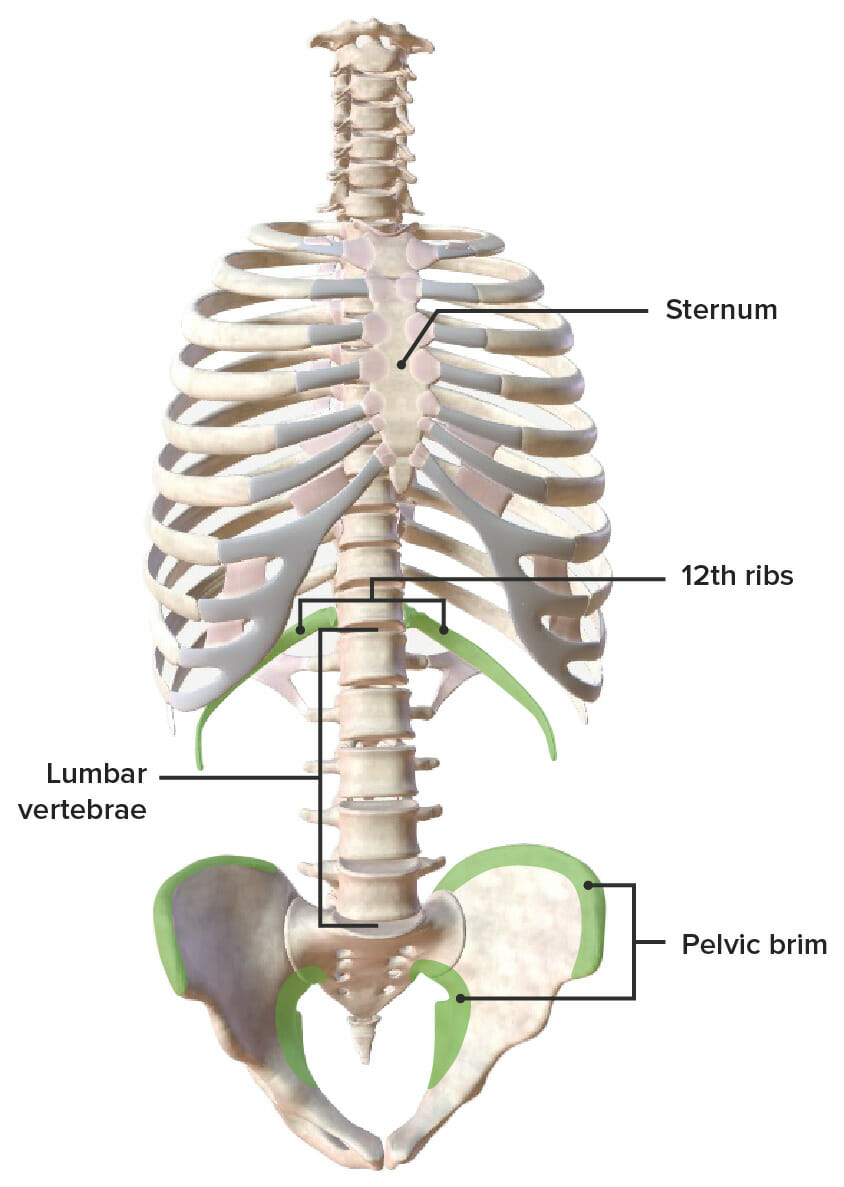
Skeletal boundaries of the posterior abdominal wall:
On the left, boundaries (marked by green): superior boundary: 12th ribs, inferior boundary: pelvic brim. The lumbar vertebrae represent the posterior boundary.
Large area of connective tissue Connective tissue Connective tissues originate from embryonic mesenchyme and are present throughout the body except inside the brain and spinal cord. The main function of connective tissues is to provide structural support to organs. Connective tissues consist of cells and an extracellular matrix. Connective Tissue: Histology that is made up of 3 layers:
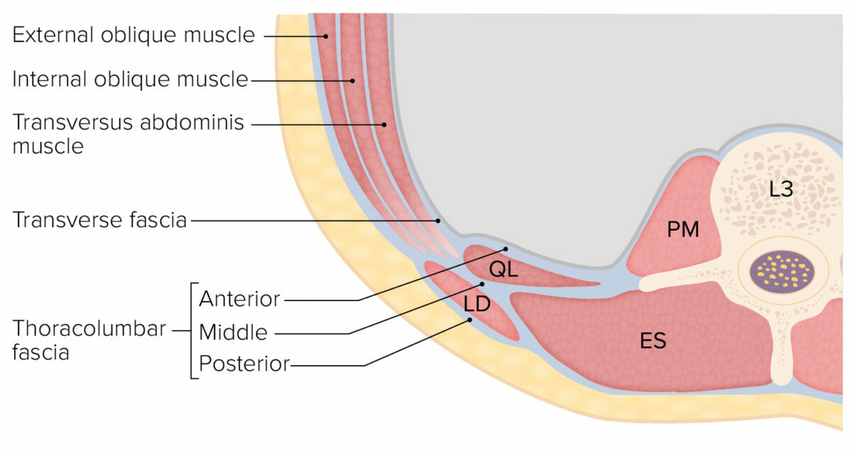
Abdominal wall muscles and the thoracolumbar fascia (cross section at L3 level):
ES: erector spinae
LD: latissimus dorsi
PM: psoas major
QL: quadratus lumborum
Similarly to the anterior abdominal wall Abdominal wall The outer margins of the abdomen, extending from the osteocartilaginous thoracic cage to the pelvis. Though its major part is muscular, the abdominal wall consists of at least seven layers: the skin, subcutaneous fat, deep fascia; abdominal muscles, transversalis fascia, extraperitoneal fat, and the parietal peritoneum. Surgical Anatomy of the Abdomen, herniation Herniation Omphalocele can occur in weakened areas in the posterior abdominal wall Abdominal wall The outer margins of the abdomen, extending from the osteocartilaginous thoracic cage to the pelvis. Though its major part is muscular, the abdominal wall consists of at least seven layers: the skin, subcutaneous fat, deep fascia; abdominal muscles, transversalis fascia, extraperitoneal fat, and the parietal peritoneum. Surgical Anatomy of the Abdomen. These herniations occur in the Grynfeltt-Lesshaft triangle (superior lumbar triangle) or Petit triangle (inferior lumbar triangle).
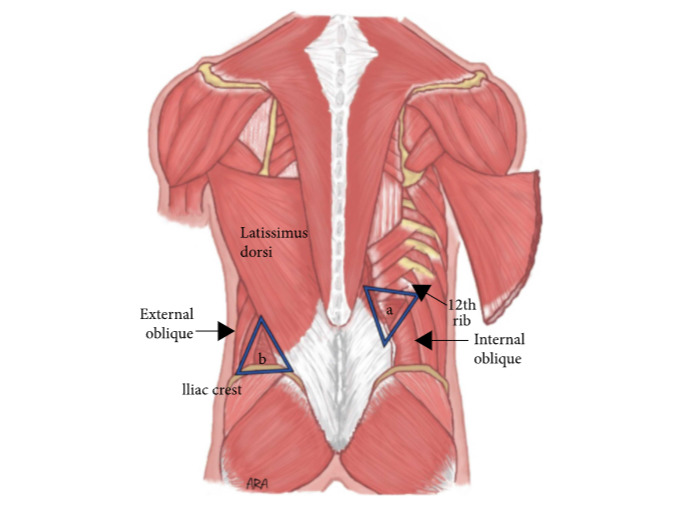
Lumbar triangles:
a: Superior lumbar triangle
b: Inferior lumbar triangle
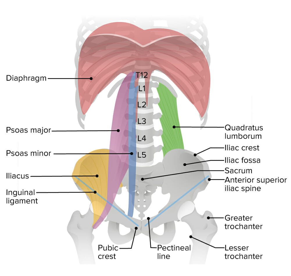
Illustration of muscles (including their origin and insertion) in the posterior abdominal wall
Image by Lecturio.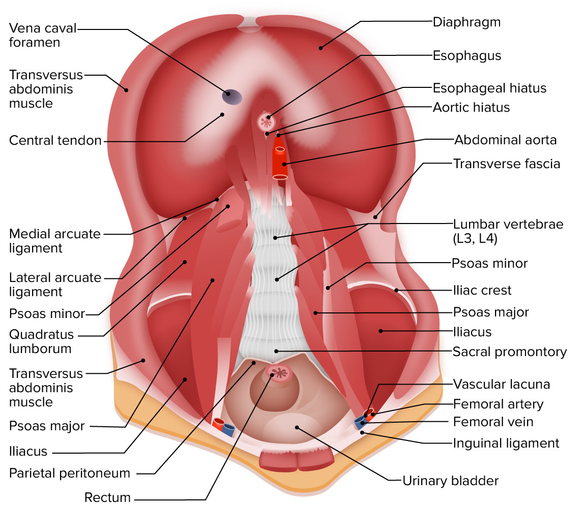
This image demonstrates all the major muscles of the posterior abdominal wall in relation to other organs, blood vessels and supporting structures:
Note that the bulk of the wall is made up of the quadratus lumborum, iliacus, and psoas muscles.
Image by Lecturio.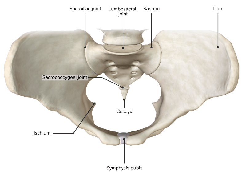
Superior view of the pelvic girdle and the 4 primary joints of the pelvis
Image by Lecturio.| Location in the abdominal aorta Aorta The main trunk of the systemic arteries. Mediastinum and Great Vessels: Anatomy | Branches ( arteries Arteries Arteries are tubular collections of cells that transport oxygenated blood and nutrients from the heart to the tissues of the body. The blood passes through the arteries in order of decreasing luminal diameter, starting in the largest artery (the aorta) and ending in the small arterioles. Arteries are classified into 3 types: large elastic arteries, medium muscular arteries, and small arteries and arterioles. Arteries: Histology) of the abdominal aorta Aorta The main trunk of the systemic arteries. Mediastinum and Great Vessels: Anatomy | Level | Paired or unpaired |
|---|---|---|---|
| Anterior | Celiac trunk | T12 | Unpaired |
| Superior mesenteric | L1 | ||
| Inferior mesenteric | L3 | ||
| Lateral | Middle suprarenal/adrenal | L1 | Paired |
| Renal | L1 to L2 | ||
| Gonadal (testicular or ovarian) | L2 | ||
| Dorsal | Inferior phrenic | T12 | Paired |
| Lumbar (4) | L1, L2, L3, L4 | ||
| Median sacral | L4 (above the bifurcation) | Unpaired | |
| Terminal | Common iliac | L4 | Paired |
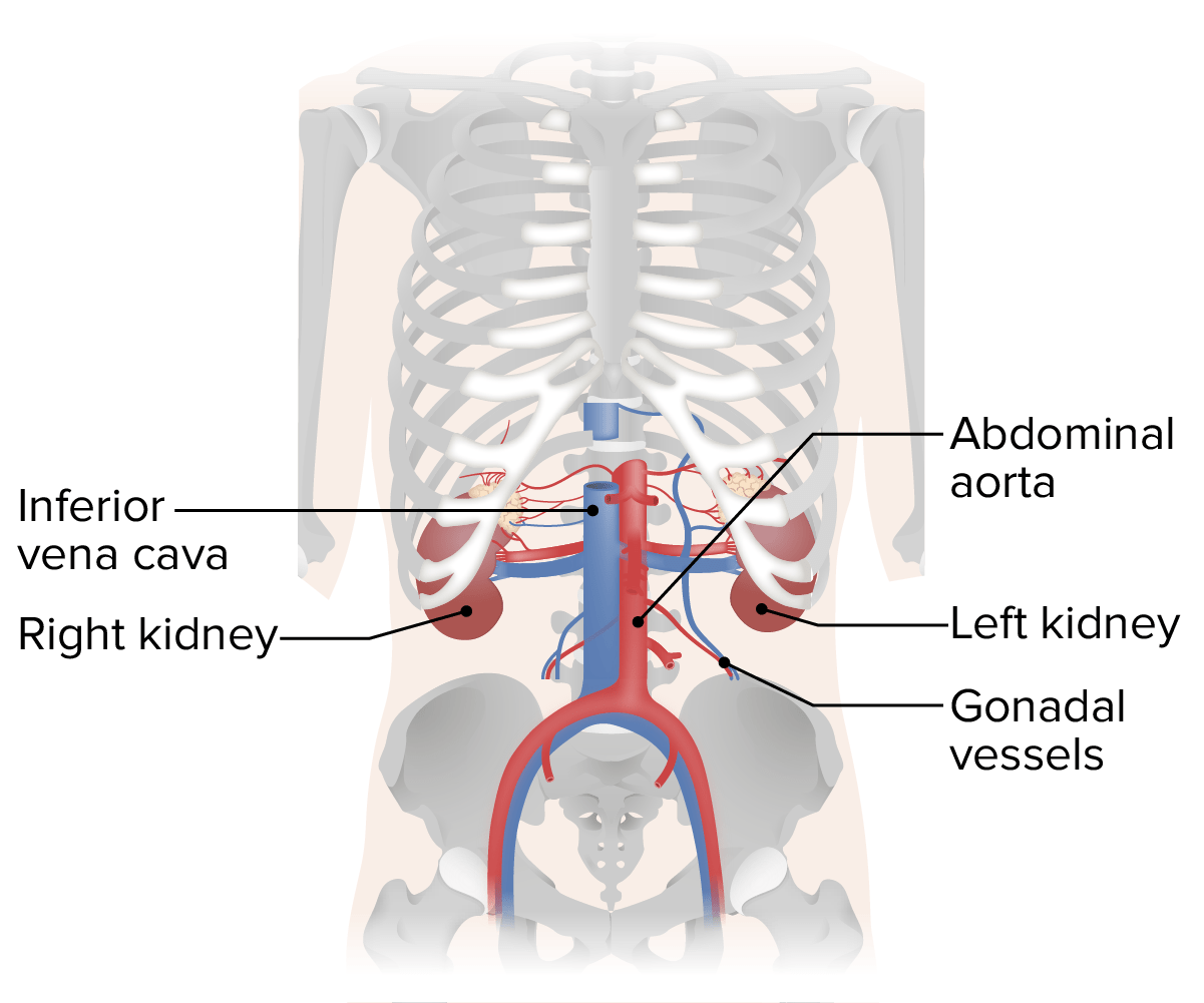
Vascular structures in the posterior abdominal wall (in relation to the kidneys and skeletal frame)
Image by Lecturio.Somatic innervation comes from the ventral rami of the subcostal and lumbar spinal nerves Spinal nerves The 31 paired peripheral nerves formed by the union of the dorsal and ventral spinal roots from each spinal cord segment. The spinal nerve plexuses and the spinal roots are also included. Spinal Cord: Anatomy.
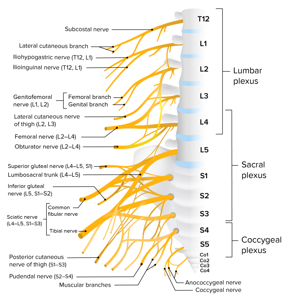
Lumbosacral plexus:
Innervation to the posterior abdominal wall comes from the ventral rami of the subcostal and lumbar spinal nerves.