Porphyrias are a group of metabolic disorders caused by a disturbance in the synthesis Synthesis Polymerase Chain Reaction (PCR) of heme. In most cases, porphyria is caused by a hereditary enzyme defect. The disease patterns differ depending on the affected enzyme, and the variants of porphyria can be clinically differentiated between acute and nonacute forms. Patients Patients Individuals participating in the health care system for the purpose of receiving therapeutic, diagnostic, or preventive procedures. Clinician–Patient Relationship with porphyria present with photosensitive skin Skin The skin, also referred to as the integumentary system, is the largest organ of the body. The skin is primarily composed of the epidermis (outer layer) and dermis (deep layer). The epidermis is primarily composed of keratinocytes that undergo rapid turnover, while the dermis contains dense layers of connective tissue. Skin: Structure and Functions eruptions and sometimes systemic symptoms such as abdominal pain Abdominal Pain Acute Abdomen and neuropathy Neuropathy Leprosy. Porphyrias are managed by avoiding triggers, such as sun exposure and consumption of alcohol. When flares occur, therapy is targeted toward symptomatic relief.
Last updated: Apr 24, 2025
Porphyrias are rare metabolic disorders caused by impairments in heme synthesis Synthesis Polymerase Chain Reaction (PCR).
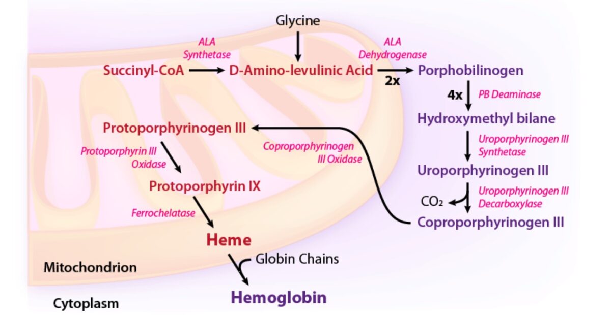
Porphyrias are due to enzymatic defects in heme biosynthesis Biosynthesis The biosynthesis of peptides and proteins on ribosomes, directed by messenger RNA, via transfer RNA that is charged with standard proteinogenic amino acids. Virology:

Enzymatic pathway of heme synthesis:
ALA: aminolevulinic acid
CoA: coenzyme A

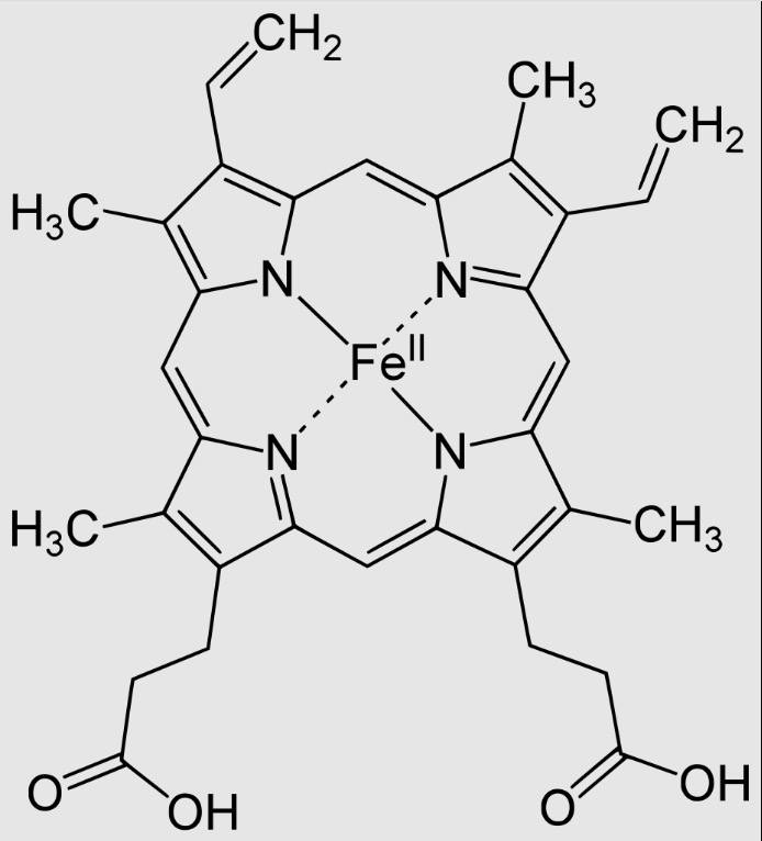
Heme B (most common type of heme)
Image: “Heme B” by Yikrazuul. License: Public Domain| Form of porphyria | Defective enzyme |
|---|---|
| Porphyria cutanea tarda | Uroporphyrinogen III decarboxylase |
| Acute intermittent porphyria | Porphobilinogen Porphobilinogen Heme Metabolism deaminase |
| X-linked X-linked Genetic diseases that are linked to gene mutations on the X chromosome in humans or the X chromosome in other species. Included here are animal models of human X-linked diseases. Common Variable Immunodeficiency (CVID) protoporphyria | δ-Aminolevulinic acid synthase 2 |
| Congenital erythropoietic porphyria | Uroporphyrinogen III synthase Uroporphyrinogen III synthase An enzyme that catalyzes the cyclization of hydroxymethylbilane to yield uroporphyrinogen III and water. It is the fourth enzyme in the 8-enzyme biosynthetic pathway of heme, and is encoded by uros gene. Heme Metabolism |
| Hereditary coproporphyria | Coproporphyrinogen oxidase Oxidase Neisseria |
| Variegate porphyria | Protoporphyrinogen Protoporphyrinogen Heme Metabolism oxidase Oxidase Neisseria |
| Erythropoietic protoporphyria | Ferrochelatase Ferrochelatase A mitochondrial enzyme found in a wide variety of cells and tissues. It is the final enzyme in the 8-enzyme biosynthetic pathway of heme. Ferrochelatase catalyzes ferrous insertion into protoporphyrin IX to form protoheme or heme. Heme Metabolism |
Most commonly, porphyrias are due to an inherited enzyme defect within the heme biosynthesis Biosynthesis The biosynthesis of peptides and proteins on ribosomes, directed by messenger RNA, via transfer RNA that is charged with standard proteinogenic amino acids. Virology pathway. They may rarely be acquired later in life:
Clinical presentation depends on the pattern of organ involvement.

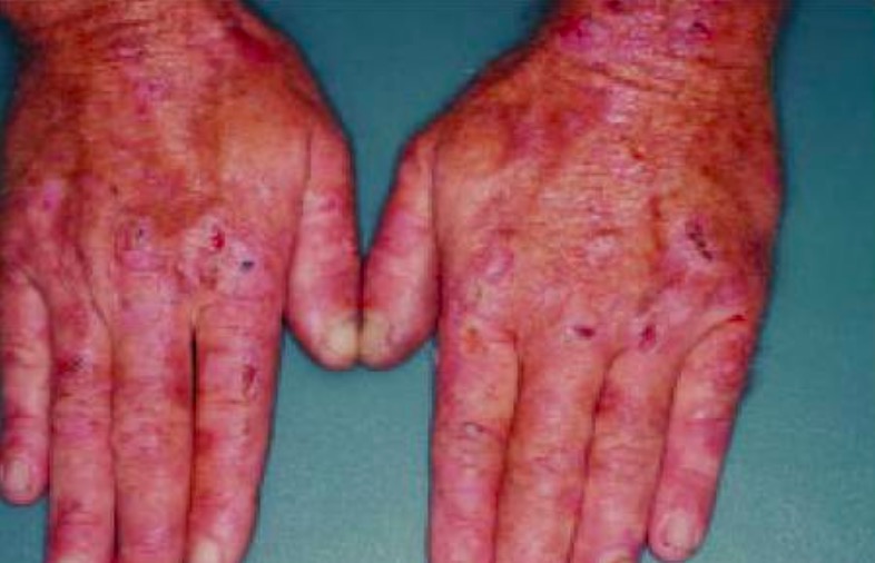
Erosions, crust, and blisters are evident on the hands of this patient with porphyria cutanea tarda.
Image: “Erosions, crust, and blisters are evident on the hands of this patient with porphyria cutanea tarda” by Department of Internal Medicine, Minia University, Minia, Egypt. License: CC BY 2.5
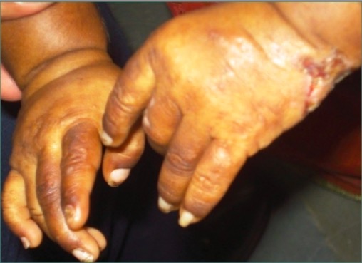
Photograph showing the presence of lesions and scars on both hands of a patient with congenital erythropoietic porphyria
Image: “Erythrodontia in congenital erythropoietic porphyria” by Department of Oral Pathology and Microbiology, Kamineni Institute of Dental Sciences, Narketpalli, Andhra Pradesh, India. License: CC BY 2.0The diagnosis of porphyrias is typically made using specialized blood tests.
There is no known cure for porphyria.