Advertisement

Advertisement

Advertisement

Advertisement

Pain: Types and Pathways

Pain has accompanied humans since they first existed, first lamented as the curse of existence and later understood as an adaptive mechanism that ensures survival. Pain is the most common symptomatic complaint and the main reason why people seek medical care Medical care Conflict of Interest. Pain symptoms are seen every day, by every physician, in every clinic and hospital in the world. Understanding pain physiology is the cornerstone to understanding how to treat it and to providing the individual with their first sigh of relief as definitive management is undertaken.

Last updated: May 17, 2024

Editorial responsibility: Stanley Oiseth, Lindsay Jones, Evelin Maza

Advertisement

Advertisement

Advertisement

Advertisement

Advertisement

Advertisement

Overview

Definitions

  • Pain:
    • According to the International Association for the Study of Pain (IASP), pain is “an unpleasant sensory Sensory Neurons which conduct nerve impulses to the central nervous system. Nervous System: Histology and emotional experience associated with actual or potential tissue damage, or described in terms of such damage.” 
    • Mental suffering or distress
  • Nociception:
    • Noxious (or toxic) stimulus or stimulus that can become noxious with prolonged exposure 
    • Process through which peripheral pain receptors Receptors Receptors are proteins located either on the surface of or within a cell that can bind to signaling molecules known as ligands (e.g., hormones) and cause some type of response within the cell. Receptors transmit information about current (or potential) tissue damage centrally as pain
  • Nociceptor: receptor Receptor Receptors are proteins located either on the surface of or within a cell that can bind to signaling molecules known as ligands (e.g., hormones) and cause some type of response within the cell. Receptors in end organ that detects biochemical changes associated with current or potential tissue damage
  • Hyperalgesia: exaggerated response to noxious stimuli
  • Allodynia Allodynia Pain due to a stimulus that does not typically provoke pain. Pain Management: sensation of pain in response to an innocuous stimulus

Types of pain

  • Physiologic (acute) pain
  • Pathologic (chronic) pain
  • Nociceptive:
    • Pain in response to actual or potentially harmful stimuli
    • Often described as aching, localized 
    • Aggravated by movement
  • Neuropathic:
Table: Differences between acute and chronic pain Chronic pain Aching sensation that persists for more than a few months. It may or may not be associated with trauma or disease, and may persist after the initial injury has healed. Its localization, character, and timing are more vague than with acute pain. Pain Management
Physiologic change Acute pain Acute pain Intensely discomforting, distressful, or agonizing sensation associated with trauma or disease, with well-defined location, character, and timing. Pain Management Chronic pain Chronic pain Aching sensation that persists for more than a few months. It may or may not be associated with trauma or disease, and may persist after the initial injury has healed. Its localization, character, and timing are more vague than with acute pain. Pain Management
Vital signs May vary consistently with degree of pain severity No or minimal change
Purpose of pain Useful Inhibits function and not useful
Central sensitization Short term; improves with healing of injury Remains present despite absence of ongoing injury
Neuropathic pain Increases likelihood of chronic pain Chronic pain Aching sensation that persists for more than a few months. It may or may not be associated with trauma or disease, and may persist after the initial injury has healed. Its localization, character, and timing are more vague than with acute pain. Pain Management when present in acute phase Acute phase Short Bowel Syndrome Common etiology of chronic pain Chronic pain Aching sensation that persists for more than a few months. It may or may not be associated with trauma or disease, and may persist after the initial injury has healed. Its localization, character, and timing are more vague than with acute pain. Pain Management
Nociceptive pain Often found during acute pain Acute pain Intensely discomforting, distressful, or agonizing sensation associated with trauma or disease, with well-defined location, character, and timing. Pain Management state Commonly presents with some neuropathic pain
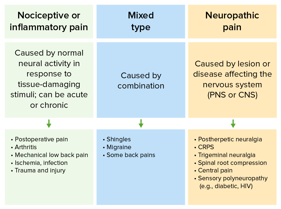
The types of pain

Differences in types of pain and their common etiologies
CRPS: complex regional pain syndrome
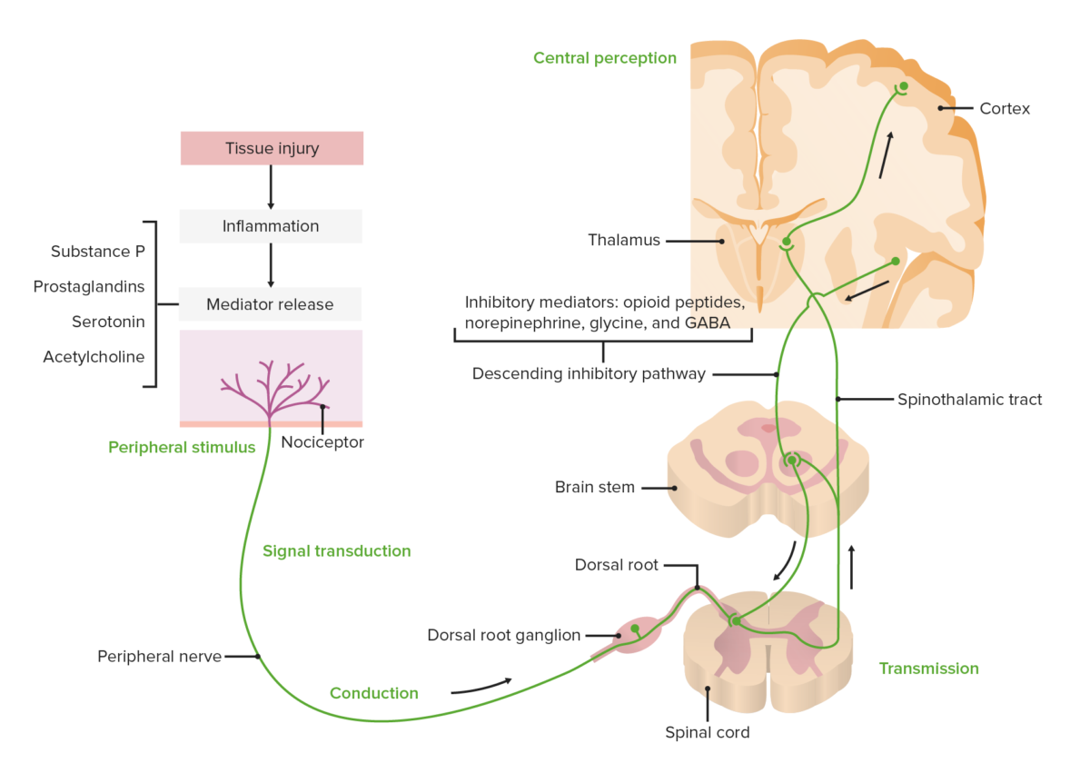
Image by Lecturio.

Embryology

  • 7 weeks: development of free nerve endings 
  • 18 weeks: hormonal stress responses to pain 
  • 23–30 weeks: thalamic projections into the somatosensory cortex Somatosensory cortex Area of the parietal lobe concerned with receiving sensations such as movement, pain, pressure, position, temperature, touch, and vibration. It lies posterior to the central sulcus. Cerebral Cortex: Anatomy
  • 26 weeks: hemodynamic and behavioral reactions to painful stimuli

Function of pain

  • Adaptive measures to minimize and avoid further tissue damage
  • Evoked responses:
    • Withdrawal from noxious stimulus (e.g., spinal reflexes)
    • Anticipatory movements (e.g., movement of the arms to protect the eyes and face, bracing before imminent impact)

Anatomy and Physiology of Pain Pathways

Phases

  1. Transduction Transduction The transfer of bacterial DNA by phages from an infected bacterium to another bacterium. This also refers to the transfer of genes into eukaryotic cells by viruses. This naturally occurring process is routinely employed as a gene transfer technique. Bacteriology
  2. Transmission
  3. Modulation
  4. Central perception Perception The process by which the nature and meaning of sensory stimuli are recognized and interpreted. Psychiatric Assessment

Nociception process

  • Thermal, mechanical, or chemical stimuli of noxious intensity comes into contact with a tissue.
  • Injured tissue releases inflammatory mediators, including:
    • Globulin
    • Protein kinases Kinases Macrolides and Ketolides
    • Arachidonic acid Arachidonic Acid An unsaturated, essential fatty acid. It is found in animal and human fat as well as in the liver, brain, and glandular organs, and is a constituent of animal phosphatides. It is formed by the synthesis from dietary linoleic acid and is a precursor in the biosynthesis of prostaglandins, thromboxanes, and leukotrienes. Nonsteroidal Antiinflammatory Drugs (NSAIDs)
    • Histamine
    • Nerve growth factor (NGF)
    • Substance P (SP)
    • Calcitonin Calcitonin A peptide hormone that lowers calcium concentration in the blood. In humans, it is released by thyroid cells and acts to decrease the formation and absorptive activity of osteoclasts. Its role in regulating plasma calcium is much greater in children and in certain diseases than in normal adults. Other Antiresorptive Drugs gene Gene A category of nucleic acid sequences that function as units of heredity and which code for the basic instructions for the development, reproduction, and maintenance of organisms. Basic Terms of Genetics–related peptide ( CGRP CGRP A 37-amino acid peptide derived from the calcitonin gene. It occurs as a result of alternative processing of mRNA from the calcitonin gene. The neuropeptide is widely distributed in the brain, gut, perivascular nerves, and other tissue. The peptide produces multiple biological effects and has both circulatory and neurotransmitter modes of action. In particular, it is a potent endogenous vasodilator. Gastrointestinal Neural and Hormonal Signaling)
  • These mediators stimulate transducer Transducer A device placed on the patient’s body to visualize a target Ultrasound (Sonography) channels Channels The Cell: Cell Membrane (similar to voltage-gated channels Channels The Cell: Cell Membrane) → initiation of receptor Receptor Receptors are proteins located either on the surface of or within a cell that can bind to signaling molecules known as ligands (e.g., hormones) and cause some type of response within the cell. Receptors potentials ( transduction Transduction The transfer of bacterial DNA by phages from an infected bacterium to another bacterium. This also refers to the transfer of genes into eukaryotic cells by viruses. This naturally occurring process is routinely employed as a gene transfer technique. Bacteriology)
  • Receptor Receptor Receptors are proteins located either on the surface of or within a cell that can bind to signaling molecules known as ligands (e.g., hormones) and cause some type of response within the cell. Receptors potentials evoke action potentials in sensory Sensory Neurons which conduct nerve impulses to the central nervous system. Nervous System: Histology nerve fibers Nerve Fibers Slender processes of neurons, including the axons and their glial envelopes (myelin sheath). Nerve fibers conduct nerve impulses to and from the central nervous system. Nervous System: Histology.
  • Action potentials are carried as afferent Afferent Neurons which conduct nerve impulses to the central nervous system. Nervous System: Histology signals via sensory Sensory Neurons which conduct nerve impulses to the central nervous system. Nervous System: Histology nerve fibers Nerve Fibers Slender processes of neurons, including the axons and their glial envelopes (myelin sheath). Nerve fibers conduct nerve impulses to and from the central nervous system. Nervous System: Histology to the dorsal root ganglia and dorsal horn Dorsal horn One of three central columns of the spinal cord. It is composed of gray matter spinal laminae i-vi. Brown-Séquard Syndrome of the spinal cord Spinal cord The spinal cord is the major conduction pathway connecting the brain to the body; it is part of the CNS. In cross section, the spinal cord is divided into an H-shaped area of gray matter (consisting of synapsing neuronal cell bodies) and a surrounding area of white matter (consisting of ascending and descending tracts of myelinated axons). Spinal Cord: Anatomy (transmission).
  • From there, the signal is transmitted up the spinal cord Spinal cord The spinal cord is the major conduction pathway connecting the brain to the body; it is part of the CNS. In cross section, the spinal cord is divided into an H-shaped area of gray matter (consisting of synapsing neuronal cell bodies) and a surrounding area of white matter (consisting of ascending and descending tracts of myelinated axons). Spinal Cord: Anatomy to the brain Brain The part of central nervous system that is contained within the skull (cranium). Arising from the neural tube, the embryonic brain is comprised of three major parts including prosencephalon (the forebrain); mesencephalon (the midbrain); and rhombencephalon (the hindbrain). The developed brain consists of cerebrum; cerebellum; and other structures in the brain stem. Nervous System: Anatomy, Structure, and Classification stem and thalamus Thalamus The thalamus is a large, ovoid structure in the dorsal part of the diencephalon that is located between the cerebral cortex and midbrain. It consists of several interconnected nuclei of grey matter separated by the laminae of white matter. The thalamus is the main conductor of information that passes between the cerebral cortex and the periphery, spinal cord, or brain stem. Thalamus: Anatomy, where significant processing (modulation) may occur.
  • The signal finally reaches the somatosensory cortex Somatosensory cortex Area of the parietal lobe concerned with receiving sensations such as movement, pain, pressure, position, temperature, touch, and vibration. It lies posterior to the central sulcus. Cerebral Cortex: Anatomy (central perception Perception The process by which the nature and meaning of sensory stimuli are recognized and interpreted. Psychiatric Assessment). The biopsychosocial interpretation of the painful experience also involves:
    • Amygdala Amygdala Almond-shaped group of basal nuclei anterior to the inferior horn of the lateral ventricle of the temporal lobe. The amygdala is part of the limbic system. Limbic System: Anatomy: involved in the emotional and affective response to pain and pain modulation
    • Hypothalamus Hypothalamus The hypothalamus is a collection of various nuclei within the diencephalon in the center of the brain. The hypothalamus plays a vital role in endocrine regulation as the primary regulator of the pituitary gland, and it is the major point of integration between the central nervous and endocrine systems. Hypothalamus: involved in the neuroendocrine corticotropin response to pain
    • Periaqueductal gray matter Gray matter Region of central nervous system that appears darker in color than the other type, white matter. It is composed of neuronal cell bodies; neuropil; glial cells and capillaries but few myelinated nerve fibers. Cerebral Cortex: Anatomy: key center for pain modulation, involved in aversive and defensive pain behaviors
    • Basal ganglia Basal Ganglia Basal ganglia are a group of subcortical nuclear agglomerations involved in movement, and are located deep to the cerebral hemispheres. Basal ganglia include the striatum (caudate nucleus and putamen), globus pallidus, substantia nigra, and subthalamic nucleus. Basal Ganglia: Anatomy: involved in the cognitive, affective, and discriminative (ability to localize sensory Sensory Neurons which conduct nerve impulses to the central nervous system. Nervous System: Histology input) aspects of pain perception Perception The process by which the nature and meaning of sensory stimuli are recognized and interpreted. Psychiatric Assessment
    • Cerebral cortex Cerebral cortex The cerebral cortex is the largest and most developed part of the human brain and CNS. Occupying the upper part of the cranial cavity, the cerebral cortex has 4 lobes and is divided into 2 hemispheres that are joined centrally by the corpus callosum. Cerebral Cortex: Anatomy: ultimate site of pain perception Perception The process by which the nature and meaning of sensory stimuli are recognized and interpreted. Psychiatric Assessment, potential for conscious activation of descending pathways for pain modulation

Modulation of pain

  • Pain signals are modulated (reduced transmission from nociceptive afferents) by endogenous opioid Opioid Compounds with activity like opiate alkaloids, acting at opioid receptors. Properties include induction of analgesia or narcosis. Constipation peptides (e.g., endorphins Endorphins One of the three major groups of endogenous opioid peptides. They are large peptides derived from the pro-opiomelanocortin precursor. The known members of this group are alpha-, beta-, and gamma-endorphin. The term endorphin is also sometimes used to refer to all opioid peptides, but the narrower sense is used here; opioid peptides is used for the broader group. Receptors and Neurotransmitters of the CNS, dynorphins Dynorphins A class of opioid peptides including dynorphin a, dynorphin b, and smaller fragments of these peptides. Dynorphins prefer kappa-opioid receptors and have been shown to play a role as central nervous system transmitters. Receptors and Neurotransmitters of the CNS, enkephalins Enkephalins One of the three major families of endogenous opioid peptides. The enkephalins are pentapeptides that are widespread in the central and peripheral nervous systems and in the adrenal medulla. Receptors and Neurotransmitters of the CNS) in the:
    • Spinal cord Spinal cord The spinal cord is the major conduction pathway connecting the brain to the body; it is part of the CNS. In cross section, the spinal cord is divided into an H-shaped area of gray matter (consisting of synapsing neuronal cell bodies) and a surrounding area of white matter (consisting of ascending and descending tracts of myelinated axons). Spinal Cord: Anatomy
    • Dorsal root ganglia
    • Midbrain Midbrain The middle of the three primitive cerebral vesicles of the embryonic brain. Without further subdivision, midbrain develops into a short, constricted portion connecting the pons and the diencephalon. Midbrain contains two major parts, the dorsal tectum mesencephali and the ventral tegmentum mesencephali, housing components of auditory, visual, and other sensorimotor systems. Brain Stem: Anatomy periaqueductal gray (PAG)
  • This mechanism occurs in the “descending” (“inhibitory”) pathways.
  • Mechanisms of action of endogenous opioid Opioid Compounds with activity like opiate alkaloids, acting at opioid receptors. Properties include induction of analgesia or narcosis. Constipation peptides:
    • Activation of mu, kappa, and delta opioid Opioid Compounds with activity like opiate alkaloids, acting at opioid receptors. Properties include induction of analgesia or narcosis. Constipation receptors Receptors Receptors are proteins located either on the surface of or within a cell that can bind to signaling molecules known as ligands (e.g., hormones) and cause some type of response within the cell. Receptors → decreased presynaptic Ca CA Condylomata acuminata are a clinical manifestation of genital HPV infection. Condylomata acuminata are described as raised, pearly, flesh-colored, papular, cauliflower-like lesions seen in the anogenital region that may cause itching, pain, or bleeding. Condylomata Acuminata (Genital Warts)2+ influx → decreased release of glutamate Glutamate Derivatives of glutamic acid. Included under this heading are a broad variety of acid forms, salts, esters, and amides that contain the 2-aminopentanedioic acid structure. Synthesis of Nonessential Amino Acids and SP
    • Increased K+ conductance in dorsal horn Dorsal horn One of three central columns of the spinal cord. It is composed of gray matter spinal laminae i-vi. Brown-Séquard Syndrome neurons Neurons The basic cellular units of nervous tissue. Each neuron consists of a body, an axon, and dendrites. Their purpose is to receive, conduct, and transmit impulses in the nervous system. Nervous System: Histology
  • Other modulators include:
Pathway of pain

Diagram showing the pathway of pain transduction, transmission, modulation, and central perception

Image by Lecturio.

Types of afferent Afferent Neurons which conduct nerve impulses to the central nervous system. Nervous System: Histology nerve fibers Nerve Fibers Slender processes of neurons, including the axons and their glial envelopes (myelin sheath). Nerve fibers conduct nerve impulses to and from the central nervous system. Nervous System: Histology

Type A fibers: 

  • Large and myelinated Myelinated Internuclear Ophthalmoplegia → fast-conducting
  • A-alpha: primary receptors Receptors Receptors are proteins located either on the surface of or within a cell that can bind to signaling molecules known as ligands (e.g., hormones) and cause some type of response within the cell. Receptors of the muscle spindle and Golgi tendon organ
  • A-beta:
    • Afferent Afferent Neurons which conduct nerve impulses to the central nervous system. Nervous System: Histology axon with the largest diameter
    • Secondary receptors Receptors Receptors are proteins located either on the surface of or within a cell that can bind to signaling molecules known as ligands (e.g., hormones) and cause some type of response within the cell. Receptors of the muscle spindle that contribute to cutaneous mechanoreceptors.
    • Perceive light touch Light touch Neurological Examination and/or moving stimuli
  • A-delta:
    • Free nerve endings that conduct stimuli related to pressure and temperature.
    • Conduction speed approximately 20 m/sec
  • A-gamma: motor Motor Neurons which send impulses peripherally to activate muscles or secretory cells. Nervous System: Histology neurons Neurons The basic cellular units of nervous tissue. Each neuron consists of a body, an axon, and dendrites. Their purpose is to receive, conduct, and transmit impulses in the nervous system. Nervous System: Histology that control the intrinsic activation of the muscle spindle.

Type B fibers:

Type C fibers:

  • Unmyelinated nociceptor slow fibers 
  • Conduction speed approximately 2 m/sec
  • Respond to combinations of thermal, mechanical, and chemical stimuli

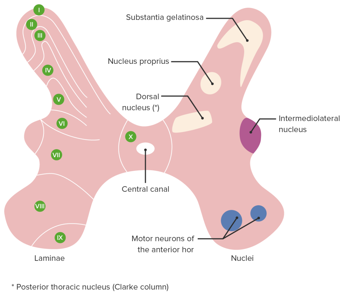
Rexed laminae

  • Organized somatosensory and motor Motor Neurons which send impulses peripherally to activate muscles or secretory cells. Nervous System: Histology map laid out in the spinal cord Spinal cord The spinal cord is the major conduction pathway connecting the brain to the body; it is part of the CNS. In cross section, the spinal cord is divided into an H-shaped area of gray matter (consisting of synapsing neuronal cell bodies) and a surrounding area of white matter (consisting of ascending and descending tracts of myelinated axons). Spinal Cord: Anatomy (primarily the dorsal horn Dorsal horn One of three central columns of the spinal cord. It is composed of gray matter spinal laminae i-vi. Brown-Séquard Syndrome) of each spinal segment
  • Different types of sensory Sensory Neurons which conduct nerve impulses to the central nervous system. Nervous System: Histology nerves (and some motor Motor Neurons which send impulses peripherally to activate muscles or secretory cells. Nervous System: Histology, laminae VI–IX) and the corresponding information they carry are organized so as to synapse Synapse The junction between 2 neurons is called a synapse. The synapse allows a neuron to pass an electrical or chemical signal to another neuron or target effector cell. Synapses and Neurotransmission in specific territories in the dorsal horn Dorsal horn One of three central columns of the spinal cord. It is composed of gray matter spinal laminae i-vi. Brown-Séquard Syndrome known as laminae.
  • There are 10 laminae, designated I–X:
    • Lamina I: receives and relays noxious and thermal stimuli
    • Lamina II: receives and relays noxious and nonnoxious physical stimuli Physical stimuli Act of eliciting a response from a person or organism through physical contact. Angioedema and is involved in pain modulation
    • Lamina III: receives and relays physical stimuli Physical stimuli Act of eliciting a response from a person or organism through physical contact. Angioedema related to light touch Light touch Neurological Examination and proprioception Proprioception Sensory functions that transduce stimuli received by proprioceptive receptors in joints, tendons, muscles, and the inner ear into neural impulses to be transmitted to the central nervous system. Proprioception provides sense of stationary positions and movements of one’s body parts, and is important in maintaining kinesthesia and postural balance. Neurological Examination
    • Lamina IV: receives and relays nonnoxious physical stimuli Physical stimuli Act of eliciting a response from a person or organism through physical contact. Angioedema
    • Lamina V: receives and relays noxious stimuli and is involved in pain modulation
    • Lamina VI: receives and relays information involved in spinal reflexes and proprioception Proprioception Sensory functions that transduce stimuli received by proprioceptive receptors in joints, tendons, muscles, and the inner ear into neural impulses to be transmitted to the central nervous system. Proprioception provides sense of stationary positions and movements of one’s body parts, and is important in maintaining kinesthesia and postural balance. Neurological Examination
    • Lamina VII: receives and relays information related to visceral function and noxious stimuli 
    • Lamina VIII: receives and relays information related to modulation of voluntary movement
    • Lamina IX: receives and relays information related to motor Motor Neurons which send impulses peripherally to activate muscles or secretory cells. Nervous System: Histology control (gross muscle contraction)
    • Lamina X: centrally located (central gray commissure Gray commissure Spinal Cord: Anatomy); where sensory Sensory Neurons which conduct nerve impulses to the central nervous system. Nervous System: Histology and motor Motor Neurons which send impulses peripherally to activate muscles or secretory cells. Nervous System: Histology neurons Neurons The basic cellular units of nervous tissue. Each neuron consists of a body, an axon, and dendrites. Their purpose is to receive, conduct, and transmit impulses in the nervous system. Nervous System: Histology cross before ascending/descending and where some degree of interneuronal cross-talk (modulation) takes place
  • There are also associated nuclei that are beyond the scope of this discussion.
  • Under physiologic conditions, sensory Sensory Neurons which conduct nerve impulses to the central nervous system. Nervous System: Histology and motor Motor Neurons which send impulses peripherally to activate muscles or secretory cells. Nervous System: Histology information (and associated modulation) is transmitted within and among the laminae in a highly organized and predictable fashion.
  • Under pathologic conditions (e.g., persistent nociceptive input, neurologic damage), there can be an abnormal rearrangement of sensory Sensory Neurons which conduct nerve impulses to the central nervous system. Nervous System: Histology inputs that contributes to the development of central sensitization and other manifestations of chronic pain Chronic pain Aching sensation that persists for more than a few months. It may or may not be associated with trauma or disease, and may persist after the initial injury has healed. Its localization, character, and timing are more vague than with acute pain. Pain Management:
    • Nociceptive neural inputs land in laminae devoted to nonnoxious or motor Motor Neurons which send impulses peripherally to activate muscles or secretory cells. Nervous System: Histology stimuli.
    • Nonnoxious or motor Motor Neurons which send impulses peripherally to activate muscles or secretory cells. Nervous System: Histology inputs land in laminae devoted to nociceptive stimuli.
    • Leads to abnormal pain perception Perception The process by which the nature and meaning of sensory stimuli are recognized and interpreted. Psychiatric Assessment to normally nonnoxious stimuli or movements:
      • Allodynia Allodynia Pain due to a stimulus that does not typically provoke pain. Pain Management
      • Hypersensitivity
      • Hyperalgesia
      • Paresthesia/ dysesthesia Dysesthesia Complex Regional Pain Syndrome (CRPS)
      • Development of neuropathic pain in an area devoid of nerve damage
      • Expansion of the receptive field beyond the normal territory of a peripheral nerve
      • Spontaneous pain in the absence of noxious stimuli or tissue damage
    • Abnormal interneuronal reflexes may develop:
      • Muscle activation or joint movement ( motor Motor Neurons which send impulses peripherally to activate muscles or secretory cells. Nervous System: Histology stimuli, proprioceptive stimuli) triggers painful stimuli.
      • Painful stimuli may trigger Trigger The type of signal that initiates the inspiratory phase by the ventilator Invasive Mechanical Ventilation abnormal motor Motor Neurons which send impulses peripherally to activate muscles or secretory cells. Nervous System: Histology activity (e.g., hypertonicity Hypertonicity Volume Depletion and Dehydration, muscle spasm).
      • Painful stimuli may trigger Trigger The type of signal that initiates the inspiratory phase by the ventilator Invasive Mechanical Ventilation autonomic or enteric phenomena and vice versa. For example:
        • Nausea Nausea An unpleasant sensation in the stomach usually accompanied by the urge to vomit. Common causes are early pregnancy, sea and motion sickness, emotional stress, intense pain, food poisoning, and various enteroviruses. Antiemetics in response to painful stimulus
        • Sweating and/or piloerection in response to painful stimulus
        • Vasoconstriction Vasoconstriction The physiological narrowing of blood vessels by contraction of the vascular smooth muscle. Vascular Resistance, Flow, and Mean Arterial Pressure in an area of painful stimulus (i.e., CRPS CRPS Complex regional pain syndrome (CRPS) is a chronic regional neuropathic pain condition characterized by excruciating pain (out of proportion to apparent tissue damage or inciting trauma), paresthesia, allodynia, temperature abnormalities, skin discoloration, edema, reduced range of motion, and bone demineralization. Complex Regional Pain Syndrome (CRPS))
A cross section of the spinal cord showing rexed laminae

Cross section of the spinal cord showing the rexed laminae (left) and associated nuclei (right)

Image by Lecturio.

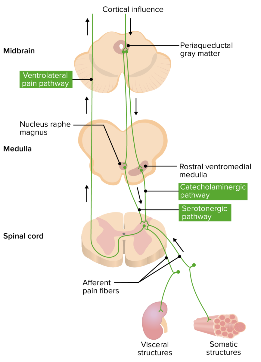
Ascending and descending pain pathways

Ascending pathway of pain:

  • Nociceptors:
    • Receptors Receptors Receptors are proteins located either on the surface of or within a cell that can bind to signaling molecules known as ligands (e.g., hormones) and cause some type of response within the cell. Receptors in the periphery respond to heat Heat Inflammation, intense cold, mechanical distortion Distortion Defense Mechanisms, changes in pH pH The quantitative measurement of the acidity or basicity of a solution. Acid-Base Balance, and chemical irritants (e.g., ADP, bradykinin Bradykinin A nonapeptide messenger that is enzymatically produced from kallidin in the blood where it is a potent but short-lived agent of arteriolar dilation and increased capillary permeability. Bradykinin is also released from mast cells during asthma attacks, from gut walls as a gastrointestinal vasodilator, from damaged tissues as a pain signal, and may be a neurotransmitter. Hereditary Angioedema (C1 Esterase Inhibitor Deficiency), serotonin Serotonin A biochemical messenger and regulator, synthesized from the essential amino acid l-tryptophan. In humans it is found primarily in the central nervous system, gastrointestinal tract, and blood platelets. Serotonin mediates several important physiological functions including neurotransmission, gastrointestinal motility, hemostasis, and cardiovascular integrity. Receptors and Neurotransmitters of the CNS, histamine)
    • Afferent Afferent Neurons which conduct nerve impulses to the central nervous system. Nervous System: Histology nerve conduction → 1st-order neuron 1st-order neuron Originates in the hypothalamus and descends to the first synapse in the cervical spinal cord, located at levels C8–T2 (also called the ciliospinal center of Budge). Horner Syndrome
  • Neuron cell bodies:
    • The cell bodies of 1st-order neurons Neurons The basic cellular units of nervous tissue. Each neuron consists of a body, an axon, and dendrites. Their purpose is to receive, conduct, and transmit impulses in the nervous system. Nervous System: Histology are located in the dorsal horn Dorsal horn One of three central columns of the spinal cord. It is composed of gray matter spinal laminae i-vi. Brown-Séquard Syndrome and dorsal root ganglia of the spinal gray matter Gray matter Region of central nervous system that appears darker in color than the other type, white matter. It is composed of neuronal cell bodies; neuropil; glial cells and capillaries but few myelinated nerve fibers. Cerebral Cortex: Anatomy (or trigeminal ganglia)
    • Glutamate Glutamate Derivatives of glutamic acid. Included under this heading are a broad variety of acid forms, salts, esters, and amides that contain the 2-aminopentanedioic acid structure. Synthesis of Nonessential Amino Acids, SP, and CGRP CGRP A 37-amino acid peptide derived from the calcitonin gene. It occurs as a result of alternative processing of mRNA from the calcitonin gene. The neuropeptide is widely distributed in the brain, gut, perivascular nerves, and other tissue. The peptide produces multiple biological effects and has both circulatory and neurotransmitter modes of action. In particular, it is a potent endogenous vasodilator. Gastrointestinal Neural and Hormonal Signaling are the main neurotransmitters released by primary afferents
  • 2nd-order neuron 2nd-order neuron Preganglionic pupillomotor fibers exit the spinal cord at T1, travel through the brachial plexus, over the lung apex, ascending to the superior cervical ganglion located near the angle of the mandible and the bifurcation of the common carotid artery. Horner Syndrome:
    • After synapsing in the spinal cord Spinal cord The spinal cord is the major conduction pathway connecting the brain to the body; it is part of the CNS. In cross section, the spinal cord is divided into an H-shaped area of gray matter (consisting of synapsing neuronal cell bodies) and a surrounding area of white matter (consisting of ascending and descending tracts of myelinated axons). Spinal Cord: Anatomy, the 1st-order neurons Neurons The basic cellular units of nervous tissue. Each neuron consists of a body, an axon, and dendrites. Their purpose is to receive, conduct, and transmit impulses in the nervous system. Nervous System: Histology project to 2nd-order neurons Neurons The basic cellular units of nervous tissue. Each neuron consists of a body, an axon, and dendrites. Their purpose is to receive, conduct, and transmit impulses in the nervous system. Nervous System: Histology
    • 2nd-order neurons Neurons The basic cellular units of nervous tissue. Each neuron consists of a body, an axon, and dendrites. Their purpose is to receive, conduct, and transmit impulses in the nervous system. Nervous System: Histology cross the midline at the anterior white commissure
    • These neurons Neurons The basic cellular units of nervous tissue. Each neuron consists of a body, an axon, and dendrites. Their purpose is to receive, conduct, and transmit impulses in the nervous system. Nervous System: Histology then ascend to the thalamus Thalamus The thalamus is a large, ovoid structure in the dorsal part of the diencephalon that is located between the cerebral cortex and midbrain. It consists of several interconnected nuclei of grey matter separated by the laminae of white matter. The thalamus is the main conductor of information that passes between the cerebral cortex and the periphery, spinal cord, or brain stem. Thalamus: Anatomy via the contralateral spinothalamic tract, carrying both pain and temperature sensations.
  • Thalamus Thalamus The thalamus is a large, ovoid structure in the dorsal part of the diencephalon that is located between the cerebral cortex and midbrain. It consists of several interconnected nuclei of grey matter separated by the laminae of white matter. The thalamus is the main conductor of information that passes between the cerebral cortex and the periphery, spinal cord, or brain stem. Thalamus: Anatomy:
    • From the thalamus Thalamus The thalamus is a large, ovoid structure in the dorsal part of the diencephalon that is located between the cerebral cortex and midbrain. It consists of several interconnected nuclei of grey matter separated by the laminae of white matter. The thalamus is the main conductor of information that passes between the cerebral cortex and the periphery, spinal cord, or brain stem. Thalamus: Anatomy, the stimulus is sent to the somatosensory cerebral cortex Cerebral cortex The cerebral cortex is the largest and most developed part of the human brain and CNS. Occupying the upper part of the cranial cavity, the cerebral cortex has 4 lobes and is divided into 2 hemispheres that are joined centrally by the corpus callosum. Cerebral Cortex: Anatomy via fibers in the posterior limb of the internal capsule Capsule An envelope of loose gel surrounding a bacterial cell which is associated with the virulence of pathogenic bacteria. Some capsules have a well-defined border, whereas others form a slime layer that trails off into the medium. Most capsules consist of relatively simple polysaccharides but there are some bacteria whose capsules are made of polypeptides. Bacteroides
    • Other thalamic neurons Neurons The basic cellular units of nervous tissue. Each neuron consists of a body, an axon, and dendrites. Their purpose is to receive, conduct, and transmit impulses in the nervous system. Nervous System: Histology project to areas of the cortex associated with emotional responses (e.g., cingulate gyrus Cingulate gyrus One of the convolutions on the medial surface of the cerebral hemispheres. It surrounds the rostral part of the brain and corpus callosum and forms part of the limbic system. Limbic System: Anatomy, insular cortex)

Descending pathway of pain:

The hypothalamus Hypothalamus The hypothalamus is a collection of various nuclei within the diencephalon in the center of the brain. The hypothalamus plays a vital role in endocrine regulation as the primary regulator of the pituitary gland, and it is the major point of integration between the central nervous and endocrine systems. Hypothalamus and cortical regions process painful stimuli and signal for the release of inhibitory mediators and hormones Hormones Hormones are messenger molecules that are synthesized in one part of the body and move through the bloodstream to exert specific regulatory effects on another part of the body. Hormones play critical roles in coordinating cellular activities throughout the body in response to the constant changes in both the internal and external environments. Hormones: Overview and Types (e.g., opioid Opioid Compounds with activity like opiate alkaloids, acting at opioid receptors. Properties include induction of analgesia or narcosis. Constipation peptides, norepinephrine Norepinephrine Precursor of epinephrine that is secreted by the adrenal medulla and is a widespread central and autonomic neurotransmitter. Norepinephrine is the principal transmitter of most postganglionic sympathetic fibers, and of the diffuse projection system in the brain that arises from the locus ceruleus. Receptors and Neurotransmitters of the CNS, glycine Glycine A non-essential amino acid. It is found primarily in gelatin and silk fibroin and used therapeutically as a nutrient. It is also a fast inhibitory neurotransmitter. Synthesis of Nonessential Amino Acids, and GABA GABA The most common inhibitory neurotransmitter in the central nervous system. Receptors and Neurotransmitters of the CNS) that make pain suppression Suppression Defense Mechanisms more effective → pain modulation
Visceral Pain

Characteristics

  • Poorly localized
  • Unpleasant
  • Associated with nausea Nausea An unpleasant sensation in the stomach usually accompanied by the urge to vomit. Common causes are early pregnancy, sea and motion sickness, emotional stress, intense pain, food poisoning, and various enteroviruses. Antiemetics and autonomic symptoms Autonomic Symptoms Cluster Headaches

Nociception process

  • Nociceptors in the walls of the viscera are sensitive to distention and inflammation Inflammation Inflammation is a complex set of responses to infection and injury involving leukocytes as the principal cellular mediators in the body’s defense against pathogenic organisms. Inflammation is also seen as a response to tissue injury in the process of wound healing. The 5 cardinal signs of inflammation are pain, heat, redness, swelling, and loss of function. Inflammation of the organ → receptor Receptor Receptors are proteins located either on the surface of or within a cell that can bind to signaling molecules known as ligands (e.g., hormones) and cause some type of response within the cell. Receptors and action potentials are evoked:
    • Pain shifts in intensity when produced in peristaltic structures (e.g., intestines).
    • Pain may be sharp upon contraction and dull upon relaxation.
  • The action potentials are carried by afferent Afferent Neurons which conduct nerve impulses to the central nervous system. Nervous System: Histology fibers via sympathetic and parasympathetic nerves of the myenteric plexus Myenteric plexus One of two ganglionated neural networks which together form the enteric nervous system. The myenteric (Auerbach’s) plexus is located between the longitudinal and circular muscle layers of the gut. Its neurons project to the circular muscle, to other myenteric ganglia, to submucosal ganglia, or directly to the epithelium, and play an important role in regulating and patterning gut motility. Gastrointestinal Neural and Hormonal Signaling.
  • The signal is transmitted to the neural bodies in the dorsal (and cranial) nerve ganglia → signal continues to the dorsal horn Dorsal horn One of three central columns of the spinal cord. It is composed of gray matter spinal laminae i-vi. Brown-Séquard Syndrome of the spinal cord Spinal cord The spinal cord is the major conduction pathway connecting the brain to the body; it is part of the CNS. In cross section, the spinal cord is divided into an H-shaped area of gray matter (consisting of synapsing neuronal cell bodies) and a surrounding area of white matter (consisting of ascending and descending tracts of myelinated axons). Spinal Cord: Anatomy.
  • Convergence-projection theory: visceral afferent Afferent Neurons which conduct nerve impulses to the central nervous system. Nervous System: Histology fibers converge with somatic afferent Afferent Neurons which conduct nerve impulses to the central nervous system. Nervous System: Histology fibers on the same neural bodies in the dorsal horn Dorsal horn One of three central columns of the spinal cord. It is composed of gray matter spinal laminae i-vi. Brown-Séquard Syndrome → the signal is sent through the spinal cord Spinal cord The spinal cord is the major conduction pathway connecting the brain to the body; it is part of the CNS. In cross section, the spinal cord is divided into an H-shaped area of gray matter (consisting of synapsing neuronal cell bodies) and a surrounding area of white matter (consisting of ascending and descending tracts of myelinated axons). Spinal Cord: Anatomy to the thalamus Thalamus The thalamus is a large, ovoid structure in the dorsal part of the diencephalon that is located between the cerebral cortex and midbrain. It consists of several interconnected nuclei of grey matter separated by the laminae of white matter. The thalamus is the main conductor of information that passes between the cerebral cortex and the periphery, spinal cord, or brain stem. Thalamus: Anatomy and the somatosensory cortex Somatosensory cortex Area of the parietal lobe concerned with receiving sensations such as movement, pain, pressure, position, temperature, touch, and vibration. It lies posterior to the central sulcus. Cerebral Cortex: Anatomy.
  • Pain is perceived to come from the somatic structure corresponding to the somatic fibers that converged with the visceral fibers ( referred pain Referred Pain Spinal Disk Herniation).
    • For example:
      • Pain in MI MI MI is ischemia and death of an area of myocardial tissue due to insufficient blood flow and oxygenation, usually from thrombus formation on a ruptured atherosclerotic plaque in the epicardial arteries. Clinical presentation is most commonly with chest pain, but women and patients with diabetes may have atypical symptoms. Myocardial Infarction is referred to the left upper limb.
      • Pain due to ureteral distention is referred to the corresponding testicle in men.
    • There may also be motor Motor Neurons which send impulses peripherally to activate muscles or secretory cells. Nervous System: Histology/sudomotor manifestations in somatic structures related to the corresponding spinal segment mediated by spinal interneurons.
      • In the osteopathic literature, this manifestation is referred to as facilitation or a viscerosomatic reflex.
      • Example: muscle spasm, fascial restriction in the paraspinal musculature at a spinal level corresponding to visceral dysfunction
Diagram illustrating the convergence-projection theory of visceral pain

Diagram illustrating the convergence-projection theory of visceral pain

Image by Lecturio.
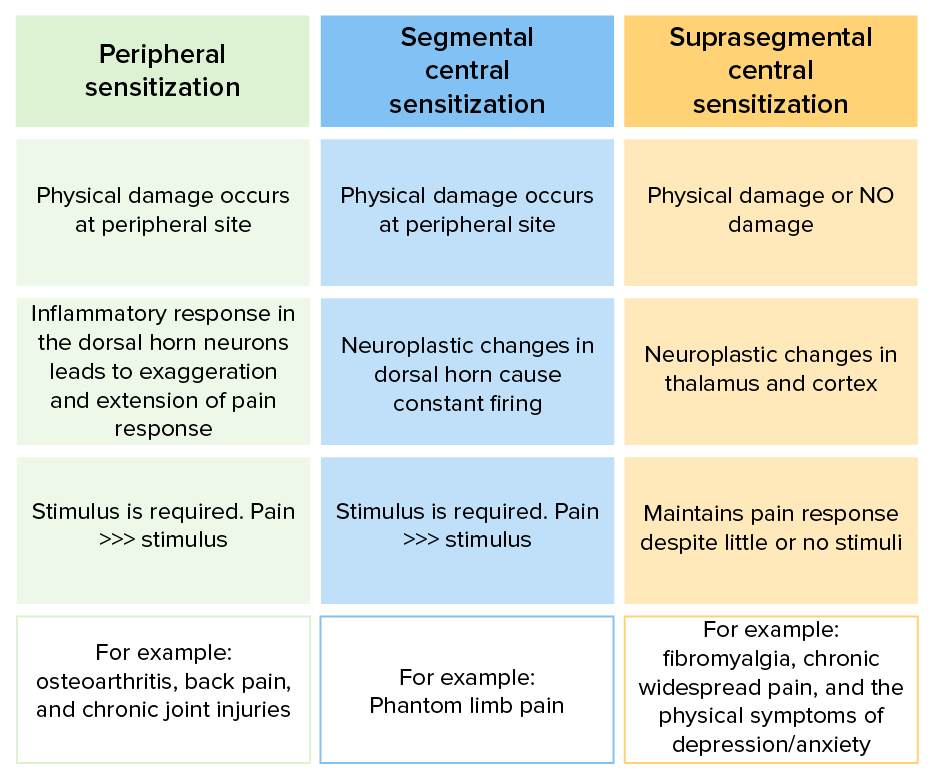
Peripheral versus Central Sensitization

Peripheral sensitization

  • Results from persistent or repetitive peripheral nociception (traditional inflammation Inflammation Inflammation is a complex set of responses to infection and injury involving leukocytes as the principal cellular mediators in the body’s defense against pathogenic organisms. Inflammation is also seen as a response to tissue injury in the process of wound healing. The 5 cardinal signs of inflammation are pain, heat, redness, swelling, and loss of function. Inflammation) or nerve injury Nerve Injury Surgical Complications
  • Hyperstimulation or damage to the 1st-order neuron 1st-order neuron Originates in the hypothalamus and descends to the first synapse in the cervical spinal cord, located at levels C8–T2 (also called the ciliospinal center of Budge). Horner Syndrome alters the electrical traits and elaboration of neurotransmitters (e.g., SP, CGRP CGRP A 37-amino acid peptide derived from the calcitonin gene. It occurs as a result of alternative processing of mRNA from the calcitonin gene. The neuropeptide is widely distributed in the brain, gut, perivascular nerves, and other tissue. The peptide produces multiple biological effects and has both circulatory and neurotransmitter modes of action. In particular, it is a potent endogenous vasodilator. Gastrointestinal Neural and Hormonal Signaling, NGF).
  • The inflammation Inflammation Inflammation is a complex set of responses to infection and injury involving leukocytes as the principal cellular mediators in the body’s defense against pathogenic organisms. Inflammation is also seen as a response to tissue injury in the process of wound healing. The 5 cardinal signs of inflammation are pain, heat, redness, swelling, and loss of function. Inflammation and the initial injury combine to create an enhanced pain sensation and perpetuates the pain response:

Central sensitization

  • Phenomenon thought to occur centrally, perhaps at the level of the rexed laminae
  • Generally a progression from peripheral sensitization but may also be produced without any known peripheral stimulus or a CNS insult.
    • Segmental central sensitization: neuroplastic changes that occur in response to a physical injury that cause constant activation of the pain pathway (i.e., progression from central sensitization)
    • Suprasegmental central sensitization: neuroplastic changes in brain Brain The part of central nervous system that is contained within the skull (cranium). Arising from the neural tube, the embryonic brain is comprised of three major parts including prosencephalon (the forebrain); mesencephalon (the midbrain); and rhombencephalon (the hindbrain). The developed brain consists of cerebrum; cerebellum; and other structures in the brain stem. Nervous System: Anatomy, Structure, and Classification sites of the pain pathway with or without known injuries (from CNS injury).
  • Often occurs in the setting of CNS insults (e.g., stroke, spinal cord Spinal cord The spinal cord is the major conduction pathway connecting the brain to the body; it is part of the CNS. In cross section, the spinal cord is divided into an H-shaped area of gray matter (consisting of synapsing neuronal cell bodies) and a surrounding area of white matter (consisting of ascending and descending tracts of myelinated axons). Spinal Cord: Anatomy injury)
  • Often associated with psychiatric conditions:
  • May occur as a primary disorder (e.g., fibromyalgia Fibromyalgia Fibromyalgia is a chronic pain syndrome characterized by widespread body pain, chronic fatigue, mood disturbance, and cognitive disturbance. It also presents with other comorbid symptoms such as migraine headaches, depression, sleep disturbance, and irritable bowel syndrome. Fibromyalgia
Table peripheral vs. Central sensitization

Mechanisms of peripheral and central pain sensitization

Image by Lecturio.

The Gate Control Theory of Pain

  • A physiologic sensory Sensory Neurons which conduct nerve impulses to the central nervous system. Nervous System: Histology “gate” exists at the level of the dorsal horn Dorsal horn One of three central columns of the spinal cord. It is composed of gray matter spinal laminae i-vi. Brown-Séquard Syndrome in any given spinal segment (or cranial nerve nucleus Nucleus Within a eukaryotic cell, a membrane-limited body which contains chromosomes and one or more nucleoli (cell nucleolus). The nuclear membrane consists of a double unit-type membrane which is perforated by a number of pores; the outermost membrane is continuous with the endoplasmic reticulum. A cell may contain more than one nucleus. The Cell: Organelles).
    • Only so much sensory Sensory Neurons which conduct nerve impulses to the central nervous system. Nervous System: Histology information can get through the gate at any given time.
    • Preferential passage through the gate is granted to incoming sensory Sensory Neurons which conduct nerve impulses to the central nervous system. Nervous System: Histology signals conducted by larger, more heavily myelinated Myelinated Internuclear Ophthalmoplegia, faster-conducting nerve fibers Nerve Fibers Slender processes of neurons, including the axons and their glial envelopes (myelin sheath). Nerve fibers conduct nerve impulses to and from the central nervous system. Nervous System: Histology.
    • An open gate is a dorsal horn Dorsal horn One of three central columns of the spinal cord. It is composed of gray matter spinal laminae i-vi. Brown-Séquard Syndrome receiving isolated noxious (painful) stimulus.
    • A closed gate is a dorsal horn Dorsal horn One of three central columns of the spinal cord. It is composed of gray matter spinal laminae i-vi. Brown-Séquard Syndrome receiving simultaneous physical (nonnoxious) physical stimuli Physical stimuli Act of eliciting a response from a person or organism through physical contact. Angioedema
  • Incoming pain signals are largely conducted by A-delta (relatively slow-conducting) and type C (slow-conducting) fibers, while A-alpha and A-beta (fast-conducting) fibers carry nonnoxious physical stimuli Physical stimuli Act of eliciting a response from a person or organism through physical contact. Angioedema (e.g., movement, vibration Vibration A continuing periodic change in displacement with respect to a fixed reference. Neurological Examination, pressure, temperature, electrical stimulation).
    • This distinction becomes very important when painful (noxious) and other physical stimuli Physical stimuli Act of eliciting a response from a person or organism through physical contact. Angioedema (nonnoxious) are simultaneously applied in the receptive field of a peripheral nerve. 
    • Nonnoxious physical stimuli Physical stimuli Act of eliciting a response from a person or organism through physical contact. Angioedema (conducted by faster, larger, more heavily myelinated Myelinated Internuclear Ophthalmoplegia fibers) are given preferential passage through the gate, lessening or eliminating the incoming pain (noxious) stimulus.
  • Humans unknowingly apply this concept on a daily basis to relieve trivial noxious insults. For example:
    • Scratching (nonnoxious physical stimulus) a mosquito bite (noxious chemical stimulus) to relieve the itch (pain)
    • Rubbing (nonnoxious physical stimulus) the elbow (“funny bone Bone Bone is a compact type of hardened connective tissue composed of bone cells, membranes, an extracellular mineralized matrix, and central bone marrow. The 2 primary types of bone are compact and spongy. Bones: Structure and Types”) after bumping it on a door frame (noxious physical stimulus) to relieve the pain
  • The “pain team” purposefully applies this concept to treat pain nonpharmacologically:
    • Massage
    • Physiotherapy Physiotherapy Spinal Stenosis
    • Peripheral nerve stimulation
    • Spinal cord Spinal cord The spinal cord is the major conduction pathway connecting the brain to the body; it is part of the CNS. In cross section, the spinal cord is divided into an H-shaped area of gray matter (consisting of synapsing neuronal cell bodies) and a surrounding area of white matter (consisting of ascending and descending tracts of myelinated axons). Spinal Cord: Anatomy stimulation

Clinical Relevance

  • Chronic pain Chronic pain Aching sensation that persists for more than a few months. It may or may not be associated with trauma or disease, and may persist after the initial injury has healed. Its localization, character, and timing are more vague than with acute pain. Pain Management: pain that extends beyond the expected period of healing (> 3 months), with an underlying pathology that is insufficient to explain the presence or extent of the pain. Chronic pain Chronic pain Aching sensation that persists for more than a few months. It may or may not be associated with trauma or disease, and may persist after the initial injury has healed. Its localization, character, and timing are more vague than with acute pain. Pain Management can disrupt sleep Sleep A readily reversible suspension of sensorimotor interaction with the environment, usually associated with recumbency and immobility. Physiology of Sleep, daily activities, and psychosocial function. Chronic pain Chronic pain Aching sensation that persists for more than a few months. It may or may not be associated with trauma or disease, and may persist after the initial injury has healed. Its localization, character, and timing are more vague than with acute pain. Pain Management is commonly seen in individuals with malignancies and requires a very personalized management approach.
  • Pain management Pain Management Pain is defined as an unpleasant sensory and emotional experience associated with actual or potential tissue damage. Pain is a subjective experience. Acute pain lasts < 3 months and typically has a specific, identifiable cause. Pain Management: Today, medical professionals have myriad—both pharmacologic and nonpharmacologic—options to alleviate pain. The specific management strategy followed depends on the type of pain and the individual’s circumstances, perspective, and underlying condition. 
  • Complex regional pain Regional Pain Complex Regional Pain Syndrome (CRPS) syndrome ( CRPS CRPS Complex regional pain syndrome (CRPS) is a chronic regional neuropathic pain condition characterized by excruciating pain (out of proportion to apparent tissue damage or inciting trauma), paresthesia, allodynia, temperature abnormalities, skin discoloration, edema, reduced range of motion, and bone demineralization. Complex Regional Pain Syndrome (CRPS)): condition characterized by excruciating pain that is out of proportion to the inciting event and is accompanied by allodynia Allodynia Pain due to a stimulus that does not typically provoke pain. Pain Management, temperature abnormalities, skin Skin The skin, also referred to as the integumentary system, is the largest organ of the body. The skin is primarily composed of the epidermis (outer layer) and dermis (deep layer). The epidermis is primarily composed of keratinocytes that undergo rapid turnover, while the dermis contains dense layers of connective tissue. Skin: Structure and Functions discoloration, and edema Edema Edema is a condition in which excess serous fluid accumulates in the body cavity or interstitial space of connective tissues. Edema is a symptom observed in several medical conditions. It can be categorized into 2 types, namely, peripheral (in the extremities) and internal (in an organ or body cavity). Edema, among other findings. This syndrome affects women more often than men and is often incited by trauma. Diagnosis is clinical, supported by the Budapest Consensus. Complex regional pain Regional Pain Complex Regional Pain Syndrome (CRPS) syndrome is closely related to litigation of work-related issues, requiring special care by the physician. 

References

  1. Yam, M.F., et al. (2018). General pathways of pain sensation and the major neurotransmitters involved in pain regulation. Int J Mol Sci 19:2164. https://doi.org/10.3390/ijms19082164
  2. Chen, J., et al. (2021). Physiology, pain. StatPearls. Retrieved October 11, 2021, from http://www.ncbi.nlm.nih.gov/books/NBK539789/ 
  3. Barrett, K. E., Barman, S. M., Boitano, S., Reckelhoff, J. F. (2017). Somatosensory neurotransmission: touch, pain, & temperature. In: Ganong’s Medical Physiology Examination & Board Review. McGraw-Hill Education. http://accessmedicine.mhmedical.com/content.aspx?aid=1142554554 
  4. House, S. A. (2021). Pain. In: Kellerman, R. D., Rakel, D. P. (Eds.), Conn’s Current Therapy. Elsevier, pp. 32–39. 
  5. Proch, R. (2019). Fibromyalgia as a biopsychosocial model for the intersections of physical and emotional pain. DeckerMed Medicine. Retrieved October 10, 2021, from https://doi.org/10.2310/PSYCH.13071
  6. Ross, E. (2018). Pain syndromes other than headache. DeckerMed Medicine. Retrieved October 11, 2021, from https://doi.org/10.2310/PSYCH.6177
  7. Mendell, L.M. (2014). Constructing and deconstructing the gate theory of pain. Pain 155:210–216. https://doi.org/10.1016/j.pain.2013.12.010

Create your free account or log in to continue reading!

Sign up now and get free access to Lecturio with concept pages, medical videos, and questions for your medical education.

User Reviews

Details