Penetrating Abdominal Injury

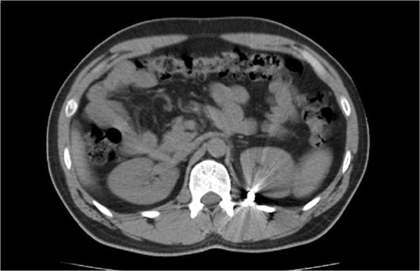
Penetrating abdominal injuries are created by an object puncturing the abdominal wall Abdominal wall The outer margins of the abdomen, extending from the osteocartilaginous thoracic cage to the pelvis. Though its major part is muscular, the abdominal wall consists of at least seven layers: the skin, subcutaneous fat, deep fascia; abdominal muscles, transversalis fascia, extraperitoneal fat, and the parietal peritoneum. Surgical Anatomy of the Abdomen. Injuries can be high velocity, like gunshot wounds Gunshot wounds Disruption of structural continuity of the body as a result of the discharge of firearms. Penetrating Chest Injury, or low velocity, like stab wounds. Different structures can be injured, including the duodenum Duodenum The shortest and widest portion of the small intestine adjacent to the pylorus of the stomach. It is named for having the length equal to about the width of 12 fingers. Small Intestine: Anatomy, spleen Spleen The spleen is the largest lymphoid organ in the body, located in the LUQ of the abdomen, superior to the left kidney and posterior to the stomach at the level of the 9th-11th ribs just below the diaphragm. The spleen is highly vascular and acts as an important blood filter, cleansing the blood of pathogens and damaged erythrocytes. Spleen: Anatomy, liver Liver The liver is the largest gland in the human body. The liver is found in the superior right quadrant of the abdomen and weighs approximately 1.5 kilograms. Its main functions are detoxification, metabolism, nutrient storage (e.g., iron and vitamins), synthesis of coagulation factors, formation of bile, filtration, and storage of blood. Liver: Anatomy, kidneys Kidneys The kidneys are a pair of bean-shaped organs located retroperitoneally against the posterior wall of the abdomen on either side of the spine. As part of the urinary tract, the kidneys are responsible for blood filtration and excretion of water-soluble waste in the urine. Kidneys: Anatomy, and pelvic organs. The extent and specific type of abdominal traumatic injury can be identified by a proper history and physical exam and supported by appropriate imaging studies. Management, which can be laparotomy Laparotomy Incision into the side of the abdomen between the ribs and pelvis. Laparotomy and Laparoscopy or a conservative approach, is dependent on the patient’s hemodynamic stability and specific type of injury.

Last updated: Jan 24, 2023

Editorial responsibility: Stanley Oiseth, Lindsay Jones, Evelin Maza

Overview

Definition

A penetrating abdominal injury is the result of trauma from an object puncturing the skin Skin The skin, also referred to as the integumentary system, is the largest organ of the body. The skin is primarily composed of the epidermis (outer layer) and dermis (deep layer). The epidermis is primarily composed of keratinocytes that undergo rapid turnover, while the dermis contains dense layers of connective tissue. Skin: Structure and Functions, entering the body, and creating a wound. 

  • Can often cause damage that results in shock Shock Shock is a life-threatening condition associated with impaired circulation that results in tissue hypoxia. The different types of shock are based on the underlying cause: distributive (↑ cardiac output (CO), ↓ systemic vascular resistance (SVR)), cardiogenic (↓ CO, ↑ SVR), hypovolemic (↓ CO, ↑ SVR), obstructive (↓ CO), and mixed. Types of Shock and infection 
  • Severity depends on which body organs are involved, the characteristics of the object, and the amount of energy transmitted. 

Epidemiology

  • Demographics:
    • 90% of cases involve men.
    • While 20-to-24-year-old individuals make up 7% of the population, 22.5% of gunshot deaths affect this age group.
  • By settings:
    • 35% of trauma patients Patients Individuals participating in the health care system for the purpose of receiving therapeutic, diagnostic, or preventive procedures. Clinician–Patient Relationship are admitted in urban trauma centers.
    • Up to 12% of those admitted are in suburban or rural centers.
  • 40% of homicides and 16% of suicides by gun involve injuries in the torso.
  • Common organs injured:
    • Small bowel Small bowel The small intestine is the longest part of the GI tract, extending from the pyloric orifice of the stomach to the ileocecal junction. The small intestine is the major organ responsible for chemical digestion and absorption of nutrients. It is divided into 3 segments: the duodenum, the jejunum, and the ileum. Small Intestine: Anatomy (50%)
    • Large bowel (40%)
    • Liver Liver The liver is the largest gland in the human body. The liver is found in the superior right quadrant of the abdomen and weighs approximately 1.5 kilograms. Its main functions are detoxification, metabolism, nutrient storage (e.g., iron and vitamins), synthesis of coagulation factors, formation of bile, filtration, and storage of blood. Liver: Anatomy (30%)
    • Intra-abdominal vascular (25%)

Etiology

  • Gunshot wounds Gunshot wounds Disruption of structural continuity of the body as a result of the discharge of firearms. Penetrating Chest Injury (65% of cases)
  • Stab wounds
  • Foreign objects from motor Motor Neurons which send impulses peripherally to activate muscles or secretory cells. Nervous System: Histology vehicle collisions or other trauma
  • Fractured bones

Pathophysiology and Clinical Presentation

Mechanism of injury

  • The projectile passes through tissue and decelerates, transferring kinetic energy into the tissue. 
  • ↑ Velocity leads to more damage than mass Mass Three-dimensional lesion that occupies a space within the breast Imaging of the Breast, and kinetic energy increases with the velocity
  • A cavity in the tissue is formed by the penetrating object → permanent cavitation Cavitation Imaging of the Lungs and Pleura
  • Medium- and high-velocity projectiles create secondary cavitation Cavitation Imaging of the Lungs and Pleura injuries as the object enters the body:

Clinical manifestations

  • Lacerations
  • Abdominal bleeding
  • Bowel evisceration Evisceration Surgical Complications
  • Abdominal bruising
  • Signs of peritonitis (abdominal tenderness, distension, rigidity Rigidity Continuous involuntary sustained muscle contraction which is often a manifestation of basal ganglia diseases. When an affected muscle is passively stretched, the degree of resistance remains constant regardless of the rate at which the muscle is stretched. This feature helps to distinguish rigidity from muscle spasticity. Megacolon, guarding, fever Fever Fever is defined as a measured body temperature of at least 38°C (100.4°F). Fever is caused by circulating endogenous and/or exogenous pyrogens that increase levels of prostaglandin E2 in the hypothalamus. Fever is commonly associated with chills, rigors, sweating, and flushing of the skin. Fever)
  • Hemodynamic instability 

Related videos

Diagnosis

Because of the wide variety and severity of injuries associated with penetrating abdominal trauma, a prompt but careful history and physical exam are necessary to direct investigation with imaging studies.
History

  • Mechanism of trauma:
    • May suggest severity of injury
    • Gunshot wounds Gunshot wounds Disruption of structural continuity of the body as a result of the discharge of firearms. Penetrating Chest Injury:
    • Stab wounds:
      • More predictable patterns of injury than gunshot wounds Gunshot wounds Disruption of structural continuity of the body as a result of the discharge of firearms. Penetrating Chest Injury
      • Type of knife and length of blade
      • Associated with lower incidence Incidence The number of new cases of a given disease during a given period in a specified population. It also is used for the rate at which new events occur in a defined population. It is differentiated from prevalence, which refers to all cases in the population at a given time. Measures of Disease Frequency of intra-abdominal injuries
  • History of previous trauma
  • Drug (illicit and prescription) or alcohol use by patient 
  • Prior surgical history
  • Amount of blood loss (on scene per paramedics and in hospital)

Physical exam

  • Airway, breathing, circulation (ABC) assessment:
    • Airway Airway ABCDE Assessment:
      • Look for foreign objects blocking the airway Airway ABCDE Assessment.
      • Assess for injury to the trachea Trachea The trachea is a tubular structure that forms part of the lower respiratory tract. The trachea is continuous superiorly with the larynx and inferiorly becomes the bronchial tree within the lungs. The trachea consists of a support frame of semicircular, or C-shaped, rings made out of hyaline cartilage and reinforced by collagenous connective tissue. Trachea: Anatomy (tracheal injury means intubation Intubation Peritonsillar Abscess will be complex).
      • Listen for unusual breathing sounds ( stridor Stridor Laryngomalacia and Tracheomalacia suggests narrowing by a foreign body Foreign Body Foreign Body Aspiration or edema Edema Edema is a condition in which excess serous fluid accumulates in the body cavity or interstitial space of connective tissues. Edema is a symptom observed in several medical conditions. It can be categorized into 2 types, namely, peripheral (in the extremities) and internal (in an organ or body cavity). Edema).
    • Breathing:
      • Look at chest wall Chest wall The chest wall consists of skin, fat, muscles, bones, and cartilage. The bony structure of the chest wall is composed of the ribs, sternum, and thoracic vertebrae. The chest wall serves as armor for the vital intrathoracic organs and provides the stability necessary for the movement of the shoulders and arms. Chest Wall: Anatomy movement for even and spontaneous breathing (uneven chest movement suggests flail chest Flail chest Flail chest is a life-threatening traumatic injury that occurs when 3 or more contiguous ribs are fractured in 2 or more different locations. Patients present with chest pain, tachypnea, hypoxia, and paradoxical chest wall movement. Flail Chest).
      • Listen to breath sounds (muffled or uneven breath sounds may suggest pneumothorax Pneumothorax A pneumothorax is a life-threatening condition in which air collects in the pleural space, causing partial or full collapse of the lung. A pneumothorax can be traumatic or spontaneous. Patients present with a sudden onset of sharp chest pain, dyspnea, and diminished breath sounds on exam. Pneumothorax or hemothorax Hemothorax A hemothorax is a collection of blood in the pleural cavity. Hemothorax most commonly occurs due to damage to the intercostal arteries or from a lung laceration following chest trauma. Hemothorax can also occur as a complication of disease, or hemothorax may be spontaneous or iatrogenic. Hemothorax).
    • Circulation Circulation The movement of the blood as it is pumped through the cardiovascular system. ABCDE Assessment:
      • Palpate pulses on all 4 extremities ( tachycardia Tachycardia Abnormally rapid heartbeat, usually with a heart rate above 100 beats per minute for adults. Tachycardia accompanied by disturbance in the cardiac depolarization (cardiac arrhythmia) is called tachyarrhythmia. Sepsis in Children suggests hemodynamic instability or pneumothorax Pneumothorax A pneumothorax is a life-threatening condition in which air collects in the pleural space, causing partial or full collapse of the lung. A pneumothorax can be traumatic or spontaneous. Patients present with a sudden onset of sharp chest pain, dyspnea, and diminished breath sounds on exam. Pneumothorax).
      • Assess capillary refill on extremities.
  • Secondary survey Secondary Survey ABCDE Assessment:
    • Examine the patient head to toe.
    • External injury should prompt investigation for corresponding internal injury:
    • Base the imaging decision on exam findings.
    • Signs concerning for severe intra-abdominal injury:
      • Hypotension Hypotension Hypotension is defined as low blood pressure, specifically < 90/60 mm Hg, and is most commonly a physiologic response. Hypotension may be mild, serious, or life threatening, depending on the cause. Hypotension (with or without abdominal distention Abdominal distention Megacolon)
      • Narrow pulse pressure
      • Tachycardia Tachycardia Abnormally rapid heartbeat, usually with a heart rate above 100 beats per minute for adults. Tachycardia accompanied by disturbance in the cardiac depolarization (cardiac arrhythmia) is called tachyarrhythmia. Sepsis in Children
      • Respiratory distress
      • Signs of inadequate perfusion
      • Peritoneal signs
      • Generalized abdominal pain Abdominal Pain Acute Abdomen that fails to resolve
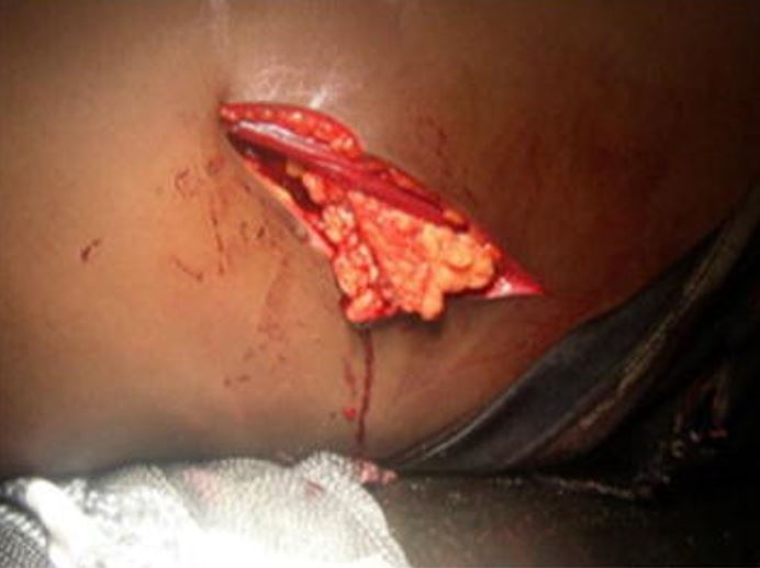
Large left thoraco-abdominal wound with epiplocele

Penetrating injury: large left thoracoabdominal wound with epiplocele

Image: “Large left thoraco-abdominal wound with epiplocele” by SpringerPlus. License: CC BY 4.0
Imaging studies

  • FAST: used in all patients Patients Individuals participating in the health care system for the purpose of receiving therapeutic, diagnostic, or preventive procedures. Clinician–Patient Relationship (stable or unstable) to look for intraperitoneal Intraperitoneal Peritoneum: Anatomy blood and pericardial effusion Pericardial effusion Fluid accumulation within the pericardium. Serous effusions are associated with pericardial diseases. Hemopericardium is associated with trauma. Lipid-containing effusion (chylopericardium) results from leakage of thoracic duct. Severe cases can lead to cardiac tamponade. Pericardial Effusion and Cardiac Tamponade
    • Hemodynamically unstable patients Hemodynamically Unstable Patients Blunt Chest Trauma:
      • Positive FAST → emergent laparotomy Laparotomy Incision into the side of the abdomen between the ribs and pelvis. Laparotomy and Laparoscopy
      • Negative FAST → search for extra-abdominal bleeding sources (e.g., femur fracture Fracture A fracture is a disruption of the cortex of any bone and periosteum and is commonly due to mechanical stress after an injury or accident. Open fractures due to trauma can be a medical emergency. Fractures are frequently associated with automobile accidents, workplace injuries, and trauma. Overview of Bone Fractures)
      • Equivocal FAST → diagnostic peritoneal lavage (DPL) or stabilize patient and obtain CT scan
    • Hemodynamically stable patients Hemodynamically Stable Patients Blunt Chest Trauma:
      • Positive FAST → emergent laparotomy Laparotomy Incision into the side of the abdomen between the ribs and pelvis. Laparotomy and Laparoscopy
      • Negative FAST, low risk for intra-abdominal injury → observation
      • Negative or equivocal FAST with high risk for intra-abdominal injury → CT scan
  • X-ray X-ray Penetrating electromagnetic radiation emitted when the inner orbital electrons of an atom are excited and release radiant energy. X-ray wavelengths range from 1 pm to 10 nm. Hard x-rays are the higher energy, shorter wavelength x-rays. Soft x-rays or grenz rays are less energetic and longer in wavelength. The short wavelength end of the x-ray spectrum overlaps the gamma rays wavelength range. The distinction between gamma rays and x-rays is based on their radiation source. Pulmonary Function Tests: directed by exam findings
    • Chest X-ray X-ray Penetrating electromagnetic radiation emitted when the inner orbital electrons of an atom are excited and release radiant energy. X-ray wavelengths range from 1 pm to 10 nm. Hard x-rays are the higher energy, shorter wavelength x-rays. Soft x-rays or grenz rays are less energetic and longer in wavelength. The short wavelength end of the x-ray spectrum overlaps the gamma rays wavelength range. The distinction between gamma rays and x-rays is based on their radiation source. Pulmonary Function Tests: can show free intraperitoneal Intraperitoneal Peritoneum: Anatomy air, herniation Herniation Omphalocele of abdominal contents
    • Pelvic X-ray X-ray Penetrating electromagnetic radiation emitted when the inner orbital electrons of an atom are excited and release radiant energy. X-ray wavelengths range from 1 pm to 10 nm. Hard x-rays are the higher energy, shorter wavelength x-rays. Soft x-rays or grenz rays are less energetic and longer in wavelength. The short wavelength end of the x-ray spectrum overlaps the gamma rays wavelength range. The distinction between gamma rays and x-rays is based on their radiation source. Pulmonary Function Tests: Pelvic bone fractures Bone fractures Breaks in bones. Bones: Remodeling and Healing can be a source of bleeding or bladder Bladder A musculomembranous sac along the urinary tract. Urine flows from the kidneys into the bladder via the ureters, and is held there until urination. Pyelonephritis and Perinephric Abscess injury.
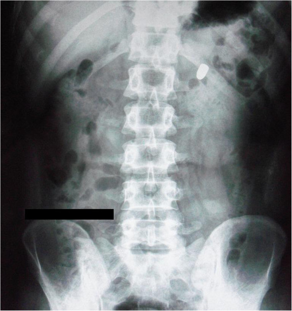
    • X-ray X-ray Penetrating electromagnetic radiation emitted when the inner orbital electrons of an atom are excited and release radiant energy. X-ray wavelengths range from 1 pm to 10 nm. Hard x-rays are the higher energy, shorter wavelength x-rays. Soft x-rays or grenz rays are less energetic and longer in wavelength. The short wavelength end of the x-ray spectrum overlaps the gamma rays wavelength range. The distinction between gamma rays and x-rays is based on their radiation source. Pulmonary Function Tests can help locate bullets and shrapnel left from a penetrating injury Penetrating Injury Brown-Séquard Syndrome.
  • CT scan abdomen with contrast:
    • Imaging of choice for stable patients Stable Patients Blunt Chest Trauma 
    • Provides information regarding retroperitoneal Retroperitoneal Peritoneum: Anatomy structures, diaphragm Diaphragm The diaphragm is a large, dome-shaped muscle that separates the thoracic cavity from the abdominal cavity. The diaphragm consists of muscle fibers and a large central tendon, which is divided into right and left parts. As the primary muscle of inspiration, the diaphragm contributes 75% of the total inspiratory muscle force. Diaphragm: Anatomy, and solid abdominal organs
    • Most sensitive and specific study in identifying liver Liver The liver is the largest gland in the human body. The liver is found in the superior right quadrant of the abdomen and weighs approximately 1.5 kilograms. Its main functions are detoxification, metabolism, nutrient storage (e.g., iron and vitamins), synthesis of coagulation factors, formation of bile, filtration, and storage of blood. Liver: Anatomy and spleen Spleen The spleen is the largest lymphoid organ in the body, located in the LUQ of the abdomen, superior to the left kidney and posterior to the stomach at the level of the 9th-11th ribs just below the diaphragm. The spleen is highly vascular and acts as an important blood filter, cleansing the blood of pathogens and damaged erythrocytes. Spleen: Anatomy injury severity

Other diagnostic studies

  • Hct:
    • < 30% suggestive of intra-abdominal injury
    • Normal Hct does not rule out severe injury.
  • Urinalysis Urinalysis Examination of urine by chemical, physical, or microscopic means. Routine urinalysis usually includes performing chemical screening tests, determining specific gravity, observing any unusual color or odor, screening for bacteriuria, and examining the sediment microscopically. Urinary Tract Infections (UTIs) in Children: blood suggestive of serious renal injury
  • Liver function tests Liver function tests Liver function tests, also known as hepatic function panels, are one of the most commonly performed screening blood tests. Such tests are also used to detect, evaluate, and monitor acute and chronic liver diseases. Liver Function Tests
  • DPL:
    • Invasive procedure used to evaluate presence of blood in abdominal cavity: A catheter is placed into the peritoneal cavity Peritoneal Cavity The space enclosed by the peritoneum. It is divided into two portions, the greater sac and the lesser sac or omental bursa, which lies behind the stomach. The two sacs are connected by the foramen of winslow, or epiploic foramen. Peritoneum: Anatomy, and then fluid is aspirated and evaluated.
    • Not often used but can be an alternative when CT scan and FAST are not available

Injury considerations

Penetrating injuries Penetrating injuries Wounds caused by objects penetrating the skin. Genitourinary Trauma may affect more than just the area that is obviously injured externally, and damage to the adjacent structures needs to be investigated.

Related videos

Management

Historical and standard management of penetrating abdominal trauma is laparotomy Laparotomy Incision into the side of the abdomen between the ribs and pelvis. Laparotomy and Laparoscopy. Recent improvements in imaging and better understanding of injury patterns have resulted in more-conservative strategies.

Initial approach

Assessment of patient stability, correlation Correlation Determination of whether or not two variables are correlated. This means to study whether an increase or decrease in one variable corresponds to an increase or decrease in the other variable. Causality, Validity, and Reliability with diagnostic tests Diagnostic tests Diagnostic tests are important aspects in making a diagnosis. Some of the most important epidemiological values of diagnostic tests include sensitivity and specificity, false positives and false negatives, positive and negative predictive values, likelihood ratios, and pre-test and post-test probabilities. Epidemiological Values of Diagnostic Tests, and determination of the need for immediate surgery:
  • Fluid resuscitation Resuscitation The restoration to life or consciousness of one apparently dead. . Neonatal Respiratory Distress Syndrome:
    • Insertion of 2 large IV lines
    • IV fluid administration
    • Replenish blood
  • Leave any foreign objects in situ until definitive management (e.g., surgical removal) is established.
  • Broad-spectrum Broad-Spectrum Fluoroquinolones antibiotics:
    • For those requiring surgical management
    • Not warranted in cases requiring nonoperative management 
  • Tetanus prophylaxis Tetanus Prophylaxis Overview of Bone Fractures
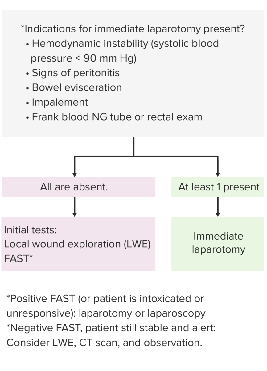
  • Indications for immediate laparotomy Laparotomy Incision into the side of the abdomen between the ribs and pelvis. Laparotomy and Laparoscopy:
    • Hemodynamic instability (systolic blood pressure < 90 mm MM Multiple myeloma (MM) is a malignant condition of plasma cells (activated B lymphocytes) primarily seen in the elderly. Monoclonal proliferation of plasma cells results in cytokine-driven osteoclastic activity and excessive secretion of IgG antibodies. Multiple Myeloma Hg)
    • Signs of peritonitis
    • Bowel evisceration Evisceration Surgical Complications
    • Impalement
    • Frank blood on rectal exam or nasogastric (NG) tube
  • Emergent laparotomy Laparotomy Incision into the side of the abdomen between the ribs and pelvis. Laparotomy and Laparoscopy:
    • The keys of laparotomy Laparotomy Incision into the side of the abdomen between the ribs and pelvis. Laparotomy and Laparoscopy are to control bleeding and prevent GI contamination. 
    • All 4 quadrants of the abdomen are packed with laparotomy Laparotomy Incision into the side of the abdomen between the ribs and pelvis. Laparotomy and Laparoscopy pads to identify localized bleeding and injuries and to prevent GI contents from spreading. 
    • Injured organs such as the liver Liver The liver is the largest gland in the human body. The liver is found in the superior right quadrant of the abdomen and weighs approximately 1.5 kilograms. Its main functions are detoxification, metabolism, nutrient storage (e.g., iron and vitamins), synthesis of coagulation factors, formation of bile, filtration, and storage of blood. Liver: Anatomy can be manually compressed to tamponade Tamponade Pericardial effusion, usually of rapid onset, exceeding ventricular filling pressures and causing collapse of the heart with a markedly reduced cardiac output. Pericarditis bleeding. 
    • Severe cases of bleeding may require ligation Ligation Application of a ligature to tie a vessel or strangulate a part. Esophageal Atresia and Tracheoesophageal Fistula of arteries Arteries Arteries are tubular collections of cells that transport oxygenated blood and nutrients from the heart to the tissues of the body. The blood passes through the arteries in order of decreasing luminal diameter, starting in the largest artery (the aorta) and ending in the small arterioles. Arteries are classified into 3 types: large elastic arteries, medium muscular arteries, and small arteries and arterioles. Arteries: Histology.
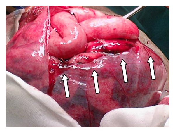
Stab wound injury with multiple small bowel perforations on laparotomy

Stab wound injury: On laparotomy, multiple small bowel perforations (arrows) were evident.

Image: “Multiple small bowel perforations (indicated by arrows)” by Naidoo K, Mewa Kinoo S, Singh B. License: CC BY 3.0
Conservative strategies

  • In hemodynamically stable patients Hemodynamically Stable Patients Blunt Chest Trauma:
    • Secondary survey Secondary Survey ABCDE Assessment
    • FAST:
      • Recommended action dependent on the result
      • If there is a positive FAST, the patient is intoxicated, or unresponsive ➝ proceed with laparotomy Laparotomy Incision into the side of the abdomen between the ribs and pelvis. Laparotomy and Laparoscopy.
    • Local wound exploration (LWE):
      • Preferably performed by 2 individuals
      • Requires sedation and local anesthesia Anesthesia A state characterized by loss of feeling or sensation. This depression of nerve function is usually the result of pharmacologic action and is induced to allow performance of surgery or other painful procedures. Anesthesiology: History and Basic Concepts
      • If wound in the anterior abdomen does not penetrate the anterior rectus fascia Fascia Layers of connective tissue of variable thickness. The superficial fascia is found immediately below the skin; the deep fascia invests muscles, nerves, and other organs. Cellulitis → wound care and consider discharge (depending on associated injuries).
    • CT scan and/or other indicated imaging
  • Alternatives to laparotomy Laparotomy Incision into the side of the abdomen between the ribs and pelvis. Laparotomy and Laparoscopy:
    • Regular Regular Insulin rechecks in stable patients Stable Patients Blunt Chest Trauma
    • Observation for at least 24 hours recommended in:
      • Age > 65 years
      • Those with medications that are anticoagulants Anticoagulants Anticoagulants are drugs that retard or interrupt the coagulation cascade. The primary classes of available anticoagulants include heparins, vitamin K-dependent antagonists (e.g., warfarin), direct thrombin inhibitors, and factor Xa inhibitors. Anticoagulants or antiplatelet therapy.
      • Other significant injuries
      • Medical comorbidities Comorbidities The presence of co-existing or additional diseases with reference to an initial diagnosis or with reference to the index condition that is the subject of study. Comorbidity may affect the ability of affected individuals to function and also their survival; it may be used as a prognostic indicator for length of hospital stay, cost factors, and outcome or survival. St. Louis Encephalitis Virus
    • Interventional radiology procedures for spleen Spleen The spleen is the largest lymphoid organ in the body, located in the LUQ of the abdomen, superior to the left kidney and posterior to the stomach at the level of the 9th-11th ribs just below the diaphragm. The spleen is highly vascular and acts as an important blood filter, cleansing the blood of pathogens and damaged erythrocytes. Spleen: Anatomy and liver Liver The liver is the largest gland in the human body. The liver is found in the superior right quadrant of the abdomen and weighs approximately 1.5 kilograms. Its main functions are detoxification, metabolism, nutrient storage (e.g., iron and vitamins), synthesis of coagulation factors, formation of bile, filtration, and storage of blood. Liver: Anatomy lacerations with active bleeding
Management of penetrating abdominal injury

Algorithm of management of penetrating abdominal injury
Abbreviations: BP: blood pressure; FAST: Focused Assessment with Sonography for Trauma; LWE: local wound exploration; CT: computed tomography

Image by Lecturio.

Clinical Relevance

  • Blunt abdominal trauma: typically involves the violation of the abdominal cavity by deceleration Deceleration A decrease in the rate of speed. Blunt Chest Trauma, crushing Crushing Blunt Chest Trauma, or external compression External Compression Blunt Chest Trauma injuries. The most injured structures are the liver Liver The liver is the largest gland in the human body. The liver is found in the superior right quadrant of the abdomen and weighs approximately 1.5 kilograms. Its main functions are detoxification, metabolism, nutrient storage (e.g., iron and vitamins), synthesis of coagulation factors, formation of bile, filtration, and storage of blood. Liver: Anatomy and spleen Spleen The spleen is the largest lymphoid organ in the body, located in the LUQ of the abdomen, superior to the left kidney and posterior to the stomach at the level of the 9th-11th ribs just below the diaphragm. The spleen is highly vascular and acts as an important blood filter, cleansing the blood of pathogens and damaged erythrocytes. Spleen: Anatomy. Management depends on the patient’s hemodynamic stability and severity of the injury.
  • ABC assessment: airway, breathing, and circulation assessment is the mainstay approach used in managing critically ill patients Patients Individuals participating in the health care system for the purpose of receiving therapeutic, diagnostic, or preventive procedures. Clinician–Patient Relationship. The ABCs are the essential first steps to perform in many situations, including unresponsive patients Patients Individuals participating in the health care system for the purpose of receiving therapeutic, diagnostic, or preventive procedures. Clinician–Patient Relationship, cardiac arrests, and critical medical or trauma patients Patients Individuals participating in the health care system for the purpose of receiving therapeutic, diagnostic, or preventive procedures. Clinician–Patient Relationship. For the trauma patient, ABC is included in the primary survey Primary Survey Thoracic Trauma in Children, the initial evaluation, and the management of injuries. 
  • Splenic injury: in blunt injuries Blunt Injuries Genitourinary Trauma, the liver Liver The liver is the largest gland in the human body. The liver is found in the superior right quadrant of the abdomen and weighs approximately 1.5 kilograms. Its main functions are detoxification, metabolism, nutrient storage (e.g., iron and vitamins), synthesis of coagulation factors, formation of bile, filtration, and storage of blood. Liver: Anatomy and spleen Spleen The spleen is the largest lymphoid organ in the body, located in the LUQ of the abdomen, superior to the left kidney and posterior to the stomach at the level of the 9th-11th ribs just below the diaphragm. The spleen is highly vascular and acts as an important blood filter, cleansing the blood of pathogens and damaged erythrocytes. Spleen: Anatomy are the most commonly injured organs. Usually, splenic injury is associated with lower left rib fractures Rib fractures Fractures of any of the ribs. Flail Chest. The features of splenic injury include hypotension Hypotension Hypotension is defined as low blood pressure, specifically < 90/60 mm Hg, and is most commonly a physiologic response. Hypotension may be mild, serious, or life threatening, depending on the cause. Hypotension, tachycardia Tachycardia Abnormally rapid heartbeat, usually with a heart rate above 100 beats per minute for adults. Tachycardia accompanied by disturbance in the cardiac depolarization (cardiac arrhythmia) is called tachyarrhythmia. Sepsis in Children, abdominal pain Abdominal Pain Acute Abdomen, left chest wall Chest wall The chest wall consists of skin, fat, muscles, bones, and cartilage. The bony structure of the chest wall is composed of the ribs, sternum, and thoracic vertebrae. The chest wall serves as armor for the vital intrathoracic organs and provides the stability necessary for the movement of the shoulders and arms. Chest Wall: Anatomy pain Pain An unpleasant sensation induced by noxious stimuli which are detected by nerve endings of nociceptive neurons. Pain: Types and Pathways, and left shoulder pain Pain An unpleasant sensation induced by noxious stimuli which are detected by nerve endings of nociceptive neurons. Pain: Types and Pathways ( referred pain Referred Pain Spinal Disk Herniation due to phrenic nerve Phrenic nerve The motor nerve of the diaphragm. The phrenic nerve fibers originate in the cervical spinal column (mostly C4) and travel through the cervical plexus to the diaphragm. Diaphragm: Anatomy irritation from splenic hemorrhage).
  • Pelvic injury: pelvic injuries and pelvic fractures Pelvic Fractures Pelvic fractures are a disruption in the cortex of a pelvic bone involving iliac wing fractures, acetabular fractures, or those causing loss of integrity of the pelvic ring (the sacrum and the 2 innominate bones). Patients often present with a history of trauma or a fall, limb length discrepancy, intense pain on palpation, and mechanical instability. Pelvic Fractures are among the worst complications of blunt abdominal injuries. Clinical features include hypotension Hypotension Hypotension is defined as low blood pressure, specifically < 90/60 mm Hg, and is most commonly a physiologic response. Hypotension may be mild, serious, or life threatening, depending on the cause. Hypotension, pain Pain An unpleasant sensation induced by noxious stimuli which are detected by nerve endings of nociceptive neurons. Pain: Types and Pathways with movement, gross hematuria Hematuria Presence of blood in the urine. Renal Cell Carcinoma, and peripelvic ecchymoses. A digital rectal exam is important to identify injury to the rectum Rectum The rectum and anal canal are the most terminal parts of the lower GI tract/large intestine that form a functional unit and control defecation. Fecal continence is maintained by several important anatomic structures including rectal folds, anal valves, the sling-like puborectalis muscle, and internal and external anal sphincters. Rectum and Anal Canal: Anatomy and locate the prostate Prostate The prostate is a gland in the male reproductive system. The gland surrounds the bladder neck and a portion of the urethra. The prostate is an exocrine gland that produces a weakly acidic secretion, which accounts for roughly 20% of the seminal fluid. . Treatment is usually limited to supportive care, but surgical stabilization may sometimes be necessary. 

References

  1. Benjamin, E. (2020). Traumatic gastrointestinal injury in the adult patient. UpToDate. Retrieved January 8, 2021, from https://www.uptodate.com/contents/traumatic-gastrointestinal-injury-in-the-adult-patient
  2. Colwell, C, & Moore, E. (2020). Initial evaluation and management of abdominal stab wounds in adults. UpToDate. Retrieved January 7, 2021, from https://www.uptodate.com/contents/initial-evaluation-and-management-of-abdominal-stab-wounds-in-adults
  3. Lotfollahzadeh, S, & Burns, B. (2021). Penetrating abdominal trauma. StatPearls. Treasure Island (FL): StatPearls Publishing. https://www.ncbi.nlm.nih.gov/books/NBK459123/
  4. Phillips, B, et al. (2017). Trauma to the bladder and ureter: A review of diagnosis, management, and prognosis. Eur J Trauma Emerg Surg. 43(6), 763–773. https://pubmed.ncbi.nlm.nih.gov/28730297/
  5. Sakamoto, R, et al. (2018). Nonoperative management of penetrating abdominal solid organ injuries in children. J Surg Res. 228, 188–193. https://pubmed.ncbi.nlm.nih.gov/29907210/

Create your free account or log in to continue reading!

Sign up now and get free access to Lecturio with concept pages, medical videos, and questions for your medical education.

User Reviews

Details