Osteomyelitis (Clinical)

Osteomyelitis is an infection of the bone that results from the spread of microorganisms from the blood (hematogenous), nearby infected tissue, or open wounds (non-hematogenous). Infections are most commonly caused by Staphylococcus aureus, but a variety of organisms have been linked to osteomyelitis. The majority of patients present with pain, redness, and swelling of the affected site, and may have associated symptoms such as fever and chills. Laboratory values will demonstrate elevated WBC, CRP, and erythrocyte sedimentation rate (ESR) in most cases. The most sensitive and specific imaging modality to diagnose osteomyelitis is MRI. Management may require long-term antibiotics and potential surgical debridement.

Last updated: Mar 4, 2024

Editorial responsibility: Stanley Oiseth, Lindsay Jones, Evelin Maza

Epidemiology and Etiology

Epidemiology[1,7,8]

  • Overall incidence Incidence The number of new cases of a given disease during a given period in a specified population. It also is used for the rate at which new events occur in a defined population. It is differentiated from prevalence, which refers to all cases in the population at a given time. Measures of Disease Frequency is unknown.
    • May account for approximately 50,000 cases annually in the United States
    • Higher incidence Incidence The number of new cases of a given disease during a given period in a specified population. It also is used for the rate at which new events occur in a defined population. It is differentiated from prevalence, which refers to all cases in the population at a given time. Measures of Disease Frequency in developing countries
  • Males > females
  • Non-hematogenous osteomyelitis Non-hematogenous osteomyelitis Osteomyelitis resulting from direct inoculation of bacteria due to surgery, prosthetic devices, trauma, hardware for fracture fixation, soft tissue infection. Osteomyelitis is more common in adults.
  • Hematogenous Hematogenous Hepatocellular Carcinoma (HCC) and Liver Metastases osteomyelitis Osteomyelitis Osteomyelitis is an infection of the bone that results from the spread of microorganisms from the blood (hematogenous), nearby infected tissue, or open wounds (non-hematogenous). Infections are most commonly caused by Staphylococcus aureus. Osteomyelitis is more common in children.

Etiology and classification[1,3,7,8]

Osteomyelitis Osteomyelitis Osteomyelitis is an infection of the bone that results from the spread of microorganisms from the blood (hematogenous), nearby infected tissue, or open wounds (non-hematogenous). Infections are most commonly caused by Staphylococcus aureus. Osteomyelitis is classified based on the route of infection.

Non-hematogenous osteomyelitis Non-hematogenous osteomyelitis Osteomyelitis resulting from direct inoculation of bacteria due to surgery, prosthetic devices, trauma, hardware for fracture fixation, soft tissue infection. Osteomyelitis (80% of cases):

  • Direct inoculation of bacteria Bacteria Bacteria are prokaryotic single-celled microorganisms that are metabolically active and divide by binary fission. Some of these organisms play a significant role in the pathogenesis of diseases. Bacteriology due to:
    • Surgery
    • Prosthetic devices
    • Trauma
    • Hardware for fracture Fracture A fracture is a disruption of the cortex of any bone and periosteum and is commonly due to mechanical stress after an injury or accident. Open fractures due to trauma can be a medical emergency. Fractures are frequently associated with automobile accidents, workplace injuries, and trauma. Overview of Bone Fractures fixation
    • Soft tissue Soft Tissue Soft Tissue Abscess infection
  • Polymicrobial:
    • Staphylococcus aureus Staphylococcus aureus Potentially pathogenic bacteria found in nasal membranes, skin, hair follicles, and perineum of warm-blooded animals. They may cause a wide range of infections and intoxications. Brain Abscess (present in > 50% of cases)
    • S. epidermidis S. epidermidis A species of staphylococcus that is a spherical, non-motile, gram-positive, chemoorganotrophic, facultative anaerobe. Mainly found on the skin and mucous membrane of warm-blooded animals, it can be primary pathogen or secondary invader. Staphylococcus
    • Streptococcus Streptococcus Streptococcus is one of the two medically important genera of gram-positive cocci, the other being Staphylococcus. Streptococci are identified as different species on blood agar on the basis of their hemolytic pattern and sensitivity to optochin and bacitracin. There are many pathogenic species of streptococci, including S. pyogenes, S. agalactiae, S. pneumoniae, and the viridans streptococci. Streptococcus
    • Gram-negative bacteria gram-negative bacteria Bacteria which lose crystal violet stain but are stained pink when treated by gram’s method. Bacteriology
    • Anaerobic bacteria Anaerobic Bacteria Nitroimidazoles

Hematogenous Hematogenous Hepatocellular Carcinoma (HCC) and Liver Metastases osteomyelitis Osteomyelitis Osteomyelitis is an infection of the bone that results from the spread of microorganisms from the blood (hematogenous), nearby infected tissue, or open wounds (non-hematogenous). Infections are most commonly caused by Staphylococcus aureus. Osteomyelitis (20% of cases):

  • Bacteria Bacteria Bacteria are prokaryotic single-celled microorganisms that are metabolically active and divide by binary fission. Some of these organisms play a significant role in the pathogenesis of diseases. Bacteriology spread via blood supply from the primary site of infection.
  • Monomicrobial:
    • S. aureus S. aureus Potentially pathogenic bacteria found in nasal membranes, skin, hair follicles, and perineum of warm-blooded animals. They may cause a wide range of infections and intoxications. Staphylococcus (most common)
    • Streptococcus Streptococcus Streptococcus is one of the two medically important genera of gram-positive cocci, the other being Staphylococcus. Streptococci are identified as different species on blood agar on the basis of their hemolytic pattern and sensitivity to optochin and bacitracin. There are many pathogenic species of streptococci, including S. pyogenes, S. agalactiae, S. pneumoniae, and the viridans streptococci. Streptococcus
    • Enteric gram-negative bacteria gram-negative bacteria Bacteria which lose crystal violet stain but are stained pink when treated by gram’s method. Bacteriology
    • Pseudomonas aeruginosa Pseudomonas aeruginosa A species of gram-negative, aerobic, rod-shaped bacteria commonly isolated from clinical specimens (wound, burn, and urinary tract infections). It is also found widely distributed in soil and water. P. Aeruginosa is a major agent of nosocomial infection. Pseudomonas
    • Serratia Serratia A genus of gram-negative, facultatively anaerobic, rod-shaped bacteria that occurs in the natural environment (soil, water, and plant surfaces) or as an opportunistic human pathogen. Acute Cholangitis
    • Salmonella Salmonella Salmonellae are gram-negative bacilli of the family Enterobacteriaceae. Salmonellae are flagellated, non-lactose-fermenting, and hydrogen sulfide-producing microbes. Salmonella enterica, the most common disease-causing species in humans, is further classified based on serotype as typhoidal (S. typhi and paratyphi) and nontyphoidal (S. enteritidis and typhimurium). Salmonella
    • Mycobacterium tuberculosis Mycobacterium tuberculosis Tuberculosis (TB) is an infectious disease caused by Mycobacterium tuberculosis complex bacteria. The bacteria usually attack the lungs but can also damage other parts of the body. Approximately 30% of people around the world are infected with this pathogen, with the majority harboring a latent infection. Tuberculosis spreads through the air when a person with active pulmonary infection coughs or sneezes. Tuberculosis
    • Neisseria gonorrhoeae Neisseria gonorrhoeae A species of gram-negative, aerobic bacteria primarily found in purulent venereal discharges. It is the causative agent of gonorrhea. Neisseria
    • Kingella
    • Bartonella henselae Bartonella henselae A species of gram-negative bacteria that is the etiologic agent of bacillary angiomatosis. This organism can also be a cause of cat-scratch disease in immunocompetent patients. Bartonella
    • Candida Candida Candida is a genus of dimorphic, opportunistic fungi. Candida albicans is part of the normal human flora and is the most common cause of candidiasis. The clinical presentation varies and can include localized mucocutaneous infections (e.g., oropharyngeal, esophageal, intertriginous, and vulvovaginal candidiasis) and invasive disease (e.g., candidemia, intraabdominal abscess, pericarditis, and meningitis). Candida/Candidiasis
    • Histoplasmosis Histoplasmosis Histoplasmosis is an infection caused by Histoplasma capsulatum, a dimorphic fungus. Transmission is through inhalation, and exposure to soils containing bird or bat droppings increases the risk of infection. Most infections are asymptomatic; however, immunocompromised individuals generally develop acute pulmonary infection, chronic infection, or even disseminated disease. Histoplasma/Histoplasmosis
    • Blastomycosis Blastomycosis Blastomycosis is an infection caused by inhalation of the spores of the fungus, Blastomyces. Blastomyces species thrive in moist soil and decaying material and are common in the Ohio and Mississippi River valleys and the Great Lakes regions of the United States and Canada. Although most patients are asymptomatic, some can develop pneumonia. Blastomyces/Blastomycosis
    • Coccidioidomycosis Coccidioidomycosis Coccidioidomycosis, commonly known as San Joaquin Valley fever, is a fungal disease caused by Coccidioides immitis or Coccidioides posadasii. When Coccidioides spores are inhaled, they transform into spherules that result in infection. Coccidioidomycosis is also a common cause of community-acquired pneumonia and can cause severe disease in the immunocompromised. Coccidioides/Coccidioidomycosis

Risk factors[2,7,12,19]

  • Non-hematogenous:
    • Trauma
    • Pressure ulcers
    • Foreign body Foreign Body Foreign Body Aspiration (prosthetics)
    • Diabetes Diabetes Diabetes mellitus (DM) is a metabolic disease characterized by hyperglycemia and dysfunction of the regulation of glucose metabolism by insulin. Type 1 DM is diagnosed mostly in children and young adults as the result of autoimmune destruction of β cells in the pancreas and the resulting lack of insulin. Type 2 DM has a significant association with obesity and is characterized by insulin resistance. Diabetes Mellitus
    • Peripheral vascular disease 
    • Peripheral neuropathy Neuropathy Leprosy
  • Hematogenous Hematogenous Hepatocellular Carcinoma (HCC) and Liver Metastases:
    • Sickle cell anemia Sickle cell anemia A disease characterized by chronic hemolytic anemia, episodic painful crises, and pathologic involvement of many organs. It is the clinical expression of homozygosity for hemoglobin S. Sickle Cell Disease
    • Diabetes Diabetes Diabetes mellitus (DM) is a metabolic disease characterized by hyperglycemia and dysfunction of the regulation of glucose metabolism by insulin. Type 1 DM is diagnosed mostly in children and young adults as the result of autoimmune destruction of β cells in the pancreas and the resulting lack of insulin. Type 2 DM has a significant association with obesity and is characterized by insulin resistance. Diabetes Mellitus
    • IV drug use
    • Indwelling catheters Indwelling catheters Catheters designed to be left within an organ or passage for an extended period of time. Pseudomonas
    • Immunodeficiency Immunodeficiency Chédiak-Higashi Syndrome
    • Endocarditis Endocarditis Endocarditis is an inflammatory disease involving the inner lining (endometrium) of the heart, most commonly affecting the cardiac valves. Both infectious and noninfectious etiologies lead to vegetations on the valve leaflets. Patients may present with nonspecific symptoms such as fever and fatigue. Endocarditis
Table: Common causes of osteomyelitis Osteomyelitis Osteomyelitis is an infection of the bone that results from the spread of microorganisms from the blood (hematogenous), nearby infected tissue, or open wounds (non-hematogenous). Infections are most commonly caused by Staphylococcus aureus. Osteomyelitis based on patients Patients Individuals participating in the health care system for the purpose of receiving therapeutic, diagnostic, or preventive procedures. Clinician–Patient Relationship’ underlying risk factors
Risk factors Infectious agents
No specific risk factor S. aureus S. aureus Potentially pathogenic bacteria found in nasal membranes, skin, hair follicles, and perineum of warm-blooded animals. They may cause a wide range of infections and intoxications. Staphylococcus
Prosthetic joint replacement Prosthetic Joint Replacement Septic Arthritis
  • S. aureus S. aureus Potentially pathogenic bacteria found in nasal membranes, skin, hair follicles, and perineum of warm-blooded animals. They may cause a wide range of infections and intoxications. Staphylococcus
  • S. epidermidis S. epidermidis A species of staphylococcus that is a spherical, non-motile, gram-positive, chemoorganotrophic, facultative anaerobe. Mainly found on the skin and mucous membrane of warm-blooded animals, it can be primary pathogen or secondary invader. Staphylococcus (creates biofilms Biofilms Encrustations formed from microbes (bacteria, algae, fungi, plankton, or protozoa) embedded in an extracellular polymeric substance matrix that is secreted by the microbes. They occur on body surfaces such as teeth (dental deposits); inanimate objects, and bodies of water. Biofilms are prevented from forming by treating surfaces with dentifrices; disinfectants; anti-infective agents; and anti-fouling agents. Proteus on prosthetics)
Sickle cell anemia Sickle cell anemia A disease characterized by chronic hemolytic anemia, episodic painful crises, and pathologic involvement of many organs. It is the clinical expression of homozygosity for hemoglobin S. Sickle Cell Disease
  • S. aureus S. aureus Potentially pathogenic bacteria found in nasal membranes, skin, hair follicles, and perineum of warm-blooded animals. They may cause a wide range of infections and intoxications. Staphylococcus
  • Salmonella Salmonella Salmonellae are gram-negative bacilli of the family Enterobacteriaceae. Salmonellae are flagellated, non-lactose-fermenting, and hydrogen sulfide-producing microbes. Salmonella enterica, the most common disease-causing species in humans, is further classified based on serotype as typhoidal (S. typhi and paratyphi) and nontyphoidal (S. enteritidis and typhimurium). Salmonella
Chronic granulomatous disease Granulomatous disease A defect of leukocyte function in which phagocytic cells ingest but fail to digest bacteria, resulting in recurring bacterial infections with granuloma formation. When chronic granulomatous disease is caused by mutations in the cybb gene, the condition is inherited in an X-linked recessive pattern. When chronic granulomatous disease is caused by cyba, ncf1, ncf2, or ncf4 gene mutations, the condition is inherited in an autosomal recessive pattern. Common Variable Immunodeficiency (CVID)
  • Serratia Serratia A genus of gram-negative, facultatively anaerobic, rod-shaped bacteria that occurs in the natural environment (soil, water, and plant surfaces) or as an opportunistic human pathogen. Acute Cholangitis
  • Aspergillus Aspergillus A genus of mitosporic fungi containing about 100 species and eleven different teleomorphs in the family trichocomaceae. Echinocandins
Vertebral osteomyelitis Vertebral Osteomyelitis Osteomyelitis
  • M. tuberculosis Tuberculosis Tuberculosis (TB) is an infectious disease caused by Mycobacterium tuberculosis complex bacteria. The bacteria usually attack the lungs but can also damage other parts of the body. Approximately 30% of people around the world are infected with this pathogen, with the majority harboring a latent infection. Tuberculosis spreads through the air when a person with active pulmonary infection coughs or sneezes. Tuberculosis (Pott’s disease)
  • S. aureus S. aureus Potentially pathogenic bacteria found in nasal membranes, skin, hair follicles, and perineum of warm-blooded animals. They may cause a wide range of infections and intoxications. Staphylococcus
Sexually active, no other risk factors N. gonorrhoeae N. gonorrhoeae A species of gram-negative, aerobic bacteria primarily found in purulent venereal discharges. It is the causative agent of gonorrhea. Neisseria (more likely to cause septic arthritis Arthritis Acute or chronic inflammation of joints. Osteoarthritis than osteomyelitis Osteomyelitis Osteomyelitis is an infection of the bone that results from the spread of microorganisms from the blood (hematogenous), nearby infected tissue, or open wounds (non-hematogenous). Infections are most commonly caused by Staphylococcus aureus. Osteomyelitis)
Cat or dog bite Pasteurella multocida Pasteurella Multocida A species of gram-negative, facultatively anaerobic, rod-shaped bacteria normally found in the flora of the mouth and respiratory tract of animals and birds. It causes shipping fever; hemorrhagic bacteremia; and intestinal disease in animals. In humans, disease usually arises from a wound infection following a bite or scratch from domesticated animals. Dog and Cat Bites
IV drug use or immunocompromised immunocompromised A human or animal whose immunologic mechanism is deficient because of an immunodeficiency disorder or other disease or as the result of the administration of immunosuppressive drugs or radiation. Gastroenteritis
  • P. aeruginosa P. aeruginosa A species of gram-negative, aerobic, rod-shaped bacteria commonly isolated from clinical specimens (wound, burn, and urinary tract infections). It is also found widely distributed in soil and water. P. Aeruginosa is a major agent of nosocomial infection. Pseudomonas
  • S. aureus S. aureus Potentially pathogenic bacteria found in nasal membranes, skin, hair follicles, and perineum of warm-blooded animals. They may cause a wide range of infections and intoxications. Staphylococcus
  • Candida Candida Candida is a genus of dimorphic, opportunistic fungi. Candida albicans is part of the normal human flora and is the most common cause of candidiasis. The clinical presentation varies and can include localized mucocutaneous infections (e.g., oropharyngeal, esophageal, intertriginous, and vulvovaginal candidiasis) and invasive disease (e.g., candidemia, intraabdominal abscess, pericarditis, and meningitis). Candida/Candidiasis

Pathophysiology

Osteomyelitis Osteomyelitis Osteomyelitis is an infection of the bone that results from the spread of microorganisms from the blood (hematogenous), nearby infected tissue, or open wounds (non-hematogenous). Infections are most commonly caused by Staphylococcus aureus. Osteomyelitis is an infection of the bone Bone Bone is a compact type of hardened connective tissue composed of bone cells, membranes, an extracellular mineralized matrix, and central bone marrow. The 2 primary types of bone are compact and spongy. Bones: Structure and Types that results from hematogenous Hematogenous Hepatocellular Carcinoma (HCC) and Liver Metastases or non-hematogenous spread of infectious organisms.[1,3,7–11]
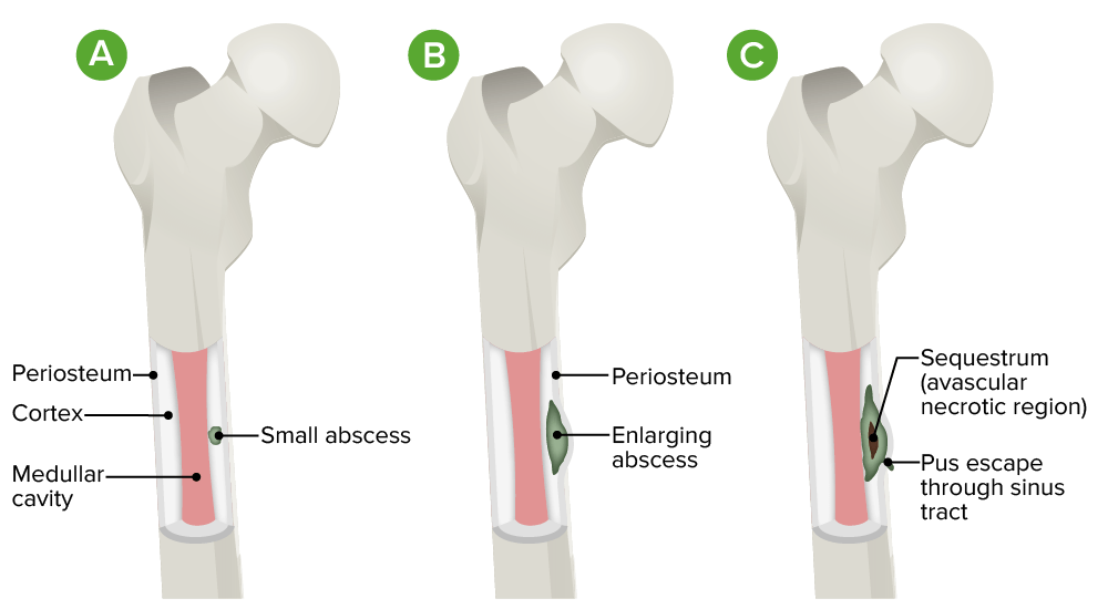
  • Since bone Bone Bone is a compact type of hardened connective tissue composed of bone cells, membranes, an extracellular mineralized matrix, and central bone marrow. The 2 primary types of bone are compact and spongy. Bones: Structure and Types is usually resistant to infection, the following is usually required:
    • A large inoculum of organisms
    • Bone Bone Bone is a compact type of hardened connective tissue composed of bone cells, membranes, an extracellular mineralized matrix, and central bone marrow. The 2 primary types of bone are compact and spongy. Bones: Structure and Types damage
    • Foreign material
  • The pathogenesis is poorly understood, but appears to be affected by several factors:
    • Host immune status
    • Underlying disease
    • Virulence Virulence The degree of pathogenicity within a group or species of microorganisms or viruses as indicated by case fatality rates and/or the ability of the organism to invade the tissues of the host. The pathogenic capacity of an organism is determined by its virulence factors. Proteus of the organisms:
      • Adherence
      • Defense mechanisms Defense mechanisms Defense mechanisms are normal subconscious means of resolving inner conflicts between an individual’s subjective moral sense and their thoughts, feelings, or actions. Defense mechanisms serve to protect the self from unpleasant feelings (anxiety, shame, and/or guilt) and are divided into pathologic, immature, mature, neurotic, and other types. Defense Mechanisms
      • Proteolytic activity
    • Vascularity and location of bone Bone Bone is a compact type of hardened connective tissue composed of bone cells, membranes, an extracellular mineralized matrix, and central bone marrow. The 2 primary types of bone are compact and spongy. Bones: Structure and Types (e.g., the metaphysis Metaphysis Bones: Structure and Types of bone Bone Bone is a compact type of hardened connective tissue composed of bone cells, membranes, an extracellular mineralized matrix, and central bone marrow. The 2 primary types of bone are compact and spongy. Bones: Structure and Types is commonly affected in hematogenous Hematogenous Hepatocellular Carcinoma (HCC) and Liver Metastases spread due to the rich vascular supply of the growth plates Growth Plates The area between the epiphysis and the diaphysis within which bone growth occurs. Osteosarcoma)
  • Timeline of infection:
    • Acute osteomyelitis Acute osteomyelitis Osteomyelitis evolves over days or weeks. Osteomyelitis evolves over days or weeks.
    • Chronic osteomyelitis Chronic osteomyelitis Osteomyelitis persists over months to years and causes bone ischemia and necrosis, bone loss, sinus tract formation. Osteomyelitis persists over months to years and causes:
      • Bone Bone Bone is a compact type of hardened connective tissue composed of bone cells, membranes, an extracellular mineralized matrix, and central bone marrow. The 2 primary types of bone are compact and spongy. Bones: Structure and Types ischemia Ischemia A hypoperfusion of the blood through an organ or tissue caused by a pathologic constriction or obstruction of its blood vessels, or an absence of blood circulation. Ischemic Cell Damage and necrosis Necrosis The death of cells in an organ or tissue due to disease, injury or failure of the blood supply. Ischemic Cell Damage
      • Bone Bone Bone is a compact type of hardened connective tissue composed of bone cells, membranes, an extracellular mineralized matrix, and central bone marrow. The 2 primary types of bone are compact and spongy. Bones: Structure and Types loss
      • Sinus tract formation
    • Chronic osteomyelitis Chronic osteomyelitis Osteomyelitis persists over months to years and causes bone ischemia and necrosis, bone loss, sinus tract formation. Osteomyelitis is not common in children.
Pathophysiology of osteomyelitis

Pathophysiology of osteomyelitis:
A. Initial infection is localized to the cortical region.
B. There is progression into the subperiosteal space with lifting of the periosteum.
C. Diffuse infection occurs with sequestrum (avascular necrotic region) and sinus tract formation.

Image by Lecturio.

Clinical Presentation

Acute osteomyelitis Acute osteomyelitis Osteomyelitis evolves over days or weeks. Osteomyelitis[1,2,8]

  • Onset may be gradual.
  • Signs and symptoms:
    • Fever Fever Fever is defined as a measured body temperature of at least 38°C (100.4°F). Fever is caused by circulating endogenous and/or exogenous pyrogens that increase levels of prostaglandin E2 in the hypothalamus. Fever is commonly associated with chills, rigors, sweating, and flushing of the skin. Fever and chills Chills The sudden sensation of being cold. It may be accompanied by shivering. Fever
    • Localized swelling Swelling Inflammation
    • Warmth
    • Erythema Erythema Redness of the skin produced by congestion of the capillaries. This condition may result from a variety of disease processes. Chalazion
    • Dull pain Pain An unpleasant sensation induced by noxious stimuli which are detected by nerve endings of nociceptive neurons. Pain: Types and Pathways

Chronic osteomyelitis Chronic osteomyelitis Osteomyelitis persists over months to years and causes bone ischemia and necrosis, bone loss, sinus tract formation. Osteomyelitis[1,2,8]

  • Similar to acute osteomyelitis Acute osteomyelitis Osteomyelitis evolves over days or weeks. Osteomyelitis
  • Intermittent bone Bone Bone is a compact type of hardened connective tissue composed of bone cells, membranes, an extracellular mineralized matrix, and central bone marrow. The 2 primary types of bone are compact and spongy. Bones: Structure and Types pain Pain An unpleasant sensation induced by noxious stimuli which are detected by nerve endings of nociceptive neurons. Pain: Types and Pathways
  • Draining sinus tract (pathognomonic)
  • Fever Fever Fever is defined as a measured body temperature of at least 38°C (100.4°F). Fever is caused by circulating endogenous and/or exogenous pyrogens that increase levels of prostaglandin E2 in the hypothalamus. Fever is commonly associated with chills, rigors, sweating, and flushing of the skin. Fever and chills Chills The sudden sensation of being cold. It may be accompanied by shivering. Fever are less common.

Physical exam[1,2,8]

  • Check for tenderness to palpation Palpation Application of fingers with light pressure to the surface of the body to determine consistency of parts beneath in physical diagnosis; includes palpation for determining the outlines of organs. Dermatologic Examination over the bone Bone Bone is a compact type of hardened connective tissue composed of bone cells, membranes, an extracellular mineralized matrix, and central bone marrow. The 2 primary types of bone are compact and spongy. Bones: Structure and Types.
  • Assess for a nidus of infection:
    • Wound healing Wound healing Wound healing is a physiological process involving tissue repair in response to injury. It involves a complex interaction of various cell types, cytokines, and inflammatory mediators. Wound healing stages include hemostasis, inflammation, granulation, and remodeling. Wound Healing
    • High likelihood of osteomyelitis Osteomyelitis Osteomyelitis is an infection of the bone that results from the spread of microorganisms from the blood (hematogenous), nearby infected tissue, or open wounds (non-hematogenous). Infections are most commonly caused by Staphylococcus aureus. Osteomyelitis if it is possible to probe Probe A device placed on the patient’s body to visualize a target Ultrasound (Sonography) a chronic ulcer to the bone Bone Bone is a compact type of hardened connective tissue composed of bone cells, membranes, an extracellular mineralized matrix, and central bone marrow. The 2 primary types of bone are compact and spongy. Bones: Structure and Types with a sterile Sterile Basic Procedures tool
  • Check sensory Sensory Neurons which conduct nerve impulses to the central nervous system. Nervous System: Histology function.
  • Evaluate for diminished pulses.

Subtypes[2,7,20]

Vertebral osteomyelitis Vertebral Osteomyelitis Osteomyelitis:

  • Most common subtype of hematogenous Hematogenous Hepatocellular Carcinoma (HCC) and Liver Metastases osteomyelitis Osteomyelitis Osteomyelitis is an infection of the bone that results from the spread of microorganisms from the blood (hematogenous), nearby infected tissue, or open wounds (non-hematogenous). Infections are most commonly caused by Staphylococcus aureus. Osteomyelitis in adults
  • The lumbar spine Spine The human spine, or vertebral column, is the most important anatomical and functional axis of the human body. It consists of 7 cervical vertebrae, 12 thoracic vertebrae, and 5 lumbar vertebrae and is limited cranially by the skull and caudally by the sacrum. Vertebral Column: Anatomy is most commonly affected.
  • Slow progression of symptoms:
    • Localized back pain Pain An unpleasant sensation induced by noxious stimuli which are detected by nerve endings of nociceptive neurons. Pain: Types and Pathways:
      • Radiation Radiation Emission or propagation of acoustic waves (sound), electromagnetic energy waves (such as light; radio waves; gamma rays; or x-rays), or a stream of subatomic particles (such as electrons; neutrons; protons; or alpha particles). Osteosarcoma to the legs, abdomen, or groin Groin The external junctural region between the lower part of the abdomen and the thigh. Male Genitourinary Examination
      • Worsened with activity and at night
    • Motor Motor Neurons which send impulses peripherally to activate muscles or secretory cells. Nervous System: Histology and sensory Sensory Neurons which conduct nerve impulses to the central nervous system. Nervous System: Histology deficits with severe progression

Sternoclavicular and pelvic osteomyelitis Pelvic Osteomyelitis Osteomyelitis:

Long bone osteomyelitis Long Bone Osteomyelitis Osteomyelitis:
  • Least common subtype in adults, but most common in children
  • Can present similar to septic arthritis Arthritis Acute or chronic inflammation of joints. Osteoarthritis of a joint
  • Weight bearing on affected long bones Long bones Length greater than width. Bones: Structure and Types may be difficult.

Diagnosis

Laboratory evaluation[1,2,8,9,13,14]

  • The following tests support the diagnosis of osteomyelitis Osteomyelitis Osteomyelitis is an infection of the bone that results from the spread of microorganisms from the blood (hematogenous), nearby infected tissue, or open wounds (non-hematogenous). Infections are most commonly caused by Staphylococcus aureus. Osteomyelitis:
  • The following cultures provide necessary identification Identification Defense Mechanisms and sensitivity data for the causative organism:
    • Blood cultures:
      • Positive in 50% of cases
      • Obtain prior to initiating antibiotics, if possible.
    • Tissue culture:
      • Should also be obtained prior to antibiotics, if possible
      • Bone Bone Bone is a compact type of hardened connective tissue composed of bone cells, membranes, an extracellular mineralized matrix, and central bone marrow. The 2 primary types of bone are compact and spongy. Bones: Structure and Types biopsy Biopsy Removal and pathologic examination of specimens from the living body. Ewing Sarcoma is the best way to identify the etiology.
      • Wound or abscess Abscess Accumulation of purulent material in tissues, organs, or circumscribed spaces, usually associated with signs of infection. Chronic Granulomatous Disease cultures are not reliable.

Imaging[5,8–10,13,14]

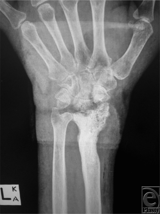
  • X-rays X-rays X-rays are high-energy particles of electromagnetic radiation used in the medical field for the generation of anatomical images. X-rays are projected through the body of a patient and onto a film, and this technique is called conventional or projectional radiography. X-rays:
    • 1st-line imaging modality
    • May not show changes in first 2 weeks of disease
    • A normal X-ray X-ray Penetrating electromagnetic radiation emitted when the inner orbital electrons of an atom are excited and release radiant energy. X-ray wavelengths range from 1 pm to 10 nm. Hard x-rays are the higher energy, shorter wavelength x-rays. Soft x-rays or grenz rays are less energetic and longer in wavelength. The short wavelength end of the x-ray spectrum overlaps the gamma rays wavelength range. The distinction between gamma rays and x-rays is based on their radiation source. Pulmonary Function Tests does not rule out osteomyelitis Osteomyelitis Osteomyelitis is an infection of the bone that results from the spread of microorganisms from the blood (hematogenous), nearby infected tissue, or open wounds (non-hematogenous). Infections are most commonly caused by Staphylococcus aureus. Osteomyelitis.
    • Rules out metastasis Metastasis The transfer of a neoplasm from one organ or part of the body to another remote from the primary site. Grading, Staging, and Metastasis or fractures
    • Findings:
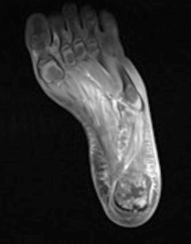
  • MRI:
    • Most widely used modality, with sensitivity and specificity Sensitivity and Specificity Binary classification measures to assess test results. Sensitivity or recall rate is the proportion of true positives. Specificity is the probability of correctly determining the absence of a condition. Epidemiological Values of Diagnostic Tests approaching 90%
    • Detects infection within 3–5 days of onset
    • Use is limited if surgical hardware is present
    • Less accurate for diagnosis of chronic osteomyelitis Chronic osteomyelitis Osteomyelitis persists over months to years and causes bone ischemia and necrosis, bone loss, sinus tract formation. Osteomyelitis
    • Findings:
      • Bone marrow Bone marrow The soft tissue filling the cavities of bones. Bone marrow exists in two types, yellow and red. Yellow marrow is found in the large cavities of large bones and consists mostly of fat cells and a few primitive blood cells. Red marrow is a hematopoietic tissue and is the site of production of erythrocytes and granular leukocytes. Bone marrow is made up of a framework of connective tissue containing branching fibers with the frame being filled with marrow cells. Bone Marrow: Composition and Hematopoiesis edema Edema Edema is a condition in which excess serous fluid accumulates in the body cavity or interstitial space of connective tissues. Edema is a symptom observed in several medical conditions. It can be categorized into 2 types, namely, peripheral (in the extremities) and internal (in an organ or body cavity). Edema is the earliest feature.
      • Cortical destruction
      • Soft tissue Soft Tissue Soft Tissue Abscess inflammation Inflammation Inflammation is a complex set of responses to infection and injury involving leukocytes as the principal cellular mediators in the body’s defense against pathogenic organisms. Inflammation is also seen as a response to tissue injury in the process of wound healing. The 5 cardinal signs of inflammation are pain, heat, redness, swelling, and loss of function. Inflammation
    • Contrast enhancement helps distinguish abscess Abscess Accumulation of purulent material in tissues, organs, or circumscribed spaces, usually associated with signs of infection. Chronic Granulomatous Disease margins and adjacent periosteum Periosteum Thin outer membrane that surrounds a bone. It contains connective tissue, capillaries, nerves, and a number of cell types. Bones: Structure and Types or soft tissue Soft Tissue Soft Tissue Abscess involvement.
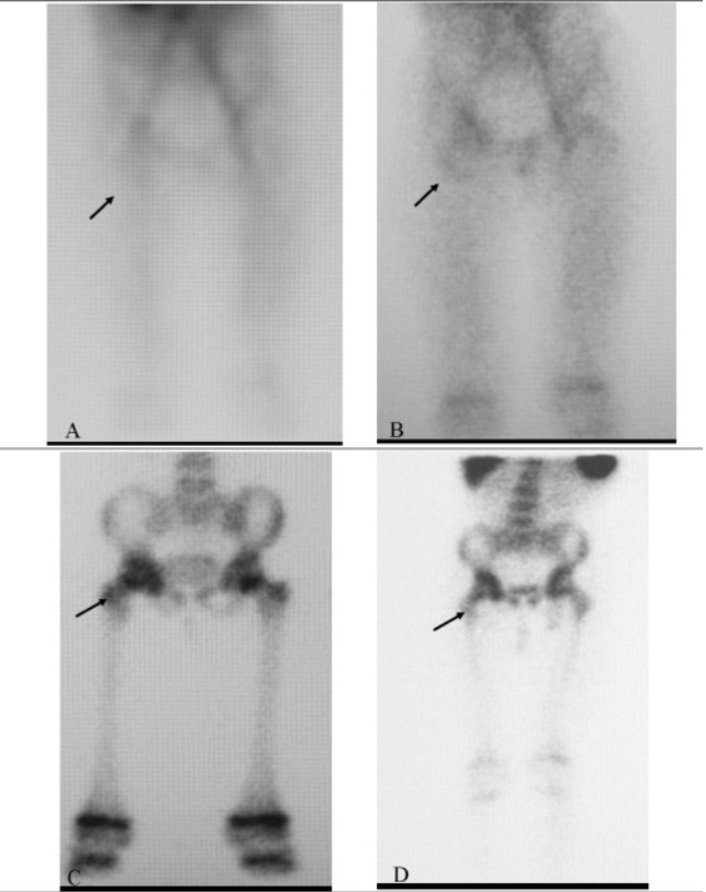
  • 3-phase bone scan Bone Scan Osteosarcoma:
    • Utilizes a radionuclide tracer, which accumulates in areas of osteoblast activity
    • Technetium-99m or gallium
    • Useful in acute osteomyelitis Acute osteomyelitis Osteomyelitis evolves over days or weeks. Osteomyelitis but less so in chronic
    • 3 phases:
    • Significant uptake in all 3 phases in osteomyelitis Osteomyelitis Osteomyelitis is an infection of the bone that results from the spread of microorganisms from the blood (hematogenous), nearby infected tissue, or open wounds (non-hematogenous). Infections are most commonly caused by Staphylococcus aureus. Osteomyelitis
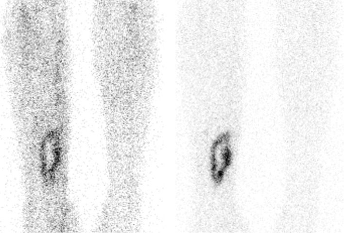
  • Tagged WBC scan:
    • Blood is drawn from the patient, and autologous WBCs are labeled with a radionuclide tracer.
    • The tagged cells are then injected back into the patient, and the scan is performed.
    • Tagged WBCs accumulate at the site of infection.
    • Best used in distal extremities
    • Can be falsely positive if other inflammatory conditions are present in the area (e.g., fracture Fracture A fracture is a disruption of the cortex of any bone and periosteum and is commonly due to mechanical stress after an injury or accident. Open fractures due to trauma can be a medical emergency. Fractures are frequently associated with automobile accidents, workplace injuries, and trauma. Overview of Bone Fractures)
    • More useful in acute than in chronic osteomyelitis Chronic osteomyelitis Osteomyelitis persists over months to years and causes bone ischemia and necrosis, bone loss, sinus tract formation. Osteomyelitis
  • CT scan:
    • Generally not useful for initial diagnosis of osteomyelitis Osteomyelitis Osteomyelitis is an infection of the bone that results from the spread of microorganisms from the blood (hematogenous), nearby infected tissue, or open wounds (non-hematogenous). Infections are most commonly caused by Staphylococcus aureus. Osteomyelitis
    • Can be used to detect the extent of bone Bone Bone is a compact type of hardened connective tissue composed of bone cells, membranes, an extracellular mineralized matrix, and central bone marrow. The 2 primary types of bone are compact and spongy. Bones: Structure and Types destruction and to guide biopsies
  • PET scan PET scan Ewing Sarcoma:
    • Highest sensitivity and specificity Sensitivity and Specificity Binary classification measures to assess test results. Sensitivity or recall rate is the proportion of true positives. Specificity is the probability of correctly determining the absence of a condition. Epidemiological Values of Diagnostic Tests for osteomyelitis Osteomyelitis Osteomyelitis is an infection of the bone that results from the spread of microorganisms from the blood (hematogenous), nearby infected tissue, or open wounds (non-hematogenous). Infections are most commonly caused by Staphylococcus aureus. Osteomyelitis (> 90%)
    • Best test for diagnosis of chronic osteomyelitis Chronic osteomyelitis Osteomyelitis persists over months to years and causes bone ischemia and necrosis, bone loss, sinus tract formation. Osteomyelitis
    • Expensive and not as widely available as the other tests

Management

Treatment of osteomyelitis Osteomyelitis Osteomyelitis is an infection of the bone that results from the spread of microorganisms from the blood (hematogenous), nearby infected tissue, or open wounds (non-hematogenous). Infections are most commonly caused by Staphylococcus aureus. Osteomyelitis involves antibiotic therapy and surgical debridement Debridement The removal of foreign material and devitalized or contaminated tissue from or adjacent to a traumatic or infected lesion until surrounding healthy tissue is exposed. Stevens-Johnson Syndrome of infected and necrotic tissue (with possible removal of infected hardware).

Antibiotic therapy[9,12,15]

General considerations:

  • Choice of antibiotics should be determined by the causative organisms.
  • If clinically feasible, antibiotic therapy should be delayed until culture and sensitivity results are available.
    • Hold antibiotics if the patient is hemodynamically stable and has no severe or progressive neurologic symptoms.
    • However, if the patient has sepsis Sepsis Systemic inflammatory response syndrome with a proven or suspected infectious etiology. When sepsis is associated with organ dysfunction distant from the site of infection, it is called severe sepsis. When sepsis is accompanied by hypotension despite adequate fluid infusion, it is called septic shock. Sepsis and Septic Shock, septic shock Septic shock Sepsis associated with hypotension or hypoperfusion despite adequate fluid resuscitation. Perfusion abnormalities may include, but are not limited to lactic acidosis; oliguria; or acute alteration in mental status. Sepsis and Septic Shock, and/or severe, progressive neurologic symptoms, give empiric therapy Empiric Therapy Meningitis in Children while obtaining microbiologic diagnosis.
  • Antibiotic therapy may be sufficient in select patients Patients Individuals participating in the health care system for the purpose of receiving therapeutic, diagnostic, or preventive procedures. Clinician–Patient Relationship with:
    • Uncomplicated disease, particularly in children
    • Osteomyelitis Osteomyelitis Osteomyelitis is an infection of the bone that results from the spread of microorganisms from the blood (hematogenous), nearby infected tissue, or open wounds (non-hematogenous). Infections are most commonly caused by Staphylococcus aureus. Osteomyelitis of a diabetic foot Foot The foot is the terminal portion of the lower limb, whose primary function is to bear weight and facilitate locomotion. The foot comprises 26 bones, including the tarsal bones, metatarsal bones, and phalanges. The bones of the foot form longitudinal and transverse arches and are supported by various muscles, ligaments, and tendons. Foot: Anatomy (without other comorbidities Comorbidities The presence of co-existing or additional diseases with reference to an initial diagnosis or with reference to the index condition that is the subject of study. Comorbidity may affect the ability of affected individuals to function and also their survival; it may be used as a prognostic indicator for length of hospital stay, cost factors, and outcome or survival. St. Louis Encephalitis Virus (e.g., peripheral arterial disease, uncontrolled diabetes Diabetes Diabetes mellitus (DM) is a metabolic disease characterized by hyperglycemia and dysfunction of the regulation of glucose metabolism by insulin. Type 1 DM is diagnosed mostly in children and young adults as the result of autoimmune destruction of β cells in the pancreas and the resulting lack of insulin. Type 2 DM has a significant association with obesity and is characterized by insulin resistance. Diabetes Mellitus))
    • No underlying hardware or prosthetics

Options:

  • Initial empiric therapy Empiric Therapy Meningitis in Children in the absence of microbiologic data should cover methicillin-resistant Staphylococcus aureus Staphylococcus aureus Potentially pathogenic bacteria found in nasal membranes, skin, hair follicles, and perineum of warm-blooded animals. They may cause a wide range of infections and intoxications. Brain Abscess ( MRSA MRSA A strain of Staphylococcus aureus that is non-susceptible to the action of methicillin. The mechanism of resistance usually involves modification of normal or the presence of acquired penicillin binding proteins. Staphylococcus) and aerobic gram-negative bacilli Bacilli Shigella in adults:
    • Vancomycin Vancomycin Antibacterial obtained from streptomyces orientalis. It is a glycopeptide related to ristocetin that inhibits bacterial cell wall assembly and is toxic to kidneys and the inner ear. Glycopeptides 15–20 mg/kg/dose every 8 to 12 hours (adjust dose based on therapeutic monitoring)
    • 3rd- or 4th-generation cephalosporin Cephalosporin Multidrug-resistant Organisms and Nosocomial Infections choices:
      • Ceftriaxone Ceftriaxone A broad-spectrum cephalosporin antibiotic and cefotaxime derivative with a very long half-life and high penetrability to meninges, eyes and inner ears. Cephalosporins 2 g IV every 24 hours
      • Ceftazidime Ceftazidime Semisynthetic, broad-spectrum antibacterial derived from cephaloridine and used especially for pseudomonas and other gram-negative infections in debilitated patients. Cephalosporins 2 g IV every 8 hours
      • Cefepime Cefepime A fourth-generation cephalosporin antibacterial agent that is used in the treatment of infections, including those of the abdomen, urinary tract, respiratory tract, and skin. It is effective against pseudomonas aeruginosa and may also be used in the empiric treatment of febrile neutropenia. Cephalosporins 2 g IV every 8–12 hours
    • Note: Piperacillin Piperacillin Semisynthetic, broad-spectrum, ampicillin derived ureidopenicillin antibiotic proposed for pseudomonas infections. It is also used in combination with other antibiotics. Penicillins tazobactam Tazobactam A penicillanic acid and sulfone derivative and potent beta-lactamase inhibitor that enhances the activity of other anti-bacterial agents against beta-lactamase producing bacteria. Cephalosporins affords good gram-negative coverage; however, combination with vancomycin Vancomycin Antibacterial obtained from streptomyces orientalis. It is a glycopeptide related to ristocetin that inhibits bacterial cell wall assembly and is toxic to kidneys and the inner ear. Glycopeptides increases the risk of renal injury.[18]
  • For organism-specific antibiotic therapy, see the summary of options in Table 1 (antibiotics listed are generally for adults).
  • For pediatric osteomyelitis Osteomyelitis Osteomyelitis is an infection of the bone that results from the spread of microorganisms from the blood (hematogenous), nearby infected tissue, or open wounds (non-hematogenous). Infections are most commonly caused by Staphylococcus aureus. Osteomyelitis, empiric treatment should cover Staphylococcus aureus Staphylococcus aureus Potentially pathogenic bacteria found in nasal membranes, skin, hair follicles, and perineum of warm-blooded animals. They may cause a wide range of infections and intoxications. Brain Abscess; options are:[19]
    • Nafcillin Nafcillin A semi-synthetic antibiotic related to penicillin. Staphylococcal Scalded Skin Syndrome (SSSS)
    • Cefazolin Cefazolin A semisynthetic cephalosporin analog with broad-spectrum antibiotic action due to inhibition of bacterial cell wall synthesis. It attains high serum levels and is excreted quickly via the urine. Cephalosporins
    • Clindamycin Clindamycin An antibacterial agent that is a semisynthetic analog of lincomycin. Lincosamides
    • Include vancomycin Vancomycin Antibacterial obtained from streptomyces orientalis. It is a glycopeptide related to ristocetin that inhibits bacterial cell wall assembly and is toxic to kidneys and the inner ear. Glycopeptides for MRSA MRSA A strain of Staphylococcus aureus that is non-susceptible to the action of methicillin. The mechanism of resistance usually involves modification of normal or the presence of acquired penicillin binding proteins. Staphylococcus coverage if local susceptibility data dictate this.
    • Add antibiotics if there is a risk for other organisms (e.g., Kingella kingae in those 6–36 months of age, Salmonella Salmonella Salmonellae are gram-negative bacilli of the family Enterobacteriaceae. Salmonellae are flagellated, non-lactose-fermenting, and hydrogen sulfide-producing microbes. Salmonella enterica, the most common disease-causing species in humans, is further classified based on serotype as typhoidal (S. typhi and paratyphi) and nontyphoidal (S. enteritidis and typhimurium). Salmonella spp. in those with hemoglobinopathies Hemoglobinopathies A group of inherited disorders characterized by structural alterations within the hemoglobin molecule. Anemia: Overview and Types).
Table: Antibiotic therapy for treatment of osteomyelitis Osteomyelitis Osteomyelitis is an infection of the bone that results from the spread of microorganisms from the blood (hematogenous), nearby infected tissue, or open wounds (non-hematogenous). Infections are most commonly caused by Staphylococcus aureus. Osteomyelitis in adults[1,4,9,12,14–16,20]
Microorganism Antibiotic options
Methicillin-sensitive Staphylococcus aureus Staphylococcus aureus Potentially pathogenic bacteria found in nasal membranes, skin, hair follicles, and perineum of warm-blooded animals. They may cause a wide range of infections and intoxications. Brain Abscess (MSSA)
  • Nafcillin Nafcillin A semi-synthetic antibiotic related to penicillin. Staphylococcal Scalded Skin Syndrome (SSSS) or oxacillin 1–2 g IV every 4 hours
  • Cefazolin Cefazolin A semisynthetic cephalosporin analog with broad-spectrum antibiotic action due to inhibition of bacterial cell wall synthesis. It attains high serum levels and is excreted quickly via the urine. Cephalosporins 2 g IV every 8 hours
  • Methicillin-resistant Staphylococcus aureus Staphylococcus aureus Potentially pathogenic bacteria found in nasal membranes, skin, hair follicles, and perineum of warm-blooded animals. They may cause a wide range of infections and intoxications. Brain Abscess ( MRSA MRSA A strain of Staphylococcus aureus that is non-susceptible to the action of methicillin. The mechanism of resistance usually involves modification of normal or the presence of acquired penicillin binding proteins. Staphylococcus)
  • Vancomycin Vancomycin Antibacterial obtained from streptomyces orientalis. It is a glycopeptide related to ristocetin that inhibits bacterial cell wall assembly and is toxic to kidneys and the inner ear. Glycopeptides 1 g IV every 12 hours (1st choice)
  • Daptomycin Daptomycin A cyclic lipopeptide antibiotic that inhibits gram-positive bacteria. Lipopeptides and Lipoglycopeptides 6 mg/kg IV daily[a]
  • Teicoplanin Teicoplanin Lipoglycopeptide antibiotic from actinoplanes teichomyceticus active against gram-positive bacteria. It consists of five major components each with a different fatty acid moiety. Glycopeptides (not available in the US) 12 mg/kg every 12 hours (adjust dose according to therapeutic monitoring)
  • Linezolid Linezolid An oxazolidinone and acetamide derived anti-bacterial agent and protein synthesis inhibitor that is used in the treatment of gram-positive bacterial infections of the skin and respiratory tract. Oxazolidinones 600 mg IV every 12 hours[b]
  • Note: Some experts advocate therapy with rifampin Rifampin A semisynthetic antibiotic produced from streptomyces mediterranei. It has a broad antibacterial spectrum, including activity against several forms of Mycobacterium. In susceptible organisms it inhibits dna-dependent RNA polymerase activity by forming a stable complex with the enzyme. It thus suppresses the initiation of RNA synthesis. Rifampin is bactericidal, and acts on both intracellular and extracellular organisms. Epiglottitis 600 mg daily added to the antibiotic chosen above.[15]
    Streptococcus Streptococcus Streptococcus is one of the two medically important genera of gram-positive cocci, the other being Staphylococcus. Streptococci are identified as different species on blood agar on the basis of their hemolytic pattern and sensitivity to optochin and bacitracin. There are many pathogenic species of streptococci, including S. pyogenes, S. agalactiae, S. pneumoniae, and the viridans streptococci. Streptococcus
  • Penicillin Penicillin Rheumatic Fever G 4 million units every 4 hours
  • Ampicillin Ampicillin Semi-synthetic derivative of penicillin that functions as an orally active broad-spectrum antibiotic. Penicillins 12 g IV every 24 hours
  • Ceftriaxone Ceftriaxone A broad-spectrum cephalosporin antibiotic and cefotaxime derivative with a very long half-life and high penetrability to meninges, eyes and inner ears. Cephalosporins 2 g IV every 24 hours
  • Enterococci
  • Penicillin Penicillin Rheumatic Fever G 4 million units IV every 4 hours
  • Ampicillin Ampicillin Semi-synthetic derivative of penicillin that functions as an orally active broad-spectrum antibiotic. Penicillins 12 g IV every 24 hours
  • Vancomycin Vancomycin Antibacterial obtained from streptomyces orientalis. It is a glycopeptide related to ristocetin that inhibits bacterial cell wall assembly and is toxic to kidneys and the inner ear. Glycopeptides 1g IV every 12 hours
  • Quinolone-sensitive Enterobacteriaceae Enterobacteriaceae A family of gram-negative, facultatively anaerobic, rod-shaped bacteria that do not form endospores. Its organisms are distributed worldwide with some being saprophytes and others being plant and animal parasites. Many species are of considerable economic importance due to their pathogenic effects on agriculture and livestock. Cephalosporins(gram-negative)
  • Ciprofloxacin Ciprofloxacin A broad-spectrum antimicrobial carboxyfluoroquinoline. Fluoroquinolones 400 mg IV every 8–12 hours (or 750 mg every 12 hours)
  • Levofloxacin Levofloxacin The l-isomer of ofloxacin. Fluoroquinolones 750 mg IV every 24 hours
  • Quinolone-resistant Enterobacteriaceae Enterobacteriaceae A family of gram-negative, facultatively anaerobic, rod-shaped bacteria that do not form endospores. Its organisms are distributed worldwide with some being saprophytes and others being plant and animal parasites. Many species are of considerable economic importance due to their pathogenic effects on agriculture and livestock. Cephalosporins (gram-negative)
  • Ceftriaxone Ceftriaxone A broad-spectrum cephalosporin antibiotic and cefotaxime derivative with a very long half-life and high penetrability to meninges, eyes and inner ears. Cephalosporins 2 g IV daily
  • Ceftazidime Ceftazidime Semisynthetic, broad-spectrum antibacterial derived from cephaloridine and used especially for pseudomonas and other gram-negative infections in debilitated patients. Cephalosporins 2 g IV every 8 hours
  • Cefepime Cefepime A fourth-generation cephalosporin antibacterial agent that is used in the treatment of infections, including those of the abdomen, urinary tract, respiratory tract, and skin. It is effective against pseudomonas aeruginosa and may also be used in the empiric treatment of febrile neutropenia. Cephalosporins 2 g IV every 8–12 hours
  • Ticarcillin Ticarcillin An antibiotic derived from penicillin similar to carbenicillin in action. Penicillins–clavulanate 3.1 g IV every 4 hours
  • Piperacillin Piperacillin Semisynthetic, broad-spectrum, ampicillin derived ureidopenicillin antibiotic proposed for pseudomonas infections. It is also used in combination with other antibiotics. Penicillins tazobactam Tazobactam A penicillanic acid and sulfone derivative and potent beta-lactamase inhibitor that enhances the activity of other anti-bacterial agents against beta-lactamase producing bacteria. Cephalosporins 3.375 g IV every 6 hours
  • Ertapenem Ertapenem A carbapenem derivative antibacterial agent that is more stable to renal dehydropeptidase I than imipenem, but does not need to be given with an enzyme inhibitor such as cilastatin. It is used in the treatment of gram-positive and gram-negative bacterial infections including intra-abdominal infections, acute gynecological infections, complicated urinary tract infections, skin infections, and respiratory tract infections. It is also used to prevent infection in colorectal surgery. Carbapenems and Aztreonam 1 g IV daily
  • Meropenem Meropenem A thienamycin derivative antibacterial agent that is more stable to renal dehydropeptidase I than imipenem, but does not need to be given with an enzyme inhibitor such as cilastatin. It is used in the treatment of bacterial infections, including infections in immunocompromised patients. Carbapenems and Aztreonam 1 g IV every 8 hours
  • Pseudomonas aeruginosa Pseudomonas aeruginosa A species of gram-negative, aerobic, rod-shaped bacteria commonly isolated from clinical specimens (wound, burn, and urinary tract infections). It is also found widely distributed in soil and water. P. Aeruginosa is a major agent of nosocomial infection. Pseudomonas Ciprofloxacin Ciprofloxacin A broad-spectrum antimicrobial carboxyfluoroquinoline. Fluoroquinolones, levofloxacin Levofloxacin The l-isomer of ofloxacin. Fluoroquinolones, cefepime Cefepime A fourth-generation cephalosporin antibacterial agent that is used in the treatment of infections, including those of the abdomen, urinary tract, respiratory tract, and skin. It is effective against pseudomonas aeruginosa and may also be used in the empiric treatment of febrile neutropenia. Cephalosporins, ceftazidime Ceftazidime Semisynthetic, broad-spectrum antibacterial derived from cephaloridine and used especially for pseudomonas and other gram-negative infections in debilitated patients. Cephalosporins, and meropenem Meropenem A thienamycin derivative antibacterial agent that is more stable to renal dehydropeptidase I than imipenem, but does not need to be given with an enzyme inhibitor such as cilastatin. It is used in the treatment of bacterial infections, including infections in immunocompromised patients. Carbapenems and Aztreonam have antipseudomonal activity (see antibiotic dosing in the antibiotics for gram-negative organisms above).
    Other recommendations:
  • Cefepime Cefepime A fourth-generation cephalosporin antibacterial agent that is used in the treatment of infections, including those of the abdomen, urinary tract, respiratory tract, and skin. It is effective against pseudomonas aeruginosa and may also be used in the empiric treatment of febrile neutropenia. Cephalosporins 2 g IV every 8–12 hours + ciprofloxacin Ciprofloxacin A broad-spectrum antimicrobial carboxyfluoroquinoline. Fluoroquinolones 400 mg IV every 8–12 hours
  • Piperacillin Piperacillin Semisynthetic, broad-spectrum, ampicillin derived ureidopenicillin antibiotic proposed for pseudomonas infections. It is also used in combination with other antibiotics. Penicillins tazobactam Tazobactam A penicillanic acid and sulfone derivative and potent beta-lactamase inhibitor that enhances the activity of other anti-bacterial agents against beta-lactamase producing bacteria. Cephalosporins 3.375 g IV every 6 hours + ciprofloxacin Ciprofloxacin A broad-spectrum antimicrobial carboxyfluoroquinoline. Fluoroquinolones 400 mg IV every 8–12 hours
  • Anaerobes Anaerobes Lincosamides
  • Clindamycin Clindamycin An antibacterial agent that is a semisynthetic analog of lincomycin. Lincosamides 600 mg IV every 6 hours
  • Ticarcillin Ticarcillin An antibiotic derived from penicillin similar to carbenicillin in action. Penicillins–clavulanate 3.1 g IV every 4 hours
  • Metronidazole Metronidazole A nitroimidazole used to treat amebiasis; vaginitis; trichomonas infections; giardiasis; anaerobic bacteria; and treponemal infections. Pyogenic Liver Abscess 500 mg IV every 6 hours
  • [a]Obtain baseline CK level, and monitor CK levels owing to increased risk of rhabdomyolysis with use of daptomycin.
    [b]Linezolid is a reasonable alternative antibiotic for MRSA osteomyelitis but prolonged use has adverse effects (thrombocytopenia, anemia, optic and peripheral neuropathy). Weekly hematologic monitoring (for therapy > 2 weeks) and periodic ophthalmologic examination (for therapy > 2 months) are recommended.[16]

    Surgery[10,11,13]

    Indications:

    • Chronic osteomyelitis Chronic osteomyelitis Osteomyelitis persists over months to years and causes bone ischemia and necrosis, bone loss, sinus tract formation. Osteomyelitis (often with diabetic foot Foot The foot is the terminal portion of the lower limb, whose primary function is to bear weight and facilitate locomotion. The foot comprises 26 bones, including the tarsal bones, metatarsal bones, and phalanges. The bones of the foot form longitudinal and transverse arches and are supported by various muscles, ligaments, and tendons. Foot: Anatomy infections Infections Invasion of the host organism by microorganisms or their toxins or by parasites that can cause pathological conditions or diseases. Chronic Granulomatous Disease)
    • Infected hardware
    • Presence of necrotic tissue or infected fluid collections

    Surgical principles:

    • Adequate debridement Debridement The removal of foreign material and devitalized or contaminated tissue from or adjacent to a traumatic or infected lesion until surrounding healthy tissue is exposed. Stevens-Johnson Syndrome:
      • Wide excision with clear vascular margins
      • Remove all scar Scar Dermatologic Examination tissue.
      • Avoid periosteal stripping of the bone Bone Bone is a compact type of hardened connective tissue composed of bone cells, membranes, an extracellular mineralized matrix, and central bone marrow. The 2 primary types of bone are compact and spongy. Bones: Structure and Types.
      • Bone Bone Bone is a compact type of hardened connective tissue composed of bone cells, membranes, an extracellular mineralized matrix, and central bone marrow. The 2 primary types of bone are compact and spongy. Bones: Structure and Types debrided until “paprika sign”—pinpoint bleeding indicating good vascular supply
    • Fractures with infected internal fixation devices:
      • Healing of the fracture Fracture A fracture is a disruption of the cortex of any bone and periosteum and is commonly due to mechanical stress after an injury or accident. Open fractures due to trauma can be a medical emergency. Fractures are frequently associated with automobile accidents, workplace injuries, and trauma. Overview of Bone Fractures is still the primary goal.
      • Complete removal of infection may not be necessary.
      • If fixation is stable, wait until infection is treated before removing the implant.
      • If fixation is not stable, proceed with removal.
    • Management of dead space Dead space That part of the respiratory tract or the air within the respiratory tract that does not exchange oxygen and carbon dioxide with pulmonary capillary blood. Ventilation: Mechanics of Breathing and soft tissue Soft Tissue Soft Tissue Abscess coverage:
      • Use of local muscle coverage, rotational or free muscle flaps
      • Vacuum-assisted closure 
      • Use of antibiotic-impregnated beads or blocks from polymethyl methacrylate (PMMA) cement
      • Healing by secondary intention Secondary intention When there are significant tissue losses and the wound surface cannot be brought together (e.g., lacerations, burns, and ulcers) Wound Healing and use of primary skin Skin The skin, also referred to as the integumentary system, is the largest organ of the body. The skin is primarily composed of the epidermis (outer layer) and dermis (deep layer). The epidermis is primarily composed of keratinocytes that undergo rapid turnover, while the dermis contains dense layers of connective tissue. Skin: Structure and Functions grafts without addressing the dead space Dead space That part of the respiratory tract or the air within the respiratory tract that does not exchange oxygen and carbon dioxide with pulmonary capillary blood. Ventilation: Mechanics of Breathing have been shown to be inadequate.
    • Skeletal stabilization and treatment of skeletal defects:
      • External fixation devices
      • Bridging defects with autogenous bone Bone Bone is a compact type of hardened connective tissue composed of bone cells, membranes, an extracellular mineralized matrix, and central bone marrow. The 2 primary types of bone are compact and spongy. Bones: Structure and Types grafts or Ilizarov fixator rings

    Duration of treatment and other therapy considerations[9, 17,19]

    • Length of antibiotic treatment varies greatly and is influenced by:
      • Presence or absence of hardware
      • Options for debridement Debridement The removal of foreign material and devitalized or contaminated tissue from or adjacent to a traumatic or infected lesion until surrounding healthy tissue is exposed. Stevens-Johnson Syndrome and ability to remove all infected bone Bone Bone is a compact type of hardened connective tissue composed of bone cells, membranes, an extracellular mineralized matrix, and central bone marrow. The 2 primary types of bone are compact and spongy. Bones: Structure and Types
    • Most treatment regimens average 6 weeks (8 weeks in MRSA MRSA A strain of Staphylococcus aureus that is non-susceptible to the action of methicillin. The mechanism of resistance usually involves modification of normal or the presence of acquired penicillin binding proteins. Staphylococcus osteomyelitis Osteomyelitis Osteomyelitis is an infection of the bone that results from the spread of microorganisms from the blood (hematogenous), nearby infected tissue, or open wounds (non-hematogenous). Infections are most commonly caused by Staphylococcus aureus. Osteomyelitis).
    • Other clinical situations:
      • Children with acute hematogenous Hematogenous Hepatocellular Carcinoma (HCC) and Liver Metastases osteomyelitis Osteomyelitis Osteomyelitis is an infection of the bone that results from the spread of microorganisms from the blood (hematogenous), nearby infected tissue, or open wounds (non-hematogenous). Infections are most commonly caused by Staphylococcus aureus. Osteomyelitis:
        • IV antibiotics for 4 days
        • Then switch to oral antibiotics, for a total of 4 weeks.
      • Immunocompromised immunocompromised A human or animal whose immunologic mechanism is deficient because of an immunodeficiency disorder or other disease or as the result of the administration of immunosuppressive drugs or radiation. Gastroenteritis children:
        • Delay transition to oral antibiotics.
        • Total therapy duration: 6 weeks
      • Chronic osteomyelitis Chronic osteomyelitis Osteomyelitis persists over months to years and causes bone ischemia and necrosis, bone loss, sinus tract formation. Osteomyelitis in adults:
        • IV antibiotics for 2–6 weeks
        • Transition to oral antibiotics, with a total treatment duration of 4–8 weeks.
      • Amputation Amputation An amputation is the separation of a portion of the limb or the entire limb from the body, along with the bone. Amputations are generally indicated for conditions that compromise the viability of the limb or promote the spread of a local process that could manifest systemically. Amputation or total resection of the infected bone Bone Bone is a compact type of hardened connective tissue composed of bone cells, membranes, an extracellular mineralized matrix, and central bone marrow. The 2 primary types of bone are compact and spongy. Bones: Structure and Types: Antibiotics can be discontinued after 2–5 days.
    • Osteomyelitis Osteomyelitis Osteomyelitis is an infection of the bone that results from the spread of microorganisms from the blood (hematogenous), nearby infected tissue, or open wounds (non-hematogenous). Infections are most commonly caused by Staphylococcus aureus. Osteomyelitis underlying a pressure ulcer:
      • < 50% of stage 4 pressure ulcers with exposed bone Bone Bone is a compact type of hardened connective tissue composed of bone cells, membranes, an extracellular mineralized matrix, and central bone marrow. The 2 primary types of bone are compact and spongy. Bones: Structure and Types have confirmed osteomyelitis Osteomyelitis Osteomyelitis is an infection of the bone that results from the spread of microorganisms from the blood (hematogenous), nearby infected tissue, or open wounds (non-hematogenous). Infections are most commonly caused by Staphylococcus aureus. Osteomyelitis on histology.[21]
      • Approach:
        • Immediate antibiotic therapy is generally not required.
        • Best to obtain bone Bone Bone is a compact type of hardened connective tissue composed of bone cells, membranes, an extracellular mineralized matrix, and central bone marrow. The 2 primary types of bone are compact and spongy. Bones: Structure and Types biopsy Biopsy Removal and pathologic examination of specimens from the living body. Ewing Sarcoma (histopathologic and microbial analysis) first to confirm the diagnosis and to have guidance on antibiotic selection Selection Lymphocyte activation by a specific antigen thus triggering clonal expansion of lymphocytes already capable of mounting an immune response to the antigen. B cells: Types and Functions.
        • Start antibiotics if associated with sepsis Sepsis Systemic inflammatory response syndrome with a proven or suspected infectious etiology. When sepsis is associated with organ dysfunction distant from the site of infection, it is called severe sepsis. When sepsis is accompanied by hypotension despite adequate fluid infusion, it is called septic shock. Sepsis and Septic Shock or surrounding soft tissue Soft Tissue Soft Tissue Abscess infection.
        • Antibiotics are also indicated if surgical wound closure is planned (i.e., flap). 
        • Surgical therapy is needed for removal of infected tissue and bone Bone Bone is a compact type of hardened connective tissue composed of bone cells, membranes, an extracellular mineralized matrix, and central bone marrow. The 2 primary types of bone are compact and spongy. Bones: Structure and Types, as well as for wound closure.
        • Coordinated wound care management is necessary to monitor treatment.
    • Long-term oral suppressive therapy should be considered for:
      • Individuals with retained hardware
      • Necrotic bone Bone Bone is a compact type of hardened connective tissue composed of bone cells, membranes, an extracellular mineralized matrix, and central bone marrow. The 2 primary types of bone are compact and spongy. Bones: Structure and Types/tissue that is not able to be debrided
    • Duration may be guided by CRP levels but is often clinically determined by the resolution of symptoms.

    Adjunctive therapies[10]

    • Hyperbaric oxygen Hyperbaric oxygen The therapeutic intermittent administration of oxygen in a chamber at greater than sea-level atmospheric pressures (three atmospheres). It is considered effective treatment for air and gas embolisms, smoke inhalation, acute carbon monoxide poisoning, caisson disease, clostridial gangrene, etc. The list of treatment modalities includes stroke. Decompression Sickness therapy:
    • Promotion of osteogenesis and bone healing Bone healing Bone, while seemingly inert, is an active, growing, and changing part of the human body, in addition to being the body’s primary calcium reservoir. In the correct homeostatic conditions, bone can remodel in response to damage, stress, or hormonal signaling (parathyroid hormone and calcitonin). Bones: Remodeling and Healing:
      • Gene Gene A category of nucleic acid sequences that function as units of heredity and which code for the basic instructions for the development, reproduction, and maintenance of organisms. Basic Terms of Genetics therapy and growth factors
      • Ultrasonography
      • Pulsed electromagnetic fields
      • Platelet-rich plasma Plasma The residual portion of blood that is left after removal of blood cells by centrifugation without prior blood coagulation. Transfusion Products ( PRP PRP Raynaud’s Phenomenon)
    • Prevention of biofilm Biofilm Encrustations formed from microbes (bacteria, algae, fungi, plankton, or protozoa) embedded in an extracellular polymeric substance matrix that is secreted by the microbes. They occur on body surfaces such as teeth (dental deposits); inanimate objects, and bodies of water. Biofilms are prevented from forming by treating surfaces with dentifrices; disinfectants; anti-infective agents; and anti-fouling agents. Staphylococcus formation:
      • Bacteriophages Bacteriophages Viruses whose hosts are bacterial cells. Virology, biofilm Biofilm Encrustations formed from microbes (bacteria, algae, fungi, plankton, or protozoa) embedded in an extracellular polymeric substance matrix that is secreted by the microbes. They occur on body surfaces such as teeth (dental deposits); inanimate objects, and bodies of water. Biofilms are prevented from forming by treating surfaces with dentifrices; disinfectants; anti-infective agents; and anti-fouling agents. Staphylococcus disruptors, antibiofilm molecules
      • Surface modifications of implants
      • Use of bioactive glass

    Differential Diagnosis

    • Leukemia: the unregulated proliferation of WBCs that causes the lymph nodes Lymph Nodes They are oval or bean shaped bodies (1 – 30 mm in diameter) located along the lymphatic system. Lymphatic Drainage System: Anatomy and spleen Spleen The spleen is the largest lymphoid organ in the body, located in the LUQ of the abdomen, superior to the left kidney and posterior to the stomach at the level of the 9th-11th ribs just below the diaphragm. The spleen is highly vascular and acts as an important blood filter, cleansing the blood of pathogens and damaged erythrocytes. Spleen: Anatomy to enlarge as well as bone marrow failure Bone marrow failure Inherited or acquired diseases characterized by insufficient and/or dysplastic blood cells. Paroxysmal Nocturnal Hemoglobinuria and anemia Anemia Anemia is a condition in which individuals have low Hb levels, which can arise from various causes. Anemia is accompanied by a reduced number of RBCs and may manifest with fatigue, shortness of breath, pallor, and weakness. Subtypes are classified by the size of RBCs, chronicity, and etiology. Anemia: Overview and Types. Osteomyelitis Osteomyelitis Osteomyelitis is an infection of the bone that results from the spread of microorganisms from the blood (hematogenous), nearby infected tissue, or open wounds (non-hematogenous). Infections are most commonly caused by Staphylococcus aureus. Osteomyelitis and leukemia may present with similar constitutional symptoms Constitutional Symptoms Antineutrophil Cytoplasmic Antibody (ANCA)-Associated Vasculitis, such as night sweats Night sweats Tuberculosis, fever Fever Fever is defined as a measured body temperature of at least 38°C (100.4°F). Fever is caused by circulating endogenous and/or exogenous pyrogens that increase levels of prostaglandin E2 in the hypothalamus. Fever is commonly associated with chills, rigors, sweating, and flushing of the skin. Fever, and bone Bone Bone is a compact type of hardened connective tissue composed of bone cells, membranes, an extracellular mineralized matrix, and central bone marrow. The 2 primary types of bone are compact and spongy. Bones: Structure and Types pain Pain An unpleasant sensation induced by noxious stimuli which are detected by nerve endings of nociceptive neurons. Pain: Types and Pathways (especially in children). Cross-sectional imaging, peripheral blood smear Peripheral Blood Smear Anemia: Overview and Types, and CBC with markedly increased WBC will provide the diagnosis. Management includes chemotherapy Chemotherapy Osteosarcoma.
    • Cancer metastasis Metastasis The transfer of a neoplasm from one organ or part of the body to another remote from the primary site. Grading, Staging, and Metastasis: cancers that metastasize to bone Bone Bone is a compact type of hardened connective tissue composed of bone cells, membranes, an extracellular mineralized matrix, and central bone marrow. The 2 primary types of bone are compact and spongy. Bones: Structure and Types include those of the prostate Prostate The prostate is a gland in the male reproductive system. The gland surrounds the bladder neck and a portion of the urethra. The prostate is an exocrine gland that produces a weakly acidic secretion, which accounts for roughly 20% of the seminal fluid. , breast, kidney, thyroid Thyroid The thyroid gland is one of the largest endocrine glands in the human body. The thyroid gland is a highly vascular, brownish-red gland located in the visceral compartment of the anterior region of the neck. Thyroid Gland: Anatomy, and lung. The presentation may be similar to osteomyelitis Osteomyelitis Osteomyelitis is an infection of the bone that results from the spread of microorganisms from the blood (hematogenous), nearby infected tissue, or open wounds (non-hematogenous). Infections are most commonly caused by Staphylococcus aureus. Osteomyelitis, with long bone Bone Bone is a compact type of hardened connective tissue composed of bone cells, membranes, an extracellular mineralized matrix, and central bone marrow. The 2 primary types of bone are compact and spongy. Bones: Structure and Types pain Pain An unpleasant sensation induced by noxious stimuli which are detected by nerve endings of nociceptive neurons. Pain: Types and Pathways or back pain Pain An unpleasant sensation induced by noxious stimuli which are detected by nerve endings of nociceptive neurons. Pain: Types and Pathways. However, constitutional symptoms Constitutional Symptoms Antineutrophil Cytoplasmic Antibody (ANCA)-Associated Vasculitis are rarely seen. On imaging, kidney, thyroid Thyroid The thyroid gland is one of the largest endocrine glands in the human body. The thyroid gland is a highly vascular, brownish-red gland located in the visceral compartment of the anterior region of the neck. Thyroid Gland: Anatomy, and lung cancer Lung cancer Lung cancer is the malignant transformation of lung tissue and the leading cause of cancer-related deaths. The majority of cases are associated with long-term smoking. The disease is generally classified histologically as either small cell lung cancer or non-small cell lung cancer. Symptoms include cough, dyspnea, weight loss, and chest discomfort. Lung Cancer can appear lytic. Biopsy Biopsy Removal and pathologic examination of specimens from the living body. Ewing Sarcoma is the most definitive way to distinguish metastasis Metastasis The transfer of a neoplasm from one organ or part of the body to another remote from the primary site. Grading, Staging, and Metastasis from osteomyelitis Osteomyelitis Osteomyelitis is an infection of the bone that results from the spread of microorganisms from the blood (hematogenous), nearby infected tissue, or open wounds (non-hematogenous). Infections are most commonly caused by Staphylococcus aureus. Osteomyelitis. Management depends on the primary cancer type.
    • Charcot arthropathy Arthropathy Osteoarthritis: results from the combination of mechanical and vascular consequences of diabetic peripheral neuropathy Neuropathy Leprosy. This process triggers a local inflammatory response that causes pain Pain An unpleasant sensation induced by noxious stimuli which are detected by nerve endings of nociceptive neurons. Pain: Types and Pathways, swelling Swelling Inflammation, and erythema Erythema Redness of the skin produced by congestion of the capillaries. This condition may result from a variety of disease processes. Chalazion of the affected joint. Patients Patients Individuals participating in the health care system for the purpose of receiving therapeutic, diagnostic, or preventive procedures. Clinician–Patient Relationship can also develop skin Skin The skin, also referred to as the integumentary system, is the largest organ of the body. The skin is primarily composed of the epidermis (outer layer) and dermis (deep layer). The epidermis is primarily composed of keratinocytes that undergo rapid turnover, while the dermis contains dense layers of connective tissue. Skin: Structure and Functions wounds from their neuropathy Neuropathy Leprosy and progress to secondary osteomyelitis Osteomyelitis Osteomyelitis is an infection of the bone that results from the spread of microorganisms from the blood (hematogenous), nearby infected tissue, or open wounds (non-hematogenous). Infections are most commonly caused by Staphylococcus aureus. Osteomyelitis. X-ray X-ray Penetrating electromagnetic radiation emitted when the inner orbital electrons of an atom are excited and release radiant energy. X-ray wavelengths range from 1 pm to 10 nm. Hard x-rays are the higher energy, shorter wavelength x-rays. Soft x-rays or grenz rays are less energetic and longer in wavelength. The short wavelength end of the x-ray spectrum overlaps the gamma rays wavelength range. The distinction between gamma rays and x-rays is based on their radiation source. Pulmonary Function Tests images will confirm the diagnosis. Management includes joint stabilization.
    • Chronic recurrent multifocal osteomyelitis Chronic Recurrent Multifocal Osteomyelitis Osteomyelitis: a sterile Sterile Basic Procedures inflammatory disorder seen in children that can cause bone Bone Bone is a compact type of hardened connective tissue composed of bone cells, membranes, an extracellular mineralized matrix, and central bone marrow. The 2 primary types of bone are compact and spongy. Bones: Structure and Types damage if not treated. The pathology is poorly understood but is presumed to be related to osteoclast dysfunction. Children may present with episodic or chronic pain Chronic pain Aching sensation that persists for more than a few months. It may or may not be associated with trauma or disease, and may persist after the initial injury has healed. Its localization, character, and timing are more vague than with acute pain. Pain Management to 1 or multiple areas. The diagnosis is based on imaging. Laboratory values will be normal, unlike in osteomyelitis Osteomyelitis Osteomyelitis is an infection of the bone that results from the spread of microorganisms from the blood (hematogenous), nearby infected tissue, or open wounds (non-hematogenous). Infections are most commonly caused by Staphylococcus aureus. Osteomyelitis. Primary treatment involves NSAID NSAID Nonsteroidal antiinflammatory drugs (NSAIDs) are a class of medications consisting of aspirin, reversible NSAIDs, and selective NSAIDs. NSAIDs are used as antiplatelet, analgesic, antipyretic, and antiinflammatory agents. Nonsteroidal Antiinflammatory Drugs (NSAIDs) therapy. Immunosuppression may be needed if primary treatment fails.
    • Septic arthritis Arthritis Acute or chronic inflammation of joints. Osteoarthritis: an infection of the joint that can be caused by bacteria Bacteria Bacteria are prokaryotic single-celled microorganisms that are metabolically active and divide by binary fission. Some of these organisms play a significant role in the pathogenesis of diseases. Bacteriology or other microorganisms. Patients Patients Individuals participating in the health care system for the purpose of receiving therapeutic, diagnostic, or preventive procedures. Clinician–Patient Relationship present with an acutely swollen, warm joint with limited range of motion Range of motion The distance and direction to which a bone joint can be extended. Range of motion is a function of the condition of the joints, muscles, and connective tissues involved. Joint flexibility can be improved through appropriate muscle strength exercises. Examination of the Upper Limbs. Constitutional symptoms Constitutional Symptoms Antineutrophil Cytoplasmic Antibody (ANCA)-Associated Vasculitis are present in some patients Patients Individuals participating in the health care system for the purpose of receiving therapeutic, diagnostic, or preventive procedures. Clinician–Patient Relationship. Diagnosis is made by synovial fluid analysis Synovial Fluid Analysis Rheumatoid Arthritis and culture. Unlike osteomyelitis Osteomyelitis Osteomyelitis is an infection of the bone that results from the spread of microorganisms from the blood (hematogenous), nearby infected tissue, or open wounds (non-hematogenous). Infections are most commonly caused by Staphylococcus aureus. Osteomyelitis, there are rarely bony changes on imaging. Management involves drainage, debridement Debridement The removal of foreign material and devitalized or contaminated tissue from or adjacent to a traumatic or infected lesion until surrounding healthy tissue is exposed. Stevens-Johnson Syndrome, and antibiotic therapy.

    References

    1. Momodu, I. I., Savaliya, V. (2021). Osteomyelitis. StatPearls. Retrieved March 3, 2021, from https://www.ncbi.nlm.nih.gov/books/NBK532250/
    2. Lalani, T., Schmitt, S. K. (2021). Nonvertebral osteomyelitis in adults: clinical manifestations and diagnosis. UpToDate. Retrieved March 3, 2021, from https://www.uptodate.com/contents/nonvertebral-osteomyelitis-in-adults-clinical-manifestations-and-diagnosis
    3. Sopirala, M. M. (2020). Pathogenesis of osteomyelitis. UpToDate. Retrieved March 3, 2021, from https://www.uptodate.com/contents/pathogenesis-of-osteomyelitis
    4. Osmon, D. R., Tande, A. J. (2021). Nonvertebral osteomyelitis in adults: treatment. UpToDate. Retrieved March 3, 2021, from https://www.uptodate.com/contents/osteomyelitis-in-adults-treatment
    5. Spritzer, C. E. (2020). Approach to imaging modalities in the setting of suspected nonvertebral osteomyelitis. UpToDate. Retrieved March 3, 2021, from https://www.uptodate.com/contents/approach-to-imaging-modalities-in-the-setting-of-suspected-nonvertebral-osteomyelitis
    6. Hordon, L. D. (2020). Diabetic neuropathic arthropathy. UpToDate. Retrieved March 3, 2021, from https://www.uptodate.com/contents/diabetic-neuropathic-arthropathy
    7. Schmitt, S. (2020). Osteomyelitis. StatPearls. Retrieved March 4, 2021, from https://www.msdmanuals.com/professional/musculoskeletal-and-connective-tissue-disorders/infections-of-joints-and-bones/osteomyelitis
    8. Kishner, S., Hart, E. K., Nixon, G. C., and Laborde, J. M. (2020). Osteomyelitis. Medscape. Retrieved March 4, 2021, from https://emedicine.medscape.com/article/1348767-overview
    9. Hatzenbuehler, J., Pulling, T. J. (2011). Diagnosis and management of osteomyelitis. American Family Physician, 84(9), 1027–1033. https://www.aafp.org/pubs/afp/issues/2011/1101/p1027.html
    10. Pande, K. (2015). Optimal management of chronic osteomyelitis: current perspectives. Orthopedic Research and Reviews, 7, 71–81. https://doi.org/10.2147/ORR.S50753
    11. Parsons, B., Strauss, E. (2004). Surgical management of chronic osteomyelitis. American Journal of Surgery, 188(1A Suppl), 57–66. doi: 10.1016/S0002-9610(03)00292-7. PMID: 15223504.
    12. Spellberg, B., Aggrey, G., Brennan, M. B., et al. (2022). Use of novel strategies to develop guidelines for management of pyogenic osteomyelitis in adults: a WikiGuidelines Group consensus statement. JAMA Network Open, 5(5), e2211321. doi:10.1001/jamanetworkopen.2022.11321
    13. Barrett, M. (2021). Osteomyelitis. BMJ Best Practice. https://bestpractice.bmj.com/topics/en-us/354
    14. Berbari, E. F., Kanj, S. S., Kowalski, T. J., et al. (2015) 2015 Infectious Diseases Society of America (IDSA) clinical practice guidelines for the diagnosis and treatment of native vertebral osteomyelitis in adults. Clinical Infectious Diseases, 61(6), e26–e46. https://doi.org/10.1093/cid/civ482
    15. Liu, C., Bayer, A., Cosgrove, S. E., Daum, R. S., Fridkin, S. K., Gorwitz, R. J., Kaplan, S. L., Karchmer, A. W., Levine, D. P., Murray, B. E., J Rybak, M., Talan, D. A., Chambers, H. F., Infectious Diseases Society of America. (2011). Clinical practice guidelines by the Infectious Diseases Society of America for the treatment of methicillin-resistant Staphylococcus aureus infections in adults and children. Clinical Infectious Diseases, 52(3), e18–e55. https://doi.org/10.1093/cid/ciq146
    16. Rao, N., Hamilton, C. W. (2007). Efficacy and safety of linezolid for gram-positive orthopedic infections: a prospective case series. Diagnostic Microbiology and Infectious Disease, 59(2), 173–179. https://doi.org/10.1016/j.diagmicrobio.2007.04.006
    17. Julien, G., Francois, M., Vernet-Garnier, V., Bajolet, O., Lebrun, D., Hentzien, M., Ohl, X., Diallo, S., Bani-Sadr, F. (2020). Short duration of post-amputation antibiotic therapy in diabetic foot patients with total resection of osteomyelitis. Medecine et Maladies Infectieuses, 50(5), 433–435. https://doi.org/10.1016/j.medmal.2020.04.015
    18. Luther, M. K., Timbrook, T. T., Caffrey, A. R., Dosa, D., Lodise, T. P., LaPlante, K. L. (2018). Vancomycin plus piperacillin-tazobactam and acute kidney injury in adults: a systematic review and meta-analysis. Critical Care Medicine, 46(1), 12–20. https://doi.org/10.1097/CCM.0000000000002769
    19. Woods, C. R., Bradley, J. S., Chatterjee, A., Copley, L. A., Robinson, J., Kronman, M. P., Arrieta, A., Fowler, S. L., Harrison, C., Carrillo-Marquez, M. A., Arnold, S. R., Eppes, S. C., Stadler, L. P., Allen, C. H., Mazur, L. J., Creech, C. B., Shah, S. S., Zaoutis, T., Feldman, D. S., Lavergne, V. (2021). Clinical practice guideline by the Pediatric Infectious Diseases Society and the Infectious Diseases Society of America: 2021 guideline on diagnosis and management of acute hematogenous osteomyelitis in pediatrics. Journal of the Pediatric Infectious Diseases Society, 10(8), 801–844. https://doi.org/10.1093/jpids/piab027
    20. Bury, D. C., Rogers, T. S., Dickman, M. M. (2021). Osteomyelitis: diagnosis and treatment. American Family Physician, 104(4), 395–402. https://www.aafp.org/pubs/afp/issues/2021/1000/p395.html
    21. Türk, E. E., Tsokos, M., Delling, G. (2003) Autopsy-based assessment of extent and type of osteomyelitis in advanced-grade sacral decubitus ulcers: a histopathologic study. Archives of Pathology and Laboratory Medicine, 127, 1599–1602. https://meridian.allenpress.com/aplm/article/127/12/1599/454614/Autopsy-Based-Assessment-of-Extent-and-Type-of

    Create your free account or log in to continue reading!

    Sign up now and get free access to Lecturio with concept pages, medical videos, and questions for your medical education.

    User Reviews

    Details