Advertisement

Advertisement

Advertisement

Advertisement

Osgood-Schlatter Disease

Osgood-Schlatter disease, or apophysitis of the tibial tubercle, is a common orthopedic condition seen in children between 10 and 15 years of age. The disease is caused by the repetitive application of mechanical forces on the knee, leading to microtrauma on the ossification Ossification The process of bone formation. Histogenesis of bone including ossification. Bones: Development and Ossification center at the site of insertion of the distal patellar ligament. Patients Patients Individuals participating in the health care system for the purpose of receiving therapeutic, diagnostic, or preventive procedures. Clinician–Patient Relationship present with localized knee pain Pain An unpleasant sensation induced by noxious stimuli which are detected by nerve endings of nociceptive neurons. Pain: Types and Pathways, tenderness, and swelling Swelling Inflammation at the proximal anterior tibia Tibia The second longest bone of the skeleton. It is located on the medial side of the lower leg, articulating with the fibula laterally, the talus distally, and the femur proximally. Knee Joint: Anatomy. Diagnosis is clinical and treatment is focused on symptomatic relief. Osgood-Schlatter disease is a self-limiting Self-Limiting Meningitis in Children condition that resolves with skeletal maturity.

Last updated: Oct 6, 2022

Editorial responsibility: Stanley Oiseth, Lindsay Jones, Evelin Maza

Advertisement

Advertisement

Advertisement

Advertisement

Advertisement

Advertisement

Advertisement

Advertisement

Overview

Definition

Osgood-Schlatter disease is an apophysitis, a painful traction injury, of the cartilage Cartilage Cartilage is a type of connective tissue derived from embryonic mesenchyme that is responsible for structural support, resilience, and the smoothness of physical actions. Perichondrium (connective tissue membrane surrounding cartilage) compensates for the absence of vasculature in cartilage by providing nutrition and support. Cartilage: Histology and bone Bone Bone is a compact type of hardened connective tissue composed of bone cells, membranes, an extracellular mineralized matrix, and central bone marrow. The 2 primary types of bone are compact and spongy. Bones: Structure and Types on the anterior, proximal tibial tubercle, where the distal patella Patella The flat, triangular bone situated at the anterior part of the knee. Knee Joint: Anatomy tendon inserts.
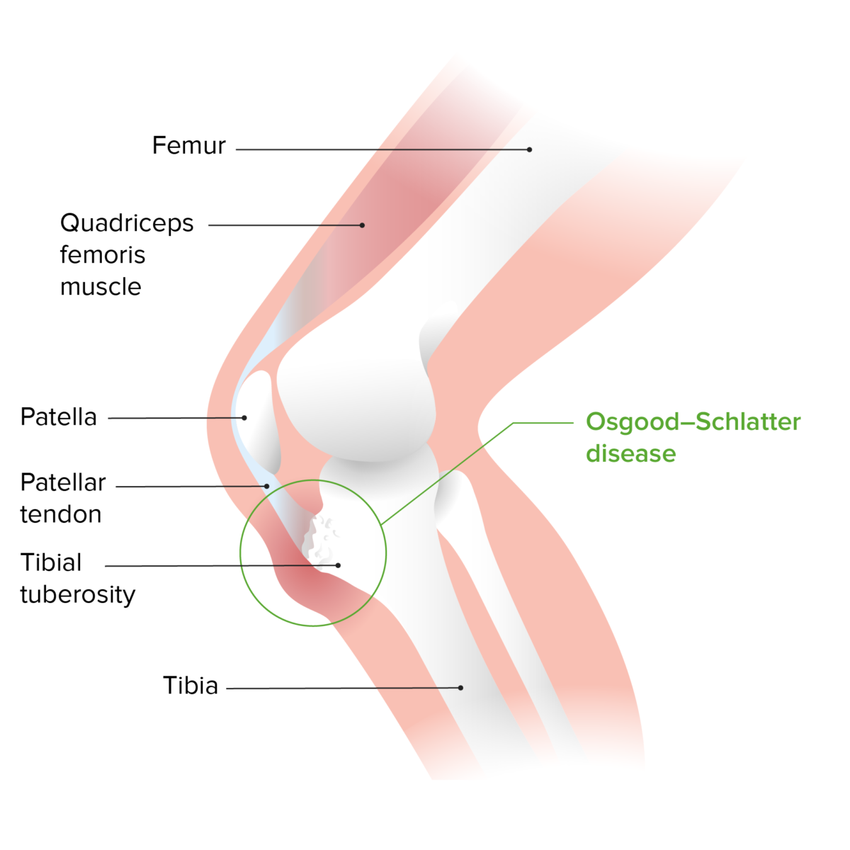
Osgood-shlatter disease

Osgood-Schlatter disease: a painful inflammation of the tibial tuberosity caused by repetitive traction by the patellar tendon due to repetitive exertion, such as running or jumping

Image by Lecturio.

Epidemiology

  • Occurs in children 10–14 years of age: 
    • Boys: 12–14 years 
    • Girls: 10–13 years 
  • More common in teens who participate in sports that involve running or jumping (e.g., soccer, football, basketball, volleyball, sprinting, gymnastics, figure skating, and ballet)
  • More common in boys than girls
  • Usually occurs during periods of accelerated relative growth
  • Bilateral in 25%–50% of cases

Pathophysiology

Osgood-Schlatter disease occurs due to overuse injury.

  • Mechanical forces applied to the knee, especially during running and jumping, cause microtrauma to the patellar tendon.
  • Repetitive microtrauma leads to small chronic avulsions of patellar tendon at the point of insertion into the tibial tubercle.
  • Swelling Swelling Inflammation and ossification Ossification The process of bone formation. Histogenesis of bone including ossification. Bones: Development and Ossification of the distal patellar tendon at point of insertion into the tibia Tibia The second longest bone of the skeleton. It is located on the medial side of the lower leg, articulating with the fibula laterally, the talus distally, and the femur proximally. Knee Joint: Anatomy follow.

Clinical Presentation

Osgood-Schlatter disease typically presents during early adolescence with the chief complaint of knee pain Pain An unpleasant sensation induced by noxious stimuli which are detected by nerve endings of nociceptive neurons. Pain: Types and Pathways.
  • Knee pain Pain An unpleasant sensation induced by noxious stimuli which are detected by nerve endings of nociceptive neurons. Pain: Types and Pathways:
    • Usually refers exclusively to tibial tubercle
    • Chronic, lasting weeks or months
    • No history of traumatic event Traumatic event An emotionally painful, shocking, stressful, and sometimes life-threatening experience. It can result from witnessing distressing events such as natural disasters, physical or sexual abuse, and terrorism or other acts of violence. Posttraumatic Stress Disorder (PTSD) marking beginning of symptoms
    • Aggravated by activity, especially jumping
  • Clinical exam findings:
    • Swelling Swelling Inflammation or increased prominence of tibial tubercle
    • Tenderness to palpation Palpation Application of fingers with light pressure to the surface of the body to determine consistency of parts beneath in physical diagnosis; includes palpation for determining the outlines of organs. Dermatologic Examination at area of swelling Swelling Inflammation
    • Pain Pain An unpleasant sensation induced by noxious stimuli which are detected by nerve endings of nociceptive neurons. Pain: Types and Pathways evoked when rising from squat or extending knee against resistance Resistance Physiologically, the opposition to flow of air caused by the forces of friction. As a part of pulmonary function testing, it is the ratio of driving pressure to the rate of air flow. Ventilation: Mechanics of Breathing
    • Findings can be unilateral or bilateral

Diagnosis

  • Clinical examination is usually sufficient to diagnose this condition. 
  • X-rays X-rays X-rays are high-energy particles of electromagnetic radiation used in the medical field for the generation of anatomical images. X-rays are projected through the body of a patient and onto a film, and this technique is called conventional or projectional radiography. X-rays may be taken to rule out other diagnoses:
    • Tumor Tumor Inflammation
    • Acute avulsion
    • Osteomyelitis Osteomyelitis Osteomyelitis is an infection of the bone that results from the spread of microorganisms from the blood (hematogenous), nearby infected tissue, or open wounds (non-hematogenous). Infections are most commonly caused by Staphylococcus aureus. Osteomyelitis
    • Other knee pathology
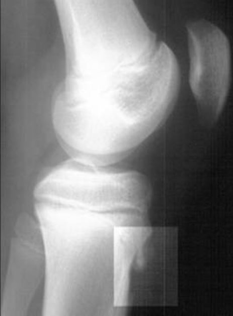
  • X-ray X-ray Penetrating electromagnetic radiation emitted when the inner orbital electrons of an atom are excited and release radiant energy. X-ray wavelengths range from 1 pm to 10 nm. Hard x-rays are the higher energy, shorter wavelength x-rays. Soft x-rays or grenz rays are less energetic and longer in wavelength. The short wavelength end of the x-ray spectrum overlaps the gamma rays wavelength range. The distinction between gamma rays and x-rays is based on their radiation source. Pulmonary Function Tests findings may include:
Osgood-schlatter disease

Osgood-Schlatter disease: lateral radiograph of the knee demonstrating fragmentation of the tibial tubercle with overlying soft tissue swelling

Image: “Review for the generalist: evaluation of anterior knee pain” by Houghton KM. License: CC BY 2.0

Management

Treatment

Most cases are treated successfully with relative rest and symptom management.

  • Reassurance Reassurance Clinician–Patient Relationship and education to the teen and parents: 
    • Explain that the condition is self-limited.
    • Discuss management and reasons to return or be reevaluated.
  • Control of pain Pain An unpleasant sensation induced by noxious stimuli which are detected by nerve endings of nociceptive neurons. Pain: Types and Pathways and swelling Swelling Inflammation:
    • Nonsteroidal anti-inflammatory drugs ( NSAIDs NSAIDS Primary vs Secondary Headaches) or acetaminophen Acetaminophen Acetaminophen is an over-the-counter nonopioid analgesic and antipyretic medication and the most commonly used analgesic worldwide. Despite the widespread use of acetaminophen, its mechanism of action is not entirely understood. Acetaminophen for limited duration 
    • Application of ice for 15–20 minutes up to 2 times/day
  • Modification of activity:
  • Physical therapy Physical Therapy Becker Muscular Dystrophy: core, hip, and lower extremity stretching and strengthening exercises
  • Surgery:
    • Rarely performed
    • Excision of ossicle after closure of growth plate if pain Pain An unpleasant sensation induced by noxious stimuli which are detected by nerve endings of nociceptive neurons. Pain: Types and Pathways persists

Complications

The majority of patients Patients Individuals participating in the health care system for the purpose of receiving therapeutic, diagnostic, or preventive procedures. Clinician–Patient Relationship are asymptomatic after cessation of growth. Uncommon sequelae include:

  • Persistence of prominence of tibial tubercle, typically pain-free:
    • Most noticeable cosmetic sequelae
    • Some patients Patients Individuals participating in the health care system for the purpose of receiving therapeutic, diagnostic, or preventive procedures. Clinician–Patient Relationship note discomfort with kneeling on prominence.
  • Residual ossicles occur in 10% of patients Patients Individuals participating in the health care system for the purpose of receiving therapeutic, diagnostic, or preventive procedures. Clinician–Patient Relationship; rarely cause persistent pain Pain An unpleasant sensation induced by noxious stimuli which are detected by nerve endings of nociceptive neurons. Pain: Types and Pathways that requires removal.
  • Rare association with genu recurvatum (knee hyperextension)

References

  1. Cassas, K. J., & Cassettari-Wayhs, A. (2006). Childhood and adolescent sports-related overuse injuries. American Family Physician, 73(6), 1014. https://www.ncbi.nlm.nih.gov/pubmed/16570735
  2. Ebraheim, N. A., Thomas, B. J., et al. (2019). Orthopedic surgery. In F. C. Brunicardiet al. (Ed.), Schwartz’s principles of surgery, 11th ed. McGraw-Hill Education.
  3. Lawrence, J. T. (2020). The knee. In R. M. Kliegman, et al. (Ed.), Nelson textbook of pediatrics, pp. 361–3623.e1.

Create your free account or log in to continue reading!

Sign up now and get free access to Lecturio with concept pages, medical videos, and questions for your medical education.

User Reviews

Details