Light travels in straight lines until it interacts with an object, which can cause it to reflect, absorb or refract. When light reflects off a surface, the angle of incidence Incidence The number of new cases of a given disease during a given period in a specified population. It also is used for the rate at which new events occur in a defined population. It is differentiated from prevalence, which refers to all cases in the population at a given time. Measures of Disease Frequency is equal to the angle of reflection. Mirrors are objects that reflect light and can create images by reflecting light in a specific way. When light is absorbed by an object, it is converted into other forms of energy, such as heat Heat Inflammation. Refraction Refraction Refractive Errors occurs when light passes through a medium, such as air or water, and its speed and direction change. Total internal reflection occurs when light is reflected back into a medium instead of refracting out of it. This effect is used in fiber optic cables, where light is transmitted over long distances without loss of signal. Microscopes and telescopes use lenses and other components to magnify and focus light for observation.
Last updated: Dec 15, 2025
Bodies that emit light, such as the sun, fixed stars, flames or a light bulb, are known as self-luminous objects or light sources. The distinction is made between artificial light sources (light bulb, arc lamp) and natural light sources (sun, fixed stars).
The light that is emitted into the surrounding space propagates in a straight line as long as it encounters no obstacle. Light beams are used for the presentation of light in an imaginary straight line along which the light is thought to propagate. Light beams do not exist in reality, because the light emitted by a light source fans out in so-called bundles of light. The light beams are demarcated by boundary or marginal rays. The linear propagation Propagation Propagation refers to how the electrical signal spreads to every myocyte in the heart. Cardiac Physiology of light is based on a shadowing effect.
| Type | Definition |
|---|---|
| Divergent | Rays emanating radially from a common point form a divergent light beam, which increases the cross-section of the light beam. |
| Convergent | Rays converging at a mutual point form a convergent light beam, which decreases the cross-section of the light beam. |
| Parallel | Parallel rays extend equally spaced, and the beam cross-section remains the same. |
| Diffuse | Diffuse rays have neither a common starting point nor a common destination, and the cross-sections are not determinable. |
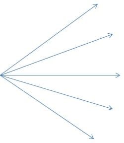
Divergent
Image by Lecturio.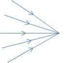
Convergent
Image by Lecturio.
Parallel
Image by Lecturio.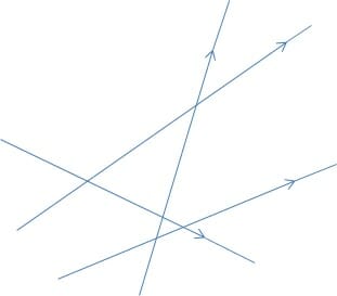
Diffuse
Image by Lecturio.Things are only visible to us when the light originating from them reaches our eye. Illuminated bodies reflect light from their surface into our eyes. There are 2 types of reflection: regular Regular Insulin and diffuse.
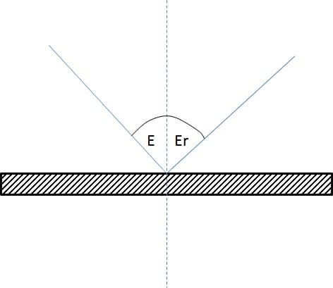
Smooth surfaces reflect light regularly, and the angle of incidence Incidence The number of new cases of a given disease during a given period in a specified population. It also is used for the rate at which new events occur in a defined population. It is differentiated from prevalence, which refers to all cases in the population at a given time. Measures of Disease Frequency is equal to the angle of reflection.
Rough surfaces scatter light in all directions and thereby produce a diffuse reflection. Such a reflection is visible from any side. Examples are room walls, the moon, and the clouds. The indirect lighting produced by diffuse reflection is characterized by reduced glare and reduced shadows.
Plane mirror is common in everyday life. In a plane mirror, the objects and images are symmetrical Symmetrical Dermatologic Examination with respect to the mirror plane. The mirror image is virtual and appears as far behind the mirror as the object in front.
Mirrors with curved surfaces are also common, e.g., car headlights.
The concave mirrors are curved inward, and the mirror surface correlates with a portion of the inside of a spherical surface. The line from the center of the sphere to the center of the mirror is known as the optical axis.
If a parallel beam of light reaches a curved mirror, the rays unite at the focal point. The distance of the focal point from the center of the mirror is called the focal distance. The focal distance is interpreted as half of the radius Radius The outer shorter of the two bones of the forearm, lying parallel to the ulna and partially revolving around it. Forearm: Anatomy of the curvature by the viewer.
f = r / 2
Incident beam parallel to the axis passes through the focal point after reflection. The parallel ray becomes the focal ray.
Incident beam through the focal point travels, after reflection, parallel to the axis. The focal ray becomes the parallel ray.
Incident beam through the spherical center returns after reflection and the central ray remains unchanged.
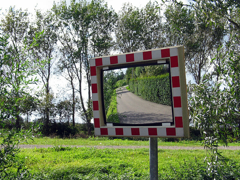
With convex mirrors, the mirror surface correlates with part of the spherical surface. The outward curved portion of the spherical surface reflects light. The images of the convex mirror are always upright and down-scaled.
The closer the object is to the mirror, the greater the image size approaches the object size. An incident parallel beam is reflected as though it originates at a point f behind the mirror—the so-called virtual focal point. An incident divergent beam is divergent after reflection, and its opening angle is greater than that of the incident beam. The peripheral rays can be constructed using the law of reflection.
The relationship between object distance, image distance and focal lengths of concave mirrors are also valid. However, the focal length and the image distance carry a negative sign, since they are behind the mirror.
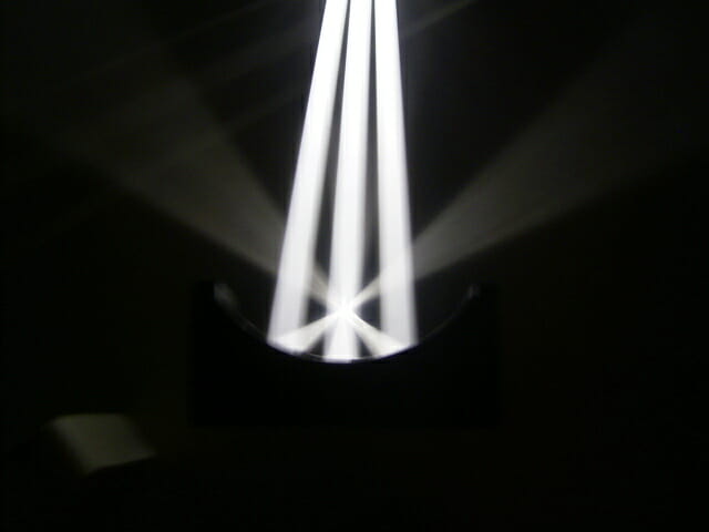
The reflecting surface of parabolic mirrors resembles the section of a parabola. All incident light beams parallel to the axis, and even off-axis, run through the focal point after reflection. Parabolic mirrors are used to reflect light in car headlights.

White walls reflect light better than grey or black. With black walls, almost no reflection of the incident light occurs. The part of the light, which is not reflected by a surface is absorbed. Absorbed light radiation Radiation Emission or propagation of acoustic waves (sound), electromagnetic energy waves (such as light; radio waves; gamma rays; or x-rays), or a stream of subatomic particles (such as electrons; neutrons; protons; or alpha particles). Osteosarcoma is usually converted into heat Heat Inflammation energy.
When a light beam crosses the boundary of two transparent media with different light velocities a part of the light is reflected, while another part is refracted. During the passage from an optically-thinner medium to an optically-denser medium, the light beam is refracted towards the perpendicular plane, and vice versa. A ray traveling perpendicular to the boundary between the 2 media is not refracted and does not change direction.
Incident ray refracted ray, and the normal is in the same plane.
The refractive ratio depends only on the nature of the 2 media. The angle of incidence Incidence The number of new cases of a given disease during a given period in a specified population. It also is used for the rate at which new events occur in a defined population. It is differentiated from prevalence, which refers to all cases in the population at a given time. Measures of Disease Frequency of the light beam does not affect the refraction Refraction Refractive Errors ratio. According to Snell’s law, it is possible to construct the ray path between 2 media when the refractive index is known.
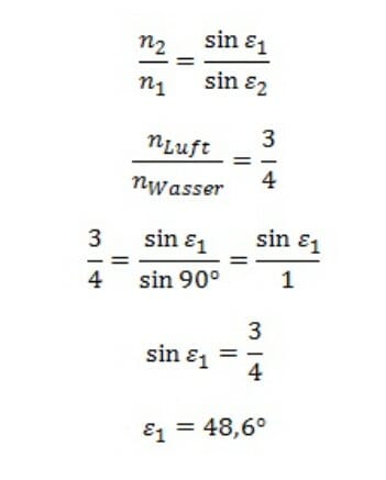
When light travels from an optically-thicker medium to the boundary with an angle of incidence Incidence The number of new cases of a given disease during a given period in a specified population. It also is used for the rate at which new events occur in a defined population. It is differentiated from prevalence, which refers to all cases in the population at a given time. Measures of Disease Frequency greater than the critical angle, the light is completely reflected. The critical angle is reached when the angle of reflection is 90°, e.g., refraction Refraction Refractive Errors at the critical angle when light crosses from water to air.
Example: refraction Refraction Refractive Errors at the critical angle when crossing from water to air
Translucent bodies that are bound by curved surfaces, usually spherical surfaces, are called optical lenses. The connection line of the center points M and M’ of the curved surface forms the optical axis of the lens Lens A transparent, biconvex structure of the eye, enclosed in a capsule and situated behind the iris and in front of the vitreous humor (vitreous body). It is slightly overlapped at its margin by the ciliary processes. Adaptation by the ciliary body is crucial for ocular accommodation. Eye: Anatomy. The points of intersection S1 S1 Heart Sounds and S2 S2 Heart Sounds of the 2 lens Lens A transparent, biconvex structure of the eye, enclosed in a capsule and situated behind the iris and in front of the vitreous humor (vitreous body). It is slightly overlapped at its margin by the ciliary processes. Adaptation by the ciliary body is crucial for ocular accommodation. Eye: Anatomy surfaces and the optical axis are known as the vertices of the lens Lens A transparent, biconvex structure of the eye, enclosed in a capsule and situated behind the iris and in front of the vitreous humor (vitreous body). It is slightly overlapped at its margin by the ciliary processes. Adaptation by the ciliary body is crucial for ocular accommodation. Eye: Anatomy. The midpoint of the segment S1S2 is called the optical midpoint 0. The plane perpendicular to the optical axis through 0 is called the plane of the lens Lens A transparent, biconvex structure of the eye, enclosed in a capsule and situated behind the iris and in front of the vitreous humor (vitreous body). It is slightly overlapped at its margin by the ciliary processes. Adaptation by the ciliary body is crucial for ocular accommodation. Eye: Anatomy or refraction Refraction Refractive Errors.
Rays traveling through the focal point of the lens Lens A transparent, biconvex structure of the eye, enclosed in a capsule and situated behind the iris and in front of the vitreous humor (vitreous body). It is slightly overlapped at its margin by the ciliary processes. Adaptation by the ciliary body is crucial for ocular accommodation. Eye: Anatomy are called focal rays.
Rays traveling through the optical midpoint are called midpoint rays.
Rays entering parallel to the optical axis are known as parallel rays.
The most common lenses are spherical. They are translucent and bound by 2 spherical surfaces. The 2 types of spherical lenses are convex and concave.
Convex lenses are thicker in the middle than on the edges. The refract lighted from a convex lens Convex Lens Refractive Errors converges parallel or moderately divergent light beams so that the rays meet at a point.
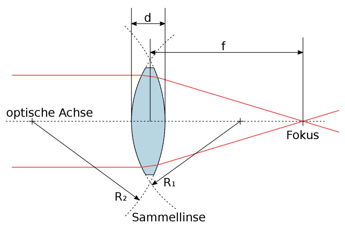
It is possible to converge divergent beam bundles with converging lenses if the divergence is not too large.
Concave lenses are thinner in the middle than on the edges. The refracted light diverges the incident beam. The incident light beam parallel to the optical axis is refracted as if the individual rays arise from a focal point in front of the lens Lens A transparent, biconvex structure of the eye, enclosed in a capsule and situated behind the iris and in front of the vitreous humor (vitreous body). It is slightly overlapped at its margin by the ciliary processes. Adaptation by the ciliary body is crucial for ocular accommodation. Eye: Anatomy. The distance of this point is the focal length (also known as the dispersal distance).
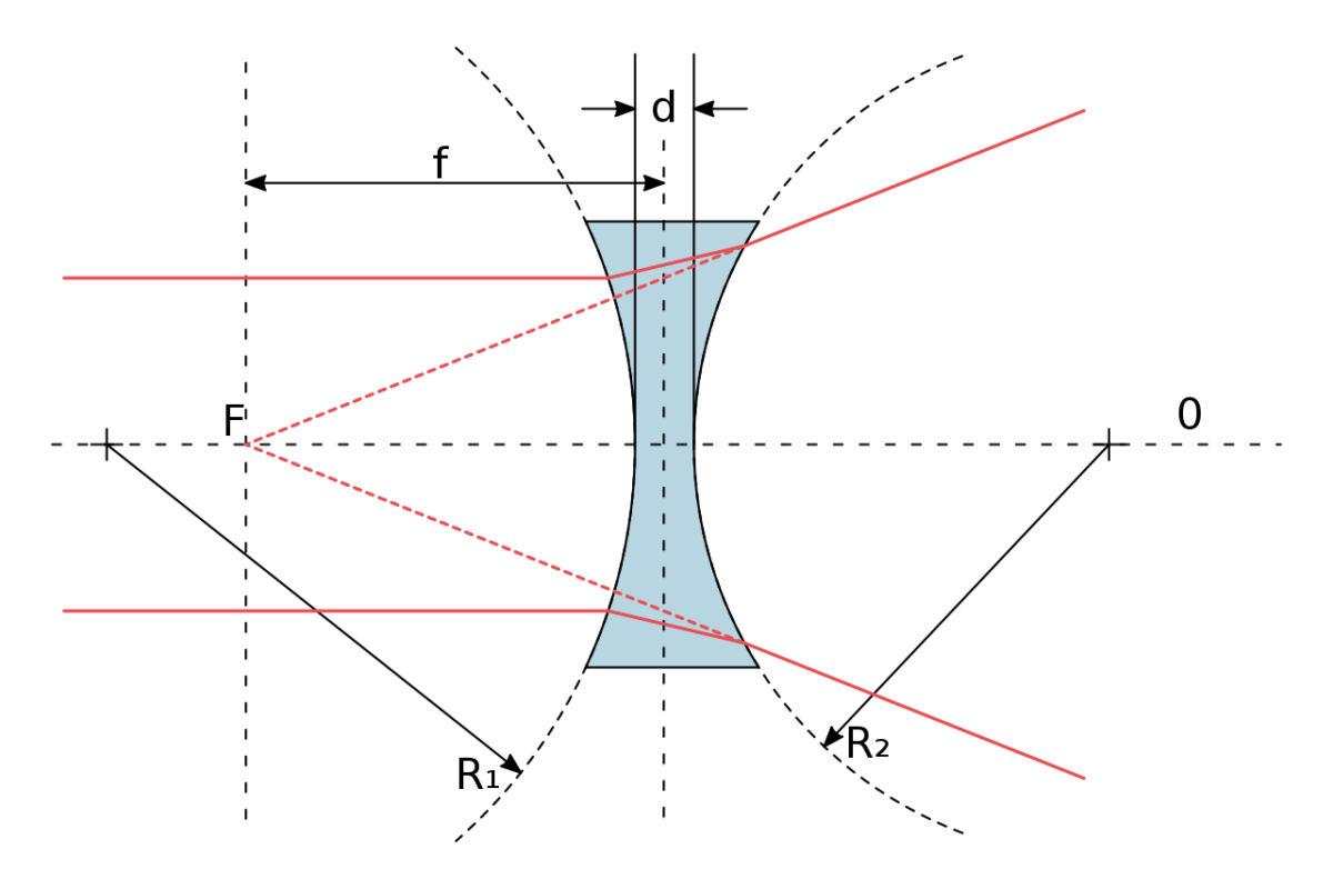
An incident light beam parallel to the optical axis is refracted as if the individual rays originate from a focal point F in front of the lens Lens A transparent, biconvex structure of the eye, enclosed in a capsule and situated behind the iris and in front of the vitreous humor (vitreous body). It is slightly overlapped at its margin by the ciliary processes. Adaptation by the ciliary body is crucial for ocular accommodation. Eye: Anatomy. The distance of this pint is the focal length f, which is negative and is also known as dispersal distance.
The human eye is essentially an optical instrument.
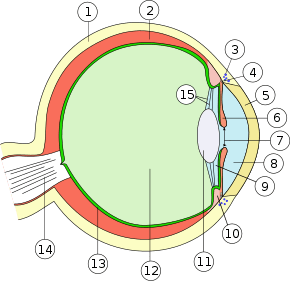
Almost spherical, the eye can be rotated by six muscles acting on it from all sides. The sclera Sclera The white, opaque, fibrous, outer tunic of the eyeball, covering it entirely excepting the segment covered anteriorly by the cornea. It is essentially avascular but contains apertures for vessels, lymphatics, and nerves. Eye: Anatomy (1) forms the outer shell and is continuous with the cornea Cornea The transparent anterior portion of the fibrous coat of the eye consisting of five layers: stratified squamous corneal epithelium; bowman membrane; corneal stroma; descemet membrane; and mesenchymal corneal endothelium. It serves as the first refracting medium of the eye. Eye: Anatomy (5). The iris (6) contains a small hole in the middle. The light rays penetrate into the interior of the eye through the pupil Pupil The pupil is the space within the eye that permits light to project onto the retina. Anatomically located in front of the lens, the pupil’s size is controlled by the surrounding iris. The pupil provides insight into the function of the central and autonomic nervous systems. Pupil: Physiology and Abnormalities (7).
The refracting system of the eye consists of the cornea Cornea The transparent anterior portion of the fibrous coat of the eye consisting of five layers: stratified squamous corneal epithelium; bowman membrane; corneal stroma; descemet membrane; and mesenchymal corneal endothelium. It serves as the first refracting medium of the eye. Eye: Anatomy (5) and the lens Lens A transparent, biconvex structure of the eye, enclosed in a capsule and situated behind the iris and in front of the vitreous humor (vitreous body). It is slightly overlapped at its margin by the ciliary processes. Adaptation by the ciliary body is crucial for ocular accommodation. Eye: Anatomy (11), which act approximately like a convex lens Convex Lens Refractive Errors refracting light onto the retina Retina The ten-layered nervous tissue membrane of the eye. It is continuous with the optic nerve and receives images of external objects and transmits visual impulses to the brain. Its outer surface is in contact with the choroid and the inner surface with the vitreous body. The outermost layer is pigmented, whereas the inner nine layers are transparent. Eye: Anatomy (13). The images of observed objects are real inverted images on the retina Retina The ten-layered nervous tissue membrane of the eye. It is continuous with the optic nerve and receives images of external objects and transmits visual impulses to the brain. Its outer surface is in contact with the choroid and the inner surface with the vitreous body. The outermost layer is pigmented, whereas the inner nine layers are transparent. Eye: Anatomy. A normal eye can see near objects as sharply as distant ones, due to the eye’s ability to change its curvature and focal length, and thus it’s refracting power. This adaptability is called accommodation Accommodation Refractive Errors.
The microscope consists of 2 lenses—the prime lens Lens A transparent, biconvex structure of the eye, enclosed in a capsule and situated behind the iris and in front of the vitreous humor (vitreous body). It is slightly overlapped at its margin by the ciliary processes. Adaptation by the ciliary body is crucial for ocular accommodation. Eye: Anatomy and the ocular system. The prime lens Lens A transparent, biconvex structure of the eye, enclosed in a capsule and situated behind the iris and in front of the vitreous humor (vitreous body). It is slightly overlapped at its margin by the ciliary processes. Adaptation by the ciliary body is crucial for ocular accommodation. Eye: Anatomy is the one facing the observed object whereas the ocular or eyepiece system is in front of the eye.
The prime lens Lens A transparent, biconvex structure of the eye, enclosed in a capsule and situated behind the iris and in front of the vitreous humor (vitreous body). It is slightly overlapped at its margin by the ciliary processes. Adaptation by the ciliary body is crucial for ocular accommodation. Eye: Anatomy can be replaced with lenses of other focal lengths, and the object distance can be adjusted.
Enlarged and inverted virtual images of the observed object can be observed microscopically. The total magnification of the microscope v is the product of the object magnification va and the magnification of the eyepiece vg.
v = va * vg