Obstetric imaging refers to imaging of the female reproductive tract and developing fetus during pregnancyPregnancyThe status during which female mammals carry their developing young (embryos or fetuses) in utero before birth, beginning from fertilization to birth.Pregnancy: Diagnosis, Physiology, and Care. Ultrasonography is the 1st-line imaging modality during pregnancyPregnancyThe status during which female mammals carry their developing young (embryos or fetuses) in utero before birth, beginning from fertilization to birth.Pregnancy: Diagnosis, Physiology, and Care as it does not emit radiationRadiationEmission or propagation of acoustic waves (sound), electromagnetic energy waves (such as light; radio waves; gamma rays; or x-rays), or a stream of subatomic particles (such as electrons; neutrons; protons; or alpha particles).Osteosarcoma; thus, it is the safest option for the developing fetus. Obstetricians depend heavily on ultrasound for the detection, monitoring, and assessment of several maternal and fetal conditions without radiationRadiationEmission or propagation of acoustic waves (sound), electromagnetic energy waves (such as light; radio waves; gamma rays; or x-rays), or a stream of subatomic particles (such as electrons; neutrons; protons; or alpha particles).Osteosarcoma exposure. For example, congenitalCongenitalChorioretinitis fetal anomalies, abnormal placentation, poor fetal growth, and abnormal fluid volumes can all be thoroughly assessed using ultrasound. Radiation-emitting imaging modalities (X-rayX-rayPenetrating electromagnetic radiation emitted when the inner orbital electrons of an atom are excited and release radiant energy. X-ray wavelengths range from 1 pm to 10 nm. Hard x-rays are the higher energy, shorter wavelength x-rays. Soft x-rays or grenz rays are less energetic and longer in wavelength. The short wavelength end of the x-ray spectrum overlaps the gamma rays wavelength range. The distinction between gamma rays and x-rays is based on their radiation source.Pulmonary Function Tests, CT) are typically reserved for nonobstetric emergency situations.
Obstetric imaging refers to imaging of the female reproductive tract and developing fetus during pregnancyPregnancyThe status during which female mammals carry their developing young (embryos or fetuses) in utero before birth, beginning from fertilization to birth.Pregnancy: Diagnosis, Physiology, and Care.
Except in rare cases, obstetric imaging is performed via ultrasound.
Ultrasound has the following advantages:
No radiationRadiationEmission or propagation of acoustic waves (sound), electromagnetic energy waves (such as light; radio waves; gamma rays; or x-rays), or a stream of subatomic particles (such as electrons; neutrons; protons; or alpha particles).Osteosarcoma exposure
Ability to view real-time images of the moving fetus
Relatively low cost and wide availability
In rare circumstances, obstetric imaging can be obtained via other modalities (e.g., MRI).
Nonobstetric imaging in pregnancyPregnancyThe status during which female mammals carry their developing young (embryos or fetuses) in utero before birth, beginning from fertilization to birth.Pregnancy: Diagnosis, Physiology, and Care should be ordered judiciously to avoid unnecessary radiationRadiationEmission or propagation of acoustic waves (sound), electromagnetic energy waves (such as light; radio waves; gamma rays; or x-rays), or a stream of subatomic particles (such as electrons; neutrons; protons; or alpha particles).Osteosarcoma exposure to the developing fetus.
Types of obstetric imaging exams
There are 2 primary types of obstetric imaging exams:
Abdominal obstetric ultrasound: good for assessing the fetus, placentaPlacentaA highly vascularized mammalian fetal-maternal organ and major site of transport of oxygen, nutrients, and fetal waste products. It includes a fetal portion (chorionic villi) derived from trophoblasts and a maternal portion (decidua) derived from the uterine endometrium. The placenta produces an array of steroid, protein and peptide hormones (placental hormones).Placenta, Umbilical Cord, and Amniotic Cavity, fluid, and uterusUterusThe uterus, cervix, and fallopian tubes are part of the internal female reproductive system. The uterus has a thick wall made of smooth muscle (the myometrium) and an inner mucosal layer (the endometrium). The most inferior portion of the uterus is the cervix, which connects the uterine cavity to the vagina.Uterus, Cervix, and Fallopian Tubes: Anatomy starting in the late 1st trimester through delivery
Transvaginal ultrasound:
Good for assessing the cervixCervixThe uterus, cervix, and fallopian tubes are part of the internal female reproductive system. The most inferior portion of the uterus is the cervix, which connects the uterine cavity to the vagina. Externally, the cervix is lined by stratified squamous cells; however, the cervical canal is lined by columnar epithelium.Uterus, Cervix, and Fallopian Tubes: Anatomy throughout pregnancyPregnancyThe status during which female mammals carry their developing young (embryos or fetuses) in utero before birth, beginning from fertilization to birth.Pregnancy: Diagnosis, Physiology, and Care
Better for evaluating the fetus and uterusUterusThe uterus, cervix, and fallopian tubes are part of the internal female reproductive system. The uterus has a thick wall made of smooth muscle (the myometrium) and an inner mucosal layer (the endometrium). The most inferior portion of the uterus is the cervix, which connects the uterine cavity to the vagina.Uterus, Cervix, and Fallopian Tubes: Anatomy in early pregnancyPregnancyThe status during which female mammals carry their developing young (embryos or fetuses) in utero before birth, beginning from fertilization to birth.Pregnancy: Diagnosis, Physiology, and Care
Specific studies
Several specific types of studies can be performed. All are ultrasound exams and may be either abdominal or transvaginal.
Anatomy survey: to assess the anatomy of both the fetus and the mother
Growth scan: Specific measurements are used to calculate the estimated weight of the fetus.
Position assessment: to determine the direction in which the fetus is facing within the uterusUterusThe uterus, cervix, and fallopian tubes are part of the internal female reproductive system. The uterus has a thick wall made of smooth muscle (the myometrium) and an inner mucosal layer (the endometrium). The most inferior portion of the uterus is the cervix, which connects the uterine cavity to the vagina.Uterus, Cervix, and Fallopian Tubes: Anatomy in preparation for delivery
Fluid assessments: measurements to help estimate the amount of amniotic fluidAmniotic fluidA clear, yellowish liquid that envelopes the fetus inside the sac of amnion. In the first trimester, it is likely a transudate of maternal or fetal plasma. In the second trimester, amniotic fluid derives primarily from fetal lung and kidney. Cells or substances in this fluid can be removed for prenatal diagnostic tests (amniocentesis).Placenta, Umbilical Cord, and Amniotic Cavity
Biophysical profile:
Assessment of fetal well-being
Determination of different types of fetal movements combined with a fluid assessment
DopplerDopplerUltrasonography applying the doppler effect, with frequency-shifted ultrasound reflections produced by moving targets (usually red blood cells) in the bloodstream along the ultrasound axis in direct proportion to the velocity of movement of the targets, to determine both direction and velocity of blood flow.Ultrasound (Sonography) studies:
Evaluation of the pulse waveforms in specific fetal arteriesArteriesArteries are tubular collections of cells that transport oxygenated blood and nutrients from the heart to the tissues of the body. The blood passes through the arteries in order of decreasing luminal diameter, starting in the largest artery (the aorta) and ending in the small arterioles. Arteries are classified into 3 types: large elastic arteries, medium muscular arteries, and small arteries and arterioles. Arteries: Histology to assess fetal well-being
May demonstrate signs of fetal anemiaAnemiaAnemia is a condition in which individuals have low Hb levels, which can arise from various causes. Anemia is accompanied by a reduced number of RBCs and may manifest with fatigue, shortness of breath, pallor, and weakness. Subtypes are classified by the size of RBCs, chronicity, and etiology. Anemia: Overview and Types or uteroplacental insufficiencyUteroplacental InsufficiencyUteroplacental insufficiency may be acute or chronic and refers to the inability of the placenta to deliver a sufficient supply of O2 and nutrients to the fetusPlacental Abnormalities
Indications for Obstetric Imaging
Routine prenatal carePrenatal carePrenatal care is a systematic and periodic assessment of pregnant women during gestation to assure the best health outcome for the mother and her fetus. Prenatal care prevents and identifies maternal and fetal problems that adversely affect the pregnancy outcome. Prenatal Care
Obstetric imaging is part of routine prenatal carePrenatal carePrenatal care is a systematic and periodic assessment of pregnant women during gestation to assure the best health outcome for the mother and her fetus. Prenatal care prevents and identifies maternal and fetal problems that adversely affect the pregnancy outcome. Prenatal Care, including:
Looking for abnormalities of the uterusUterusThe uterus, cervix, and fallopian tubes are part of the internal female reproductive system. The uterus has a thick wall made of smooth muscle (the myometrium) and an inner mucosal layer (the endometrium). The most inferior portion of the uterus is the cervix, which connects the uterine cavity to the vagina.Uterus, Cervix, and Fallopian Tubes: Anatomy and/or ovariesOvariesOvaries are the paired gonads of the female reproductive system that contain haploid gametes known as oocytes. The ovaries are located intraperitoneally in the pelvis, just posterior to the broad ligament, and are connected to the pelvic sidewall and to the uterus by ligaments. These organs function to secrete hormones (estrogen and progesterone) and to produce the female germ cells (oocytes).Ovaries: Anatomy
Location of the placentaPlacentaA highly vascularized mammalian fetal-maternal organ and major site of transport of oxygen, nutrients, and fetal waste products. It includes a fetal portion (chorionic villi) derived from trophoblasts and a maternal portion (decidua) derived from the uterine endometrium. The placenta produces an array of steroid, protein and peptide hormones (placental hormones).Placenta, Umbilical Cord, and Amniotic Cavity (confirming it is away from the internal cervical os)
Looking for signs of placental invasion into the myometrium (e.g., placenta accretaPlacenta AccretaAbnormal placentation in which all or parts of the placenta are attached directly to the myometrium due to a complete or partial absence of decidua. It is associated with postpartum hemorrhage because of the failure of placental separation.Placental Abnormalities spectrum (PAS))
Assessing the cervical length
3rd trimester:
Estimating fluid volumes
Determining fetal positionFetal positionDirection of the fetal head in relation to the maternal pelvis in vertex presentationsNormal and Abnormal Labor prior to delivery
Assessing fetal status in higher-risk pregnancies (e.g., biophysical profile or dopplerDopplerUltrasonography applying the doppler effect, with frequency-shifted ultrasound reflections produced by moving targets (usually red blood cells) in the bloodstream along the ultrasound axis in direct proportion to the velocity of movement of the targets, to determine both direction and velocity of blood flow.Ultrasound (Sonography) studies in an individual with known preeclampsiaPreeclampsiaA complication of pregnancy, characterized by a complex of symptoms including maternal hypertension and proteinuria with or without pathological edema. Symptoms may range between mild and severe. Pre-eclampsia usually occurs after the 20th week of gestation, but may develop before this time in the presence of trophoblastic disease.Hypertensive Pregnancy Disorders)
Following fetuses at high risk for developing hydropsHydropsCholecystitis fetalis
Emergency care
Individuals may present with a number of symptoms in pregnancyPregnancyThe status during which female mammals carry their developing young (embryos or fetuses) in utero before birth, beginning from fertilization to birth.Pregnancy: Diagnosis, Physiology, and Care that warrant ultrasound evaluation:
Bleeding and/or painPainAn unpleasant sensation induced by noxious stimuli which are detected by nerve endings of nociceptive neurons.Pain: Types and Pathways in early pregnancyPregnancyThe status during which female mammals carry their developing young (embryos or fetuses) in utero before birth, beginning from fertilization to birth.Pregnancy: Diagnosis, Physiology, and Care:
Rule out ectopic and molar pregnancies.
Assess fetal viability/evaluate for potential fetal loss (i.e., spontaneous abortionAbortionExpulsion of the product of fertilization before completing the term of gestation and without deliberate interference.Spontaneous Abortion).
Bleeding in later pregnancyPregnancyThe status during which female mammals carry their developing young (embryos or fetuses) in utero before birth, beginning from fertilization to birth.Pregnancy: Diagnosis, Physiology, and Care: Look for signs of placental abruptionPlacental AbruptionPremature separation of the normally implanted placenta from the uterus. Signs of varying degree of severity include uterine bleeding, uterine muscle hypertonia, and fetal distress or fetal death.Antepartum Hemorrhage (prematurePrematureChildbirth before 37 weeks of pregnancy (259 days from the first day of the mother’s last menstrual period, or 245 days after fertilization).Necrotizing Enterocolitis separation of the placentaPlacentaA highly vascularized mammalian fetal-maternal organ and major site of transport of oxygen, nutrients, and fetal waste products. It includes a fetal portion (chorionic villi) derived from trophoblasts and a maternal portion (decidua) derived from the uterine endometrium. The placenta produces an array of steroid, protein and peptide hormones (placental hormones).Placenta, Umbilical Cord, and Amniotic Cavity).
Preterm contractions or pelvic painPainAn unpleasant sensation induced by noxious stimuli which are detected by nerve endings of nociceptive neurons.Pain: Types and Pathways:
Cervical length measurement to assess for cervical insufficiencyCervical insufficiencyCervical dilation without contractions (not labor), due to structural weakness of the cervixPreterm Labor and Birth or signs of cervical change
Growth scan: important to help the pediatrics team prepare for delivery and provide appropriate counseling to parents (especially in anticipated cases of very prematurePrematureChildbirth before 37 weeks of pregnancy (259 days from the first day of the mother’s last menstrual period, or 245 days after fertilization).Necrotizing Enterocolitis delivery)
Loss of fluid: Assess fluid levels.
Decreased fetal movement: biophysical profile for the assessment of fetal movement
During procedures
Ultrasound is often used to assist the physician during procedures such as:
AmniocentesisAmniocentesisPercutaneous transabdominal puncture of the uterus during pregnancy to obtain amniotic fluid. It is commonly used for fetal karyotype determination in order to diagnose abnormal fetal conditions.Polyhydramnios
2nd/3rd trimesters: A hip roll (rolled-up sheet) should be placed under 1 of the hips of the individual so that they are not lying flat on their back.
Transvaginal scans: lower lithotomy
Tips for obtaining good images:
Maximize contact between the individual’s skinSkinThe skin, also referred to as the integumentary system, is the largest organ of the body. The skin is primarily composed of the epidermis (outer layer) and dermis (deep layer). The epidermis is primarily composed of keratinocytes that undergo rapid turnover, while the dermis contains dense layers of connective tissue.Skin: Structure and Functions and ultrasound probeProbeA device placed on the patient’s body to visualize a targetUltrasound (Sonography).
Use plenty of ultrasound gel.
Depth and gain:
Determines the field of view and echogenicity characteristics of the tissue
In early pregnancyPregnancyThe status during which female mammals carry their developing young (embryos or fetuses) in utero before birth, beginning from fertilization to birth.Pregnancy: Diagnosis, Physiology, and Care, the entire gestational sac should be viewed at once.
Components of the exam
Components that should be assessed during all 2nd and 3rd trimester obstetric ultrasounds:
Fetal heart rateHeart rateThe number of times the heart ventricles contract per unit of time, usually per minute.Cardiac Physiology
Fetal positionFetal positionDirection of the fetal head in relation to the maternal pelvis in vertex presentationsNormal and Abnormal Labor: What part of the fetus is in the lower uterine segment (and thus will deliver 1st)?
Amniotic fluidAmniotic fluidA clear, yellowish liquid that envelopes the fetus inside the sac of amnion. In the first trimester, it is likely a transudate of maternal or fetal plasma. In the second trimester, amniotic fluid derives primarily from fetal lung and kidney. Cells or substances in this fluid can be removed for prenatal diagnostic tests (amniocentesis).Placenta, Umbilical Cord, and Amniotic Cavity volume
Placental location in relation to the cervixCervixThe uterus, cervix, and fallopian tubes are part of the internal female reproductive system. The most inferior portion of the uterus is the cervix, which connects the uterine cavity to the vagina. Externally, the cervix is lined by stratified squamous cells; however, the cervical canal is lined by columnar epithelium.Uterus, Cervix, and Fallopian Tubes: Anatomy
Any other objective of the study (e.g., anatomic survey, growth assessment, etcETCThe electron transport chain (ETC) sends electrons through a series of proteins, which generate an electrochemical proton gradient that produces energy in the form of adenosine triphosphate (ATP).Electron Transport Chain (ETC).)
Preparing for general image interpretation
Prior to interpretation of any image, the physician should take certain preparatory steps. The same systematic approach should be followed every time.
Confirm name, date, and time on all images.
Review the individual’s medical history and physical examination findings.
Confirm that the appropriate exams and techniques that can best assess the suspected pathology were ordered/performed.
Earliest sign of intrauterine pregnancyPregnancyThe status during which female mammals carry their developing young (embryos or fetuses) in utero before birth, beginning from fertilization to birth.Pregnancy: Diagnosis, Physiology, and Care, seen around 4.5‒5 weeks gestational ageGestational ageThe age of the conceptus, beginning from the time of fertilization. In clinical obstetrics, the gestational age is often estimated as the time from the last day of the last menstruation which is about 2 weeks before ovulation and fertilization.Pregnancy: Diagnosis, Physiology, and Care (wga)
Should be visible in the uterusUterusThe uterus, cervix, and fallopian tubes are part of the internal female reproductive system. The uterus has a thick wall made of smooth muscle (the myometrium) and an inner mucosal layer (the endometrium). The most inferior portion of the uterus is the cervix, which connects the uterine cavity to the vagina.Uterus, Cervix, and Fallopian Tubes: Anatomy if the quantitative serum β-hCG is > 2,000
AnechoicAnechoicA structure that produces no echo at all (looks completely black)Ultrasound (Sonography), well-defined round structure
Surrounded by an echogenic rim, representing the decidual reaction
Yolk sacYolk SacThe first of four extra-embryonic membranes to form during embryogenesis. In reptiles and birds, it arises from endoderm and mesoderm to incorporate the egg yolk into the digestive tract for nourishing the embryo. In placental mammals, its nutritional function is vestigial; however, it is the source of intestinal mucosa; blood cells; and germ cells. It is sometimes called the vitelline sac, which should not be confused with the vitelline membrane of the egg.Embryoblast and Trophoblast Development:
HyperechoicHyperechoicA structure that produces a high-amplitude echo (lighter grays and white)Ultrasound (Sonography) ring-like structure within the gestational sac
1st seen at approximately 5‒6 wga and disappears at approximately 10 wga
Fetal pole:
The fetus itself
Visible around 5.5‒6 wga
A heartbeat is usually visible as soon as the fetal pole is visible.
An adnexal massMassThree-dimensional lesion that occupies a space within the breastImaging of the Breast representing the follicle from which the oocyte ovulated, which persists throughout the 1st trimester of pregnancyPregnancyThe status during which female mammals carry their developing young (embryos or fetuses) in utero before birth, beginning from fertilization to birth.Pregnancy: Diagnosis, Physiology, and Care
Produces progesteroneProgesteroneThe major progestational steroid that is secreted primarily by the corpus luteum and the placenta. Progesterone acts on the uterus, the mammary glands and the brain. It is required in embryo implantation; pregnancy maintenance, and the development of mammary tissue for milk production. Progesterone, converted from pregnenolone, also serves as an intermediate in the biosynthesis of gonadal steroid hormones and adrenal corticosteroids.Gonadal Hormones, which is vital for survival of the pregnancyPregnancyThe status during which female mammals carry their developing young (embryos or fetuses) in utero before birth, beginning from fertilization to birth.Pregnancy: Diagnosis, Physiology, and Care
Sonographic appearance:
Cyst may be simple or complex.
Typically surrounded by ↑ vasculature, seen on DopplerDopplerUltrasonography applying the doppler effect, with frequency-shifted ultrasound reflections produced by moving targets (usually red blood cells) in the bloodstream along the ultrasound axis in direct proportion to the velocity of movement of the targets, to determine both direction and velocity of blood flow.Ultrasound (Sonography) studies as a circumferential rim of color known as the “ring of fire”
Determining viability
Establishing that a pregnancyPregnancyThe status during which female mammals carry their developing young (embryos or fetuses) in utero before birth, beginning from fertilization to birth.Pregnancy: Diagnosis, Physiology, and Care is viable requires:
Intrauterine location:
Should be within the main uterine body endometriumEndometriumThe mucous membrane lining of the uterine cavity that is hormonally responsive during the menstrual cycle and pregnancy. The endometrium undergoes cyclic changes that characterize menstruation. After successful fertilization, it serves to sustain the developing embryo.Embryoblast and Trophoblast Development
At least a gestational sac and yolk sacYolk SacThe first of four extra-embryonic membranes to form during embryogenesis. In reptiles and birds, it arises from endoderm and mesoderm to incorporate the egg yolk into the digestive tract for nourishing the embryo. In placental mammals, its nutritional function is vestigial; however, it is the source of intestinal mucosa; blood cells; and germ cells. It is sometimes called the vitelline sac, which should not be confused with the vitelline membrane of the egg.Embryoblast and Trophoblast Development must be seen in order to establish the pregnancyPregnancyThe status during which female mammals carry their developing young (embryos or fetuses) in utero before birth, beginning from fertilization to birth.Pregnancy: Diagnosis, Physiology, and Care location (a gestational sac alone is not enough).
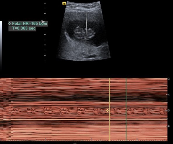
A detectable fetal heart rateHeart rateThe number of times the heart ventricles contract per unit of time, usually per minute.Cardiac Physiology, usually between about 120‒160 per minute (may be slightly higher at certain points in early pregnancyPregnancyThe status during which female mammals carry their developing young (embryos or fetuses) in utero before birth, beginning from fertilization to birth.Pregnancy: Diagnosis, Physiology, and Care)
Calculation of the fetal heart rate by ultrasound
Image by Hetal Verma.
PregnancyPregnancyThe status during which female mammals carry their developing young (embryos or fetuses) in utero before birth, beginning from fertilization to birth.Pregnancy: Diagnosis, Physiology, and Care dating via ultrasound
1st trimester obstetric ultrasound is the best and the most accurate tool to estimate gestational ageGestational ageThe age of the conceptus, beginning from the time of fertilization. In clinical obstetrics, the gestational age is often estimated as the time from the last day of the last menstruation which is about 2 weeks before ovulation and fertilization.Pregnancy: Diagnosis, Physiology, and Care and calculate the EDDEDDPregnancy: Diagnosis, Physiology, and Care.
Most accurate in the 1st trimester
Ultrasound gets less and less accurate as gestation progresses due to normal genetic variations (e.g., height of parents) and due to effects of the intrauterine environment (e.g., smokers have worse placental perfusion).
1st-trimester dating:
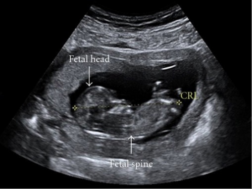
Measure the crown-rump length.
Crown-rump length should be consistent with the expected gestational ageGestational ageThe age of the conceptus, beginning from the time of fertilization. In clinical obstetrics, the gestational age is often estimated as the time from the last day of the last menstruation which is about 2 weeks before ovulation and fertilization.Pregnancy: Diagnosis, Physiology, and Care based on the last menstrual periodLast menstrual periodThe 1st day of a woman’s last menstrual period. By convention, this date is usually used to date pregnancies.Pregnancy: Diagnosis, Physiology, and Care (LMPLMPThe 1st day of a woman’s LMPPregnancy: Diagnosis, Physiology, and Care).
Ultrasound image of a 12-week fetus: The broken yellow line is the crown-rump length measurement.
Image: “Ultrasound image of a 12-week fetus” by Child Health, Royal Aberdeen Children’s Hospital, University of Aberdeen, Foresterhill, Aberdeen AB25 2ZG, UK. License: CC BY 3.0
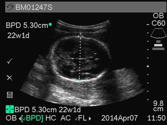
Biparietal diameter measurement (a component of the growth scan used to help calculate the estimated fetal weight)
Image: “Representative sample of biparietal diameter from Ghana Randomized Air Pollution and Health Study (GRAPHS) participant” by Boamah EA. License: CC BY 2.0
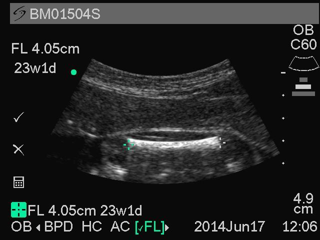
Femur length (a component of the growth scan used to help calculate the estimated fetal weight)
Image: “Representative sample image of femur length from Ghana Randomized Air Pollution and Health Study (GRAPHS) participant” by Boamah EA. License: CC BY 2.0
Determining the number of embryos
The uterusUterusThe uterus, cervix, and fallopian tubes are part of the internal female reproductive system. The uterus has a thick wall made of smooth muscle (the myometrium) and an inner mucosal layer (the endometrium). The most inferior portion of the uterus is the cervix, which connects the uterine cavity to the vagina.Uterus, Cervix, and Fallopian Tubes: Anatomy should be fully evaluated in all planes to get an accurate fetal count.
Multiple gestation: when > 1 fetus is present
Twins
Higher-order multiples (e.g., triplets, quadruplets, etcETCThe electron transport chain (ETC) sends electrons through a series of proteins, which generate an electrochemical proton gradient that produces energy in the form of adenosine triphosphate (ATP).Electron Transport Chain (ETC).)
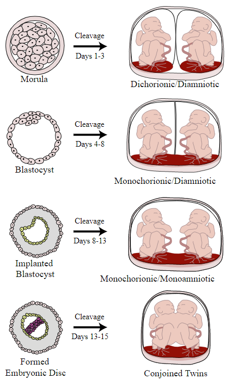
Determining chorionicity in multiple gestations
Chorionicity describes whether the fetuses share a chorionChorionThe outermost extraembryonic membrane surrounding the developing embryo. In reptiles and birds, it adheres to the shell and allows exchange of gases between the egg and its environment. In mammals, the chorion evolves into the fetal contribution of the placenta.Placenta, Umbilical Cord, and Amniotic Cavity or amnionAmnionThe innermost membranous sac that surrounds and protects the developing embryo which is bathed in the amniotic fluid. Amnion cells are secretory epithelial cells and contribute to the amniotic fluid.Placenta, Umbilical Cord, and Amniotic Cavity. Chorionicity can be established by different ultrasound findings in different types of twins:
Dichorionic/diamniotic twins (each twin is in their own chorioamnion and has their own placentaPlacentaA highly vascularized mammalian fetal-maternal organ and major site of transport of oxygen, nutrients, and fetal waste products. It includes a fetal portion (chorionic villi) derived from trophoblasts and a maternal portion (decidua) derived from the uterine endometrium. The placenta produces an array of steroid, protein and peptide hormones (placental hormones).Placenta, Umbilical Cord, and Amniotic Cavity):
Thick intertwin membrane
Lambda sign: a thick, triangular protrusion of tissue leading up to the intertwin membrane
2 separate placentas (however, if they are right next to each other, they may appear as a single placentaPlacentaA highly vascularized mammalian fetal-maternal organ and major site of transport of oxygen, nutrients, and fetal waste products. It includes a fetal portion (chorionic villi) derived from trophoblasts and a maternal portion (decidua) derived from the uterine endometrium. The placenta produces an array of steroid, protein and peptide hormones (placental hormones).Placenta, Umbilical Cord, and Amniotic Cavity)
Monochorionic/diamniotic (twins are in their own amniotic sac, but share a chorionChorionThe outermost extraembryonic membrane surrounding the developing embryo. In reptiles and birds, it adheres to the shell and allows exchange of gases between the egg and its environment. In mammals, the chorion evolves into the fetal contribution of the placenta.Placenta, Umbilical Cord, and Amniotic Cavity and placentaPlacentaA highly vascularized mammalian fetal-maternal organ and major site of transport of oxygen, nutrients, and fetal waste products. It includes a fetal portion (chorionic villi) derived from trophoblasts and a maternal portion (decidua) derived from the uterine endometrium. The placenta produces an array of steroid, protein and peptide hormones (placental hormones).Placenta, Umbilical Cord, and Amniotic Cavity):
Thin intertwin membrane
T sign: The intertwin membrane comes straight into the sac wall, without the thick triangular protrusion of tissue that is seen in dichorionic diamniotic twins.
Single placentaPlacentaA highly vascularized mammalian fetal-maternal organ and major site of transport of oxygen, nutrients, and fetal waste products. It includes a fetal portion (chorionic villi) derived from trophoblasts and a maternal portion (decidua) derived from the uterine endometrium. The placenta produces an array of steroid, protein and peptide hormones (placental hormones).Placenta, Umbilical Cord, and Amniotic Cavity
Monochorionic/monoamniotic (twins share a chorioamnion and placentaPlacentaA highly vascularized mammalian fetal-maternal organ and major site of transport of oxygen, nutrients, and fetal waste products. It includes a fetal portion (chorionic villi) derived from trophoblasts and a maternal portion (decidua) derived from the uterine endometrium. The placenta produces an array of steroid, protein and peptide hormones (placental hormones).Placenta, Umbilical Cord, and Amniotic Cavity)
No intertwin membrane
Single placentaPlacentaA highly vascularized mammalian fetal-maternal organ and major site of transport of oxygen, nutrients, and fetal waste products. It includes a fetal portion (chorionic villi) derived from trophoblasts and a maternal portion (decidua) derived from the uterine endometrium. The placenta produces an array of steroid, protein and peptide hormones (placental hormones).Placenta, Umbilical Cord, and Amniotic Cavity
Diagram depicting twin chorionicity
Image: “Illustrates various types of chorionicity and amniosity (how the baby’s sac looks) in monozygotic (one egg/identical) twins as a result of when the blastocyst or embryo splits.” by Kevin Dufendach. License: CC BY 3.0
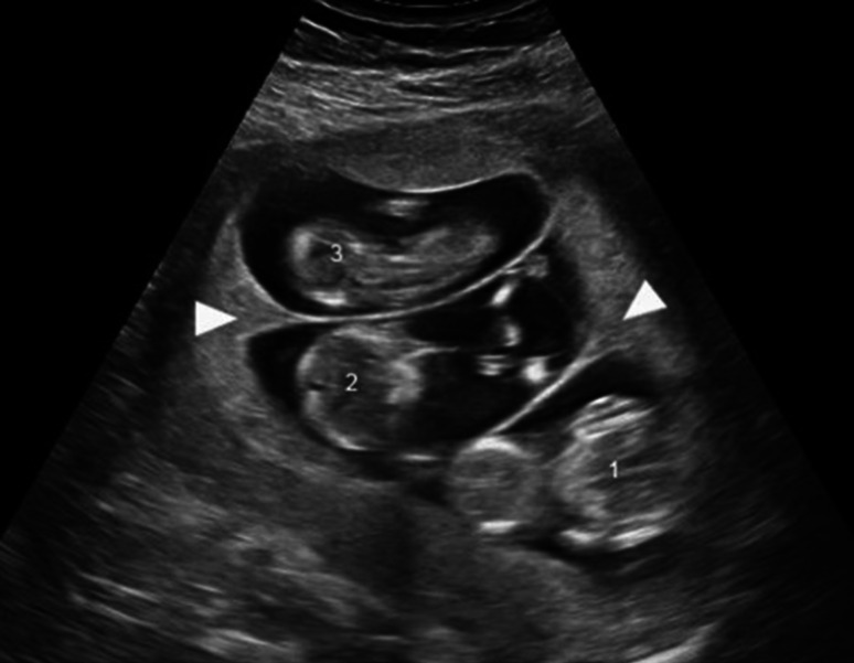
Ultrasound picture of trichorionic triplets at 12-weeks gestational age: Each fetus has their own chorion, amnion, and placenta. The numbers (1‒3) indicate each of the 3 fetuses. Arrowheads show the lambda sign, the triangular portion of tissue leading into the intertwin membrane.
Image: “Ultrasound picture of trichorionic triplets at 12 weeks gestational age” by Kristine Marceau. License: CC BY 4.0, cropped by Lecturio.
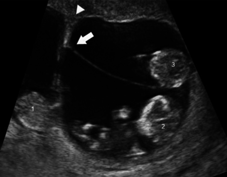
Ultrasound picture of a dichorionic, triamniotic triplets at 13-weeks gestational age: Arrowhead shows the lambda sign between the 2 chorions. The full arrow shows the T sign between the 2 monochorionic-diamniotic infants. The numbers 1‒3 show the 3 fetuses. Fetus 1 has their own placenta, while fetuses 2 and 3 share a placenta.
Image: “Ultrasound picture of a dichorionic, triamniotic triplets at 13 weeks gestational age” by Kristine Marceau. License: CC BY 4.0, cropped by Lecturio.
Anatomic survey
A complete anatomic survey assesses both the maternal reproductive tract and looks for fetal anomalies. Some of the important features evaluated include:
Maternal anatomy:
Cervical length: should be > 25 mmMMMultiple myeloma (MM) is a malignant condition of plasma cells (activated B lymphocytes) primarily seen in the elderly. Monoclonal proliferation of plasma cells results in cytokine-driven osteoclastic activity and excessive secretion of IgG antibodies.Multiple Myeloma until at least 24 wga
Presence of any uterine fibroidsUterine FibroidsGynecological Imaging distorting the cavity, or in the lower uterine segment, which may be in the way of a potential cesarean incision
Placental and umbilical cordUmbilical cordThe flexible rope-like structure that connects a developing fetus to the placenta in mammals. The cord contains blood vessels which carry oxygen and nutrients from the mother to the fetus and waste products away from the fetus.Placenta, Umbilical Cord, and Amniotic Cavity assessment:
PlacentaPlacentaA highly vascularized mammalian fetal-maternal organ and major site of transport of oxygen, nutrients, and fetal waste products. It includes a fetal portion (chorionic villi) derived from trophoblasts and a maternal portion (decidua) derived from the uterine endometrium. The placenta produces an array of steroid, protein and peptide hormones (placental hormones).Placenta, Umbilical Cord, and Amniotic Cavity:
Should not cover the internal cervical os
Should not invade into the underlying myometrium
Umbilical cordUmbilical cordThe flexible rope-like structure that connects a developing fetus to the placenta in mammals. The cord contains blood vessels which carry oxygen and nutrients from the mother to the fetus and waste products away from the fetus.Placenta, Umbilical Cord, and Amniotic Cavity:
Should have 3 visible vessels
Should insert near the middle of the placentaPlacentaA highly vascularized mammalian fetal-maternal organ and major site of transport of oxygen, nutrients, and fetal waste products. It includes a fetal portion (chorionic villi) derived from trophoblasts and a maternal portion (decidua) derived from the uterine endometrium. The placenta produces an array of steroid, protein and peptide hormones (placental hormones).Placenta, Umbilical Cord, and Amniotic Cavity and at the fetal umbilicus
Vessels should be surrounded by protective jelly all the way down to the placental insertion.
Fetal anatomy: Multiple structures, including all major organs, are measured and assessed.
BrainBrainThe part of central nervous system that is contained within the skull (cranium). Arising from the neural tube, the embryonic brain is comprised of three major parts including prosencephalon (the forebrain); mesencephalon (the midbrain); and rhombencephalon (the hindbrain). The developed brain consists of cerebrum; cerebellum; and other structures in the brain stem.Nervous System: Anatomy, Structure, and Classification
Face
Heart/lungsLungsLungs are the main organs of the respiratory system. Lungs are paired viscera located in the thoracic cavity and are composed of spongy tissue. The primary function of the lungs is to oxygenate blood and eliminate CO2. Lungs: Anatomy, including 4-chamber and outflow-tract views of the heart
Abdomen
Limbs
Genitalia
Amniotic fluidAmniotic fluidA clear, yellowish liquid that envelopes the fetus inside the sac of amnion. In the first trimester, it is likely a transudate of maternal or fetal plasma. In the second trimester, amniotic fluid derives primarily from fetal lung and kidney. Cells or substances in this fluid can be removed for prenatal diagnostic tests (amniocentesis).Placenta, Umbilical Cord, and Amniotic Cavity assessment
Amniotic fluidAmniotic fluidA clear, yellowish liquid that envelopes the fetus inside the sac of amnion. In the first trimester, it is likely a transudate of maternal or fetal plasma. In the second trimester, amniotic fluid derives primarily from fetal lung and kidney. Cells or substances in this fluid can be removed for prenatal diagnostic tests (amniocentesis).Placenta, Umbilical Cord, and Amniotic Cavity can be assessed in 2 ways:
Single deepest pocket (SDP):
Measures the single deepest vertical pocket of fluid
The measured pocket must be free of the umbilical cordUmbilical cordThe flexible rope-like structure that connects a developing fetus to the placenta in mammals. The cord contains blood vessels which carry oxygen and nutrients from the mother to the fetus and waste products away from the fetus.Placenta, Umbilical Cord, and Amniotic Cavity and fetal parts.
Normal range (2nd and 3rd trimesters): 2‒8 cm
Amniotic fluidAmniotic fluidA clear, yellowish liquid that envelopes the fetus inside the sac of amnion. In the first trimester, it is likely a transudate of maternal or fetal plasma. In the second trimester, amniotic fluid derives primarily from fetal lung and kidney. Cells or substances in this fluid can be removed for prenatal diagnostic tests (amniocentesis).Placenta, Umbilical Cord, and Amniotic Cavity index (AFI):
Divide the uterusUterusThe uterus, cervix, and fallopian tubes are part of the internal female reproductive system. The uterus has a thick wall made of smooth muscle (the myometrium) and an inner mucosal layer (the endometrium). The most inferior portion of the uterus is the cervix, which connects the uterine cavity to the vagina.Uterus, Cervix, and Fallopian Tubes: Anatomy into 4 quadrants and obtain an SDP for each quadrant; the AFI is the sum of the 4 SDP measurements.
Normal range (2nd and 3rd trimesters): 5‒24 cm
Summary of normal findings on obstetric ultrasound
Single intrauterine pregnancyPregnancyThe status during which female mammals carry their developing young (embryos or fetuses) in utero before birth, beginning from fertilization to birth.Pregnancy: Diagnosis, Physiology, and Care
Fetal heart rateHeart rateThe number of times the heart ventricles contract per unit of time, usually per minute.Cardiac Physiology between 120 and 160 per minute
Normal placental attachment, away from the cervical os
3-vessel umbilical cordUmbilical cordThe flexible rope-like structure that connects a developing fetus to the placenta in mammals. The cord contains blood vessels which carry oxygen and nutrients from the mother to the fetus and waste products away from the fetus.Placenta, Umbilical Cord, and Amniotic Cavity
Normal volume of amniotic fluidAmniotic fluidA clear, yellowish liquid that envelopes the fetus inside the sac of amnion. In the first trimester, it is likely a transudate of maternal or fetal plasma. In the second trimester, amniotic fluid derives primarily from fetal lung and kidney. Cells or substances in this fluid can be removed for prenatal diagnostic tests (amniocentesis).Placenta, Umbilical Cord, and Amniotic Cavity
Cervical length > 25 mmMMMultiple myeloma (MM) is a malignant condition of plasma cells (activated B lymphocytes) primarily seen in the elderly. Monoclonal proliferation of plasma cells results in cytokine-driven osteoclastic activity and excessive secretion of IgG antibodies.Multiple Myeloma until at least 24 wga
Appropriate fetal weight for gestational ageGestational ageThe age of the conceptus, beginning from the time of fertilization. In clinical obstetrics, the gestational age is often estimated as the time from the last day of the last menstruation which is about 2 weeks before ovulation and fertilization.Pregnancy: Diagnosis, Physiology, and Care
Vertex fetal positioning in the late 3rd trimester (not important earlier)
Abnormal Findings on Obstetric Ultrasound
Numerous abnormalities can be identified on obstetric ultrasound.
Abnormal/nonviable pregnancies
Threatened and missed abortions:
Threatened abortionThreatened abortionUterine bleeding from a gestation of less than 20 weeks without any cervical dilatation. It is characterized by vaginal bleeding, lower back discomfort, or midline pelvic cramping and a risk factor for miscarriage.Spontaneous Abortion:
A pregnancyPregnancyThe status during which female mammals carry their developing young (embryos or fetuses) in utero before birth, beginning from fertilization to birth.Pregnancy: Diagnosis, Physiology, and Care with clinical signs indicating the possibility of a miscarriageMiscarriageSpontaneous abortion, also known as miscarriage, is the loss of a pregnancy before 20 weeks’ gestation. However, the layperson use of the term “abortion” is often intended to refer to induced termination of a pregnancy, whereas “miscarriage” is preferred for spontaneous loss.Spontaneous Abortion (e.g., bleeding and cramping)
Fetal heart rateHeart rateThe number of times the heart ventricles contract per unit of time, usually per minute.Cardiac Physiology (FHR) will still be present.
Hyper- or hypo-echoic areas may be visible near the placentaPlacentaA highly vascularized mammalian fetal-maternal organ and major site of transport of oxygen, nutrients, and fetal waste products. It includes a fetal portion (chorionic villi) derived from trophoblasts and a maternal portion (decidua) derived from the uterine endometrium. The placenta produces an array of steroid, protein and peptide hormones (placental hormones).Placenta, Umbilical Cord, and Amniotic Cavity or behind the membranes, suggestive of bleeding.
A fetus is present in the uterusUterusThe uterus, cervix, and fallopian tubes are part of the internal female reproductive system. The uterus has a thick wall made of smooth muscle (the myometrium) and an inner mucosal layer (the endometrium). The most inferior portion of the uterus is the cervix, which connects the uterine cavity to the vagina.Uterus, Cervix, and Fallopian Tubes: Anatomy, but no longer viable.
FHR will be absent.
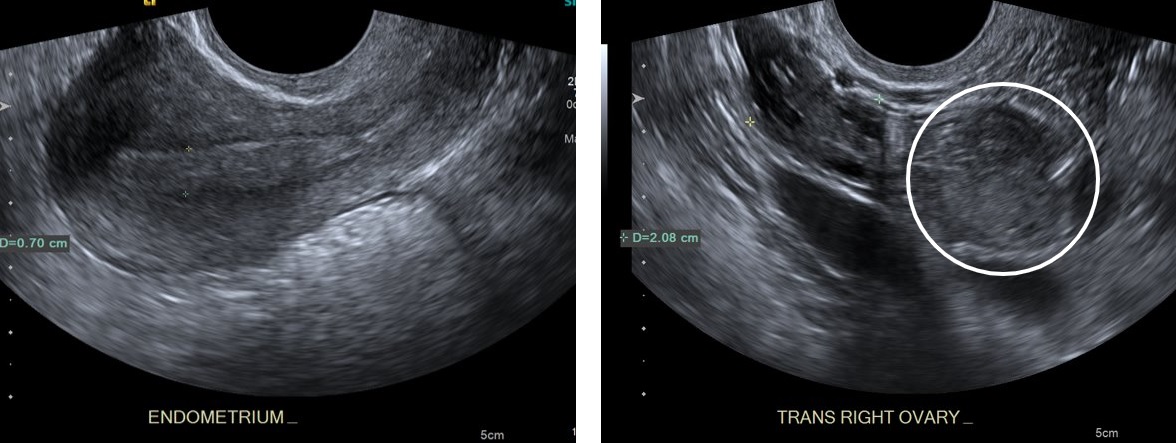
Ectopic pregnancyEctopic pregnancyEctopic pregnancy refers to the implantation of a fertilized egg (embryo) outside the uterine cavity. The main cause is disruption of the normal anatomy of the fallopian tube. Ectopic Pregnancy:
An ectopic pregnancyEctopic pregnancyEctopic pregnancy refers to the implantation of a fertilized egg (embryo) outside the uterine cavity. The main cause is disruption of the normal anatomy of the fallopian tube. Ectopic Pregnancy is characterized by implantationImplantationEndometrial implantation of embryo, mammalian at the blastocyst stage.Fertilization and First Week outside the uterine cavity. Ultrasound findings include:
Heterogeneous adnexal massMassThree-dimensional lesion that occupies a space within the breastImaging of the Breast
Tubal ring sign: an echogenic ring separating the ectopic pregnancyEctopic pregnancyEctopic pregnancy refers to the implantation of a fertilized egg (embryo) outside the uterine cavity. The main cause is disruption of the normal anatomy of the fallopian tube. Ectopic Pregnancy from the ovary
Pseudogestational sac:
CysticCysticFibrocystic Change sac within the uterusUterusThe uterus, cervix, and fallopian tubes are part of the internal female reproductive system. The uterus has a thick wall made of smooth muscle (the myometrium) and an inner mucosal layer (the endometrium). The most inferior portion of the uterus is the cervix, which connects the uterine cavity to the vagina.Uterus, Cervix, and Fallopian Tubes: Anatomy, with no embryoEmbryoThe entity of a developing mammal, generally from the cleavage of a zygote to the end of embryonic differentiation of basic structures. For the human embryo, this represents the first two months of intrauterine development preceding the stages of the fetus.Fertilization and First Week
Decidual reaction present: thickened echogenic endometriumEndometriumThe mucous membrane lining of the uterine cavity that is hormonally responsive during the menstrual cycle and pregnancy. The endometrium undergoes cyclic changes that characterize menstruation. After successful fertilization, it serves to sustain the developing embryo.Embryoblast and Trophoblast Development surrounding the intrauterine sac (because pregnancyPregnancyThe status during which female mammals carry their developing young (embryos or fetuses) in utero before birth, beginning from fertilization to birth.Pregnancy: Diagnosis, Physiology, and CarehormonesHormonesHormones are messenger molecules that are synthesized in one part of the body and move through the bloodstream to exert specific regulatory effects on another part of the body. Hormones play critical roles in coordinating cellular activities throughout the body in response to the constant changes in both the internal and external environments. Hormones: Overview and Types are still being produced by the ectopic pregnancyEctopic pregnancyEctopic pregnancy refers to the implantation of a fertilized egg (embryo) outside the uterine cavity. The main cause is disruption of the normal anatomy of the fallopian tube. Ectopic Pregnancy)
Misleading, because it can appear identical to an early gestational sac before the yolk sacYolk SacThe first of four extra-embryonic membranes to form during embryogenesis. In reptiles and birds, it arises from endoderm and mesoderm to incorporate the egg yolk into the digestive tract for nourishing the embryo. In placental mammals, its nutritional function is vestigial; however, it is the source of intestinal mucosa; blood cells; and germ cells. It is sometimes called the vitelline sac, which should not be confused with the vitelline membrane of the egg.Embryoblast and Trophoblast Development appears
No identifiable pregnancyPregnancyThe status during which female mammals carry their developing young (embryos or fetuses) in utero before birth, beginning from fertilization to birth.Pregnancy: Diagnosis, Physiology, and Care when the HCG is > 2,000
Free peritoneal fluid, possibly with low-level internal echos suggests hemorrhage from ruptured ectopic pregnancyEctopic pregnancyEctopic pregnancy refers to the implantation of a fertilized egg (embryo) outside the uterine cavity. The main cause is disruption of the normal anatomy of the fallopian tube. Ectopic Pregnancy.
Note on heterotopicHeterotopicTransplantation of tissue typical of one area to a different recipient site. The tissue may be autologous, heterologous, or homologous.Organ Transplantation pregnancies (twin gestations with 1 fetus in the uterusUterusThe uterus, cervix, and fallopian tubes are part of the internal female reproductive system. The uterus has a thick wall made of smooth muscle (the myometrium) and an inner mucosal layer (the endometrium). The most inferior portion of the uterus is the cervix, which connects the uterine cavity to the vagina.Uterus, Cervix, and Fallopian Tubes: Anatomy and 1 ectopic):
Possible, but exceedingly rare
If an intrauterine gestation is identified, the adnexa should still be evaluated for masses; if it is not seen, heterotopicHeterotopicTransplantation of tissue typical of one area to a different recipient site. The tissue may be autologous, heterologous, or homologous.Organ TransplantationpregnancyPregnancyThe status during which female mammals carry their developing young (embryos or fetuses) in utero before birth, beginning from fertilization to birth.Pregnancy: Diagnosis, Physiology, and Care can be excluded.
Transvaginal ultrasound showing an empty uterus (left) with endometrial thickening and an echogenic mass (right) representing an ectopic pregnancy adjacent to normal ovarian tissue
Image by Hetal Verma.
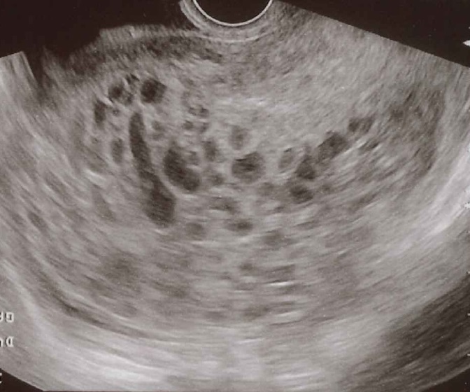
Molar pregnancyPregnancyThe status during which female mammals carry their developing young (embryos or fetuses) in utero before birth, beginning from fertilization to birth.Pregnancy: Diagnosis, Physiology, and Care:
Molar pregnancies are a type of gestational trophoblastic diseaseGestational trophoblastic diseaseGestational trophoblastic diseases are a spectrum of placental disorders resulting from abnormal placental trophoblastic growth. These disorders range from benign molar pregnancies (complete and partial moles) to neoplastic conditions such as invasive moles and choriocarcinoma. Gestational Trophoblastic Disease that occur due to abnormal fertilizationFertilizationTo undergo fertilization, the sperm enters the uterus, travels towards the ampulla of the fallopian tube, and encounters the oocyte. The zona pellucida (the outer layer of the oocyte) deteriorates along with the zygote, which travels towards the uterus and eventually forms a blastocyst, allowing for implantation to occur. Fertilization and First Week.
How they occur:
Complete moleMoleNevi (singular nevus), also known as “moles,” are benign neoplasms of the skin. Nevus is a non-specific medical term because it encompasses both congenital and acquired lesions, hyper- and hypopigmented lesions, and raised or flat lesions.Nevus/Nevi: An enucleated ovum (i.e., an egg without any DNADNAA deoxyribonucleotide polymer that is the primary genetic material of all cells. Eukaryotic and prokaryotic organisms normally contain DNA in a double-stranded state, yet several important biological processes transiently involve single-stranded regions. DNA, which consists of a polysugar-phosphate backbone possessing projections of purines (adenine and guanine) and pyrimidines (thymine and cytosine), forms a double helix that is held together by hydrogen bonds between these purines and pyrimidines (adenine to thymine and guanine to cytosine).DNA Types and Structure) is fertilized by 1 sperm (that duplicates) or 2 sperm (rare).
Partial moleMoleNevi (singular nevus), also known as “moles,” are benign neoplasms of the skin. Nevus is a non-specific medical term because it encompasses both congenital and acquired lesions, hyper- and hypopigmented lesions, and raised or flat lesions.Nevus/Nevi: 2 sperm fertilize a haploidHaploidThe chromosomal constitution of cells, in which each type of chromosome is represented once. Symbol: n.Basic Terms of Genetics ovum.
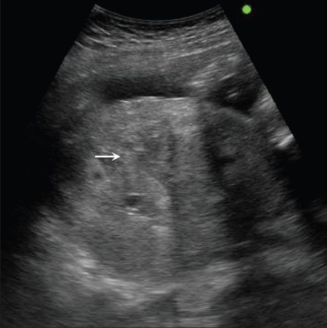
Ultrasound findings:
Enlarged uterusUterusThe uterus, cervix, and fallopian tubes are part of the internal female reproductive system. The uterus has a thick wall made of smooth muscle (the myometrium) and an inner mucosal layer (the endometrium). The most inferior portion of the uterus is the cervix, which connects the uterine cavity to the vagina.Uterus, Cervix, and Fallopian Tubes: Anatomy
Heterogeneous tissue within the uterusUterusThe uterus, cervix, and fallopian tubes are part of the internal female reproductive system. The uterus has a thick wall made of smooth muscle (the myometrium) and an inner mucosal layer (the endometrium). The most inferior portion of the uterus is the cervix, which connects the uterine cavity to the vagina.Uterus, Cervix, and Fallopian Tubes: Anatomy with a classic “snowstorm” appearance
Placental tissue: hyperechoicHyperechoicA structure that produces a high-amplitude echo (lighter grays and white)Ultrasound (Sonography)
Fetus/fetal parts may or may not be present.
Large bilateral ovarian cystsCystsAny fluid-filled closed cavity or sac that is lined by an epithelium. Cysts can be of normal, abnormal, non-neoplastic, or neoplastic tissues.Fibrocystic Change may be present.
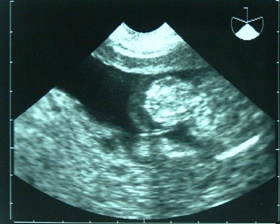
Transvaginal ultrasonography showing a molar pregnancy: The pattern is described as a “cluster of grapes.”
Image: “Molar pregnancy” by Mikael Häggström. License: CC0 1.0
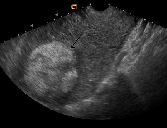
Retained products of conception:
After an abortionAbortionExpulsion of the product of fertilization before completing the term of gestation and without deliberate interference.Spontaneous Abortion (either spontaneous or induced), or postpartum after delivery of the placentaPlacentaA highly vascularized mammalian fetal-maternal organ and major site of transport of oxygen, nutrients, and fetal waste products. It includes a fetal portion (chorionic villi) derived from trophoblasts and a maternal portion (decidua) derived from the uterine endometrium. The placenta produces an array of steroid, protein and peptide hormones (placental hormones).Placenta, Umbilical Cord, and Amniotic Cavity, tissue may be retained within the uterusUterusThe uterus, cervix, and fallopian tubes are part of the internal female reproductive system. The uterus has a thick wall made of smooth muscle (the myometrium) and an inner mucosal layer (the endometrium). The most inferior portion of the uterus is the cervix, which connects the uterine cavity to the vagina.Uterus, Cervix, and Fallopian Tubes: Anatomy. This phenomenon is known as retained products of conception and can lead to hemorrhage and infection. Ultrasound findings include:
Intrauterine, heterogeneous material (typically hyperechoicHyperechoicA structure that produces a high-amplitude echo (lighter grays and white)Ultrasound (Sonography))
Enlarged uterusUterusThe uterus, cervix, and fallopian tubes are part of the internal female reproductive system. The uterus has a thick wall made of smooth muscle (the myometrium) and an inner mucosal layer (the endometrium). The most inferior portion of the uterus is the cervix, which connects the uterine cavity to the vagina.Uterus, Cervix, and Fallopian Tubes: Anatomy
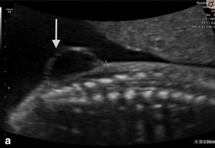
A transvaginal ultrasound demonstrates a heterogeneous echogenic mass in the endometrial cavity (black arrow), representing retained products of conception.
Image: “Gray-scale US demonstrates an echogenic mass in the endometrial cavity (black arrow)” by Maureen P. Kohi et al. License: CC BY 3.0
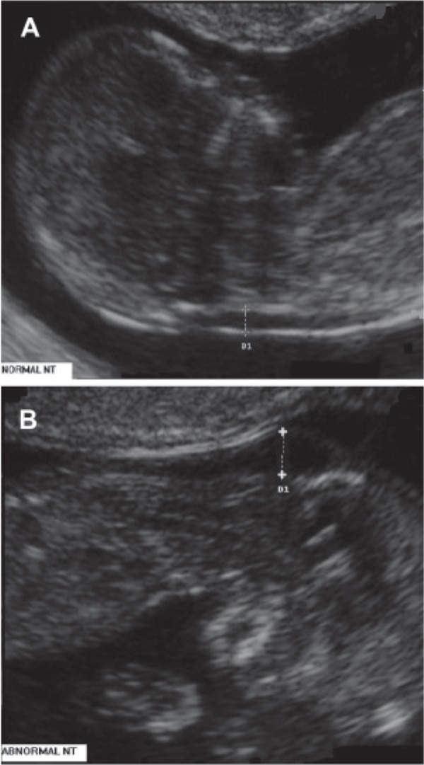
An assessment of the nuchal translucency (or thickness of the nuchal fold at the back of the neckNeckThe part of a human or animal body connecting the head to the rest of the body.Peritonsillar Abscess) is a part of common aneuploidy screening testsAneuploidy Screening TestsPrenatal Care.
Measures the hypoechoicHypoechoicA structure that produces a low-amplitude echo (darker grays)Ultrasound (Sonography) region between the skinSkinThe skin, also referred to as the integumentary system, is the largest organ of the body. The skin is primarily composed of the epidermis (outer layer) and dermis (deep layer). The epidermis is primarily composed of keratinocytes that undergo rapid turnover, while the dermis contains dense layers of connective tissue.Skin: Structure and Functions and soft tissueSoft TissueSoft Tissue Abscess behind the cervical spineSpineThe human spine, or vertebral column, is the most important anatomical and functional axis of the human body. It consists of 7 cervical vertebrae, 12 thoracic vertebrae, and 5 lumbar vertebrae and is limited cranially by the skull and caudally by the sacrum.Vertebral Column: Anatomy
A thickened nuchal fold increases the risk for:
Trisomy 21Trisomy 21Down syndrome, or trisomy 21, is the most common chromosomal aberration and the most frequent genetic cause of developmental delay. Both boys and girls are affected and have characteristic craniofacial and musculoskeletal features, as well as multiple medical anomalies involving the cardiac, gastrointestinal, ocular, and auditory systems.Down syndrome (Trisomy 21) (most common)
Trisomies 13 and 18
Turner syndromeTurner syndromeTurner syndrome is a genetic condition affecting women, in which 1 X chromosome is partly or completely missing. The classic result is the karyotype 45,XO with a female phenotype. Turner syndrome is associated with decreased sex hormone levels and is the most common cause of primary amenorrhea.Turner Syndrome
HydrocephalusHydrocephalusExcessive accumulation of cerebrospinal fluid within the cranium which may be associated with dilation of cerebral ventricles, intracranial.Subarachnoid Hemorrhage
> 100 different developmental and genetic syndromes have also been associated with an increased nuchal fold
Nuchal translucency (NT) measurements: Figure (A) shows a normal fetus (looking up). Figure (B) shows a fetus with trisomy 21 (looking down), demonstrating increased NT thickness.
Image: “Nuchal translucency measurements” by Barati M et al. License: CC BY 2.5
Almost any area of the body can develop incorrectly, leading to congenitalCongenitalChorioretinitis anomalies. Many of them are visible on ultrasound. Some of the clinically important anomalies and their associated ultrasound findings include:
Cardiac defects (most common, found in approximately 1% of births):
A full fetal echo can be performed in utero → full spectrum of lesions can be identified
Clinically important defects include:
Tetralogy of FallotTetralogy of FallotTetralogy of Fallot is the most common cyanotic congenital heart disease. The disease is the confluence of 4 pathologic cardiac features: overriding aorta, ventricular septal defect, right ventricular outflow obstruction, and right ventricular hypertrophy. Tetralogy of Fallot
Transposition of the great vesselsTransposition of the Great VesselsTransposition of the great vessels (TGV) is a cyanotic congenital heart disease characterized by “switching” of the great arteries. There are 2 presentations: the dextro (D)- and levo (L)-looped forms. The L-looped form is rare and congenitally corrected, as the ventricles are also switched. Transposition of the Great Arteries
Truncus arteriosusTruncus arteriosusTruncus arteriosus (TA) is a congenital heart defect characterized by the persistence of a common cardiac arterial trunk tract that fails to divide into the pulmonary artery and aorta during embryonic development. Truncus arteriosus is a rare congenital malformation with a high mortality rate within the 1st 5 weeks of life if not managed promptly. Truncus Arteriosus
Neural tubeNeural tubeA tube of ectodermal tissue in an embryo that will give rise to the central nervous system, including the spinal cord and the brain. Lumen within the neural tube is called neural canal which gives rise to the central canal of the spinal cord and the ventricles of the brain.Gastrulation and Neurulation defects (2nd most common):
AnencephalyAnencephalyA malformation of the nervous system caused by failure of the anterior neuropore to close. Infants are born with intact spinal cords, cerebellums, and brainstems, but lack formation of neural structures above this level. The skull is only partially formed but the eyes are usually normal. This condition may be associated with folate deficiency. Affected infants are only capable of primitive (brain stem) reflexes and usually do not survive for more than two weeks.Neural Tube Defects (most common neural tubeNeural tubeA tube of ectodermal tissue in an embryo that will give rise to the central nervous system, including the spinal cord and the brain. Lumen within the neural tube is called neural canal which gives rise to the central canal of the spinal cord and the ventricles of the brain.Gastrulation and Neurulation defect): absence of the brainBrainThe part of central nervous system that is contained within the skull (cranium). Arising from the neural tube, the embryonic brain is comprised of three major parts including prosencephalon (the forebrain); mesencephalon (the midbrain); and rhombencephalon (the hindbrain). The developed brain consists of cerebrum; cerebellum; and other structures in the brain stem.Nervous System: Anatomy, Structure, and Classification
Cephalocele: cranial defects through which the brainBrainThe part of central nervous system that is contained within the skull (cranium). Arising from the neural tube, the embryonic brain is comprised of three major parts including prosencephalon (the forebrain); mesencephalon (the midbrain); and rhombencephalon (the hindbrain). The developed brain consists of cerebrum; cerebellum; and other structures in the brain stem.Nervous System: Anatomy, Structure, and Classification or meningesMeningesThe brain and the spinal cord are enveloped by 3 overlapping layers of connective tissue called the meninges. The layers are, from the most external layer to the most internal layer, the dura mater, arachnoid mater, and pia mater. Between these layers are 3 potential spaces called the epidural, subdural, and subarachnoid spaces. Meninges: Anatomy herniate outside the skullSkullThe skull (cranium) is the skeletal structure of the head supporting the face and forming a protective cavity for the brain. The skull consists of 22 bones divided into the viscerocranium (facial skeleton) and the neurocranium.Skull: Anatomy
Spina bifida/meningoceleMeningoceleA congenital or acquired protrusion of the meninges, unaccompanied by neural tissue, through a bony defect in the skull or vertebral column.Neural Tube Defects/myelomeningocele: protrusion of the spinal contents through bony defects in the spineSpineThe human spine, or vertebral column, is the most important anatomical and functional axis of the human body. It consists of 7 cervical vertebrae, 12 thoracic vertebrae, and 5 lumbar vertebrae and is limited cranially by the skull and caudally by the sacrum.Vertebral Column: Anatomy
Abdominal wallAbdominal wallThe outer margins of the abdomen, extending from the osteocartilaginous thoracic cage to the pelvis. Though its major part is muscular, the abdominal wall consists of at least seven layers: the skin, subcutaneous fat, deep fascia; abdominal muscles, transversalis fascia, extraperitoneal fat, and the parietal peritoneum.Surgical Anatomy of the Abdomen defects:
OmphaloceleOmphaloceleOmphalocele is a congenital anterior abdominal wall defect in which the intestines are covered by peritoneum and amniotic membranes. The condition results from the failure of the midgut to return to the abdominal cavity by 10 weeks’ gestation. Omphalocele: Multiple bowel loops (+/- liverLiverThe liver is the largest gland in the human body. The liver is found in the superior right quadrant of the abdomen and weighs approximately 1.5 kilograms. Its main functions are detoxification, metabolism, nutrient storage (e.g., iron and vitamins), synthesis of coagulation factors, formation of bile, filtration, and storage of blood. Liver: Anatomy) are seen herniating through a membrane-covered midline abdominal defect.
GastroschisisGastroschisisGastroschisis is a congenital abdominal wall defect characterized by the complete lack of closure of the abdominal musculature. A portion of intestine does not return to the abdominal cavity, thereby remaining in its early embryonic herniated state but with no coverings. Gastroschisis: Bowel loops protrude outside the abdominal cavity without an overlying membrane, through a lateralabdominal wallAbdominal wallThe outer margins of the abdomen, extending from the osteocartilaginous thoracic cage to the pelvis. Though its major part is muscular, the abdominal wall consists of at least seven layers: the skin, subcutaneous fat, deep fascia; abdominal muscles, transversalis fascia, extraperitoneal fat, and the parietal peritoneum.Surgical Anatomy of the Abdomen defect.
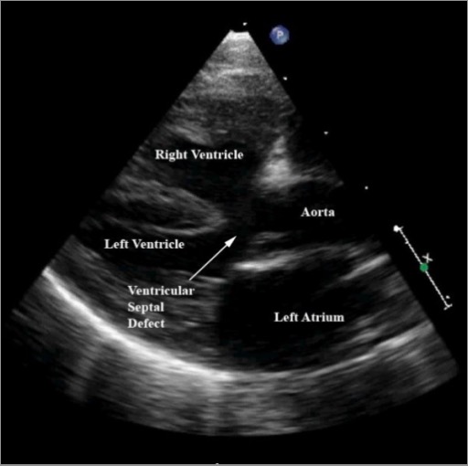
Ultrasound image of tetralogy of Fallot: This figure demonstrates are large ventricular septal defect, aortic override, and right ventricular hypertrophy, all characteristic findings in tetralogy of Fallot.
Image: “Tetralogy of Fallot” by North Carolina Children’s Heart Center, Department of Pediatrics, University of North Carolina at Chapel Hill, Chapel Hill, NC, USA. License: CC BY 2.0
A 2-dimensional ultrasound showing a meningocele, a type of neural tube defect
Image: “Two-dimensional ultrasound showing a meningocele” by Cavalheiro S et al. License: CC BY 4.0
Ultrasound of a fetal omphalocele
Image: “Ultrasound of a fetal omphalocele” by Agarwal R et al. License: CC BY 3.0
Intrauterine growth restriction (IUGR):
Abnormally low EFW on a growth scan
Typically defined as an EFW < 10th percentile for the estimated gestational ageGestational ageThe age of the conceptus, beginning from the time of fertilization. In clinical obstetrics, the gestational age is often estimated as the time from the last day of the last menstruation which is about 2 weeks before ovulation and fertilization.Pregnancy: Diagnosis, Physiology, and Care
Hydrops fetalis refers to abnormal fluid collections in ≥ 2 of the following fetal compartments:
Significant skinSkinThe skin, also referred to as the integumentary system, is the largest organ of the body. The skin is primarily composed of the epidermis (outer layer) and dermis (deep layer). The epidermis is primarily composed of keratinocytes that undergo rapid turnover, while the dermis contains dense layers of connective tissue.Skin: Structure and FunctionsedemaEdemaEdema is a condition in which excess serous fluid accumulates in the body cavity or interstitial space of connective tissues. Edema is a symptom observed in several medical conditions. It can be categorized into 2 types, namely, peripheral (in the extremities) and internal (in an organ or body cavity). Edema (present in almost all hydropic infants) > 5 mmMMMultiple myeloma (MM) is a malignant condition of plasma cells (activated B lymphocytes) primarily seen in the elderly. Monoclonal proliferation of plasma cells results in cytokine-driven osteoclastic activity and excessive secretion of IgG antibodies.Multiple Myeloma
AscitesAscitesAscites is the pathologic accumulation of fluid within the peritoneal cavity that occurs due to an osmotic and/or hydrostatic pressure imbalance secondary to portal hypertension (cirrhosis, heart failure) or non-portal hypertension (hypoalbuminemia, malignancy, infection).Ascites
Pleural effusions
Pericardial effusions
Other potential ultrasound findings:
PolyhydramniosPolyhydramniosPolyhydramnios is a pathological excess of amniotic fluid. Common causes of polyhydramnios include fetal anomalies, gestational diabetes, multiple gestations, and congenital infections. Patients are often asymptomatic but may present with dyspnea, extremity swelling, or abdominal distention. Polyhydramnios
Increased nuchal translucency
Increased placental thickness
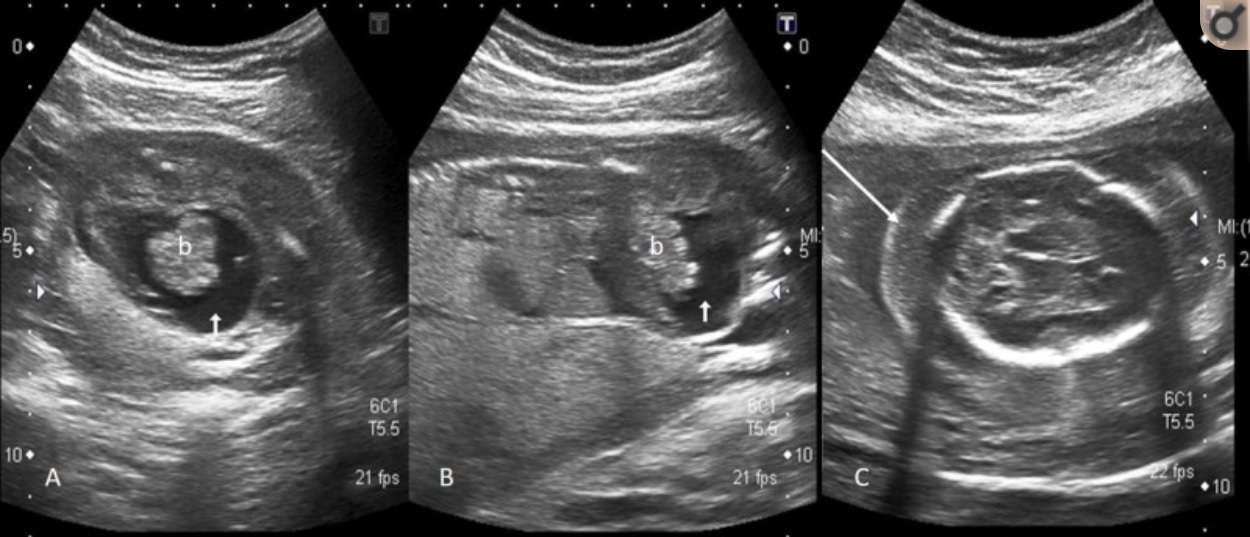
Ultrasound images of an infant with hydrops fetalis: Axial (A) and oblique sagittal (B) images showing fetal ascites (short white arrow) and floating bowel loops (b); axial image of the fetal head (C) showing significant scalp edema (long white arrow)
Image: “Ultrasound images of an infant with hydrops fetalis” by Afzal et al. License: CC BY 3.0
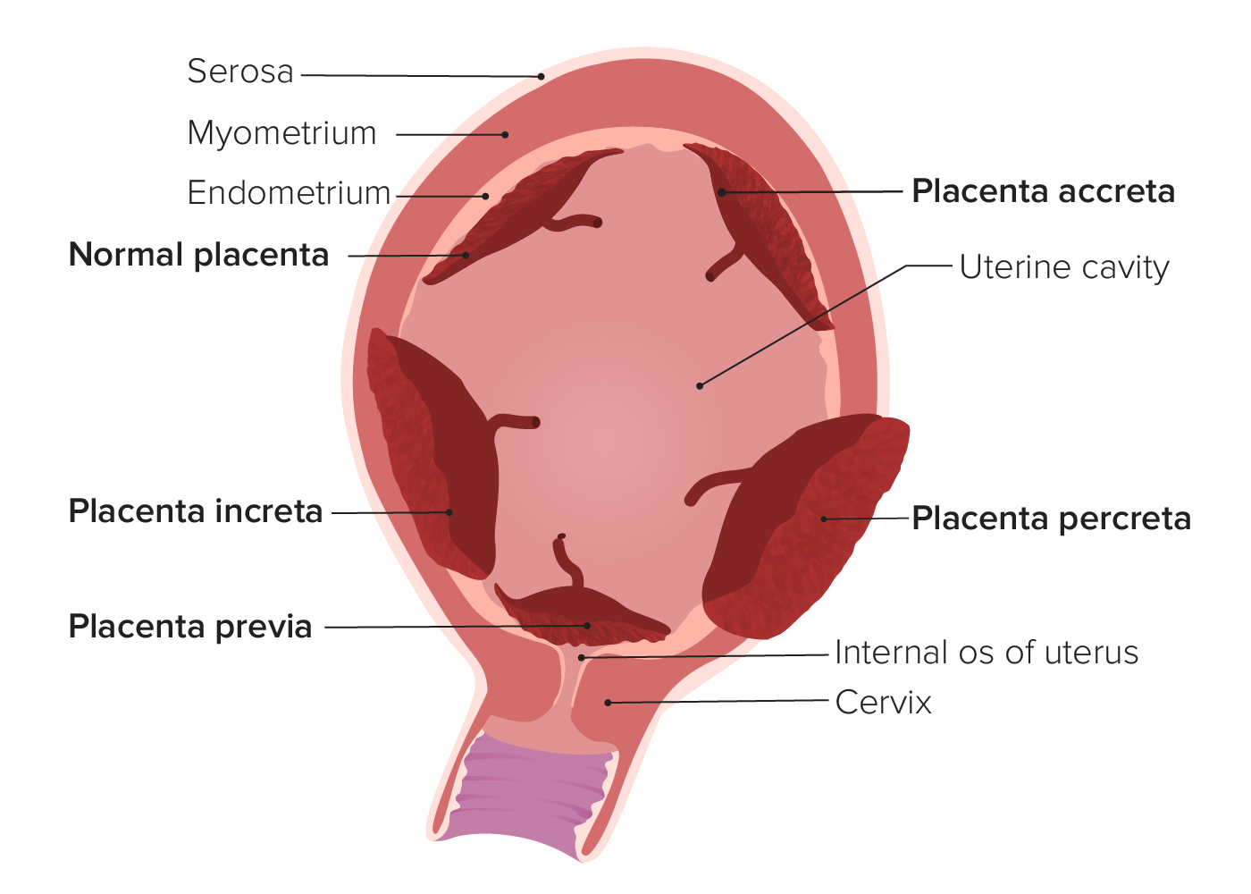
Placental abnormalitiesPlacental abnormalitiesNormal placental structure and function are essential for a healthy pregnancy. Some of the most common placental abnormalities include structural anomalies (such as a succenturiate lobe or velamentous cord insertion), implantation anomalies (such as placenta accreta and placenta previa), and functional anomalies (such as placental insufficiency). Placental Abnormalities
Abnormal placentation:
Abnormal placentation refers to abnormal implantationImplantationEndometrial implantation of embryo, mammalian at the blastocyst stage.Fertilization and First Week of the placentaPlacentaA highly vascularized mammalian fetal-maternal organ and major site of transport of oxygen, nutrients, and fetal waste products. It includes a fetal portion (chorionic villi) derived from trophoblasts and a maternal portion (decidua) derived from the uterine endometrium. The placenta produces an array of steroid, protein and peptide hormones (placental hormones).Placenta, Umbilical Cord, and Amniotic Cavity. Ultrasound findings may show an abnormal placental location, or show it invading into the uterine wall.
Placenta previaPlacenta PreviaAbnormal placentation in which the placenta implants in the lower segment of the uterus (the zone of dilation) and may cover part or all of the opening of the cervix. It is often associated with serious antepartum bleeding and premature labor.Placental Abnormalities: PlacentaPlacentaA highly vascularized mammalian fetal-maternal organ and major site of transport of oxygen, nutrients, and fetal waste products. It includes a fetal portion (chorionic villi) derived from trophoblasts and a maternal portion (decidua) derived from the uterine endometrium. The placenta produces an array of steroid, protein and peptide hormones (placental hormones).Placenta, Umbilical Cord, and Amniotic Cavity covers the internal cervical os.
Low-lying placentaPlacentaA highly vascularized mammalian fetal-maternal organ and major site of transport of oxygen, nutrients, and fetal waste products. It includes a fetal portion (chorionic villi) derived from trophoblasts and a maternal portion (decidua) derived from the uterine endometrium. The placenta produces an array of steroid, protein and peptide hormones (placental hormones).Placenta, Umbilical Cord, and Amniotic Cavity: PlacentaPlacentaA highly vascularized mammalian fetal-maternal organ and major site of transport of oxygen, nutrients, and fetal waste products. It includes a fetal portion (chorionic villi) derived from trophoblasts and a maternal portion (decidua) derived from the uterine endometrium. The placenta produces an array of steroid, protein and peptide hormones (placental hormones).Placenta, Umbilical Cord, and Amniotic Cavity is within 2 cm of the internal cervical os.
PAS:PlacentaPlacentaA highly vascularized mammalian fetal-maternal organ and major site of transport of oxygen, nutrients, and fetal waste products. It includes a fetal portion (chorionic villi) derived from trophoblasts and a maternal portion (decidua) derived from the uterine endometrium. The placenta produces an array of steroid, protein and peptide hormones (placental hormones).Placenta, Umbilical Cord, and Amniotic Cavity is abnormally adherent to the uterine wall.
Placenta accretaPlacenta AccretaAbnormal placentation in which all or parts of the placenta are attached directly to the myometrium due to a complete or partial absence of decidua. It is associated with postpartum hemorrhage because of the failure of placental separation.Placental Abnormalities (approximately 65%): PlacentaPlacentaA highly vascularized mammalian fetal-maternal organ and major site of transport of oxygen, nutrients, and fetal waste products. It includes a fetal portion (chorionic villi) derived from trophoblasts and a maternal portion (decidua) derived from the uterine endometrium. The placenta produces an array of steroid, protein and peptide hormones (placental hormones).Placenta, Umbilical Cord, and Amniotic Cavity attaches directly to the myometrium due to the partial or total absence of the decidua basalis.
PlacentaPlacentaA highly vascularized mammalian fetal-maternal organ and major site of transport of oxygen, nutrients, and fetal waste products. It includes a fetal portion (chorionic villi) derived from trophoblasts and a maternal portion (decidua) derived from the uterine endometrium. The placenta produces an array of steroid, protein and peptide hormones (placental hormones).Placenta, Umbilical Cord, and Amniotic Cavity increta (15%): Placental villi invade into the myometrium.
PlacentaPlacentaA highly vascularized mammalian fetal-maternal organ and major site of transport of oxygen, nutrients, and fetal waste products. It includes a fetal portion (chorionic villi) derived from trophoblasts and a maternal portion (decidua) derived from the uterine endometrium. The placenta produces an array of steroid, protein and peptide hormones (placental hormones).Placenta, Umbilical Cord, and Amniotic Cavity percreta (approximately 20%): Placental villi penetrate through the entire myometrium and may invade other surrounding structures.
Placental abruptionPlacental AbruptionPremature separation of the normally implanted placenta from the uterus. Signs of varying degree of severity include uterine bleeding, uterine muscle hypertonia, and fetal distress or fetal death.Antepartum Hemorrhage:
Placental abruptionPlacental AbruptionPremature separation of the normally implanted placenta from the uterus. Signs of varying degree of severity include uterine bleeding, uterine muscle hypertonia, and fetal distress or fetal death.Antepartum Hemorrhagerefers to the prematurePrematureChildbirth before 37 weeks of pregnancy (259 days from the first day of the mother’s last menstrual period, or 245 days after fertilization).Necrotizing Enterocolitis separation of the placentaPlacentaA highly vascularized mammalian fetal-maternal organ and major site of transport of oxygen, nutrients, and fetal waste products. It includes a fetal portion (chorionic villi) derived from trophoblasts and a maternal portion (decidua) derived from the uterine endometrium. The placenta produces an array of steroid, protein and peptide hormones (placental hormones).Placenta, Umbilical Cord, and Amniotic Cavity, leading to maternal-fetal hemorrhage. Ultrasound findings are usually only seen in large abruptions and may include:
Hyper- or iso-echoic retroplacental hematomaHematomaA collection of blood outside the blood vessels. Hematoma can be localized in an organ, space, or tissue.Intussusception
Heterogeneity within the placentaPlacentaA highly vascularized mammalian fetal-maternal organ and major site of transport of oxygen, nutrients, and fetal waste products. It includes a fetal portion (chorionic villi) derived from trophoblasts and a maternal portion (decidua) derived from the uterine endometrium. The placenta produces an array of steroid, protein and peptide hormones (placental hormones).Placenta, Umbilical Cord, and Amniotic Cavity
Separation of placental edges from the uterusUterusThe uterus, cervix, and fallopian tubes are part of the internal female reproductive system. The uterus has a thick wall made of smooth muscle (the myometrium) and an inner mucosal layer (the endometrium). The most inferior portion of the uterus is the cervix, which connects the uterine cavity to the vagina.Uterus, Cervix, and Fallopian Tubes: Anatomy
Placental thickening
Acute placental abruption: Note the bulky heterogeneous placenta (arrows) in this hypertensive, 29-week gestational age pregnant individual.
Image: “Acute placental abruption” by Kinare A. License: CC BY 2.0
Fluid abnormalities
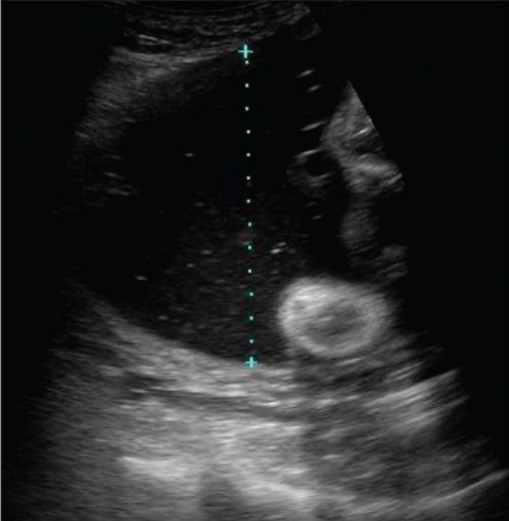
Fluid assessment, at least with an SDP, should be part of every obstetric ultrasound. Fluid abnormalities include:
PolyhydramniosPolyhydramniosPolyhydramnios is a pathological excess of amniotic fluid. Common causes of polyhydramnios include fetal anomalies, gestational diabetes, multiple gestations, and congenital infections. Patients are often asymptomatic but may present with dyspnea, extremity swelling, or abdominal distention. Polyhydramnios: too much fluid (SDP ≥ 8 cm or AFI ≥ 24 cm)
OligohydramniosOligohydramniosOligohydramnios refers to amniotic fluid volume less than expected for the current gestational age. Oligohydramnios is diagnosed by ultrasound and defined as an amniotic fluid index (AFI) of ‰¤ 5 cm or a single deep pocket (SDP) of < 2 cm in the 2nd or 3rd trimester. Oligohydramnios: too little fluid (SDP < 2 cm or AFI ≤ 5 cm)
AnhydramniosAnhydramniosAn extreme case of oligohydramnios with no measurable pockets of amniotic fluid present.Oligohydramnios: no fluid (no measurable pockets of fluid)
Single deepest vertical pocket of fluid is measured using ultrasound to assess amniotic fluid volume. Polyhydramnios is present in this case (SDP > 8 cm).
Image: “Demonstration of the technique to measure a single vertical pocket of liquor” by Kinare A. License: CC BY 2.0
Characterization of maternal pelvic anatomy in cases with unusual or complex abnormalities
CT is almost never indicated for evaluation of the fetus or maternal pelvic anatomy for obstetric indications.
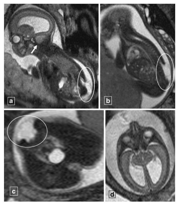
Magnetic resonance imaging findings in a fetus at 23 wga, suggestive of a fetal Chiari II malformation: a: T2-weighted sagittal image demonstrating a lumbosacral neural tube defect (encircled) with cerebellar tonsillar herniation (arrow) b: T2-weighted sagittal image demonstrating a myelomeningocele (encircled) from L2 to the end of the sacrum c: T2-weighted axial image showing the myelomeningocele (encircled) d: T2-weighted axial image demonstrating hydrocephalus
Image: “Neural tube defect with tonsillar herniation” by Loomba R. License: CC BY 3.0
Nonobstetric imaging during pregnancyPregnancyThe status during which female mammals carry their developing young (embryos or fetuses) in utero before birth, beginning from fertilization to birth.Pregnancy: Diagnosis, Physiology, and Care
Ultrasound and MRI are the preferred modalities due to the lack of radiationRadiationEmission or propagation of acoustic waves (sound), electromagnetic energy waves (such as light; radio waves; gamma rays; or x-rays), or a stream of subatomic particles (such as electrons; neutrons; protons; or alpha particles).Osteosarcoma exposure.
Example: Abdominal ultrasound is the preferred initial test for appendicitisAppendicitisAppendicitis is the acute inflammation of the vermiform appendix and the most common abdominal surgical emergency globally. The condition has a lifetime risk of 8%. Characteristic features include periumbilical abdominal pain that migrates to the right lower quadrant, fever, anorexia, nausea, and vomiting.Appendicitis over CT.
Chest X-rayX-rayPenetrating electromagnetic radiation emitted when the inner orbital electrons of an atom are excited and release radiant energy. X-ray wavelengths range from 1 pm to 10 nm. Hard x-rays are the higher energy, shorter wavelength x-rays. Soft x-rays or grenz rays are less energetic and longer in wavelength. The short wavelength end of the x-ray spectrum overlaps the gamma rays wavelength range. The distinction between gamma rays and x-rays is based on their radiation source.Pulmonary Function Tests:
Abdomen should be shielded.
Only done when absolutely necessary (e.g., clinical deterioration with concern for pneumoniaPneumoniaPneumonia or pulmonary inflammation is an acute or chronic inflammation of lung tissue. Causes include infection with bacteria, viruses, or fungi. In more rare cases, pneumonia can also be caused through toxic triggers through inhalation of toxic substances, immunological processes, or in the course of radiotherapy.Pneumonia).
Indicated in cases of choriocarcinomaChoriocarcinomaA malignant metastatic form of trophoblastic tumors. Unlike the hydatidiform mole, choriocarcinoma contains no chorionic villi but rather sheets of undifferentiated cytotrophoblasts and syncytiotrophoblasts (trophoblasts). It is characterized by the large amounts of chorionic gonadotropin produced. Tissue origins can be determined by DNA analyses: placental (fetal) origin or non-placental origin.Gestational Trophoblastic Disease to evaluate for lung metastases
If CT is the modality required to make an important diagnosis (e.g., an individual presenting with stroke symptoms):
A single CT scan in pregnancyPregnancyThe status during which female mammals carry their developing young (embryos or fetuses) in utero before birth, beginning from fertilization to birth.Pregnancy: Diagnosis, Physiology, and Care is considered relatively safe.
Risks/benefits should be carefully weighed and discussed with the individual.
Round, left midlung airspace opacity in an individual reported to have gestational trophoblastic disease
Image: “The appearance of lung metastasis in patient that was reported” by Ghaemmaghami F. License: CC BY 2.5
Clinical Relevance
Obstetric imaging is a critical part of almost all obstetric care.
Diagnosing abnormal pregnancies
Ectopic pregnancyEctopic pregnancyEctopic pregnancy refers to the implantation of a fertilized egg (embryo) outside the uterine cavity. The main cause is disruption of the normal anatomy of the fallopian tube. Ectopic Pregnancy: pregnancies that have implanted outside the uterine cavity, most commonly in the fallopian tubesFallopian tubesThe uterus, cervix, and fallopian tubes are part of the internal female reproductive system. The fallopian tubes receive an ovum after ovulation and help move it and/or a fertilized embryo toward the uterus via ciliated cells lining the tubes and peristaltic movements of its smooth muscle. Uterus, Cervix, and Fallopian Tubes: Anatomy. Ectopic pregnancies are nonviable and may rupture as they grow, leading to life-threatening maternal hemorrhage. Individuals typically present with lower abdominal painAbdominal PainAcute Abdomen and/or abnormal bleeding; a urine pregnancyPregnancyThe status during which female mammals carry their developing young (embryos or fetuses) in utero before birth, beginning from fertilization to birth.Pregnancy: Diagnosis, Physiology, and Care test will be positive. Management is either with methotrexateMethotrexateAn antineoplastic antimetabolite with immunosuppressant properties. It is an inhibitor of tetrahydrofolate dehydrogenase and prevents the formation of tetrahydrofolate, necessary for synthesis of thymidylate, an essential component of DNA.Antimetabolite Chemotherapy or surgical excision, depending on the case.
Molar pregnancyPregnancyThe status during which female mammals carry their developing young (embryos or fetuses) in utero before birth, beginning from fertilization to birth.Pregnancy: Diagnosis, Physiology, and Care: arises from abnormal fertilizationFertilizationTo undergo fertilization, the sperm enters the uterus, travels towards the ampulla of the fallopian tube, and encounters the oocyte. The zona pellucida (the outer layer of the oocyte) deteriorates along with the zygote, which travels towards the uterus and eventually forms a blastocyst, allowing for implantation to occur. Fertilization and First Week, resulting in abnormal embryonic geneticsGeneticsGenetics is the study of genes and their functions and behaviors.Basic Terms of Genetics. The classic clinical presentation is hyperemesis (severe nauseaNauseaAn unpleasant sensation in the stomach usually accompanied by the urge to vomit. Common causes are early pregnancy, sea and motion sickness, emotional stress, intense pain, food poisoning, and various enteroviruses.Antiemetics and vomitingVomitingThe forcible expulsion of the contents of the stomach through the mouth.Hypokalemia), 1st trimester vaginal bleeding, and enlarged uterusUterusThe uterus, cervix, and fallopian tubes are part of the internal female reproductive system. The uterus has a thick wall made of smooth muscle (the myometrium) and an inner mucosal layer (the endometrium). The most inferior portion of the uterus is the cervix, which connects the uterine cavity to the vagina.Uterus, Cervix, and Fallopian Tubes: Anatomy relative to the individual’s estimated gestational ageGestational ageThe age of the conceptus, beginning from the time of fertilization. In clinical obstetrics, the gestational age is often estimated as the time from the last day of the last menstruation which is about 2 weeks before ovulation and fertilization.Pregnancy: Diagnosis, Physiology, and Care. Diagnosis is by ultrasound and abnormally elevated hCG levels. Suction and curettageCurettageA scraping, usually of the interior of a cavity or tract, for removal of new growth or other abnormal tissue, or to obtain material for tissue diagnosis. It is performed with a curet (curette), a spoon-shaped instrument designed for that purpose.Benign Bone Tumors are the mainstays of treatment and post-procedural monitoring is essential to ensure that invasive disease does not develop.
Table: Characteristics of complete versus partial molar pregnancies
Complete moleMoleNevi (singular nevus), also known as “moles,” are benign neoplasms of the skin. Nevus is a non-specific medical term because it encompasses both congenital and acquired lesions, hyper- and hypopigmented lesions, and raised or flat lesions.Nevus/Nevi
Partial moleMoleNevi (singular nevus), also known as “moles,” are benign neoplasms of the skin. Nevus is a non-specific medical term because it encompasses both congenital and acquired lesions, hyper- and hypopigmented lesions, and raised or flat lesions.Nevus/Nevi
KaryotypeKaryotypeThe full set of chromosomes presented as a systematized array of metaphase chromosomes from a photomicrograph of a single cell nucleus arranged in pairs in descending order of size and according to the position of the centromere.Congenital Malformations of the Female Reproductive System
46,XX or 46,XY
Triploid (69,XXX, 69,XXYXXYKlinefelter syndrome is a chromosomal aneuploidy characterized by the presence of 1 or more extra X chromosomes in a male karyotype, most commonly leading to karyotype 47,XXY. Klinefelter syndrome is associated with decreased levels of testosterone and is the most common cause of congenital hypogonadism.Klinefelter Syndrome, or 69,XYY)
Formed from
Enucleated egg and a single sperm
2 sperm and 1 egg
Fetal parts
Absent
Present
Human chorionic gonadotropin levels
↑↑↑
↑
Ultrasound findings
Honeycomb uterusUterusThe uterus, cervix, and fallopian tubes are part of the internal female reproductive system. The uterus has a thick wall made of smooth muscle (the myometrium) and an inner mucosal layer (the endometrium). The most inferior portion of the uterus is the cervix, which connects the uterine cavity to the vagina.Uterus, Cervix, and Fallopian Tubes: Anatomy
“Clusters of grapes”
“Snowstorm” uterusUterusThe uterus, cervix, and fallopian tubes are part of the internal female reproductive system. The uterus has a thick wall made of smooth muscle (the myometrium) and an inner mucosal layer (the endometrium). The most inferior portion of the uterus is the cervix, which connects the uterine cavity to the vagina.Uterus, Cervix, and Fallopian Tubes: Anatomy
Higher risk for choriocarcinomaChoriocarcinomaA malignant metastatic form of trophoblastic tumors. Unlike the hydatidiform mole, choriocarcinoma contains no chorionic villi but rather sheets of undifferentiated cytotrophoblasts and syncytiotrophoblasts (trophoblasts). It is characterized by the large amounts of chorionic gonadotropin produced. Tissue origins can be determined by DNA analyses: placental (fetal) origin or non-placental origin.Gestational Trophoblastic Disease
Rare
Fetal issues
Fetal anomalies: abnormalities in the fetus due to either genetic, chromosomal, or developmental abnormalities. Fetal anomalies can occur in any organ system throughout the body; clinical presentation and management will depend on the specific anomalies. The most common are cardiac and neural tubeNeural tubeA tube of ectodermal tissue in an embryo that will give rise to the central nervous system, including the spinal cord and the brain. Lumen within the neural tube is called neural canal which gives rise to the central canal of the spinal cord and the ventricles of the brain.Gastrulation and Neurulation defects.
Fetal growth restrictionFetal growth restrictionFetal growth restriction (FGR), also known as intrauterine fetal growth restriction (IUGR), is an estimated fetal weight (EFW) or abdominal circumference < 10th percentile for gestational age. The term small for gestational age (SGA) is sometimes erroneously used interchangeably with FGR. Fetal Growth Restriction (FGRFGRFetal growth restriction (FGR), also known as intrauterine fetal growth restriction (IUGR), is an estimated fetal weight (EFW) or abdominal circumference < 10th percentile for gestational age. The term small for gestational age (sga) is sometimes erroneously used interchangeably with FGR.Fetal Growth Restriction): a condition, also known as IUGR, referring to poor fetal growth in utero due to environmental factors. Fetal growth restrictionFetal growth restrictionFetal growth restriction (FGR), also known as intrauterine fetal growth restriction (IUGR), is an estimated fetal weight (EFW) or abdominal circumference < 10th percentile for gestational age. The term small for gestational age (SGA) is sometimes erroneously used interchangeably with FGR. Fetal Growth Restriction is typically defined as an EFW < the 10th percentile based on the estimated gestational ageGestational ageThe age of the conceptus, beginning from the time of fertilization. In clinical obstetrics, the gestational age is often estimated as the time from the last day of the last menstruation which is about 2 weeks before ovulation and fertilization.Pregnancy: Diagnosis, Physiology, and Care. The primary pathogenesis is due to poor perfusion through the placentaPlacentaA highly vascularized mammalian fetal-maternal organ and major site of transport of oxygen, nutrients, and fetal waste products. It includes a fetal portion (chorionic villi) derived from trophoblasts and a maternal portion (decidua) derived from the uterine endometrium. The placenta produces an array of steroid, protein and peptide hormones (placental hormones).Placenta, Umbilical Cord, and Amniotic Cavity, limiting the oxygen and nutrients available to the fetus. There are numerous fetal, maternal, and placental etiologies.
HydropsHydropsCholecystitis fetalis: an abnormal collection of fluid within the fetus. Diagnosis requires at least 2 of the following (all visible on obstetric ultrasound) presentations: skinSkinThe skin, also referred to as the integumentary system, is the largest organ of the body. The skin is primarily composed of the epidermis (outer layer) and dermis (deep layer). The epidermis is primarily composed of keratinocytes that undergo rapid turnover, while the dermis contains dense layers of connective tissue.Skin: Structure and FunctionsedemaEdemaEdema is a condition in which excess serous fluid accumulates in the body cavity or interstitial space of connective tissues. Edema is a symptom observed in several medical conditions. It can be categorized into 2 types, namely, peripheral (in the extremities) and internal (in an organ or body cavity). Edema, ascitesAscitesAscites is the pathologic accumulation of fluid within the peritoneal cavity that occurs due to an osmotic and/or hydrostatic pressure imbalance secondary to portal hypertension (cirrhosis, heart failure) or non-portal hypertension (hypoalbuminemia, malignancy, infection).Ascites, pleural effusions, or pericardial effusions. HydropsHydropsCholecystitis fetalis is often caused by maternal antibodiesAntibodiesImmunoglobulins (Igs), also known as antibodies, are glycoprotein molecules produced by plasma cells that act in immune responses by recognizing and binding particular antigens. The various Ig classes are IgG (the most abundant), IgM, IgE, IgD, and IgA, which differ in their biologic features, structure, target specificity, and distribution.Immunoglobulins: Types and Functions against fetal RBCsRBCsErythrocytes, or red blood cells (RBCs), are the most abundant cells in the blood. While erythrocytes in the fetus are initially produced in the yolk sac then the liver, the bone marrow eventually becomes the main site of production.Erythrocytes: Histology leading to severe fetal anemiaAnemiaAnemia is a condition in which individuals have low Hb levels, which can arise from various causes. Anemia is accompanied by a reduced number of RBCs and may manifest with fatigue, shortness of breath, pallor, and weakness. Subtypes are classified by the size of RBCs, chronicity, and etiology. Anemia: Overview and Types, although there are several nonimmune etiologies as well (e.g., infection with parvovirus B19Parvovirus B19Primate erythroparvovirus 1 (generally referred to as parvovirus B19, B19 virus, or sometimes erythrovirus B19) ranks among the smallest DNA viruses. Parvovirus B19 is of the family Parvoviridae and genus Erythrovirus. In immunocompetent humans, parvovirus B19 classically results in erythema infectiosum (5th disease) or “slapped cheek syndrome.”Parvovirus B19). Often, the primary management is close monitoring, with induced preterm delivery when fetal status begins to deteriorate.
Abnormal placentation
Placenta previaPlacenta PreviaAbnormal placentation in which the placenta implants in the lower segment of the uterus (the zone of dilation) and may cover part or all of the opening of the cervix. It is often associated with serious antepartum bleeding and premature labor.Placental Abnormalities: the presence of placental tissue covering the internal cervical os. When the cervixCervixThe uterus, cervix, and fallopian tubes are part of the internal female reproductive system. The most inferior portion of the uterus is the cervix, which connects the uterine cavity to the vagina. Externally, the cervix is lined by stratified squamous cells; however, the cervical canal is lined by columnar epithelium.Uterus, Cervix, and Fallopian Tubes: Anatomy begins dilating, the placentaPlacentaA highly vascularized mammalian fetal-maternal organ and major site of transport of oxygen, nutrients, and fetal waste products. It includes a fetal portion (chorionic villi) derived from trophoblasts and a maternal portion (decidua) derived from the uterine endometrium. The placenta produces an array of steroid, protein and peptide hormones (placental hormones).Placenta, Umbilical Cord, and Amniotic Cavity will become “detached” over the opening cervical os, resulting in life-threatening maternal-fetal hemorrhage. Diagnosis is using obstetric ultrasound (usually transvaginal). Management is by avoiding any digital cervical exams and delivering the infant via cesarean deliveryCesarean DeliveryCesarean delivery (CD) is the operative delivery of ≥ 1 infants through a surgical incision in the maternal abdomen and uterus. Cesarean deliveries may be indicated for a number of either maternal or fetal reasons, most commonly including fetal intolerance to labor, arrest of labor, a history of prior uterine surgery, fetal malpresentation, and placental abnormalities. Cesarean Delivery prior to the onset of labor.
PAS: describes a spectrum in which the placentaPlacentaA highly vascularized mammalian fetal-maternal organ and major site of transport of oxygen, nutrients, and fetal waste products. It includes a fetal portion (chorionic villi) derived from trophoblasts and a maternal portion (decidua) derived from the uterine endometrium. The placenta produces an array of steroid, protein and peptide hormones (placental hormones).Placenta, Umbilical Cord, and Amniotic Cavity is abnormally and firmly adherent to the uterine wall. In this situation, the placentaPlacentaA highly vascularized mammalian fetal-maternal organ and major site of transport of oxygen, nutrients, and fetal waste products. It includes a fetal portion (chorionic villi) derived from trophoblasts and a maternal portion (decidua) derived from the uterine endometrium. The placenta produces an array of steroid, protein and peptide hormones (placental hormones).Placenta, Umbilical Cord, and Amniotic Cavity is unable to detach postpartum and continues to bleed, often heavily, resulting in life-threatening hemorrhage. The 3 degrees of PAS are placenta accretaPlacenta AccretaAbnormal placentation in which all or parts of the placenta are attached directly to the myometrium due to a complete or partial absence of decidua. It is associated with postpartum hemorrhage because of the failure of placental separation.Placental Abnormalities, increta, and percreta. Diagnosis is via ultrasound, and management is usually surgical, often including a planned hysterectomy at the time of cesarean deliveryCesarean DeliveryCesarean delivery (CD) is the operative delivery of ≥ 1 infants through a surgical incision in the maternal abdomen and uterus. Cesarean deliveries may be indicated for a number of either maternal or fetal reasons, most commonly including fetal intolerance to labor, arrest of labor, a history of prior uterine surgery, fetal malpresentation, and placental abnormalities. Cesarean Delivery.
Abnormal amniotic fluidAmniotic fluidA clear, yellowish liquid that envelopes the fetus inside the sac of amnion. In the first trimester, it is likely a transudate of maternal or fetal plasma. In the second trimester, amniotic fluid derives primarily from fetal lung and kidney. Cells or substances in this fluid can be removed for prenatal diagnostic tests (amniocentesis).Placenta, Umbilical Cord, and Amniotic Cavity volumes
PolyhydramniosPolyhydramniosPolyhydramnios is a pathological excess of amniotic fluid. Common causes of polyhydramnios include fetal anomalies, gestational diabetes, multiple gestations, and congenital infections. Patients are often asymptomatic but may present with dyspnea, extremity swelling, or abdominal distention. Polyhydramnios: a pathological excess of amniotic fluidAmniotic fluidA clear, yellowish liquid that envelopes the fetus inside the sac of amnion. In the first trimester, it is likely a transudate of maternal or fetal plasma. In the second trimester, amniotic fluid derives primarily from fetal lung and kidney. Cells or substances in this fluid can be removed for prenatal diagnostic tests (amniocentesis).Placenta, Umbilical Cord, and Amniotic Cavity. Common causes include fetal anomalies, gestational diabetesDiabetesDiabetes mellitus (DM) is a metabolic disease characterized by hyperglycemia and dysfunction of the regulation of glucose metabolism by insulin. Type 1 DM is diagnosed mostly in children and young adults as the result of autoimmune destruction of β cells in the pancreas and the resulting lack of insulin. Type 2 DM has a significant association with obesity and is characterized by insulin resistance.Diabetes Mellitus, multiple gestations, and congenitalCongenitalChorioretinitisinfectionsInfectionsInvasion of the host organism by microorganisms or their toxins or by parasites that can cause pathological conditions or diseases.Chronic Granulomatous Disease. Diagnosis is made based on ultrasound with an AFI ≥ 24 cm or an SDP ≥ 8 cm. Management of moderate-to-severe cases includes amnioreductionAmnioreductionPolyhydramnios, medical therapy, and careful management during labor. Major risks include cord prolapse and placental abruptionPlacental AbruptionPremature separation of the normally implanted placenta from the uterus. Signs of varying degree of severity include uterine bleeding, uterine muscle hypertonia, and fetal distress or fetal death.Antepartum Hemorrhage at the time of membrane rupture.
OligohydramniosOligohydramniosOligohydramnios refers to amniotic fluid volume less than expected for the current gestational age. Oligohydramnios is diagnosed by ultrasound and defined as an amniotic fluid index (AFI) of ‰¤ 5 cm or a single deep pocket (SDP) of < 2 cm in the 2nd or 3rd trimester. Oligohydramnios: a condition characterized by pathologically low amniotic fluidAmniotic fluidA clear, yellowish liquid that envelopes the fetus inside the sac of amnion. In the first trimester, it is likely a transudate of maternal or fetal plasma. In the second trimester, amniotic fluid derives primarily from fetal lung and kidney. Cells or substances in this fluid can be removed for prenatal diagnostic tests (amniocentesis).Placenta, Umbilical Cord, and Amniotic Cavity volumes. OligohydramniosOligohydramniosOligohydramnios refers to amniotic fluid volume less than expected for the current gestational age. Oligohydramnios is diagnosed by ultrasound and defined as an amniotic fluid index (AFI) of ‰¤ 5 cm or a single deep pocket (SDP) of < 2 cm in the 2nd or 3rd trimester. Oligohydramnios is diagnosed by ultrasound and defined as an AFI of ≤ 5 cm or an SDP of < 2 cm in the 2nd and 3rd trimesters. Etiologies include uteroplacental insufficiencyUteroplacental InsufficiencyUteroplacental insufficiency may be acute or chronic and refers to the inability of the placenta to deliver a sufficient supply of O2 and nutrients to the fetusPlacental Abnormalities, drugs, fetal malformations (especially those related to renal/urinary systems), and maternal TORCH infectionsInfectionsInvasion of the host organism by microorganisms or their toxins or by parasites that can cause pathological conditions or diseases.Chronic Granulomatous Disease. Management involves close antenatal monitoring because significant complications are possible, including FGRFGRFetal growth restriction (FGR), also known as intrauterine fetal growth restriction (IUGR), is an estimated fetal weight (EFW) or abdominal circumference < 10th percentile for gestational age. The term small for gestational age (sga) is sometimes erroneously used interchangeably with FGR.Fetal Growth Restriction, preterm delivery, and developmental deformities.