Neonatal Polycythemia

Neonatal polycythemia Polycythemia An increase in the total red cell mass of the blood. Renal Cell Carcinoma is a hematocrit (HCT) that is 2 standard deviations above the average values for gestation and postnatal age. Neonatal polycythemia Polycythemia An increase in the total red cell mass of the blood. Renal Cell Carcinoma can develop from increased fetal hematopoiesis Hematopoiesis The development and formation of various types of blood cells. Hematopoiesis can take place in the bone marrow (medullary) or outside the bone marrow (extramedullary hematopoiesis). Bone Marrow: Composition and Hematopoiesis (secondary to placental insufficiency, maternal endocrinopathies, genetic disorders, etc ETC The electron transport chain (ETC) sends electrons through a series of proteins, which generate an electrochemical proton gradient that produces energy in the form of adenosine triphosphate (ATP). Electron Transport Chain (ETC).) or passive erythrocyte transfusion (placental-, feto-, or maternal-fetal transfusion). Patients Patients Individuals participating in the health care system for the purpose of receiving therapeutic, diagnostic, or preventive procedures. Clinician–Patient Relationship may be asymptomatic or present with plethora, cardiorespiratory distress, and other symptoms. Continuous monitoring of vital signs and metabolic derangements is important. Treatment includes partial exchange transfusion.

Last updated: May 17, 2024

Editorial responsibility: Stanley Oiseth, Lindsay Jones, Evelin Maza

Overview

Definitions

  • Neonatal polycythemia Polycythemia An increase in the total red cell mass of the blood. Renal Cell Carcinoma (absolute polycythemia Polycythemia An increase in the total red cell mass of the blood. Renal Cell Carcinoma): increased erythrocyte mass Mass Three-dimensional lesion that occupies a space within the breast Imaging of the Breast defined as hematocrit (HCT) > 65% or hemoglobin ( Hb Hb The oxygen-carrying proteins of erythrocytes. They are found in all vertebrates and some invertebrates. The number of globin subunits in the hemoglobin quaternary structure differs between species. Structures range from monomeric to a variety of multimeric arrangements. Gas Exchange) > 22 g/dL
  • Hemoconcentration (relative polycythemia Polycythemia An increase in the total red cell mass of the blood. Renal Cell Carcinoma): a decrease in plasma Plasma The residual portion of blood that is left after removal of blood cells by centrifugation without prior blood coagulation. Transfusion Products volume resulting in an increased concentration of  erythrocytes Erythrocytes Erythrocytes, or red blood cells (RBCs), are the most abundant cells in the blood. While erythrocytes in the fetus are initially produced in the yolk sac then the liver, the bone marrow eventually becomes the main site of production. Erythrocytes: Histology

Epidemiology

  •  1%2% of children born at sea level 
  •  5% of children born at high altitudes

Etiology

  • Increased fetal erythropoiesis Erythropoiesis The production of red blood cells (erythrocytes). In humans, erythrocytes are produced by the yolk sac in the first trimester; by the liver in the second trimester; by the bone marrow in the third trimester and after birth. In normal individuals, the erythrocyte count in the peripheral blood remains relatively constant implying a balance between the rate of erythrocyte production and rate of destruction. Erythrocytes: Histology
    • Placental insufficiency
      • Preeclampsia Preeclampsia A complication of pregnancy, characterized by a complex of symptoms including maternal hypertension and proteinuria with or without pathological edema. Symptoms may range between mild and severe. Pre-eclampsia usually occurs after the 20th week of gestation, but may develop before this time in the presence of trophoblastic disease. Hypertensive Pregnancy Disorders
      • Maternal hypertension Hypertension Hypertension, or high blood pressure, is a common disease that manifests as elevated systemic arterial pressures. Hypertension is most often asymptomatic and is found incidentally as part of a routine physical examination or during triage for an unrelated medical encounter. Hypertension
      • Chronic or recurrent placental abruption Placental Abruption Premature separation of the normally implanted placenta from the uterus. Signs of varying degree of severity include uterine bleeding, uterine muscle hypertonia, and fetal distress or fetal death. Antepartum Hemorrhage
      • Maternal cyanotic heart or severe pulmonary disease Pulmonary disease Diseases involving the respiratory system. Blastomyces/Blastomycosis
      • Postdate pregnancy Pregnancy The status during which female mammals carry their developing young (embryos or fetuses) in utero before birth, beginning from fertilization to birth. Pregnancy: Diagnosis, Physiology, and Care
      • Maternal smoking Smoking Willful or deliberate act of inhaling and exhaling smoke from burning substances or agents held by hand. Interstitial Lung Diseases
      • Mother residing at a high altitude
    • Endocrinopathies
      • Poorly controlled maternal diabetes Diabetes Diabetes mellitus (DM) is a metabolic disease characterized by hyperglycemia and dysfunction of the regulation of glucose metabolism by insulin. Type 1 DM is diagnosed mostly in children and young adults as the result of autoimmune destruction of β cells in the pancreas and the resulting lack of insulin. Type 2 DM has a significant association with obesity and is characterized by insulin resistance. Diabetes Mellitus
      • Congenital Congenital Chorioretinitis thyrotoxicosis Thyrotoxicosis A hypermetabolic syndrome caused by excess thyroid hormones which may come from endogenous or exogenous sources. The endogenous source of hormone may be thyroid hyperplasia; thyroid neoplasms; or hormone-producing extrathyroidal tissue. Thyrotoxicosis is characterized by nervousness; tachycardia; fatigue; weight loss; heat intolerance; and excessive sweating. Thyrotoxicosis and Hyperthyroidism
    • Genetic disorders
      • Trisomies 13, 18, 21
      • Beckwith-Weidemann syndrome
    • Adrenal hyperplasia Hyperplasia An increase in the number of cells in a tissue or organ without tumor formation. It differs from hypertrophy, which is an increase in bulk without an increase in the number of cells. Cellular Adaptation
    • Perinatal asphyxia Perinatal Asphyxia Neonatal Respiratory Distress Syndrome
    • Intrauterine growth restriction
    • Intrauterine chronic hypoxia Hypoxia Sub-optimal oxygen levels in the ambient air of living organisms. Ischemic Cell Damage
  • Passive erythrocyte transfusion
    • Placental-fetal transfusion (delayed cord clamping, newborn Newborn An infant during the first 28 days after birth. Physical Examination of the Newborn held below mother’s level before cord clamping)
    • Feto-fetal transfusion (twin-twin transfusion syndrome)
    • Mother-fetal hemorrhage

Pathophysiology

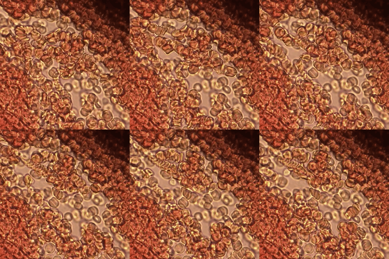
Red blood cells stacking

Blood cells on a drying slide flow in rouleau stacks

Image: “Rouleau stacking” by Rozzychan. License: CC BY 4.0

Clinical Presentation

  • The majority of cases are asymptomatic. 
  • If present, symptoms usually appear by 2 hours after birth.
  • Most characteristic findings: plethora and ruddiness
  • Neurologic manifestations:
    • Poor feeding and lethargy Lethargy A general state of sluggishness, listless, or uninterested, with being tired, and having difficulty concentrating and doing simple tasks. It may be related to depression or drug addiction. Hyponatremia
    • Irritability, jitteriness, and tremor Tremor Cyclical movement of a body part that can represent either a physiologic process or a manifestation of disease. Intention or action tremor, a common manifestation of cerebellar diseases, is aggravated by movement. In contrast, resting tremor is maximal when there is no attempt at voluntary movement, and occurs as a relatively frequent manifestation of parkinson disease. Myotonic Dystrophies
    • Seizures Seizures A seizure is abnormal electrical activity of the neurons in the cerebral cortex that can manifest in numerous ways depending on the region of the brain affected. Seizures consist of a sudden imbalance that occurs between the excitatory and inhibitory signals in cortical neurons, creating a net excitation. The 2 major classes of seizures are focal and generalized. Seizures
    • Cerebrovascular accidents
  • Cardiopulmonary manifestations:
    • Respiratory distress and tachypnea Tachypnea Increased respiratory rate. Pulmonary Examination
    • Cyanosis Cyanosis A bluish or purplish discoloration of the skin and mucous membranes due to an increase in the amount of deoxygenated hemoglobin in the blood or a structural defect in the hemoglobin molecule. Pulmonary Examination and apnea
  • Gastrointestinal manifestations:
    • Necrotizing enterocolitis Enterocolitis Inflammation of the mucosa of both the small intestine and the large intestine. Etiology includes ischemia, infections, allergic, and immune responses. Yersinia spp./Yersiniosis with feeding intolerance, abdominal distension and tenderness, rectorrhagia, bradycardia Bradycardia Bradyarrhythmia is a rhythm in which the heart rate is less than 60/min. Bradyarrhythmia can be physiologic, without symptoms or hemodynamic change. Pathologic bradyarrhythmia results in reduced cardiac output and hemodynamic instability causing syncope, dizziness, or dyspnea. Bradyarrhythmias, and apnea
  • Genitourinary manifestations:
    • Oliguria Oliguria Decreased urine output that is below the normal range. Oliguria can be defined as urine output of less than or equal to 0. 5 or 1 ml/kg/hr depending on the age. Renal Potassium Regulation
    • Hematuria Hematuria Presence of blood in the urine. Renal Cell Carcinoma
    • Priapism Priapism A prolonged painful erection that may lasts hours and is not associated with sexual activity. It is seen in patients with sickle cell anemia, advanced malignancy, spinal trauma; and certain drug treatments. Penile Anomalies and Conditions

Diagnosis and Management

Laboratory studies

  • Diagnosis is confirmed with a complete blood count showing HCT > 65% or Hb Hb The oxygen-carrying proteins of erythrocytes. They are found in all vertebrates and some invertebrates. The number of globin subunits in the hemoglobin quaternary structure differs between species. Structures range from monomeric to a variety of multimeric arrangements. Gas Exchange > 22 mg/dL (capillary values must be confirmed by venous values).
  • Other common findings:
    • Thrombocytopenia Thrombocytopenia Thrombocytopenia occurs when the platelet count is < 150,000 per microliter. The normal range for platelets is usually 150,000-450,000/µL of whole blood. Thrombocytopenia can be a result of decreased production, increased destruction, or splenic sequestration of platelets. Patients are often asymptomatic until platelet counts are < 50,000/µL. Thrombocytopenia 
    • Hypoglycemia Hypoglycemia Hypoglycemia is an emergency condition defined as a serum glucose level ≤ 70 mg/dL (≤ 3.9 mmol/L) in diabetic patients. In nondiabetic patients, there is no specific or defined limit for normal serum glucose levels, and hypoglycemia is defined mainly by its clinical features. Hypoglycemia
    • Hyperbilirubinemia Hyperbilirubinemia A condition characterized by an abnormal increase of bilirubin in the blood, which may result in jaundice. Bilirubin, a breakdown product of heme, is normally excreted in the bile or further catabolized before excretion in the urine. Jaundice 
    • Hypocalcemia Hypocalcemia Hypocalcemia, a serum calcium < 8.5 mg/dL, can result from various conditions. The causes may include hypoparathyroidism, drugs, disorders leading to vitamin D deficiency, and more. Calcium levels are regulated and affected by different elements such as dietary intake, parathyroid hormone (PTH), vitamin D, pH, and albumin. Presentation can range from an asymptomatic (mild deficiency) to a life-threatening condition (acute, significant deficiency). Hypocalcemia
    • Hypoxia Hypoxia Sub-optimal oxygen levels in the ambient air of living organisms. Ischemic Cell Damage and acidosis Acidosis A pathologic condition of acid accumulation or depletion of base in the body. The two main types are respiratory acidosis and metabolic acidosis, due to metabolic acid build up. Respiratory Acidosis in arterial blood gas Arterial blood gas Respiratory Alkalosis

Imaging studies

  • May be required to exclude differential diagnoses or complications
  • Cranial ultrasound, computed tomography (CT) scan, or magnetic resonance imaging (MRI) to rule out cerebrovascular accident Cerebrovascular accident An ischemic stroke (also known as cerebrovascular accident) is an acute neurologic injury that occurs as a result of brain ischemia; this condition may be due to cerebral blood vessel occlusion by thrombosis or embolism, or rarely due to systemic hypoperfusion. Ischemic Stroke (CVA) including intracranial hemorrhage Intracranial hemorrhage Subarachnoid hemorrhage (SAH) is a type of cerebrovascular accident (stroke) resulting from intracranial hemorrhage into the subarachnoid space between the arachnoid and the pia mater layers of the meninges surrounding the brain. Most sahs originate from a saccular aneurysm in the circle of willis but may also occur as a result of trauma, uncontrolled hypertension, vasculitis, anticoagulant use, or stimulant use. Subarachnoid Hemorrhage
  • Echocardiography Echocardiography Ultrasonic recording of the size, motion, and composition of the heart and surrounding tissues. The standard approach is transthoracic. Tricuspid Valve Atresia (TVA) to rule out cardiopulmonary disorders
  • Chest X-ray X-ray Penetrating electromagnetic radiation emitted when the inner orbital electrons of an atom are excited and release radiant energy. X-ray wavelengths range from 1 pm to 10 nm. Hard x-rays are the higher energy, shorter wavelength x-rays. Soft x-rays or grenz rays are less energetic and longer in wavelength. The short wavelength end of the x-ray spectrum overlaps the gamma rays wavelength range. The distinction between gamma rays and x-rays is based on their radiation source. Pulmonary Function Tests to rule out transient tachypnea of the newborn Transient tachypnea of the newborn Abnormal increase in respiratory rate in the newborn. It is self-limiting and attributed to the delayed fetal lung fluid clearance often in caesarean section delivery. Physical Examination of the Newborn, respiratory distress syndrome, pneumonia Pneumonia Pneumonia or pulmonary inflammation is an acute or chronic inflammation of lung tissue. Causes include infection with bacteria, viruses, or fungi. In more rare cases, pneumonia can also be caused through toxic triggers through inhalation of toxic substances, immunological processes, or in the course of radiotherapy. Pneumonia
  • Abdominal X-ray X-ray Penetrating electromagnetic radiation emitted when the inner orbital electrons of an atom are excited and release radiant energy. X-ray wavelengths range from 1 pm to 10 nm. Hard x-rays are the higher energy, shorter wavelength x-rays. Soft x-rays or grenz rays are less energetic and longer in wavelength. The short wavelength end of the x-ray spectrum overlaps the gamma rays wavelength range. The distinction between gamma rays and x-rays is based on their radiation source. Pulmonary Function Tests or CT scan for signs of necrotizing enterocolitis Enterocolitis Inflammation of the mucosa of both the small intestine and the large intestine. Etiology includes ischemia, infections, allergic, and immune responses. Yersinia spp./Yersiniosis

Management

  • Careful monitoring for vital signs, hypoglycemia Hypoglycemia Hypoglycemia is an emergency condition defined as a serum glucose level ≤ 70 mg/dL (≤ 3.9 mmol/L) in diabetic patients. In nondiabetic patients, there is no specific or defined limit for normal serum glucose levels, and hypoglycemia is defined mainly by its clinical features. Hypoglycemia, hypoxia Hypoxia Sub-optimal oxygen levels in the ambient air of living organisms. Ischemic Cell Damage, electrolyte abnormalities, hyperbilirubinemia Hyperbilirubinemia A condition characterized by an abnormal increase of bilirubin in the blood, which may result in jaundice. Bilirubin, a breakdown product of heme, is normally excreted in the bile or further catabolized before excretion in the urine. Jaundice, and urine output
  • Mainstay of treatment is partial exchange transfusion ( PET PET An imaging technique that combines a positron-emission tomography (PET) scanner and a ct X ray scanner. This establishes a precise anatomic localization in the same session. Nuclear Imaging):
    • PET PET An imaging technique that combines a positron-emission tomography (PET) scanner and a ct X ray scanner. This establishes a precise anatomic localization in the same session. Nuclear Imaging involves slowly removing blood and infusing normal saline Normal saline A crystalloid solution that contains 9. 0g of sodium chloride per liter of water. It has a variety of uses, including: as a contact lens solution, in ophthalmic solutions and nasal lavage, in wound irrigation, and for fluid therapy. Intravenous Fluids.
    • Asymptomatic patients Patients Individuals participating in the health care system for the purpose of receiving therapeutic, diagnostic, or preventive procedures. Clinician–Patient Relationship with HCT > 75% may benefit from PET PET An imaging technique that combines a positron-emission tomography (PET) scanner and a ct X ray scanner. This establishes a precise anatomic localization in the same session. Nuclear Imaging.
    • Symptomatic patients Patients Individuals participating in the health care system for the purpose of receiving therapeutic, diagnostic, or preventive procedures. Clinician–Patient Relationship with HCT 60%–65%: consider alternative diagnoses
    • Symptomatic patients Patients Individuals participating in the health care system for the purpose of receiving therapeutic, diagnostic, or preventive procedures. Clinician–Patient Relationship with HCT > 65%: consider PET PET An imaging technique that combines a positron-emission tomography (PET) scanner and a ct X ray scanner. This establishes a precise anatomic localization in the same session. Nuclear Imaging
    • It is unclear whether polycythemia Polycythemia An increase in the total red cell mass of the blood. Renal Cell Carcinoma or PET PET An imaging technique that combines a positron-emission tomography (PET) scanner and a ct X ray scanner. This establishes a precise anatomic localization in the same session. Nuclear Imaging have long-term health consequences.

References

  1. Brandow, A. M., & Camitta, B. M. (2020). Polycythemia. In R. M. Kliegman MD, J. W. St Geme MD, N. J. Blum MD, Shah, Samir S., MD, MSCE, Tasker, Robert C., MBBS, MD & Wilson, Karen M., MD, MPH (Eds.), Nelson textbook of pediatrics (pp. 256-2567.e1). https://www.clinicalkey.es/#!/content/3-s2.0-B9780323529501004934
  2. Watchko, J. F., M.D. (2015). Common hematologic problems in the newborn nursery. Pediatric Clinics of North America, 62(2), 509-524. doi:http://dx.doi.org/10.1016/j.pcl.2014.11.011
  3. Neonatal polycythemia. UpToDate. Retrieved September 23, 2020, from https://www.uptodate.com/contents/neonatal-polycythemia?search=neonatal%20polycythemia&source=search_result&selectedTitle=1~13&usage_type=default&display_rank=1#H25988283
  4. Kremyanskaya, M., Najfeld, V., Mascarenhas, J., & Hoffman, R. (2018). The polycythemias. In R. Hoffman MD, E. J. Benz MD, L. E. Silberstein MD, Heslop, Helen E., MD, DSc (Hon), J. I. Weitz MD, J. Anastasi MD. Hematology: Basic principles and practice (pp. 1071-1105). https://www.clinicalkey.es/#!/content/3-s2.0-B9780323357623000688

Create your free account or log in to continue reading!

Sign up now and get free access to Lecturio with concept pages, medical videos, and questions for your medical education.

User Reviews

Details