Myocarditis is an inflammatory disease of the myocardium, which may occur alone or in association with a systemic process. It can be acute or chronic, and the clinical presentation is highly variable. Myocarditis often presents with chest pain, dyspnea, or arrhythmias; the diagnosis should be suspected in patients without cardiovascular risk factors or cardiac symptoms after a recent viral upper respiratory infection. In the absence of cardiac symptoms, some patients have abnormal findings on ECG or cardiac biomarkers. There are numerous etiologies of myocarditis, but all lead to inflammation and myocyte injury, most often leading to signs and symptoms of heart failure. The course of myocarditis may vary based on the etiology and timeline of symptom progression. The diagnosis is supported by clinical findings, laboratory evaluation, and cardiac imaging. Cardiomegaly and contractile dysfunction are seen on echocardiography. A definitive diagnosis by endomyocardial biopsy is not always required. Management is supportive and aimed at addressing complications.
MyocarditisMyocarditisMyocarditis is an inflammatory disease of the myocardium, which may occur alone or in association with a systemic process. There are numerous etiologies of myocarditis, but all lead to inflammation and myocyte injury, most often leading to signs and symptoms of heart failure. Myocarditis is an inflammatory disease of the myocardiumMyocardiumThe muscle tissue of the heart. It is composed of striated, involuntary muscle cells connected to form the contractile pump to generate blood flow.Heart: Anatomy.
Epidemiology[5,11]
Global prevalencePrevalenceThe total number of cases of a given disease in a specified population at a designated time. It is differentiated from incidence, which refers to the number of new cases in the population at a given time.Measures of Disease Frequency: approximately varies between 10.2 and 105.6 cases per 100,000 people [19]
About 1%–5% of viral infectionsInfectionsInvasion of the host organism by microorganisms or their toxins or by parasites that can cause pathological conditions or diseases.Chronic Granulomatous Disease involve the myocardiumMyocardiumThe muscle tissue of the heart. It is composed of striated, involuntary muscle cells connected to form the contractile pump to generate blood flow.Heart: Anatomy.
An early comorbidity associated with COVID-19COVID-19Coronavirus disease 2019 (COVID-19) is an infectious disease caused by the severe acute respiratory syndrome coronavirus 2 (SARS-CoV-2) that mainly affects the respiratory system but can also cause damage to other body systems (cardiovascular, gastrointestinal, renal, and central nervous systems)., with 15-fold increase in incidenceIncidenceThe number of new cases of a given disease during a given period in a specified population. It also is used for the rate at which new events occur in a defined population. It is differentiated from prevalence, which refers to all cases in the population at a given time.Measures of Disease Frequency of myocarditisMyocarditisMyocarditis is an inflammatory disease of the myocardium, which may occur alone or in association with a systemic process. There are numerous etiologies of myocarditis, but all lead to inflammation and myocyte injury, most often leading to signs and symptoms of heart failure. Myocarditis compared to pre-COVID levels
Affects more men than women
More common in young adults
Etiology[2,10,11]
The most frequent causes of myocarditisMyocarditisMyocarditis is an inflammatory disease of the myocardium, which may occur alone or in association with a systemic process. There are numerous etiologies of myocarditis, but all lead to inflammation and myocyte injury, most often leading to signs and symptoms of heart failure. Myocarditis are presumed viral infectionsInfectionsInvasion of the host organism by microorganisms or their toxins or by parasites that can cause pathological conditions or diseases.Chronic Granulomatous Disease, cardiotoxins such as those found in chemotherapyChemotherapyOsteosarcoma regimens and recreational drugs, and immune systemImmune systemThe body’s defense mechanism against foreign organisms or substances and deviant native cells. It includes the humoral immune response and the cell-mediated response and consists of a complex of interrelated cellular, molecular, and genetic components.Primary Lymphatic Organs activation.
Most common cause of lymphocytic myocarditisMyocarditisMyocarditis is an inflammatory disease of the myocardium, which may occur alone or in association with a systemic process. There are numerous etiologies of myocarditis, but all lead to inflammation and myocyte injury, most often leading to signs and symptoms of heart failure. Myocarditis in North America and Europe
It is recognized that SARS-CoV-2 causes myocarditisMyocarditisMyocarditis is an inflammatory disease of the myocardium, which may occur alone or in association with a systemic process. There are numerous etiologies of myocarditis, but all lead to inflammation and myocyte injury, most often leading to signs and symptoms of heart failure. Myocarditis (both direct viral injury and immune-mediated mechanisms).[6,8]
Bacterial
Protozoal (most common in Africa, AsiaASIASpinal Cord Injuries, and South America): Chagas diseaseChagas diseaseInfection with the protozoan parasite trypanosoma cruzi, a form of trypanosomiasis endemic in central and south america. It is named after the brazilian physician carlos chagas, who discovered the parasite. Infection by the parasite (positive serologic result only) is distinguished from the clinical manifestations that develop years later, such as destruction of parasympathetic ganglia; chagas cardiomyopathy; and dysfunction of the esophagus or colon.Trypanosoma cruzi/Chagas disease caused by Trypanosoma cruziTrypanosoma cruziChagas disease is an infection caused by the American trypanosome Trypanosoma cruzi. This parasitic protozoan is transmitted in the feces of reduviid bugs in South and Central America. Acute infection may present with inflammation at the inoculation site (chagoma), fever, and lymphadenopathy. Untreated, chronic infection can progress to severe complications.Trypanosoma cruzi/Chagas diseaseis the most common infective cause worldwide; endemic in rural South and Central America
Fungal
Immune-mediated disorders:[10]
Rheumatic feverRheumatic feverAcute rheumatic fever (ARF) is an autoimmune inflammatory process that usually follows Streptococcal pharyngitis. Acute rheumatic fever usually occurs 2-4 weeks after an untreated infection and affects the heart, skin, joints, and nervous system. Rheumatic Fever
Kawasaki diseaseKawasaki diseaseAn acute, febrile, mucocutaneous condition accompanied by swelling of cervical lymph nodes in infants and young children. The principal symptoms are fever, congestion of the ocular conjunctivae, reddening of the lips and oral cavity, protuberance of tongue papillae, and edema or erythema of the extremities.Kawasaki Disease
SarcoidosisSarcoidosisSarcoidosis is a multisystem inflammatory disease that causes noncaseating granulomas. The exact etiology is unknown. Sarcoidosis usually affects the lungs and thoracic lymph nodes, but it can also affect almost every system in the body, including the skin, heart, and eyes, most commonly. Sarcoidosis
Systemic lupus erythematosusSystemic lupus erythematosusSystemic lupus erythematosus (SLE) is a chronic autoimmune, inflammatory condition that causes immune-complex deposition in organs, resulting in systemic manifestations. Women, particularly those of African American descent, are more commonly affected.Systemic Lupus Erythematosus
PolymyositisPolymyositisPolymyositis (PM) is an autoimmune inflammatory myopathy caused by T cell-mediated muscle injury. The etiology of PM is unclear, but there are several genetic and environmental associations. Polymyositis is most common in middle-aged women and rarely affects children. Polymyositis
DermatomyositisDermatomyositisA subacute or chronic inflammatory disease of muscle and skin, marked by proximal muscle weakness and a characteristic skin rash. The illness occurs with approximately equal frequency in children and adults. The skin lesions usually take the form of a purplish rash (or less often an exfoliative dermatitis) involving the nose, cheeks, forehead, upper trunk, and arms. The disease is associated with a complement mediated intramuscular microangiopathy, leading to loss of capillaries, muscle ischemia, muscle-fiber necrosis, and perifascicular atrophy. The childhood form of this disease tends to evolve into a systemic vasculitis. Dermatomyositis may occur in association with malignant neoplasms.Paraneoplastic Syndromes
SclerodermaSclerodermaScleroderma (systemic sclerosis) is an autoimmune condition characterized by diffuse collagen deposition and fibrosis. The clinical presentation varies from limited skin involvement to diffuse involvement of internal organs. Scleroderma
VasculitisVasculitisInflammation of any one of the blood vessels, including the arteries; veins; and rest of the vasculature system in the body.Systemic Lupus Erythematosus
Exogenous agents:[13–15]
SmallpoxSmallpoxAn acute, highly contagious, often fatal infectious disease caused by an orthopoxvirus characterized by a biphasic febrile course and distinctive progressive skin eruptions. Vaccination has succeeded in eradicating smallpox worldwide.OrthopoxvirusvaccineVaccineSuspensions of killed or attenuated microorganisms (bacteria, viruses, fungi, protozoa), antigenic proteins, synthetic constructs, or other bio-molecular derivatives, administered for the prevention, amelioration, or treatment of infectious and other diseases.Vaccination (subclinical, 1:200; clinically suspected, 1:5,500)
mRNAmRNARNA sequences that serve as templates for protein synthesis. Bacterial mRNAs are generally primary transcripts in that they do not require post-transcriptional processing. Eukaryotic mRNA is synthesized in the nucleus and must be exported to the cytoplasm for translation. Most eukaryotic mRNAs have a sequence of polyadenylic acid at the 3′ end, referred to as the poly(a) tail. The function of this tail is not known for certain, but it may play a role in the export of mature mRNA from the nucleus as well as in helping stabilize some mRNA molecules by retarding their degradation in the cytoplasm.RNA Types and StructureCOVID-19COVID-19Coronavirus disease 2019 (COVID-19) is an infectious disease caused by the severe acute respiratory syndrome coronavirus 2 (SARS-CoV-2) that mainly affects the respiratory system but can also cause damage to other body systems (cardiovascular, gastrointestinal, renal, and central nervous systems).vaccineVaccineSuspensions of killed or attenuated microorganisms (bacteria, viruses, fungi, protozoa), antigenic proteins, synthetic constructs, or other bio-molecular derivatives, administered for the prevention, amelioration, or treatment of infectious and other diseases.Vaccination (while rare, myocarditisMyocarditisMyocarditis is an inflammatory disease of the myocardium, which may occur alone or in association with a systemic process. There are numerous etiologies of myocarditis, but all lead to inflammation and myocyte injury, most often leading to signs and symptoms of heart failure. Myocarditis has been seen in adolescent and young adults (more in males) within 7 days after the second dose)
Genetic predisposition:
Genetic defects in the structural proteinsStructural proteinsProteins and Peptides of myocytesMyocytesMature contractile cells, commonly known as myocytes, that form one of three kinds of muscle. The three types of muscle cells are skeletal, cardiac, and smooth. They are derived from embryonic (precursor) muscle cells called myoblasts.Muscle Tissue: Histology (detected in 16% of cases)
↑ Susceptibility to myocyte damage
Environmental causes:
Exposure to carbon monoxideCarbon monoxideCarbon monoxide (CO). A poisonous colorless, odorless, tasteless gas. It combines with hemoglobin to form carboxyhemoglobin, which has no oxygen carrying capacity. The resultant oxygen deprivation causes headache, dizziness, decreased pulse and respiratory rates, unconsciousness, and death.Carbon Monoxide Poisoning or phosphorus
Heavy metal toxicityToxicityDosage Calculation (e.g., arsenicArsenicA shiny gray element with atomic symbol as, atomic number 33, and atomic weight 75. It occurs throughout the universe, mostly in the form of metallic arsenides. Most forms are toxic. According to the fourth annual report on carcinogens, arsenic and certain arsenic compounds have been listed as known carcinogens.Metal Poisoning (Lead, Arsenic, Iron))
Black widow venom
Scorpion and snakeSnakeLimbless reptiles of the suborder serpentes.Snakebites bites
Drugs:
Doxorubicin
CyclophosphamideCyclophosphamidePrecursor of an alkylating nitrogen mustard antineoplastic and immunosuppressive agent that must be activated in the liver to form the active aldophosphamide. It has been used in the treatment of lymphoma and leukemia. Its side effect, alopecia, has been used for defleecing sheep. Cyclophosphamide may also cause sterility, birth defects, mutations, and cancer.Immunosuppressants
CocaineCocaineAn alkaloid ester extracted from the leaves of plants including coca. It is a local anesthetic and vasoconstrictor and is clinically used for that purpose, particularly in the eye, ear, nose, and throat. It also has powerful central nervous system effects similar to the amphetamines and is a drug of abuse. Cocaine, like amphetamines, acts by multiple mechanisms on brain catecholaminergic neurons; the mechanism of its reinforcing effects is thought to involve inhibition of dopamine uptake.Local Anesthetics
AmphetaminesAmphetaminesAnalogs or derivatives of amphetamine. Many are sympathomimetics and central nervous system stimulators causing excitation, vasopressin, bronchodilation, and to varying degrees, anorexia, analepsis, nasal decongestion, and some smooth muscle relaxation.Stimulants
EthanolEthanolA clear, colorless liquid rapidly absorbed from the gastrointestinal tract and distributed throughout the body. It has bactericidal activity and is used often as a topical disinfectant. It is widely used as a solvent and preservative in pharmaceutical preparations as well as serving as the primary ingredient in alcoholic beverages.Ethanol Metabolism
Antibiotics:
PenicillinsPenicillinsBeta-lactam antibiotics contain a beta-lactam ring as a part of their chemical structure. Drugs in this class include penicillin G and V, penicillinase-sensitive and penicillinase-resistant penicillins, cephalosporins, carbapenems, and aztreonam. Penicillins
CephalosporinsCephalosporinsCephalosporins are a group of bactericidal beta-lactam antibiotics (similar to penicillins) that exert their effects by preventing bacteria from producing their cell walls, ultimately leading to cell death. Cephalosporins are categorized by generation and all drug names begin with “cef-” or “ceph-.” Cephalosporins
Interleukin-2Interleukin-2A soluble substance elaborated by antigen- or mitogen-stimulated T-lymphocytes which induces DNA synthesis in naive lymphocytes.Interleukins
Novel cancer therapies/immune checkpoint inhibitorsImmune Checkpoint InhibitorsDrugs that block negative regulator immune checkpoint proteins (e.g., PD-1 receptor and CTLA-4 antigen) thereby increasing suppressed immune activation in immunotherapies.Cancer Immunotherapy:
NivolumabNivolumabA genetically engineered, fully humanized immunoglobulin g4 monoclonal antibody that binds to the pd-1 receptor, activating an immune response to tumor cells. It is used as monotherapy or in combination with ipilimumab for the treatment of advanced malignant melanoma. It is also used in the treatment of advanced or recurring non-small cell lung cancer; renal cell carcinoma; and Hodgkin’s lymphoma.Melanoma
The following table summarizes the infectious causes of myocarditisMyocarditisMyocarditis is an inflammatory disease of the myocardium, which may occur alone or in association with a systemic process. There are numerous etiologies of myocarditis, but all lead to inflammation and myocyte injury, most often leading to signs and symptoms of heart failure. Myocarditis. Keep in mind that this list is not exhaustive.
Coxsackie B virusVirusViruses are infectious, obligate intracellular parasites composed of a nucleic acid core surrounded by a protein capsid. Viruses can be either naked (non-enveloped) or enveloped. The classification of viruses is complex and based on many factors, including type and structure of the nucleoid and capsid, the presence of an envelope, the replication cycle, and the host range. Virology
AdenovirusAdenovirusAdenovirus (member of the family Adenoviridae) is a nonenveloped, double-stranded DNA virus. Adenovirus is transmitted in a variety of ways, and it can have various presentations based on the site of entry. Presentation can include febrile pharyngitis, conjunctivitis, acute respiratory disease, atypical pneumonia, and gastroenteritis. Adenovirus
Parvovirus B19Parvovirus B19Primate erythroparvovirus 1 (generally referred to as parvovirus B19, B19 virus, or sometimes erythrovirus B19) ranks among the smallest DNA viruses. Parvovirus B19 is of the family Parvoviridae and genus Erythrovirus. In immunocompetent humans, parvovirus B19 classically results in erythema infectiosum (5th disease) or “slapped cheek syndrome.”Parvovirus B19
Human herpesvirus 6
Epstein-Barr virusEpstein-Barr VirusEpstein-Barr virus (EBV) is a linear, double-stranded DNA virus belonging to the Herpesviridae family. This highly prevalent virus is mostly transmitted through contact with oropharyngeal secretions from an infected individual. The virus can infect epithelial cells and B lymphocytes, where it can undergo lytic replication or latency. Epstein-Barr Virus
CytomegalovirusCytomegalovirusCMV is a ubiquitous double-stranded DNA virus belonging to the Herpesviridae family. CMV infections can be transmitted in bodily fluids, such as blood, saliva, urine, semen, and breast milk. The initial infection is usually asymptomatic in the immunocompetent host, or it can present with symptoms of mononucleosis. Cytomegalovirus
Hepatitis CHepatitis CHepatitis C is an infection of the liver caused by the hepatitis C virus (HCV). The infection can be transmitted through infectious blood or body fluids and may be transmitted during childbirth or through IV drug use or sexual intercourse. Hepatitis C virus can cause both acute and chronic hepatitis, ranging from a mild to a serious, lifelong illness including liver cirrhosis and hepatocellular carcinoma (HCC).Hepatitis C Virus
InfluenzaInfluenzaInfluenza viruses are members of the Orthomyxoviridae family and the causative organisms of influenza, a highly contagious febrile respiratory disease. There are 3 primary influenza viruses (A, B, and C) and various subtypes, which are classified based on their virulent surface antigens, hemagglutinin (HA) and neuraminidase (NA). Influenza typically presents with a fever, myalgia, headache, and symptoms of an upper respiratory infection. Influenza Viruses/Influenza
PoliovirusPoliovirusPoliomyelitis is an infectious disease caused by the poliovirus. This virus is a member of the Picornaviridae family. It is a small, single-stranded, positive-sense RNA virus without a lipid envelope. Transmission occurs through the fecal-oral route and, occasionally, through respiratory aerosols. Poliovirus/Poliomyelitis
Borrelia burgdorferiBorrelia burgdorferiA specific species of bacteria, part of the borrelia burgdorferi group, whose common name is lyme disease spirochete.Borrelia
Mycoplasma pneumoniaeMycoplasma pneumoniaeShort filamentous organism of the genus mycoplasma, which binds firmly to the cells of the respiratory epithelium. It is one of the etiologic agents of non-viral primary atypical pneumonia in man.Mycoplasma
Mycobacterium tuberculosisMycobacterium tuberculosisTuberculosis (TB) is an infectious disease caused by Mycobacterium tuberculosis complex bacteria. The bacteria usually attack the lungs but can also damage other parts of the body. Approximately 30% of people around the world are infected with this pathogen, with the majority harboring a latent infection. Tuberculosis spreads through the air when a person with active pulmonary infection coughs or sneezes.Tuberculosis
Corynebacterium diphtheriaeCorynebacterium diphtheriaeDiphtheria is an infectious disease caused by corynebacterium diphtheriae that most often results in respiratory disease with membranous inflammation of the pharynx, sore throat, fever, swollen glands, and weakness. The hallmark sign is a sheet of thick, gray material covering the back of the throat.Diphtheria
StaphylococcusStaphylococcusStaphylococcus is a medically important genera of Gram-positive, aerobic cocci. These bacteria form clusters resembling grapes on culture plates. Staphylococci are ubiquitous for humans, and many strains compose the normal skin flora.Staphylococcus
Neisseria gonorrhoeaeNeisseria gonorrhoeaeA species of gram-negative, aerobic bacteria primarily found in purulent venereal discharges. It is the causative agent of gonorrhea.Neisseria
StreptococcusStreptococcusStreptococcus is one of the two medically important genera of gram-positive cocci, the other being Staphylococcus. Streptococci are identified as different species on blood agar on the basis of their hemolytic pattern and sensitivity to optochin and bacitracin. There are many pathogenic species of streptococci, including S. pyogenes, S. agalactiae, S. pneumoniae, and the viridans streptococci.Streptococcus
BrucellaBrucellaBrucellosis (also known as undulant fever, Mediterranean fever, or Malta fever) is a zoonotic infection that spreads predominantly through ingestion of unpasteurized dairy products or direct contact with infected animal products. Clinical manifestations include fever, arthralgias, malaise, lymphadenopathy, and hepatosplenomegaly. Brucella/Brucellosis
Haemophilus influenzaeHaemophilus InfluenzaeA species of Haemophilus found on the mucous membranes of humans and a variety of animals. The species is further divided into biotypes I through viii.Haemophilus
Treponema pallidumTreponema pallidumThe causative agent of venereal and non-venereal syphilis as well as yaws.Treponema
Coxiella burnetti
Rickettsia rickettsiiRickettsia rickettsiiA species of gram-negative, aerobic bacteria that is the etiologic agent of rocky mountain spotted fever. Its cells are slightly smaller and more uniform in size than those of rickettsia prowazekii.Rickettsia
Trypanosoma cruziTrypanosoma cruziChagas disease is an infection caused by the American trypanosome Trypanosoma cruzi. This parasitic protozoan is transmitted in the feces of reduviid bugs in South and Central America. Acute infection may present with inflammation at the inoculation site (chagoma), fever, and lymphadenopathy. Untreated, chronic infection can progress to severe complications.Trypanosoma cruzi/Chagas disease (Chagas diseaseChagas diseaseInfection with the protozoan parasite trypanosoma cruzi, a form of trypanosomiasis endemic in central and south america. It is named after the brazilian physician carlos chagas, who discovered the parasite. Infection by the parasite (positive serologic result only) is distinguished from the clinical manifestations that develop years later, such as destruction of parasympathetic ganglia; chagas cardiomyopathy; and dysfunction of the esophagus or colon.Trypanosoma cruzi/Chagas disease)
ToxoplasmaToxoplasmaToxoplasmosis is an infectious disease caused by Toxoplasma gondii, an obligate intracellular protozoan parasite. Felines are the definitive host, but transmission to humans can occur through contact with cat feces or the consumption of contaminated foods. The clinical presentation and complications depend on the host’s immune status. Toxoplasma/Toxoplasmosis gondii
Entamoeba histolyticaEntamoeba HistolyticaA species of parasitic protozoa causing entamoebiasis and amebic dysentery (dysentery, amebic). Characteristics include a single nucleus containing a small central karyosome and peripheral chromatin that is finely and regularly beaded.Amebicides
LeishmaniaLeishmaniaLeishmania species are obligate intracellular parasites that are transmitted by an infected sandfly. The disease is endemic to Asia, the Middle East, Africa, the Mediterranean, and South and Central America. Clinical presentation varies, dependent on the pathogenicity of the species and the host’s immune response. Leishmania/Leishmaniasis
AspergillusAspergillusA genus of mitosporic fungi containing about 100 species and eleven different teleomorphs in the family trichocomaceae.Echinocandins
CandidaCandidaCandida is a genus of dimorphic, opportunistic fungi. Candida albicans is part of the normal human flora and is the most common cause of candidiasis. The clinical presentation varies and can include localized mucocutaneous infections (e.g., oropharyngeal, esophageal, intertriginous, and vulvovaginal candidiasis) and invasive disease (e.g., candidemia, intraabdominal abscess, pericarditis, and meningitis). Candida/Candidiasis
ActinomycesActinomycesActinomyces is an anaerobic, gram-positive, branching, filamentous rod. Actinomyces israelii is the most common species involved in human disease. The organism is commonly found as part of the normal flora in the oral cavity, gastrointestinal tract, and reproductive tract. Actinomyces/Actinomycosis
BlastomycesBlastomycesBlastomycosis is an infection caused by inhalation of the spores of the fungus, Blastomyces. Blastomyces species thrive in moist soil and decaying material and are common in the Ohio and Mississippi River valleys and the Great Lakes regions of the United States and Canada. Although most patients are asymptomatic, some can develop pneumonia.Blastomyces/Blastomycosis
CoccidioidesCoccidioidesCoccidioidomycosis, commonly known as San Joaquin Valley fever, is a fungal disease caused by Coccidioides immitis or Coccidioides posadasii. When Coccidioides spores are inhaled, they transform into spherules that result in infection. Coccidioidomycosis is also a common cause of community-acquired pneumonia and can cause severe disease in the immunocompromised.Coccidioides/Coccidioidomycosis
HistoplasmaHistoplasmaHistoplasmosis is an infection caused by Histoplasma capsulatum, a dimorphic fungus. The fungus exists as a mold at low temperatures and as yeast at high temperatures. H. capsulatum is the most common endemic fungal infection in the US and is most prevalent in the midwestern and central states along the Ohio and Mississippi River valleys.Histoplasma/Histoplasmosis
CryptococcusCryptococcusCryptococcosis is an opportunistic, fungal infection caused by the Cryptococcus species. The principal pathogens in humans are C. neoformans (primary) and C. gattii. Cryptococcus neoformans is typically found in pigeon droppings and acquired by inhaling dust from contaminated soil. The majority of affected patients are immunocompromised. Cryptococcus/Cryptococcosis
MucormycosisMucormycosisMucormycosis is an angioinvasive fungal infection caused by multiple fungi within the order, Mucorales. The fungi are ubiquitous in the environment, but mucormycosis is very rare and almost always occurs in patients who are immunocompromised. Inhalation of fungal spores can cause rhinocerebral or pulmonary mucormycosis, direct inoculation can cause cutaneous mucormycosis, and ingestion can cause gastrointestinal mucormycosis. Mucorales/Mucormycosis or Mucormycetes
NocardiaNocardiaNocardia is a branching, filamentous, gram-positive bacilli. It is partially acid fast due to the presence of mycolic acids in the cell wall. Nocardia is a ubiquitous soil organism that most commonly affects immunocompromised patients. Nocardia is transmitted via inhalation of aerosolized bacteria or less commonly, via direct contact with wounds. Nocardia/Nocardiosis
InflammationInflammationInflammation is a complex set of responses to infection and injury involving leukocytes as the principal cellular mediators in the body’s defense against pathogenic organisms. Inflammation is also seen as a response to tissue injury in the process of wound healing. The 5 cardinal signs of inflammation are pain, heat, redness, swelling, and loss of function. Inflammation due to various etiologies leads to enlargement of the heart and dilation of all chambers.[1,2,8,11,17]
Initiation of myocyte injury:
Viral: direct viral toxicityToxicityDosage Calculation (causing cell deathCell deathInjurious stimuli trigger the process of cellular adaptation, whereby cells respond to withstand the harmful changes in their environment. Overwhelmed adaptive mechanisms lead to cell injury. Mild stimuli produce reversible injury. If the stimulus is severe or persistent, injury becomes irreversible. Apoptosis is programmed cell death, a mechanism with both physiologic and pathologic effects.Cell Injury and Death), immune response (lymphocytic infiltration, and cytokinesCytokinesNon-antibody proteins secreted by inflammatory leukocytes and some non-leukocytic cells, that act as intercellular mediators. They differ from classical hormones in that they are produced by a number of tissue or cell types rather than by specialized glands. They generally act locally in a paracrine or autocrine rather than endocrine manner.Adaptive Immune Response) exacerbates the injury.
Bacterial: direct invasion or bacterial toxins → myocyte damage
Autoimmune: autoantibodiesAutoantibodiesAntibodies that react with self-antigens (autoantigens) of the organism that produced them.Blotting Techniques to myocyte components
Toxins: hypersensitivity or direct effects
Myocardial inflammationInflammationInflammation is a complex set of responses to infection and injury involving leukocytes as the principal cellular mediators in the body’s defense against pathogenic organisms. Inflammation is also seen as a response to tissue injury in the process of wound healing. The 5 cardinal signs of inflammation are pain, heat, redness, swelling, and loss of function. Inflammation and injury (T and B lymphocytesB lymphocytesLymphoid cells concerned with humoral immunity. They are short-lived cells resembling bursa-derived lymphocytes of birds in their production of immunoglobulin upon appropriate stimulation.B cells: Types and Functions) → myocardial necrosisNecrosisThe death of cells in an organ or tissue due to disease, injury or failure of the blood supply.Ischemic Cell Damage and inflammationInflammationInflammation is a complex set of responses to infection and injury involving leukocytes as the principal cellular mediators in the body’s defense against pathogenic organisms. Inflammation is also seen as a response to tissue injury in the process of wound healing. The 5 cardinal signs of inflammation are pain, heat, redness, swelling, and loss of function. Inflammation is perpetuated.
Continued inflammationInflammationInflammation is a complex set of responses to infection and injury involving leukocytes as the principal cellular mediators in the body’s defense against pathogenic organisms. Inflammation is also seen as a response to tissue injury in the process of wound healing. The 5 cardinal signs of inflammation are pain, heat, redness, swelling, and loss of function. Inflammation → severe and prolonged damage → fibrosisFibrosisAny pathological condition where fibrous connective tissue invades any organ, usually as a consequence of inflammation or other injury.Bronchiolitis Obliterans (fibroblastsFibroblastsConnective tissue cells which secrete an extracellular matrix rich in collagen and other macromolecules.Sarcoidosis are activated and extracellular matrixExtracellular matrixA meshwork-like substance found within the extracellular space and in association with the basement membrane of the cell surface. It promotes cellular proliferation and provides a supporting structure to which cells or cell lysates in culture dishes adhere.Hypertrophic and Keloid ScarsproteinsProteinsLinear polypeptides that are synthesized on ribosomes and may be further modified, crosslinked, cleaved, or assembled into complex proteins with several subunits. The specific sequence of amino acids determines the shape the polypeptide will take, during protein folding, and the function of the protein.Energy Homeostasis are deposited) → chamber remodeling → dilated cardiomyopathyDilated CardiomyopathyDilated cardiomyopathy (DCM) is the most common type of non-ischemic cardiomyopathy and a common cause of heart failure (HF). The cause may be idiopathic, familial, or secondary to a variety of underlying conditions. The disease is characterized by the enlargement of 1 or both ventricles and reduced systolic function. Dilated Cardiomyopathy
Consequences:
Heart failureHeart FailureA heterogeneous condition in which the heart is unable to pump out sufficient blood to meet the metabolic need of the body. Heart failure can be caused by structural defects, functional abnormalities (ventricular dysfunction), or a sudden overload beyond its capacity. Chronic heart failure is more common than acute heart failure which results from sudden insult to cardiac function, such as myocardial infarction.Total Anomalous Pulmonary Venous Return (TAPVR)
Cardiac arrhythmia
ExtensionExtensionExamination of the Upper Limbs to the pericardiumPericardiumA conical fibroserous sac surrounding the heart and the roots of the great vessels (aorta; venae cavae; pulmonary artery). Pericardium consists of two sacs: the outer fibrous pericardium and the inner serous pericardium. The latter consists of an outer parietal layer facing the fibrous pericardium, and an inner visceral layer (epicardium) resting next to the heart, and a pericardial cavity between these two layers.Heart: Anatomy → pericarditisPericarditisPericarditis is an inflammation of the pericardium, often with fluid accumulation. It can be caused by infection (often viral), myocardial infarction, drugs, malignancies, metabolic disorders, autoimmune disorders, or trauma. Acute, subacute, and chronic forms exist. Pericarditis
The clinical presentation can vary based on the severity and temporal progression of symptoms. Most signs and symptoms are related to heart failureHeart FailureA heterogeneous condition in which the heart is unable to pump out sufficient blood to meet the metabolic need of the body. Heart failure can be caused by structural defects, functional abnormalities (ventricular dysfunction), or a sudden overload beyond its capacity. Chronic heart failure is more common than acute heart failure which results from sudden insult to cardiac function, such as myocardial infarction.Total Anomalous Pulmonary Venous Return (TAPVR) in fulminant myocarditisMyocarditisMyocarditis is an inflammatory disease of the myocardium, which may occur alone or in association with a systemic process. There are numerous etiologies of myocarditis, but all lead to inflammation and myocyte injury, most often leading to signs and symptoms of heart failure. Myocarditis.
Clinical classification of myocarditisMyocarditisMyocarditis is an inflammatory disease of the myocardium, which may occur alone or in association with a systemic process. There are numerous etiologies of myocarditis, but all lead to inflammation and myocyte injury, most often leading to signs and symptoms of heart failure. Myocarditis[17,23]
Acute myocarditisAcute myocarditisMyocarditis: presentation and diagnosis within a short period of time (< 1 month)
Chronic myocarditisChronic myocarditisMyocarditis (chronic inflammatory cardiomyopathyCardiomyopathyCardiomyopathy refers to a group of myocardial diseases associated with structural changes of the heart muscles (myocardium) and impaired systolic and/or diastolic function in the absence of other heart disorders (coronary artery disease, hypertension, valvular disease, and congenital heart disease). Cardiomyopathy: Overview and Types): myocardial inflammationInflammationInflammation is a complex set of responses to infection and injury involving leukocytes as the principal cellular mediators in the body’s defense against pathogenic organisms. Inflammation is also seen as a response to tissue injury in the process of wound healing. The 5 cardinal signs of inflammation are pain, heat, redness, swelling, and loss of function. Inflammation with established dilated cardiomyopathyDilated CardiomyopathyDilated cardiomyopathy (DCM) is the most common type of non-ischemic cardiomyopathy and a common cause of heart failure (HF). The cause may be idiopathic, familial, or secondary to a variety of underlying conditions. The disease is characterized by the enlargement of 1 or both ventricles and reduced systolic function. Dilated Cardiomyopathy generally > 1 month
Classic presentation:
Chest painPainAn unpleasant sensation induced by noxious stimuli which are detected by nerve endings of nociceptive neurons.Pain: Types and Pathways
Heart failureHeart FailureA heterogeneous condition in which the heart is unable to pump out sufficient blood to meet the metabolic need of the body. Heart failure can be caused by structural defects, functional abnormalities (ventricular dysfunction), or a sudden overload beyond its capacity. Chronic heart failure is more common than acute heart failure which results from sudden insult to cardiac function, such as myocardial infarction.Total Anomalous Pulmonary Venous Return (TAPVR)/cardiogenic shockCardiogenic shockShock resulting from diminution of cardiac output in heart disease.Types of Shock
Arrhythmia
Symptoms[3,11]
Viral or infectious illness:
FeverFeverFever is defined as a measured body temperature of at least 38°C (100.4°F). Fever is caused by circulating endogenous and/or exogenous pyrogens that increase levels of prostaglandin E2 in the hypothalamus. Fever is commonly associated with chills, rigors, sweating, and flushing of the skin. Fever and rashRashRocky Mountain Spotted Fever (may signify eosinophilic myocarditisMyocarditisMyocarditis is an inflammatory disease of the myocardium, which may occur alone or in association with a systemic process. There are numerous etiologies of myocarditis, but all lead to inflammation and myocyte injury, most often leading to signs and symptoms of heart failure. Myocarditis)
Chest painPainAn unpleasant sensation induced by noxious stimuli which are detected by nerve endings of nociceptive neurons.Pain: Types and Pathways
Heart failureHeart FailureA heterogeneous condition in which the heart is unable to pump out sufficient blood to meet the metabolic need of the body. Heart failure can be caused by structural defects, functional abnormalities (ventricular dysfunction), or a sudden overload beyond its capacity. Chronic heart failure is more common than acute heart failure which results from sudden insult to cardiac function, such as myocardial infarction.Total Anomalous Pulmonary Venous Return (TAPVR):
DyspneaDyspneaDyspnea is the subjective sensation of breathing discomfort. Dyspnea is a normal manifestation of heavy physical or psychological exertion, but also may be caused by underlying conditions (both pulmonary and extrapulmonary). Dyspnea
FatigueFatigueThe state of weariness following a period of exertion, mental or physical, characterized by a decreased capacity for work and reduced efficiency to respond to stimuli.Fibromyalgia
SyncopeSyncopeSyncope is a short-term loss of consciousness and loss of postural stability followed by spontaneous return of consciousness to the previous neurologic baseline without the need for resuscitation. The condition is caused by transient interruption of cerebral blood flow that may be benign or related to a underlying life-threatening condition. Syncope
Recognize signs of right heart failureHeart FailureA heterogeneous condition in which the heart is unable to pump out sufficient blood to meet the metabolic need of the body. Heart failure can be caused by structural defects, functional abnormalities (ventricular dysfunction), or a sudden overload beyond its capacity. Chronic heart failure is more common than acute heart failure which results from sudden insult to cardiac function, such as myocardial infarction.Total Anomalous Pulmonary Venous Return (TAPVR) including right upper quadrantRight upper quadrantAnterior Abdominal Wall: AnatomypainPainAn unpleasant sensation induced by noxious stimuli which are detected by nerve endings of nociceptive neurons.Pain: Types and Pathways.
TachycardiaTachycardiaAbnormally rapid heartbeat, usually with a heart rate above 100 beats per minute for adults. Tachycardia accompanied by disturbance in the cardiac depolarization (cardiac arrhythmia) is called tachyarrhythmia.Sepsis in Children
Arrhythmia
Peripheral edemaPeripheral edemaPeripheral edema is the swelling of the lower extremities, namely, legs, feet, and ankles.Edema
Pericardial friction rubPericardial friction rubA rasping, scratching, or grating sound with up to 3 components per cardiac cycle and best heard during expiration with the patient leaning forward.Pericarditis (pericarditisPericarditisPericarditis is an inflammation of the pericardium, often with fluid accumulation. It can be caused by infection (often viral), myocardial infarction, drugs, malignancies, metabolic disorders, autoimmune disorders, or trauma. Acute, subacute, and chronic forms exist. Pericarditis)
Myocardial infarctionMyocardial infarctionMI is ischemia and death of an area of myocardial tissue due to insufficient blood flow and oxygenation, usually from thrombus formation on a ruptured atherosclerotic plaque in the epicardial arteries. Clinical presentation is most commonly with chest pain, but women and patients with diabetes may have atypical symptoms.Myocardial Infarction
Dilated cardiomyopathyDilated CardiomyopathyDilated cardiomyopathy (DCM) is the most common type of non-ischemic cardiomyopathy and a common cause of heart failure (HF). The cause may be idiopathic, familial, or secondary to a variety of underlying conditions. The disease is characterized by the enlargement of 1 or both ventricles and reduced systolic function. Dilated Cardiomyopathy
Sudden cardiac arrestCardiac arrestCardiac arrest is the sudden, complete cessation of cardiac output with hemodynamic collapse. Patients present as pulseless, unresponsive, and apneic. Rhythms associated with cardiac arrest are ventricular fibrillation/tachycardia, asystole, or pulseless electrical activity. Cardiac Arrest
The European Society of Cardiology (ESC) and the American Heart AssociationAmerican Heart AssociationA voluntary organization concerned with the prevention and treatment of heart and vascular diseases.Heart Failure (AHA) scientific statements recommend measuring the erythrocyte sedimentation rateErythrocyte Sedimentation RateSoft Tissue Abscess (ESRESRSoft Tissue Abscess) and C-reactive protein (CRP) and high-sensitivity cardiac troponin (hs-cTn) levels to help diagnose myocarditisMyocarditisMyocarditis is an inflammatory disease of the myocardium, which may occur alone or in association with a systemic process. There are numerous etiologies of myocarditis, but all lead to inflammation and myocyte injury, most often leading to signs and symptoms of heart failure. Myocarditis; however, a normal level for any of these does not exclude myocarditisMyocarditisMyocarditis is an inflammatory disease of the myocardium, which may occur alone or in association with a systemic process. There are numerous etiologies of myocarditis, but all lead to inflammation and myocyte injury, most often leading to signs and symptoms of heart failure. Myocarditis.
↑ high-sensitivity cardiac troponin (hs-cTn) and CK
↑ BrainBrainThe part of central nervous system that is contained within the skull (cranium). Arising from the neural tube, the embryonic brain is comprised of three major parts including prosencephalon (the forebrain); mesencephalon (the midbrain); and rhombencephalon (the hindbrain). The developed brain consists of cerebrum; cerebellum; and other structures in the brain stem.Nervous System: Anatomy, Structure, and Classification natriuretic peptide (BNPBNPA peptide that is secreted by the brain and the heart atria, stored mainly in cardiac ventricular myocardium. It can cause natriuresis; diuresis; vasodilation; and inhibits secretion of renin and aldosterone. It improves heart function. It contains 32 amino acids.Renal Sodium and Water Regulation): useful prognostically
Order in all patientsPatientsIndividuals participating in the health care system for the purpose of receiving therapeutic, diagnostic, or preventive procedures.Clinician–Patient Relationship with suspected myocarditisMyocarditisMyocarditis is an inflammatory disease of the myocardium, which may occur alone or in association with a systemic process. There are numerous etiologies of myocarditis, but all lead to inflammation and myocyte injury, most often leading to signs and symptoms of heart failure. Myocarditis:
CBC with differential count
Chemistries
Liver function testsLiver function testsLiver function tests, also known as hepatic function panels, are one of the most commonly performed screening blood tests. Such tests are also used to detect, evaluate, and monitor acute and chronic liver diseases.Liver Function Tests
Peripheral eosinophiliaEosinophiliaAbnormal increase of eosinophils in the blood, tissues or organs.Autosomal Dominant Hyperimmunoglobulin E Syndrome > 65% seen with hypersensitivity/eosinophilic myocarditisMyocarditisMyocarditis is an inflammatory disease of the myocardium, which may occur alone or in association with a systemic process. There are numerous etiologies of myocarditis, but all lead to inflammation and myocyte injury, most often leading to signs and symptoms of heart failure. Myocarditis
↑ Lactate level: impending circulatory failure
Viral testing may aid in identifying the causative agent; routine viral serologies are not recommended (low sensitivity and specificitySensitivity and SpecificityBinary classification measures to assess test results. Sensitivity or recall rate is the proportion of true positives. Specificity is the probability of correctly determining the absence of a condition.Epidemiological Values of Diagnostic Tests compared to PCRPCRPolymerase chain reaction (PCR) is a technique that amplifies DNA fragments exponentially for analysis. The process is highly specific, allowing for the targeting of specific genomic sequences, even with minuscule sample amounts. The PCR cycles multiple times through 3 phases: denaturation of the template DNA, annealing of a specific primer to the individual DNA strands, and synthesis/elongation of new DNA molecules.Polymerase Chain Reaction (PCR) on biopsyBiopsyRemoval and pathologic examination of specimens from the living body.Ewing Sarcoma tissue).
ECGECGAn electrocardiogram (ECG) is a graphic representation of the electrical activity of the heart plotted against time. Adhesive electrodes are affixed to the skin surface allowing measurement of cardiac impulses from many angles. The ECG provides 3-dimensional information about the conduction system of the heart, the myocardium, and other cardiac structures. Electrocardiogram (ECG):[9,10,12,17]
ST changes:
Often nonspecific
Diffuse ST elevation signals seen in pericarditisPericarditisPericarditis is an inflammation of the pericardium, often with fluid accumulation. It can be caused by infection (often viral), myocardial infarction, drugs, malignancies, metabolic disorders, autoimmune disorders, or trauma. Acute, subacute, and chronic forms exist. Pericarditis, often in a nonvascular distribution.
Injury with ST-segment elevations in segmental contiguous leads may mimic acute coronary arteryCoronary ArteryTruncus Arteriosus occlusion.
PR prolongation or PR-segment depression: may be seen with concomitant evidence of pericarditisPericarditisPericarditis is an inflammation of the pericardium, often with fluid accumulation. It can be caused by infection (often viral), myocardial infarction, drugs, malignancies, metabolic disorders, autoimmune disorders, or trauma. Acute, subacute, and chronic forms exist. Pericarditis
Arrhythmia
Conduction delays (e.g., QRS prolongation)
Low QRS voltage with fulminant myocarditisMyocarditisMyocarditis is an inflammatory disease of the myocardium, which may occur alone or in association with a systemic process. There are numerous etiologies of myocarditis, but all lead to inflammation and myocyte injury, most often leading to signs and symptoms of heart failure. Myocarditis because of myocardial edemaEdemaEdema is a condition in which excess serous fluid accumulates in the body cavity or interstitial space of connective tissues. Edema is a symptom observed in several medical conditions. It can be categorized into 2 types, namely, peripheral (in the extremities) and internal (in an organ or body cavity). Edema
A normal ECGECGAn electrocardiogram (ECG) is a graphic representation of the electrical activity of the heart plotted against time. Adhesive electrodes are affixed to the skin surface allowing measurement of cardiac impulses from many angles. The ECG provides 3-dimensional information about the conduction system of the heart, the myocardium, and other cardiac structures. Electrocardiogram (ECG) does not automatically rule out myocarditisMyocarditisMyocarditis is an inflammatory disease of the myocardium, which may occur alone or in association with a systemic process. There are numerous etiologies of myocarditis, but all lead to inflammation and myocyte injury, most often leading to signs and symptoms of heart failure. Myocarditis.
Chest X-rayX-rayPenetrating electromagnetic radiation emitted when the inner orbital electrons of an atom are excited and release radiant energy. X-ray wavelengths range from 1 pm to 10 nm. Hard x-rays are the higher energy, shorter wavelength x-rays. Soft x-rays or grenz rays are less energetic and longer in wavelength. The short wavelength end of the x-ray spectrum overlaps the gamma rays wavelength range. The distinction between gamma rays and x-rays is based on their radiation source.Pulmonary Function Tests:[3,12]
Nonspecific; normal to enlarged heart
Indications of heart failureHeart FailureA heterogeneous condition in which the heart is unable to pump out sufficient blood to meet the metabolic need of the body. Heart failure can be caused by structural defects, functional abnormalities (ventricular dysfunction), or a sudden overload beyond its capacity. Chronic heart failure is more common than acute heart failure which results from sudden insult to cardiac function, such as myocardial infarction.Total Anomalous Pulmonary Venous Return (TAPVR):
Pulmonary venous congestion
Pleural effusionPleural EffusionPleural effusion refers to the accumulation of fluid between the layers of the parietal and visceral pleura. Common causes of this condition include infection, malignancy, autoimmune disorders, or volume overload. Clinical manifestations include chest pain, cough, and dyspnea. Pleural Effusion
Should be performed in all patientsPatientsIndividuals participating in the health care system for the purpose of receiving therapeutic, diagnostic, or preventive procedures.Clinician–Patient Relationship with suspected myocarditisMyocarditisMyocarditis is an inflammatory disease of the myocardium, which may occur alone or in association with a systemic process. There are numerous etiologies of myocarditis, but all lead to inflammation and myocyte injury, most often leading to signs and symptoms of heart failure. Myocarditis
May be normal in early or mild disease
Possible findings:
Left ventricular (LV) dilation:
Greater dilation suggests a chronic insult.
Small left ventricle suggests a fulminant myocarditisMyocarditisMyocarditis is an inflammatory disease of the myocardium, which may occur alone or in association with a systemic process. There are numerous etiologies of myocarditis, but all lead to inflammation and myocyte injury, most often leading to signs and symptoms of heart failure. Myocarditis (rapid onset severe heart failureHeart FailureA heterogeneous condition in which the heart is unable to pump out sufficient blood to meet the metabolic need of the body. Heart failure can be caused by structural defects, functional abnormalities (ventricular dysfunction), or a sudden overload beyond its capacity. Chronic heart failure is more common than acute heart failure which results from sudden insult to cardiac function, such as myocardial infarction.Total Anomalous Pulmonary Venous Return (TAPVR) in myocarditisMyocarditisMyocarditis is an inflammatory disease of the myocardium, which may occur alone or in association with a systemic process. There are numerous etiologies of myocarditis, but all lead to inflammation and myocyte injury, most often leading to signs and symptoms of heart failure. Myocarditis). LV is often non- or mildly dilated, with thick walls due to myocardial edemaEdemaEdema is a condition in which excess serous fluid accumulates in the body cavity or interstitial space of connective tissues. Edema is a symptom observed in several medical conditions. It can be categorized into 2 types, namely, peripheral (in the extremities) and internal (in an organ or body cavity). Edema.
Pericardial effusionPericardial effusionFluid accumulation within the pericardium. Serous effusions are associated with pericardial diseases. Hemopericardium is associated with trauma. Lipid-containing effusion (chylopericardium) results from leakage of thoracic duct. Severe cases can lead to cardiac tamponade.Pericardial Effusion and Cardiac Tamponade
Abnormal myocardial echogenicity (increased in myocardial inflammationInflammationInflammation is a complex set of responses to infection and injury involving leukocytes as the principal cellular mediators in the body’s defense against pathogenic organisms. Inflammation is also seen as a response to tissue injury in the process of wound healing. The 5 cardinal signs of inflammation are pain, heat, redness, swelling, and loss of function. Inflammation)
Excludes other causes of heart failureHeart FailureA heterogeneous condition in which the heart is unable to pump out sufficient blood to meet the metabolic need of the body. Heart failure can be caused by structural defects, functional abnormalities (ventricular dysfunction), or a sudden overload beyond its capacity. Chronic heart failure is more common than acute heart failure which results from sudden insult to cardiac function, such as myocardial infarction.Total Anomalous Pulmonary Venous Return (TAPVR)
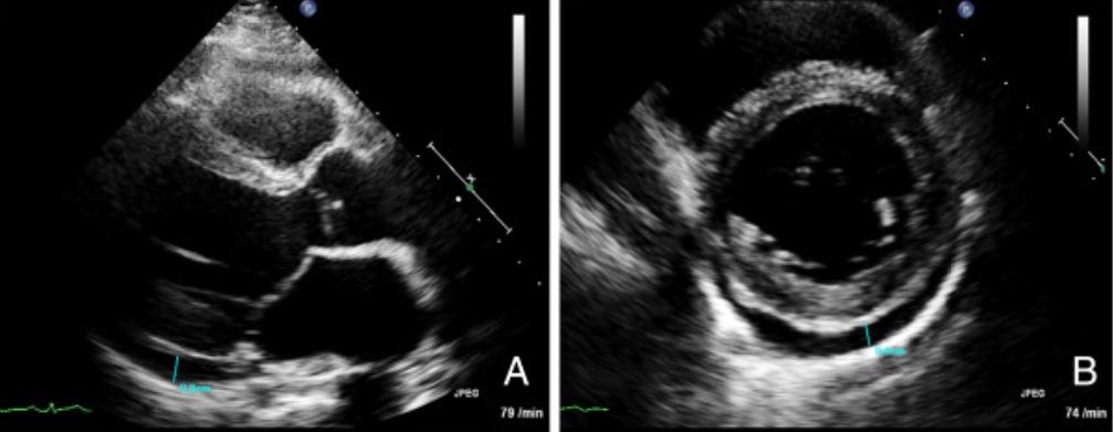
Transthoracic echocardiogram images for a patient with myocarditis: (A) Long axis view and (B) short axis view demonstrating a pericardial effusion (marked in blue)
Image: “Recurrent lymphocytic myocarditis in a young male with ulcerative colitis” by Varnavas, V.C. et al. License: CC BY 2.0
Indicated in patientsPatientsIndividuals participating in the health care system for the purpose of receiving therapeutic, diagnostic, or preventive procedures.Clinician–Patient Relationship with clinically suspected myocarditisMyocarditisMyocarditis is an inflammatory disease of the myocardium, which may occur alone or in association with a systemic process. There are numerous etiologies of myocarditis, but all lead to inflammation and myocyte injury, most often leading to signs and symptoms of heart failure. Myocarditis, with diagnosis made based on abnormalities found in T1 and T2 imaging
An accurate noninvasive tool used to detect myocardial inflammationInflammationInflammation is a complex set of responses to infection and injury involving leukocytes as the principal cellular mediators in the body’s defense against pathogenic organisms. Inflammation is also seen as a response to tissue injury in the process of wound healing. The 5 cardinal signs of inflammation are pain, heat, redness, swelling, and loss of function. Inflammation and replacement fibrosisFibrosisAny pathological condition where fibrous connective tissue invades any organ, usually as a consequence of inflammation or other injury.Bronchiolitis Obliterans
Characteristic gadoliniumGadoliniumAn element of the rare earth family of metals. It has the atomic symbol gd, atomic number 64, and atomic weight 157. 25. Its oxide is used in the control rods of some nuclear reactors.Magnetic Resonance Imaging (MRI) enhancement of the myocardial wall
Myocardial edemaEdemaEdema is a condition in which excess serous fluid accumulates in the body cavity or interstitial space of connective tissues. Edema is a symptom observed in several medical conditions. It can be categorized into 2 types, namely, peripheral (in the extremities) and internal (in an organ or body cavity). Edema
Myocardial hyperemia
InflammationInflammationInflammation is a complex set of responses to infection and injury involving leukocytes as the principal cellular mediators in the body’s defense against pathogenic organisms. Inflammation is also seen as a response to tissue injury in the process of wound healing. The 5 cardinal signs of inflammation are pain, heat, redness, swelling, and loss of function. Inflammation or scarScarDermatologic Examination
FibrosisFibrosisAny pathological condition where fibrous connective tissue invades any organ, usually as a consequence of inflammation or other injury.Bronchiolitis Obliterans
Used for follow-up as well; performed 6–12 months after the index event
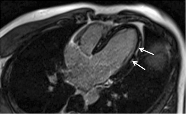
Cardiac MRI suggestive of myocarditis. White arrows mark enhancement of the myocardium, which is suggestive of edema
Image: “Myocarditis in Patients With Antisynthetase Syndrome: Prevalence, Presentation, and Outcomes” by Dieval, C. et al. License: CC BY 4.0
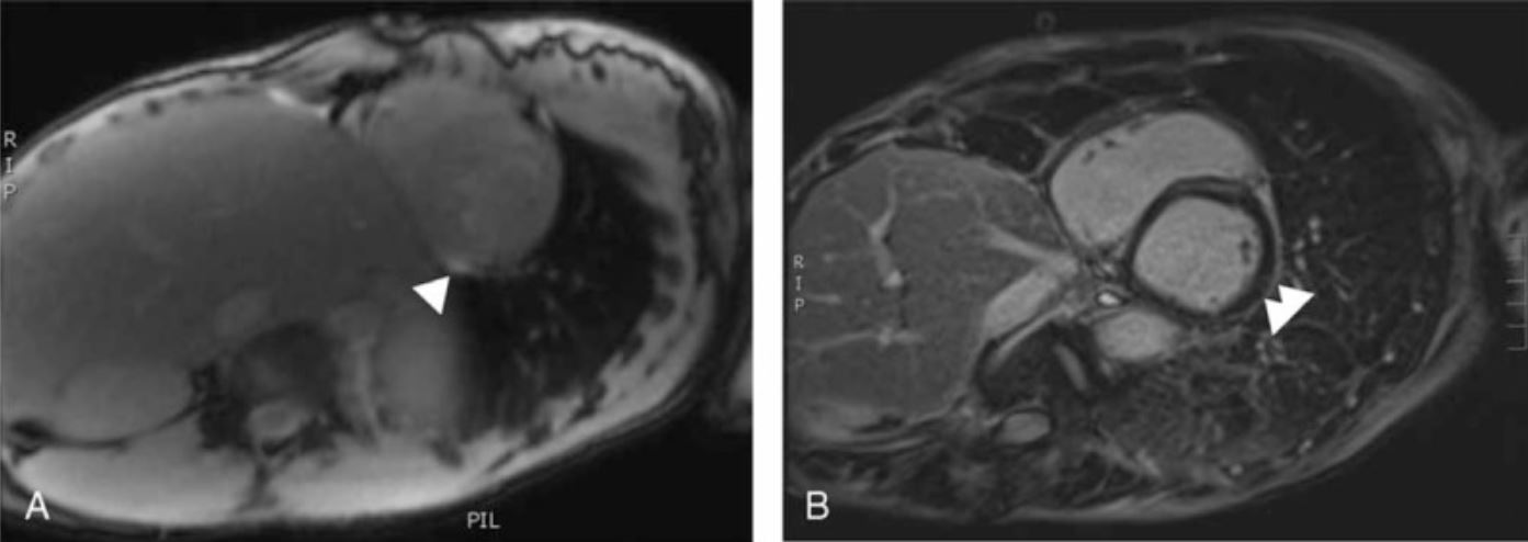
Cardiac MRI demonstrating mid-wall gadolinium enhancement of the lateral wall, which is a characteristic finding
Image: “Myopericarditis complicated by pulmonary embolism in an immunocompetent patient with acute cytomegalovirus infection: A case report” by Vandamme, Y.M. et al. License: CC BY 2.0
Other imaging:
Positron emission tomography (PETPETAn imaging technique that combines a positron-emission tomography (PET) scanner and a ct X ray scanner. This establishes a precise anatomic localization in the same session.Nuclear Imaging):
Rarely used (not as widely available and is cost-prohibitive)
Alternative tool when:
Cardiac MRMRCalculated as the ratio of the total number of people who die due to all causes over a specific time period to the total number of people in the selected population.Measures of Health Status contraindicated
Systemic autoimmune disease suspected (and there is extracardiac involvement), especially in detecting inflammationInflammationInflammation is a complex set of responses to infection and injury involving leukocytes as the principal cellular mediators in the body’s defense against pathogenic organisms. Inflammation is also seen as a response to tissue injury in the process of wound healing. The 5 cardinal signs of inflammation are pain, heat, redness, swelling, and loss of function. Inflammation in cardiac sarcoidosisSarcoidosisSarcoidosis is a multisystem inflammatory disease that causes noncaseating granulomas. The exact etiology is unknown. Sarcoidosis usually affects the lungs and thoracic lymph nodes, but it can also affect almost every system in the body, including the skin, heart, and eyes, most commonly. Sarcoidosis
Invasive coronary angiographyAngiographyRadiography of blood vessels after injection of a contrast medium.Cardiac Surgery:
Often performed to rule out acute coronary syndrome (ACS), especially in patientsPatientsIndividuals participating in the health care system for the purpose of receiving therapeutic, diagnostic, or preventive procedures.Clinician–Patient Relationship at high risk for ischemic heart diseaseIschemic heart diseaseCoronary heart disease (CHD), or ischemic heart disease, describes a situation in which an inadequate supply of blood to the myocardium exists due to a stenosis of the coronary arteries, typically from atherosclerosis. Coronary Heart Disease
Additionally, the most common cause of elevated cardiac biomarkers is ACS, so there is a low thresholdThresholdMinimum voltage necessary to generate an action potential (an all-or-none response)Skeletal Muscle Contraction for performing this test.
Endomyocardial biopsyEndomyocardial biopsyMyocarditis (EMB) is the gold standard for diagnosis; however, it is rarely needed. According to the AHA, the American College of Cardiology (ACC), and the ESC, in patientsPatientsIndividuals participating in the health care system for the purpose of receiving therapeutic, diagnostic, or preventive procedures.Clinician–Patient Relationship with rapidly progressing heart failureHeart FailureA heterogeneous condition in which the heart is unable to pump out sufficient blood to meet the metabolic need of the body. Heart failure can be caused by structural defects, functional abnormalities (ventricular dysfunction), or a sudden overload beyond its capacity. Chronic heart failure is more common than acute heart failure which results from sudden insult to cardiac function, such as myocardial infarction.Total Anomalous Pulmonary Venous Return (TAPVR) (and the cause can be confirmed only by myocardial histology, as with myocarditisMyocarditisMyocarditis is an inflammatory disease of the myocardium, which may occur alone or in association with a systemic process. There are numerous etiologies of myocarditis, but all lead to inflammation and myocyte injury, most often leading to signs and symptoms of heart failure. Myocarditis), EMB should be considered. It is especially important to perform in cases where etiology-directed therapies (such as immunosuppressive agents) are required.[9]
Indications:
Acute deterioration of cardiac function without a known etiology
Failure to respond to therapy within 1–2 weeks after diagnosis
New-onset heart failureHeart FailureA heterogeneous condition in which the heart is unable to pump out sufficient blood to meet the metabolic need of the body. Heart failure can be caused by structural defects, functional abnormalities (ventricular dysfunction), or a sudden overload beyond its capacity. Chronic heart failure is more common than acute heart failure which results from sudden insult to cardiac function, such as myocardial infarction.Total Anomalous Pulmonary Venous Return (TAPVR) associated with a dilated left ventricle (LV) and new ventricular arrhythmia or bradyarrhythmiaBradyarrhythmiaBradyarrhythmia is a rhythm in which the heart rate is less than 60/min. Bradyarrhythmia can be physiologic, without symptoms or hemodynamic change. Pathologic bradyarrhythmia results in reduced cardiac output and hemodynamic instability causing syncope, dizziness, or dyspnea. Bradyarrhythmias
Suspected hypersensitivity (subset of eosinophilic) myocarditisMyocarditisMyocarditis is an inflammatory disease of the myocardium, which may occur alone or in association with a systemic process. There are numerous etiologies of myocarditis, but all lead to inflammation and myocyte injury, most often leading to signs and symptoms of heart failure. Myocarditis
Fulminant myocarditisMyocarditisMyocarditis is an inflammatory disease of the myocardium, which may occur alone or in association with a systemic process. There are numerous etiologies of myocarditis, but all lead to inflammation and myocyte injury, most often leading to signs and symptoms of heart failure. Myocarditis when MRI is not possible
In patientsPatientsIndividuals participating in the health care system for the purpose of receiving therapeutic, diagnostic, or preventive procedures.Clinician–Patient Relationship receiving immune checkpoint inhibitorsImmune Checkpoint InhibitorsDrugs that block negative regulator immune checkpoint proteins (e.g., PD-1 receptor and CTLA-4 antigen) thereby increasing suppressed immune activation in immunotherapies.Cancer Immunotherapy who develop cardiac dysfunction
Major histopathologic types (on biopsyBiopsyRemoval and pathologic examination of specimens from the living body.Ewing Sarcoma):[1]
Lymphocytic myocarditisMyocarditisMyocarditis is an inflammatory disease of the myocardium, which may occur alone or in association with a systemic process. There are numerous etiologies of myocarditis, but all lead to inflammation and myocyte injury, most often leading to signs and symptoms of heart failure. Myocarditis (most common)
Neutrophilic myocarditisMyocarditisMyocarditis is an inflammatory disease of the myocardium, which may occur alone or in association with a systemic process. There are numerous etiologies of myocarditis, but all lead to inflammation and myocyte injury, most often leading to signs and symptoms of heart failure. Myocarditis
Eosinophilic myocarditisMyocarditisMyocarditis is an inflammatory disease of the myocardium, which may occur alone or in association with a systemic process. There are numerous etiologies of myocarditis, but all lead to inflammation and myocyte injury, most often leading to signs and symptoms of heart failure. Myocarditis
Giant cell myocarditisMyocarditisMyocarditis is an inflammatory disease of the myocardium, which may occur alone or in association with a systemic process. There are numerous etiologies of myocarditis, but all lead to inflammation and myocyte injury, most often leading to signs and symptoms of heart failure. Myocarditis
Cardiac sarcoidosisSarcoidosisSarcoidosis is a multisystem inflammatory disease that causes noncaseating granulomas. The exact etiology is unknown. Sarcoidosis usually affects the lungs and thoracic lymph nodes, but it can also affect almost every system in the body, including the skin, heart, and eyes, most commonly. Sarcoidosis
Other granulomatous myocarditisMyocarditisMyocarditis is an inflammatory disease of the myocardium, which may occur alone or in association with a systemic process. There are numerous etiologies of myocarditis, but all lead to inflammation and myocyte injury, most often leading to signs and symptoms of heart failure. Myocarditis
Findings:
Cellular infiltrates may be:
Lymphocytic
Eosinophilic
Neutrophilic
Mononuclear
Myocyte necrosisNecrosisThe death of cells in an organ or tissue due to disease, injury or failure of the blood supply.Ischemic Cell Damage
Interstitial fibrosisFibrosisAny pathological condition where fibrous connective tissue invades any organ, usually as a consequence of inflammation or other injury.Bronchiolitis Obliterans
Myofiber hypertrophyHypertrophyGeneral increase in bulk of a part or organ due to cell enlargement and accumulation of fluids and secretions, not due to tumor formation, nor to an increase in the number of cells (hyperplasia).Cellular Adaptation
Granulomatous changes:
MycobacteriumMycobacteriumMycobacterium is a genus of the family Mycobacteriaceae in the phylum Actinobacteria. Mycobacteria comprise more than 150 species of facultative intracellular bacilli that are mostly obligate aerobes. Mycobacteria are responsible for multiple human infections including serious diseases, such as tuberculosis (M. tuberculosis), leprosy (M. leprae), and M. avium complex infections.Mycobacterium
FungiFungiA kingdom of eukaryotic, heterotrophic organisms that live parasitically as saprobes, including mushrooms; yeasts; smuts, molds, etc. They reproduce either sexually or asexually, and have life cycles that range from simple to complex. Filamentous fungi, commonly known as molds, refer to those that grow as multicellular colonies.Mycology
Giant cellsGiant cellsMultinucleated masses produced by the fusion of many cells; often associated with viral infections. In aids, they are induced when the envelope glycoprotein of the HIV virus binds to the CD4 antigen of uninfected neighboring T4 cells. The resulting syncytium leads to cell death and thus may account for the cytopathic effect of the virus.Giant Cell Arteritis: giant-cell myocarditisGiant-cell myocarditisMyocarditis (a rapidly progressive necrotizing myocardial destruction, implying worse prognosisPrognosisA prediction of the probable outcome of a disease based on a individual’s condition and the usual course of the disease as seen in similar situations.Non-Hodgkin Lymphomas)
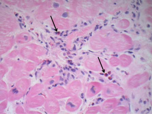
Endomyocardial biopsy in eosinophilic myocarditis: There is infiltration of eosinophils (arrows) seen on hematoxylin and eosin staining.
Image: “Eosinophilic myocarditis: Two case reports and review of the literature” by Rizkallah, J. et al. License: CC BY 2.0
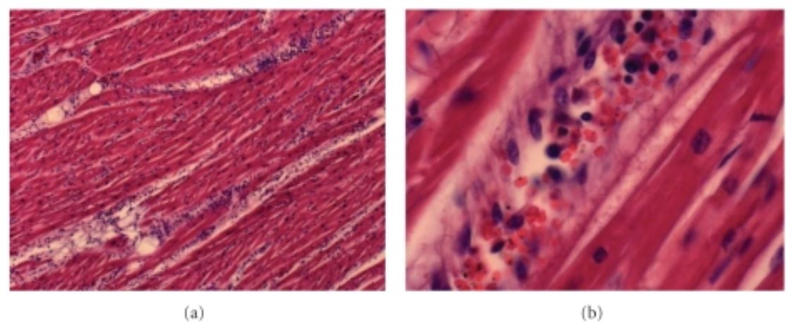
Acute myocarditis due to Plasmodium falciparum: There is extensive lymphocytic infiltration.
Image: “Fatal Myocarditis in Course of Plasmodium falciparum Infection: Case Report and Review of Cardiac Complications in Malaria” by Costenaro P, Benedetti P, Facchin C, Mengoli C, Pellizzer G. License: CC BY 3.0
Management
PatientsPatientsIndividuals participating in the health care system for the purpose of receiving therapeutic, diagnostic, or preventive procedures.Clinician–Patient Relationship with myocarditisMyocarditisMyocarditis is an inflammatory disease of the myocardium, which may occur alone or in association with a systemic process. There are numerous etiologies of myocarditis, but all lead to inflammation and myocyte injury, most often leading to signs and symptoms of heart failure. Myocarditis presenting with hypotensionHypotensionHypotension is defined as low blood pressure, specifically < 90/60 mm Hg, and is most commonly a physiologic response. Hypotension may be mild, serious, or life threatening, depending on the cause. Hypotension are often young and have no prior cardiac disease and are thus treated with IV fluidsIV fluidsIntravenous fluids are one of the most common interventions administered in medicine to approximate physiologic bodily fluids. Intravenous fluids are divided into 2 categories: crystalloid and colloid solutions. Intravenous fluids have a wide variety of indications, including intravascular volume expansion, electrolyte manipulation, and maintenance fluids. Intravenous Fluids, which can cause worsening of acute heart failureHeart FailureA heterogeneous condition in which the heart is unable to pump out sufficient blood to meet the metabolic need of the body. Heart failure can be caused by structural defects, functional abnormalities (ventricular dysfunction), or a sudden overload beyond its capacity. Chronic heart failure is more common than acute heart failure which results from sudden insult to cardiac function, such as myocardial infarction.Total Anomalous Pulmonary Venous Return (TAPVR) and cardiogenic shockCardiogenic shockShock resulting from diminution of cardiac output in heart disease.Types of Shock. Early invasive hemodynamic monitoring and right-sided heart catheterization are indicated if myocarditisMyocarditisMyocarditis is an inflammatory disease of the myocardium, which may occur alone or in association with a systemic process. There are numerous etiologies of myocarditis, but all lead to inflammation and myocyte injury, most often leading to signs and symptoms of heart failure. Myocarditis is suspected.
General principles[9,12,17]
Management for myocarditisMyocarditisMyocarditis is an inflammatory disease of the myocardium, which may occur alone or in association with a systemic process. There are numerous etiologies of myocarditis, but all lead to inflammation and myocyte injury, most often leading to signs and symptoms of heart failure. Myocarditis is generally supportive but often includes:
Heart failureHeart FailureA heterogeneous condition in which the heart is unable to pump out sufficient blood to meet the metabolic need of the body. Heart failure can be caused by structural defects, functional abnormalities (ventricular dysfunction), or a sudden overload beyond its capacity. Chronic heart failure is more common than acute heart failure which results from sudden insult to cardiac function, such as myocardial infarction.Total Anomalous Pulmonary Venous Return (TAPVR) management
Arrhythmia management
Treating the underlying etiology (when possible)
Referral to cardiology for all patientsPatientsIndividuals participating in the health care system for the purpose of receiving therapeutic, diagnostic, or preventive procedures.Clinician–Patient Relationship with myocarditisMyocarditisMyocarditis is an inflammatory disease of the myocardium, which may occur alone or in association with a systemic process. There are numerous etiologies of myocarditis, but all lead to inflammation and myocyte injury, most often leading to signs and symptoms of heart failure. Myocarditis:
Myocardial dysfunction may worsen.
Transfer to a tertiary care center with ventricular support devices available.
Consider transfer to a facility with transplantation available if early signs of fulminant myocarditisMyocarditisMyocarditis is an inflammatory disease of the myocardium, which may occur alone or in association with a systemic process. There are numerous etiologies of myocarditis, but all lead to inflammation and myocyte injury, most often leading to signs and symptoms of heart failure. Myocarditis are present.
Early invasive hemodynamic monitoring is recommended in those suspected with fulminant myocarditisMyocarditisMyocarditis is an inflammatory disease of the myocardium, which may occur alone or in association with a systemic process. There are numerous etiologies of myocarditis, but all lead to inflammation and myocyte injury, most often leading to signs and symptoms of heart failure. Myocarditis.
PatientsPatientsIndividuals participating in the health care system for the purpose of receiving therapeutic, diagnostic, or preventive procedures.Clinician–Patient Relationship with myocarditisMyocarditisMyocarditis is an inflammatory disease of the myocardium, which may occur alone or in association with a systemic process. There are numerous etiologies of myocarditis, but all lead to inflammation and myocyte injury, most often leading to signs and symptoms of heart failure. Myocarditis due to toxic agents can be monitored safely if:
No symptoms of heart failureHeart FailureA heterogeneous condition in which the heart is unable to pump out sufficient blood to meet the metabolic need of the body. Heart failure can be caused by structural defects, functional abnormalities (ventricular dysfunction), or a sudden overload beyond its capacity. Chronic heart failure is more common than acute heart failure which results from sudden insult to cardiac function, such as myocardial infarction.Total Anomalous Pulmonary Venous Return (TAPVR)
Most cases resolve spontaneously.
Treat myocarditisMyocarditisMyocarditis is an inflammatory disease of the myocardium, which may occur alone or in association with a systemic process. There are numerous etiologies of myocarditis, but all lead to inflammation and myocyte injury, most often leading to signs and symptoms of heart failure. Myocarditis associated with a condition (e.g., autoimmune disease, Lyme diseaseLyme diseaseLyme disease is a tick-borne infection caused by the gram-negative spirochete Borrelia burgdorferi. Lyme disease is transmitted by the black-legged Ixodes tick (known as a deer tick), which is only found in specific geographic regions. Patient presentation can vary depending on the stage of the disease and may include a characteristic erythema migrans rash. Lyme Disease) according to standard guidelines.
Approach based on the proposed 4-stage classification of MyocarditisMyocarditisMyocarditis is an inflammatory disease of the myocardium, which may occur alone or in association with a systemic process. There are numerous etiologies of myocarditis, but all lead to inflammation and myocyte injury, most often leading to signs and symptoms of heart failure. Myocarditis[17]
To aid in risk stratification, treatment decisions and monitoring of disease progression:
Stage A (At Risk):
(+) exposure to risk factors but without evidence of myocardial injury
Need to monitor symptom development and provide patient education
Asymptomatic inflammationInflammationInflammation is a complex set of responses to infection and injury involving leukocytes as the principal cellular mediators in the body’s defense against pathogenic organisms. Inflammation is also seen as a response to tissue injury in the process of wound healing. The 5 cardinal signs of inflammation are pain, heat, redness, swelling, and loss of function. Inflammation of the myocardiumMyocardiumThe muscle tissue of the heart. It is composed of striated, involuntary muscle cells connected to form the contractile pump to generate blood flow.Heart: Anatomy (detected through imaging or biomarkers)
Need close follow-up and further diagnostic testing
Stage C (Symptomatic MyocarditisMyocarditisMyocarditis is an inflammatory disease of the myocardium, which may occur alone or in association with a systemic process. There are numerous etiologies of myocarditis, but all lead to inflammation and myocyte injury, most often leading to signs and symptoms of heart failure. Myocarditis):
Symptoms such as chest painPainAn unpleasant sensation induced by noxious stimuli which are detected by nerve endings of nociceptive neurons.Pain: Types and Pathways, dyspneaDyspneaDyspnea is the subjective sensation of breathing discomfort. Dyspnea is a normal manifestation of heavy physical or psychological exertion, but also may be caused by underlying conditions (both pulmonary and extrapulmonary). Dyspnea, or arrhythmias are noted, along with evidence of myocardial injury.
Initiate medical therapy/intervention.
Stage D (Advanced MyocarditisMyocarditisMyocarditis is an inflammatory disease of the myocardium, which may occur alone or in association with a systemic process. There are numerous etiologies of myocarditis, but all lead to inflammation and myocyte injury, most often leading to signs and symptoms of heart failure. Myocarditis):
Severe manifestations including cardiogenic shockCardiogenic shockShock resulting from diminution of cardiac output in heart disease.Types of Shock, hemodynamic instability, or life-threatening arrhythmias.
Need to refer to specialized centers.
Heart failureHeart FailureA heterogeneous condition in which the heart is unable to pump out sufficient blood to meet the metabolic need of the body. Heart failure can be caused by structural defects, functional abnormalities (ventricular dysfunction), or a sudden overload beyond its capacity. Chronic heart failure is more common than acute heart failure which results from sudden insult to cardiac function, such as myocardial infarction.Total Anomalous Pulmonary Venous Return (TAPVR) management[9,12,17]
The 1st-line treatment of heart failureHeart FailureA heterogeneous condition in which the heart is unable to pump out sufficient blood to meet the metabolic need of the body. Heart failure can be caused by structural defects, functional abnormalities (ventricular dysfunction), or a sudden overload beyond its capacity. Chronic heart failure is more common than acute heart failure which results from sudden insult to cardiac function, such as myocardial infarction.Total Anomalous Pulmonary Venous Return (TAPVR) due to myocarditisMyocarditisMyocarditis is an inflammatory disease of the myocardium, which may occur alone or in association with a systemic process. There are numerous etiologies of myocarditis, but all lead to inflammation and myocyte injury, most often leading to signs and symptoms of heart failure. Myocarditis is afterloadAfterloadAfterload is the resistance in the aorta that prevents blood from leaving the heart. Afterload represents the pressure the LV needs to overcome to eject blood into the aorta.Cardiac Mechanics reduction and diuresis, along with addressing the underlying cause. Following guideline-directed medical therapy is standard. Some patientsPatientsIndividuals participating in the health care system for the purpose of receiving therapeutic, diagnostic, or preventive procedures.Clinician–Patient Relationship will require inotropic support or mechanical assist devices. Cardiac transplantation may be needed for intractable heart failureHeart FailureA heterogeneous condition in which the heart is unable to pump out sufficient blood to meet the metabolic need of the body. Heart failure can be caused by structural defects, functional abnormalities (ventricular dysfunction), or a sudden overload beyond its capacity. Chronic heart failure is more common than acute heart failure which results from sudden insult to cardiac function, such as myocardial infarction.Total Anomalous Pulmonary Venous Return (TAPVR) (HF).
Medical therapy for heart failureHeart FailureA heterogeneous condition in which the heart is unable to pump out sufficient blood to meet the metabolic need of the body. Heart failure can be caused by structural defects, functional abnormalities (ventricular dysfunction), or a sudden overload beyond its capacity. Chronic heart failure is more common than acute heart failure which results from sudden insult to cardiac function, such as myocardial infarction.Total Anomalous Pulmonary Venous Return (TAPVR):
Renin-angiotensin system (RASRASRenal artery stenosis (RAS) is the narrowing of one or both renal arteries, usually caused by atherosclerotic disease or by fibromuscular dysplasia. If the stenosis is severe enough, the stenosis causes decreased renal blood flow, which activates the renin-angiotensin-aldosterone system (RAAS) and leads to renovascular hypertension (RVH).Renal Artery Stenosis) inhibitors (ACE inhibitorsACE inhibitorsTruncus Arteriosus/Angiotensin IIAngiotensin IIAn octapeptide that is a potent but labile vasoconstrictor. It is produced from angiotensin I after the removal of two amino acids at the c-terminal by angiotensin converting enzyme. The amino acid in position 5 varies in different species. To block vasoconstriction and hypertension effect of angiotensin II, patients are often treated with ace inhibitors or with angiotensin II type 1 receptor blockers.Renal Sodium and Water RegulationreceptorReceptorReceptors are proteins located either on the surface of or within a cell that can bind to signaling molecules known as ligands (e.g., hormones) and cause some type of response within the cell.Receptors blockers/angiotensin receptor-neprilysin inhibitorAngiotensin Receptor-Neprilysin InhibitorRenin-Angiotensin-Aldosterone System Inhibitors)
For fulminant heart failureHeart FailureA heterogeneous condition in which the heart is unable to pump out sufficient blood to meet the metabolic need of the body. Heart failure can be caused by structural defects, functional abnormalities (ventricular dysfunction), or a sudden overload beyond its capacity. Chronic heart failure is more common than acute heart failure which results from sudden insult to cardiac function, such as myocardial infarction.Total Anomalous Pulmonary Venous Return (TAPVR):
VasopressorVasopressorAcute Cholangitis therapy with norepinephrineNorepinephrinePrecursor of epinephrine that is secreted by the adrenal medulla and is a widespread central and autonomic neurotransmitter. Norepinephrine is the principal transmitter of most postganglionic sympathetic fibers, and of the diffuse projection system in the brain that arises from the locus ceruleus.Receptors and Neurotransmitters of the CNS
GlucocorticoidsGlucocorticoidsGlucocorticoids are a class within the corticosteroid family. Glucocorticoids are chemically and functionally similar to endogenous cortisol. There are a wide array of indications, which primarily benefit from the antiinflammatory and immunosuppressive effects of this class of drugs.Glucocorticoids:
Beneficial for:
Eosinophilic myocarditisMyocarditisMyocarditis is an inflammatory disease of the myocardium, which may occur alone or in association with a systemic process. There are numerous etiologies of myocarditis, but all lead to inflammation and myocyte injury, most often leading to signs and symptoms of heart failure. Myocarditis
Immune checkpoint inhibitor–associated myocarditisMyocarditisMyocarditis is an inflammatory disease of the myocardium, which may occur alone or in association with a systemic process. There are numerous etiologies of myocarditis, but all lead to inflammation and myocyte injury, most often leading to signs and symptoms of heart failure. Myocarditis
Giant cell myocarditisMyocarditisMyocarditis is an inflammatory disease of the myocardium, which may occur alone or in association with a systemic process. There are numerous etiologies of myocarditis, but all lead to inflammation and myocyte injury, most often leading to signs and symptoms of heart failure. Myocarditis
Cardiac sarcoidosisSarcoidosisSarcoidosis is a multisystem inflammatory disease that causes noncaseating granulomas. The exact etiology is unknown. Sarcoidosis usually affects the lungs and thoracic lymph nodes, but it can also affect almost every system in the body, including the skin, heart, and eyes, most commonly. Sarcoidosis
Glucocorticoid dosing: solumedrol 1 g IV for 3 days, then oral prednisonePrednisoneA synthetic anti-inflammatory glucocorticoid derived from cortisone. It is biologically inert and converted to prednisolone in the liver.Immunosuppressants 1–2 mg/kg daily for a few months (no clear dose established). Slow weaningWeaningTechniques for effecting the transition of the respiratory-failure patient from mechanical ventilation to spontaneous ventilation, while meeting the criteria that tidal volume be above a given threshold (greater than 5 ml/kg), respiratory frequency be below a given count (less than 30 breaths/min), and oxygen partial pressure be above a given threshold (pao2 greater than 50mm hg). Weaning studies focus on finding methods to monitor and predict the outcome of mechanical ventilator weaning as well as finding ventilatory support techniques which will facilitate successful weaning. Present methods include intermittent mandatory ventilation, intermittent positive pressure ventilation, and mandatory minute volume ventilation.Invasive Mechanical Ventilation over 1 year has been recommended.
In some cases, combined with other agents (e.g. azathioprineAzathioprineAn immunosuppressive agent used in combination with cyclophosphamide and hydroxychloroquine in the treatment of rheumatoid arthritis. According to the fourth annual report on carcinogens, this substance has been listed as a known carcinogen.Immunosuppressants)
In giant cell myocarditisMyocarditisMyocarditis is an inflammatory disease of the myocardium, which may occur alone or in association with a systemic process. There are numerous etiologies of myocarditis, but all lead to inflammation and myocyte injury, most often leading to signs and symptoms of heart failure. Myocarditis:
Cases respond to immunosuppressive therapy (suggesting that the condition is mostly autoimmune, and not an active infection).
Antithymocyte globulin and calcineurin inhibitorCalcineurin inhibitorCompounds that inhibit or block the phosphatase activity of calcineurin.Vitiligo therapy have been found to be beneficial in giant cell myocarditisMyocarditisMyocarditis is an inflammatory disease of the myocardium, which may occur alone or in association with a systemic process. There are numerous etiologies of myocarditis, but all lead to inflammation and myocyte injury, most often leading to signs and symptoms of heart failure. Myocarditis and can lead to clinical remissionRemissionA spontaneous diminution or abatement of a disease over time, without formal treatment.Cluster Headaches in ⅔ of patientsPatientsIndividuals participating in the health care system for the purpose of receiving therapeutic, diagnostic, or preventive procedures.Clinician–Patient Relationship.
Above treatment is given with corticosteroidsCorticosteroidsChorioretinitis and cyclosporineCyclosporineA cyclic undecapeptide from an extract of soil fungi. It is a powerful immunosupressant with a specific action on T-lymphocytes. It is used for the prophylaxis of graft rejection in organ and tissue transplantation.Immunosuppressants.
Arrhythmia management[9,12,17]
Antiarrhythmic therapy
Avoid treatment of sinus tachycardiaTachycardiaAbnormally rapid heartbeat, usually with a heart rate above 100 beats per minute for adults. Tachycardia accompanied by disturbance in the cardiac depolarization (cardiac arrhythmia) is called tachyarrhythmia.Sepsis in Children with negative inotropic rate-control agents:
MetoprololMetoprololA selective adrenergic beta-1 blocking agent that is commonly used to treat angina pectoris; hypertension; and cardiac arrhythmias.Antiadrenergic Drugs
Implantable cardiac defibrillatorDefibrillatorCardiac electrical stimulators that apply brief high-voltage electroshocks to the heart. These stimulators are used to restore normal rhythm and contractile function in hearts of patients who are experiencing ventricular fibrillation or ventricular tachycardia that is not accompanied by a palpable pulse. Some defibrillators may also be used to correct certain noncritical dysrhythmias (called synchronized defibrillation or cardioversion), using relatively low-level discharges synchronized to the patient’s ECG waveform.Cardiac Arrest (ICD): recommended for giant cell myocarditisMyocarditisMyocarditis is an inflammatory disease of the myocardium, which may occur alone or in association with a systemic process. There are numerous etiologies of myocarditis, but all lead to inflammation and myocyte injury, most often leading to signs and symptoms of heart failure. Myocarditis owing to a high risk of ventricular tachycardiaTachycardiaAbnormally rapid heartbeat, usually with a heart rate above 100 beats per minute for adults. Tachycardia accompanied by disturbance in the cardiac depolarization (cardiac arrhythmia) is called tachyarrhythmia.Sepsis in Children
Other considerations[5,7,8]
AnticoagulantsAnticoagulantsAnticoagulants are drugs that retard or interrupt the coagulation cascade. The primary classes of available anticoagulants include heparins, vitamin K-dependent antagonists (e.g., warfarin), direct thrombin inhibitors, and factor Xa inhibitors. Anticoagulants are indicated for:
Atrial fibrillationAtrial fibrillationAtrial fibrillation (AF or Afib) is a supraventricular tachyarrhythmia and the most common kind of arrhythmia. It is caused by rapid, uncontrolled atrial contractions and uncoordinated ventricular responses. Atrial Fibrillation
Most patientsPatientsIndividuals participating in the health care system for the purpose of receiving therapeutic, diagnostic, or preventive procedures.Clinician–Patient Relationship will have a partial or full recovery. However, long-term follow-up and monitoring are recommended.
Gradual resumption of physical activities:
No competitive sports for 3–6 months (risk of ventricular arrhythmias)
EchocardiographyEchocardiographyUltrasonic recording of the size, motion, and composition of the heart and surrounding tissues. The standard approach is transthoracic.Tricuspid Valve Atresia (TVA)
Holter monitoring
Serial echocardiographyEchocardiographyUltrasonic recording of the size, motion, and composition of the heart and surrounding tissues. The standard approach is transthoracic.Tricuspid Valve Atresia (TVA) monitoring
Re-evaluation and titration of medications, as appropriate
Summary of Diagnosis and Treatment (5-step process)
Outline of approaching a patient with symptoms of myocarditisMyocarditisMyocarditis is an inflammatory disease of the myocardium, which may occur alone or in association with a systemic process. There are numerous etiologies of myocarditis, but all lead to inflammation and myocyte injury, most often leading to signs and symptoms of heart failure. Myocarditis:[17]
Step 1: clinical suspicion
Obtain appropriate labs (including cardiac and inflammatory biomarkers, CBC with differential and others), ECGECGAn electrocardiogram (ECG) is a graphic representation of the electrical activity of the heart plotted against time. Adhesive electrodes are affixed to the skin surface allowing measurement of cardiac impulses from many angles. The ECG provides 3-dimensional information about the conduction system of the heart, the myocardium, and other cardiac structures. Electrocardiogram (ECG), echocardiogramEchocardiogramTransposition of the Great Arteries.
Assess stability of the patient and the need for hospitalizationHospitalizationThe confinement of a patient in a hospital.Delirium.
Referral to advanced heart failureHeart FailureA heterogeneous condition in which the heart is unable to pump out sufficient blood to meet the metabolic need of the body. Heart failure can be caused by structural defects, functional abnormalities (ventricular dysfunction), or a sudden overload beyond its capacity. Chronic heart failure is more common than acute heart failure which results from sudden insult to cardiac function, such as myocardial infarction.Total Anomalous Pulmonary Venous Return (TAPVR) center
Assess need for arrhythmia treatment and circulatory support.
Step 3: pivotal diagnostic testsDiagnostic testsDiagnostic tests are important aspects in making a diagnosis. Some of the most important epidemiological values of diagnostic tests include sensitivity and specificity, false positives and false negatives, positive and negative predictive values, likelihood ratios, and pre-test and post-test probabilities. Epidemiological Values of Diagnostic Tests to establish the diagnosis of myocarditisMyocarditisMyocarditis is an inflammatory disease of the myocardium, which may occur alone or in association with a systemic process. There are numerous etiologies of myocarditis, but all lead to inflammation and myocyte injury, most often leading to signs and symptoms of heart failure. Myocarditis
Outpatient genetic testingGenetic TestingDetection of a mutation; genotype; karyotype; or specific alleles associated with genetic traits, heritable diseases, or predisposition to a disease, or that may lead to the disease in descendants. It includes prenatal genetic testing.Myotonic Dystrophies to consenting patientsPatientsIndividuals participating in the health care system for the purpose of receiving therapeutic, diagnostic, or preventive procedures.Clinician–Patient Relationship
PericarditisPericarditisPericarditis is an inflammation of the pericardium, often with fluid accumulation. It can be caused by infection (often viral), myocardial infarction, drugs, malignancies, metabolic disorders, autoimmune disorders, or trauma. Acute, subacute, and chronic forms exist. Pericarditis: an inflammationInflammationInflammation is a complex set of responses to infection and injury involving leukocytes as the principal cellular mediators in the body’s defense against pathogenic organisms. Inflammation is also seen as a response to tissue injury in the process of wound healing. The 5 cardinal signs of inflammation are pain, heat, redness, swelling, and loss of function. Inflammation of the outer lining of the heart resulting from infection, autoimmune disease, radiationRadiationEmission or propagation of acoustic waves (sound), electromagnetic energy waves (such as light; radio waves; gamma rays; or x-rays), or a stream of subatomic particles (such as electrons; neutrons; protons; or alpha particles).Osteosarcoma, surgery, or myocardial infarctionMyocardial infarctionMI is ischemia and death of an area of myocardial tissue due to insufficient blood flow and oxygenation, usually from thrombus formation on a ruptured atherosclerotic plaque in the epicardial arteries. Clinical presentation is most commonly with chest pain, but women and patients with diabetes may have atypical symptoms.Myocardial Infarction. PericarditisPericarditisPericarditis is an inflammation of the pericardium, often with fluid accumulation. It can be caused by infection (often viral), myocardial infarction, drugs, malignancies, metabolic disorders, autoimmune disorders, or trauma. Acute, subacute, and chronic forms exist. Pericarditis clinically presents with feverFeverFever is defined as a measured body temperature of at least 38°C (100.4°F). Fever is caused by circulating endogenous and/or exogenous pyrogens that increase levels of prostaglandin E2 in the hypothalamus. Fever is commonly associated with chills, rigors, sweating, and flushing of the skin. Fever and pleuritic chest painPainAn unpleasant sensation induced by noxious stimuli which are detected by nerve endings of nociceptive neurons.Pain: Types and Pathways that increases with lying supine, and pericardial rub on auscultation. An ECGECGAn electrocardiogram (ECG) is a graphic representation of the electrical activity of the heart plotted against time. Adhesive electrodes are affixed to the skin surface allowing measurement of cardiac impulses from many angles. The ECG provides 3-dimensional information about the conduction system of the heart, the myocardium, and other cardiac structures. Electrocardiogram (ECG) with diffuse ST-segment elevation and an echocardiogramEchocardiogramTransposition of the Great Arteries showing pericardial effusionPericardial effusionFluid accumulation within the pericardium. Serous effusions are associated with pericardial diseases. Hemopericardium is associated with trauma. Lipid-containing effusion (chylopericardium) results from leakage of thoracic duct. Severe cases can lead to cardiac tamponade.Pericardial Effusion and Cardiac Tamponade can confirm the diagnosis. Management is supportive.
Myocardial infarctionMyocardial infarctionMI is ischemia and death of an area of myocardial tissue due to insufficient blood flow and oxygenation, usually from thrombus formation on a ruptured atherosclerotic plaque in the epicardial arteries. Clinical presentation is most commonly with chest pain, but women and patients with diabetes may have atypical symptoms.Myocardial Infarction:ischemiaIschemiaA hypoperfusion of the blood through an organ or tissue caused by a pathologic constriction or obstruction of its blood vessels, or an absence of blood circulation.Ischemic Cell Damage of the myocardiumMyocardiumThe muscle tissue of the heart. It is composed of striated, involuntary muscle cells connected to form the contractile pump to generate blood flow.Heart: Anatomy due to partial or complete obstruction of the coronary arteriesArteriesArteries are tubular collections of cells that transport oxygenated blood and nutrients from the heart to the tissues of the body. The blood passes through the arteries in order of decreasing luminal diameter, starting in the largest artery (the aorta) and ending in the small arterioles. Arteries are classified into 3 types: large elastic arteries, medium muscular arteries, and small arteries and arterioles. Arteries: Histology. PatientsPatientsIndividuals participating in the health care system for the purpose of receiving therapeutic, diagnostic, or preventive procedures.Clinician–Patient Relationship present with acute onset chest painPainAn unpleasant sensation induced by noxious stimuli which are detected by nerve endings of nociceptive neurons.Pain: Types and Pathways. As in the case of myocarditisMyocarditisMyocarditis is an inflammatory disease of the myocardium, which may occur alone or in association with a systemic process. There are numerous etiologies of myocarditis, but all lead to inflammation and myocyte injury, most often leading to signs and symptoms of heart failure. Myocarditis, heart failureHeart FailureA heterogeneous condition in which the heart is unable to pump out sufficient blood to meet the metabolic need of the body. Heart failure can be caused by structural defects, functional abnormalities (ventricular dysfunction), or a sudden overload beyond its capacity. Chronic heart failure is more common than acute heart failure which results from sudden insult to cardiac function, such as myocardial infarction.Total Anomalous Pulmonary Venous Return (TAPVR) can develop, which is usually accompanied by elevated troponins and typical ischemic ECGECGAn electrocardiogram (ECG) is a graphic representation of the electrical activity of the heart plotted against time. Adhesive electrodes are affixed to the skin surface allowing measurement of cardiac impulses from many angles. The ECG provides 3-dimensional information about the conduction system of the heart, the myocardium, and other cardiac structures. Electrocardiogram (ECG) changes (ST elevations or depressions). Wall-motion abnormalities can be diagnosed based on a transthoracic echo. Management is with antiplatelet medications, nitratesNitratesNitrates are a class of medications that cause systemic vasodilation (veins > arteries) by smooth muscle relaxation. Nitrates are primarily indicated for the treatment of angina, where preferential venodilation causes pooling of blood, decreased preload, and ultimately decreased myocardial O2 demand.Nitrates, beta blockers, and revascularizationRevascularizationThromboangiitis Obliterans (Buerger Disease).
Pericardial effusionPericardial effusionFluid accumulation within the pericardium. Serous effusions are associated with pericardial diseases. Hemopericardium is associated with trauma. Lipid-containing effusion (chylopericardium) results from leakage of thoracic duct. Severe cases can lead to cardiac tamponade.Pericardial Effusion and Cardiac Tamponade and tamponadeTamponadePericardial effusion, usually of rapid onset, exceeding ventricular filling pressures and causing collapse of the heart with a markedly reduced cardiac output.Pericarditis: the presence of fluid in the pericardial sac, which can cause compressionCompressionBlunt Chest Trauma of the heart and lead to tamponadeTamponadePericardial effusion, usually of rapid onset, exceeding ventricular filling pressures and causing collapse of the heart with a markedly reduced cardiac output.Pericarditis physiology. TamponadeTamponadePericardial effusion, usually of rapid onset, exceeding ventricular filling pressures and causing collapse of the heart with a markedly reduced cardiac output.Pericarditis physiology prevents the heart from filling and results in hemodynamic collapse. Beck’s triadBeck’s triadThe triad describes the classic findings in cardiac tamponade: hypotension, jugular venous distension, muffled heart sounds on auscultation.Pericardial Effusion and Cardiac Tamponade of distended neckNeckThe part of a human or animal body connecting the head to the rest of the body.Peritonsillar AbscessveinsVeinsVeins are tubular collections of cells, which transport deoxygenated blood and waste from the capillary beds back to the heart. Veins are classified into 3 types: small veins/venules, medium veins, and large veins. Each type contains 3 primary layers: tunica intima, tunica media, and tunica adventitia. Veins: Histology, hypotensionHypotensionHypotension is defined as low blood pressure, specifically < 90/60 mm Hg, and is most commonly a physiologic response. Hypotension may be mild, serious, or life threatening, depending on the cause. Hypotension, and muffled heart soundsMuffled Heart SoundsPericardial Effusion and Cardiac Tamponade is often noted on physical exam. Diagnosis is confirmed using transthoracic echocardiographyTransthoracic EchocardiographyImaging of the Heart and Great Vessels, and management involves drainage of the pericardiumPericardiumA conical fibroserous sac surrounding the heart and the roots of the great vessels (aorta; venae cavae; pulmonary artery). Pericardium consists of two sacs: the outer fibrous pericardium and the inner serous pericardium. The latter consists of an outer parietal layer facing the fibrous pericardium, and an inner visceral layer (epicardium) resting next to the heart, and a pericardial cavity between these two layers.Heart: Anatomy.
Pulmonary embolismPulmonary EmbolismPulmonary embolism (PE) is a potentially fatal condition that occurs as a result of intraluminal obstruction of the main pulmonary artery or its branches. The causative factors include thrombi, air, amniotic fluid, and fat. In PE, gas exchange is impaired due to the decreased return of deoxygenated blood to the lungs. Pulmonary Embolism: obstruction of the pulmonary arteriesArteriesArteries are tubular collections of cells that transport oxygenated blood and nutrients from the heart to the tissues of the body. The blood passes through the arteries in order of decreasing luminal diameter, starting in the largest artery (the aorta) and ending in the small arterioles. Arteries are classified into 3 types: large elastic arteries, medium muscular arteries, and small arteries and arterioles. Arteries: Histology, which is most often due to thrombus migration from the deep venous system. Signs and symptoms include pleuritic chest painPainAn unpleasant sensation induced by noxious stimuli which are detected by nerve endings of nociceptive neurons.Pain: Types and Pathways, dyspneaDyspneaDyspnea is the subjective sensation of breathing discomfort. Dyspnea is a normal manifestation of heavy physical or psychological exertion, but also may be caused by underlying conditions (both pulmonary and extrapulmonary). Dyspnea, tachypneaTachypneaIncreased respiratory rate.Pulmonary Examination, and tachycardiaTachycardiaAbnormally rapid heartbeat, usually with a heart rate above 100 beats per minute for adults. Tachycardia accompanied by disturbance in the cardiac depolarization (cardiac arrhythmia) is called tachyarrhythmia.Sepsis in Children. Severe cases of pulmonary embolismPulmonary EmbolismPulmonary embolism (PE) is a potentially fatal condition that occurs as a result of intraluminal obstruction of the main pulmonary artery or its branches. The causative factors include thrombi, air, amniotic fluid, and fat. In PE, gas exchange is impaired due to the decreased return of deoxygenated blood to the lungs. Pulmonary Embolism can result in hemodynamic instability or cardiopulmonary arrestCardiopulmonary arrestCardiac arrest is the sudden, complete cessation of cardiac output with hemodynamic collapse. Patients present as pulseless, unresponsive, and apneic. Rhythms associated with cardiac arrest are ventricular fibrillation/tachycardia, asystole, or pulseless electrical activity.Cardiac Arrest. A chest CT with angiographyAngiographyRadiography of blood vessels after injection of a contrast medium.Cardiac Surgery is the primary method of diagnosis. Management includes oxygen and anticoagulationAnticoagulationPulmonary Hypertension Drugs for most patientsPatientsIndividuals participating in the health care system for the purpose of receiving therapeutic, diagnostic, or preventive procedures.Clinician–Patient Relationship; thrombolytic therapies may be indicated for unstable patientsUnstable PatientsBlunt Chest Trauma.
Billing and Coding
Diagnosis Codes:
These codes are used to formally document a diagnosis of myocarditisMyocarditisMyocarditis is an inflammatory disease of the myocardium, which may occur alone or in association with a systemic process. There are numerous etiologies of myocarditis, but all lead to inflammation and myocyte injury, most often leading to signs and symptoms of heart failure. Myocarditis, an inflammationInflammationInflammation is a complex set of responses to infection and injury involving leukocytes as the principal cellular mediators in the body’s defense against pathogenic organisms. Inflammation is also seen as a response to tissue injury in the process of wound healing. The 5 cardinal signs of inflammation are pain, heat, redness, swelling, and loss of function. Inflammation of the heart muscle, which can impair the heart’s ability to pumpPumpACES and RUSH: Resuscitation Ultrasound Protocols and cause arrhythmias.
MyocarditisMyocarditisMyocarditis is an inflammatory disease of the myocardium, which may occur alone or in association with a systemic process. There are numerous etiologies of myocarditis, but all lead to inflammation and myocyte injury, most often leading to signs and symptoms of heart failure. Myocarditis, unspecified
MyocarditisMyocarditisMyocarditis is an inflammatory disease of the myocardium, which may occur alone or in association with a systemic process. There are numerous etiologies of myocarditis, but all lead to inflammation and myocyte injury, most often leading to signs and symptoms of heart failure. Myocarditis (disorder)
Evaluation & Workup:
These codes are used to order diagnostic testsDiagnostic testsDiagnostic tests are important aspects in making a diagnosis. Some of the most important epidemiological values of diagnostic tests include sensitivity and specificity, false positives and false negatives, positive and negative predictive values, likelihood ratios, and pre-test and post-test probabilities. Epidemiological Values of Diagnostic Tests to evaluate myocarditisMyocarditisMyocarditis is an inflammatory disease of the myocardium, which may occur alone or in association with a systemic process. There are numerous etiologies of myocarditis, but all lead to inflammation and myocyte injury, most often leading to signs and symptoms of heart failure. Myocarditis, including blood tests for cardiac enzymesEnzymesEnzymes are complex protein biocatalysts that accelerate chemical reactions without being consumed by them. Due to the body’s constant metabolic needs, the absence of enzymes would make life unsustainable, as reactions would occur too slowly without these molecules. Basics of Enzymes like troponin, an echocardiogramEchocardiogramTransposition of the Great Arteries to assess heart function, and an endomyocardial biopsyEndomyocardial biopsyMyocarditis for definitive diagnosis.
Domain
Code
Description
CPT
84484
Troponin, quantitative
CPT
93306
EchocardiographyEchocardiographyUltrasonic recording of the size, motion, and composition of the heart and surrounding tissues. The standard approach is transthoracic.Tricuspid Valve Atresia (TVA), transthoracic, real-time with image documentationDocumentationSystematic organization, storage, retrieval, and dissemination of specialized information, especially of a scientific or technical nature. It often involves authenticating or validating information.Advance Directives
These codes are used to prescribe medications for supportive care, primarily aimed at treating the consequences of myocarditisMyocarditisMyocarditis is an inflammatory disease of the myocardium, which may occur alone or in association with a systemic process. There are numerous etiologies of myocarditis, but all lead to inflammation and myocyte injury, most often leading to signs and symptoms of heart failure. Myocarditis such as heart failureHeart FailureA heterogeneous condition in which the heart is unable to pump out sufficient blood to meet the metabolic need of the body. Heart failure can be caused by structural defects, functional abnormalities (ventricular dysfunction), or a sudden overload beyond its capacity. Chronic heart failure is more common than acute heart failure which results from sudden insult to cardiac function, such as myocardial infarction.Total Anomalous Pulmonary Venous Return (TAPVR), using drugs like ACE inhibitorsACE inhibitorsTruncus Arteriosus and beta-blockersBeta-blockersDrugs that bind to but do not activate beta-adrenergic receptors thereby blocking the actions of beta-adrenergic agonists. Adrenergic beta-antagonists are used for treatment of hypertension, cardiac arrhythmias, angina pectoris, glaucoma, migraine headaches, and anxiety.Class 2 Antiarrhythmic Drugs (Beta Blockers).
Domain
Code
Description
RxNorm
6472
LisinoprilLisinoprilOne of the angiotensin-converting enzyme inhibitors (ACE inhibitors), orally active, that has been used in the treatment of hypertension and congestive heart failure.Renin-Angiotensin-Aldosterone System Inhibitors (ingredient)
RxNorm
20428
CarvedilolCarvedilolA carbazole and propanol derivative that acts as a non-cardioselective beta blocker and vasodilator. It has blocking activity for alpha 1 adrenergic receptors and, at higher doses, may function as a blocker of calcium channels; it also has antioxidant properties. Carvedilol is used in the treatment of hypertension; angina pectoris; and heart failure. It can also reduce the risk of death following myocardial infarction.Class 2 Antiarrhythmic Drugs (Beta Blockers) (ingredient)
RxNorm
4603
FurosemideFurosemideA benzoic-sulfonamide-furan. It is a diuretic with fast onset and short duration that is used for edema and chronic renal insufficiency.Loop Diuretics (ingredient)
These codes are used to document the serious potential outcomes of myocarditisMyocarditisMyocarditis is an inflammatory disease of the myocardium, which may occur alone or in association with a systemic process. There are numerous etiologies of myocarditis, but all lead to inflammation and myocyte injury, most often leading to signs and symptoms of heart failure. Myocarditis, including the development of dilated cardiomyopathyDilated CardiomyopathyDilated cardiomyopathy (DCM) is the most common type of non-ischemic cardiomyopathy and a common cause of heart failure (HF). The cause may be idiopathic, familial, or secondary to a variety of underlying conditions. The disease is characterized by the enlargement of 1 or both ventricles and reduced systolic function. Dilated Cardiomyopathy, severe heart failureHeart FailureA heterogeneous condition in which the heart is unable to pump out sufficient blood to meet the metabolic need of the body. Heart failure can be caused by structural defects, functional abnormalities (ventricular dysfunction), or a sudden overload beyond its capacity. Chronic heart failure is more common than acute heart failure which results from sudden insult to cardiac function, such as myocardial infarction.Total Anomalous Pulmonary Venous Return (TAPVR), and life-threatening ventricular arrhythmias.
Domain
Code
Description
ICD-10-CM
I42.0
Dilated cardiomyopathyDilated CardiomyopathyDilated cardiomyopathy (DCM) is the most common type of non-ischemic cardiomyopathy and a common cause of heart failure (HF). The cause may be idiopathic, familial, or secondary to a variety of underlying conditions. The disease is characterized by the enlargement of 1 or both ventricles and reduced systolic function. Dilated Cardiomyopathy
ICD-10-CM
I50.9
Heart failureHeart FailureA heterogeneous condition in which the heart is unable to pump out sufficient blood to meet the metabolic need of the body. Heart failure can be caused by structural defects, functional abnormalities (ventricular dysfunction), or a sudden overload beyond its capacity. Chronic heart failure is more common than acute heart failure which results from sudden insult to cardiac function, such as myocardial infarction.Total Anomalous Pulmonary Venous Return (TAPVR), unspecified
ICD-10-CM
I49.01
Ventricular fibrillationVentricular fibrillationVentricular fibrillation (VF or V-fib) is a type of ventricular tachyarrhythmia (> 300/min) often preceded by ventricular tachycardia. In this arrhythmia, the ventricle beats rapidly and sporadically. The ventricular contraction is uncoordinated, leading to a decrease in cardiac output and immediate hemodynamic collapse. Ventricular Fibrillation (V-fib)
References
Leone, O., Pieroni, M., Rapezzi, C., et al. (2019). The spectrum of myocarditis: from pathology to the clinics. Virchows Archiv, 475, 279–301. https://doi.org/10.1007/s00428-019-02615-8
Kociol, R. D., Cooper, L. T., et al. (2020). Recognition and initial management of fulminant myocarditis: a scientific statement from the American Heart Association Circulation, 141(6). https://doi.org/10.1161/CIR.0000000000000745
Caforio, A. L. P., Adler, Y., et al. (2017). Diagnosis and management of myocardial involvement in systemic immune-mediated diseases: a position statement of the European Society of Cardiology Working Group on Myocardial and Pericardial Disease. European Heart Journal, 38(35), 2649–2662. https://doi.org/10.1093/eurheartj/ehx321
McKenna, W. J., Elliott, P. M. (2019). Diseases of the myocardium and endocardium. In Crow, M. K., et al. (Eds.), Goldman-Cecil Medicine (26th ed., vol. 1, pp. 301–314).
Kiefer, T., Granger, C. B., Jackson, K. P. (2023). Heart disease. In Papadakis, M. A., et al. (Eds.), Current Medical Diagnosis & Treatment (62nd ed., pp. 414–417).
Engler, R. J. M., Nelson, M. R., et al. (2015). A prospective study of the incidence of myocarditis/pericarditis and new onset cardiac symptoms following smallpox and influenza vaccination. PloS One, 10(3), e0118283. https://doi.org/10.1371/journal.pone.0118283
Oster, M. E., Shay, D. K., et al. (2022). Myocarditis cases reported after mRNA-based COVID-19 vaccination in the US from December 2020 to August 2021. JAMA, 327(4), 331–340. https://doi.org/10.1001/jama.2021.24110
Gargano, J. W., Wallace, M., et al. (2021). Use of mRNA COVID-19 vaccine after reports of myocarditis among vaccine recipients: update from the Advisory Committee on Immunization Practices—United States, June 2021. Morbidity and Mortality Weekly Report, 70(27), 977–982. https://doi.org/10.15585/mmwr.mm7027e2
Roth, G. A., Mensah, G. A., et al. (2020). Global burden of cardiovascular diseases and risk factors, 1990–2019: update from the GBD 2019 study. Journal of the American College of Cardiology, 76(25), 2982–3021. https://doi.org/10.1016/j.jacc.2020.11.010
Writing Committee, Drazner, M. H., Bozkurt, B., Cooper, L. T., Aggarwal, N. R., Basso, C., Bhave, N. M., Caforio, A. L. P., Ferreira, V. M., Heidecker, B., Kontorovich, A. R., Martín, P., Roth, G. A., & Van Eyk, J. E. (2025). 2024 ACC Expert Consensus Decision Pathway on Strategies and Criteria for the Diagnosis and Management of Myocarditis: A Report of the American College of Cardiology Solution Set Oversight Committee. Journal of the American College of Cardiology, 85(4), 391–431. https://doi.org/10.1016/j.jacc.2024.10.080
Caforio, A. L. P., Baritussio, A., Basso, C., Marcolongo, R. (2022). Clinically suspected and biopsy-proven myocarditis temporally associated with SARS-CoV-2 infection. Annual Review of Medicine, 73, 149–166. https://doi.org/10.1146/annurev-med-042220-023859
Li, J., Fan, H., Yang, Y., Huang, Z., Yuan, Y., & Liang, B. (2024). Global burden of myocarditis from 1990 to 2021: findings from the Global Burden of Disease Study 2021. BMC cardiovascular disorders, 24(1), 720. https://doi.org/10.1186/s12872-024-04402-z
Lu, C., Qin, F., et al. (2016). Immunosuppressive treatment for myocarditis: a meta-analysis of randomized controlled trials. Journal of Cardiovascular Medicine, 17(8), 631–637. https://doi.org/10.2459/JCM.0000000000000134
Ammirati, E., Frigerio, M., Adler, E. D., Basso, C., Birnie, D. H., Brambatti, M., Friedrich, M. G., Klingel, K., Lehtonen, J., Moslehi, J. J., Pedrotti, P., Rimoldi, O. E., Schultheiss, H. P., Tschöpe, C., Cooper, L. T., Jr, & Camici, P. G. (2020). Management of Acute Myocarditis and Chronic Inflammatory Cardiomyopathy: An Expert Consensus Document. Circulation. Heart failure, 13(11), e007405. https://doi.org/10.1161/CIRCHEARTFAILURE.120.007405
Create your free account or log in to continue reading!