The back is composed of several muscles of varying sizes and functions, which are grouped into intrinsic (or primary) back muscles and extrinsic (or secondary) back muscles. This division is based on the functionality and embryologic origin of these muscle groups. The extrinsic muscles comprise the superficial and intermediate muscle groups, while the intrinsic muscles comprise the deep muscles. The deep muscles are further subdivided into superficial, intermediate, and deep muscle layers.
Last updated: Dec 15, 2025
Extrinsic back muscles:
Intrinsic back muscles:
| Group | Layer | Function |
|---|---|---|
| Extrinsic muscles of the back | Superficial layer:
|
|
Intermediate layer:
|
Synergistic muscles of respiration Respiration The act of breathing with the lungs, consisting of inhalation, or the taking into the lungs of the ambient air, and of exhalation, or the expelling of the modified air which contains more carbon dioxide than the air taken in. Nose Anatomy (External & Internal) | |
| Intrinsic or deep (or primary) muscles of the back | Superficial or spinotransversales group:
|
Extensors of the cervical spine Spine The human spine, or vertebral column, is the most important anatomical and functional axis of the human body. It consists of 7 cervical vertebrae, 12 thoracic vertebrae, and 5 lumbar vertebrae and is limited cranially by the skull and caudally by the sacrum. Vertebral Column: Anatomy |
Intermediate layer or erector spinae group:
|
|
|
Deep group:
|
Rotation Rotation Motion of an object in which either one or more points on a line are fixed. It is also the motion of a particle about a fixed point. X-rays and extension Extension Examination of the Upper Limbs of the vertebral column Vertebral column The human spine, or vertebral column, is the most important anatomical and functional axis of the human body. It consists of 7 cervical vertebrae, 12 thoracic vertebrae, and 5 lumbar vertebrae and is limited cranially by the skull and caudally by the sacrum. Vertebral Column: Anatomy |
Superficial extrinsic back muscles are primarily associated with the movement of the shoulder/ arm Arm The arm, or “upper arm” in common usage, is the region of the upper limb that extends from the shoulder to the elbow joint and connects inferiorly to the forearm through the cubital fossa. It is divided into 2 fascial compartments (anterior and posterior). Arm: Anatomy.
| Muscle | Origin | Insertion | Innervation | Action |
|---|---|---|---|---|
| Trapezius |
|
|
Accessory nerve (CN XI) and the cervical plexus Cervical Plexus A network of nerve fibers originating in the upper four cervical spinal cord segments. The cervical plexus distributes cutaneous nerves to parts of the neck, shoulders, and back of the head. It also distributes motor fibers to muscles of the cervical spinal column, infrahyoid muscles, and the diaphragm. Peripheral Nerve Injuries in the Cervicothoracic Region (C2–C4) |
|
| Latissimus dorsi | Spinous processes of T7–L5, dorsal sacrum Sacrum Five fused vertebrae forming a triangle-shaped structure at the back of the pelvis. It articulates superiorly with the lumbar vertebrae, inferiorly with the coccyx, and anteriorly with the ilium of the pelvis. The sacrum strengthens and stabilizes the pelvis. Vertebral Column: Anatomy, medial ⅓ of iliac crest, and 9th‒12th ribs Ribs A set of twelve curved bones which connect to the vertebral column posteriorly, and terminate anteriorly as costal cartilage. Together, they form a protective cage around the internal thoracic organs. Chest Wall: Anatomy | Crest of lesser tubercle of the humerus Humerus Bone in humans and primates extending from the shoulder joint to the elbow joint. Arm: Anatomy | Thoracodorsal nerve Thoracodorsal nerve Axilla and Brachial Plexus: Anatomy (C6–C8) |
|
| Rhomboid major | Spinous processes of T2–T5 | Medial border of the scapula | Dorsal scapular nerve Dorsal scapular nerve Axilla and Brachial Plexus: Anatomy (C5) | Fixes and moves the scapula cranially and medially |
| Rhomboid minor | Spinous processes of C7‒T1 | Medial border of the scapula | ||
| Levator scapulae | Transverse processes and posterior tubercles of C1–C4 | Superior angle of the scapula | Dorsal scapular nerve Dorsal scapular nerve Axilla and Brachial Plexus: Anatomy (C5) and ventral rami C3–C4 |
|
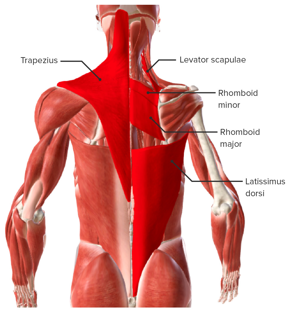
Extrinsic back muscles: superficial muscles
Image by BioDigital, edited by Lecturio
Extrinsic back muscles: trapezius
Image by BioDigital, edited by Lecturio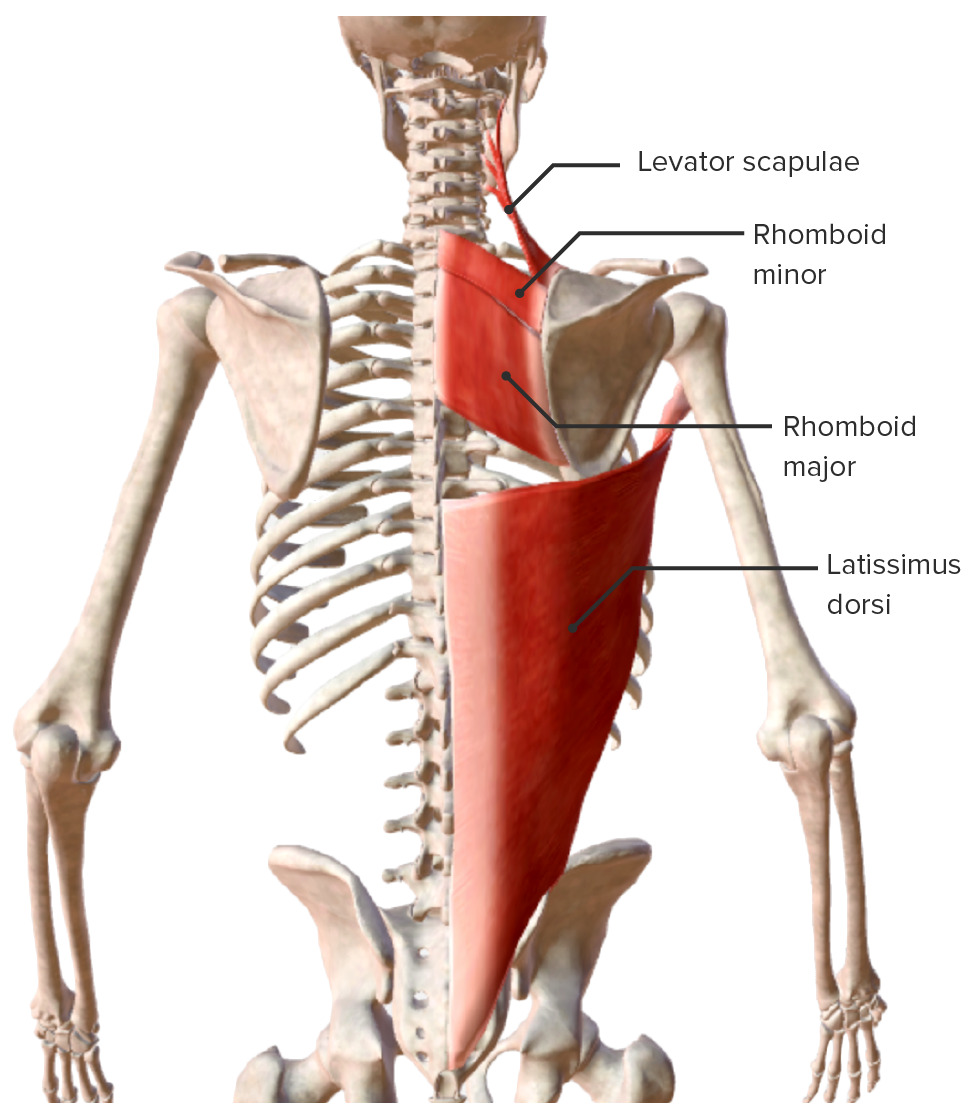
Extrinsic back muscles: superficial muscles (latissimus dorsi, rhomboid major, rhomboid minor, and levator scapulae)
Image by BioDigital, edited by LecturioIntermediate extrinsic back muscles are primarily involved with rib/thoracic cage motion.
| Muscle | Origin | Insertion | Innervation | Action |
|---|---|---|---|---|
| Serratus posterior superior | Spinous processes of C6–T2 | Costal angle of ribs Ribs A set of twelve curved bones which connect to the vertebral column posteriorly, and terminate anteriorly as costal cartilage. Together, they form a protective cage around the internal thoracic organs. Chest Wall: Anatomy 2–5 | Ventral rami of spinal nerves Spinal nerves The 31 paired peripheral nerves formed by the union of the dorsal and ventral spinal roots from each spinal cord segment. The spinal nerve plexuses and the spinal roots are also included. Spinal Cord: Anatomy T1– T4 T4 The major hormone derived from the thyroid gland. Thyroxine is synthesized via the iodination of tyrosines (monoiodotyrosine) and the coupling of iodotyrosines (diiodotyrosine) in the thyroglobulin. Thyroxine is released from thyroglobulin by proteolysis and secreted into the blood. Thyroxine is peripherally deiodinated to form triiodothyronine which exerts a broad spectrum of stimulatory effects on cell metabolism. Thyroid Hormones (intercostal nerves) |
|
| Serratus posterior inferior | Spinous processes T11–L2 | Lower edge of ribs Ribs A set of twelve curved bones which connect to the vertebral column posteriorly, and terminate anteriorly as costal cartilage. Together, they form a protective cage around the internal thoracic organs. Chest Wall: Anatomy 9–12 | Ventral rami of spinal nerves Spinal nerves The 31 paired peripheral nerves formed by the union of the dorsal and ventral spinal roots from each spinal cord segment. The spinal nerve plexuses and the spinal roots are also included. Spinal Cord: Anatomy T9–T12 (intercostal nerves) |
|
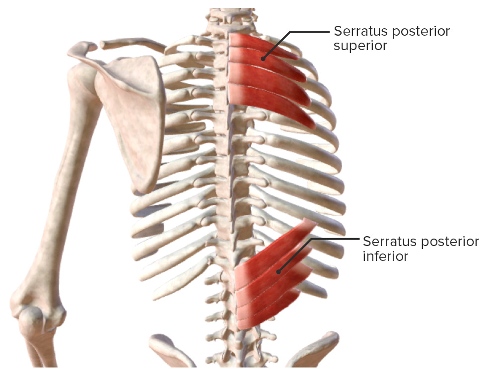
Extrinsic back muscles: intermediate muscles (serratus posterior superior and serratus posterior inferior)
Image by BioDigital, edited by Lecturio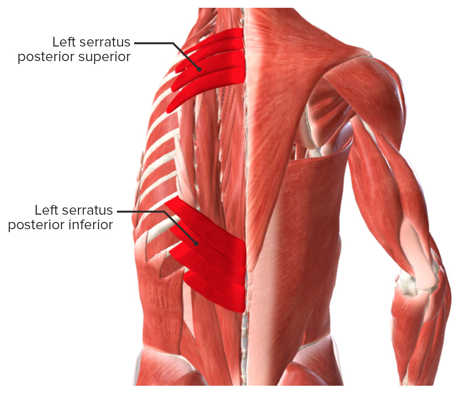
Extrinsic back muscles: intermediate muscles
Image by BioDigital, edited by LecturioThe intrinsic, or deep, back muscles are grouped into 3 layers: superficial, intermediate, and deep; they are primarily involved in moving the vertebral column Vertebral column The human spine, or vertebral column, is the most important anatomical and functional axis of the human body. It consists of 7 cervical vertebrae, 12 thoracic vertebrae, and 5 lumbar vertebrae and is limited cranially by the skull and caudally by the sacrum. Vertebral Column: Anatomy. The superficial layer is primarily responsible for the movement of the cervical spine Spine The human spine, or vertebral column, is the most important anatomical and functional axis of the human body. It consists of 7 cervical vertebrae, 12 thoracic vertebrae, and 5 lumbar vertebrae and is limited cranially by the skull and caudally by the sacrum. Vertebral Column: Anatomy.
| Muscle | Origin | Insertion | Innervation | Action |
|---|---|---|---|---|
| Splenius capitis | Spinous processes of C7– T3 T3 A T3 thyroid hormone normally synthesized and secreted by the thyroid gland in much smaller quantities than thyroxine (T4). Most T3 is derived from peripheral monodeiodination of T4 at the 5′ position of the outer ring of the iodothyronine nucleus. The hormone finally delivered and used by the tissues is mainly t3. Thyroid Hormones and the supraspinal ligament | Lateral half of superior nuchal line and the mastoid process | Dorsal rami of C1–C6 | Extensor and lateral bending of the cervical spine Spine The human spine, or vertebral column, is the most important anatomical and functional axis of the human body. It consists of 7 cervical vertebrae, 12 thoracic vertebrae, and 5 lumbar vertebrae and is limited cranially by the skull and caudally by the sacrum. Vertebral Column: Anatomy and the head |
| Splenius cervicis | Spinous processes of T3 T3 A T3 thyroid hormone normally synthesized and secreted by the thyroid gland in much smaller quantities than thyroxine (T4). Most T3 is derived from peripheral monodeiodination of T4 at the 5′ position of the outer ring of the iodothyronine nucleus. The hormone finally delivered and used by the tissues is mainly t3. Thyroid Hormones–T6 and the supraspinal ligament | Posterior tubercle of the transverse processes of C1–C3 |
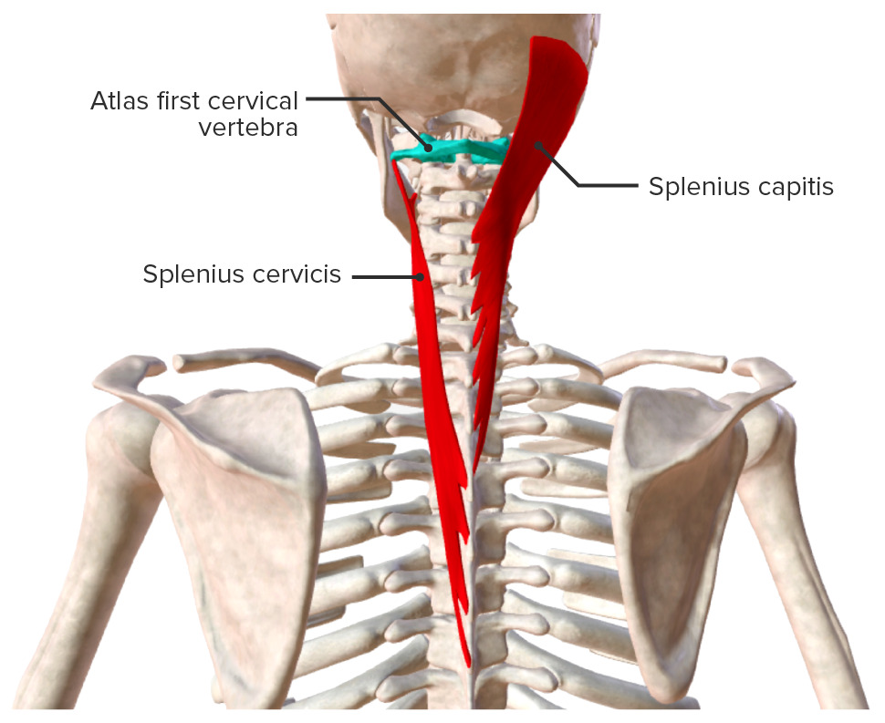
The superficial layer of the intrinsic back muscles
Image by BioDigital, edited by LecturioIntermediate layer (intrinsic muscles): The 3 muscles of the intermediate layer make up the erector spinae group.
| Muscle | Origin | Insertion | Innervation | Action |
|---|---|---|---|---|
| Spinalis | Thoracis: spinous process of upper lumbar and lower thoracic vertebrae Thoracic vertebrae A group of twelve vertebrae connected to the ribs that support the upper trunk region. Vertebral Column: Anatomy | Spinous process of upper thoracic vertebrae Thoracic vertebrae A group of twelve vertebrae connected to the ribs that support the upper trunk region. Vertebral Column: Anatomy | Dorsal rami of spinal nerves Spinal nerves The 31 paired peripheral nerves formed by the union of the dorsal and ventral spinal roots from each spinal cord segment. The spinal nerve plexuses and the spinal roots are also included. Spinal Cord: Anatomy |
|
| Cervicis: nuchal ligament and C7 spinous process | Spinous process of C2–C7 | |||
| Capitis: transverse processes of C4–T6 | Superior and inferior nuchal lines | |||
| Longissimus | Thoracic: dorsal sacrum Sacrum Five fused vertebrae forming a triangle-shaped structure at the back of the pelvis. It articulates superiorly with the lumbar vertebrae, inferiorly with the coccyx, and anteriorly with the ilium of the pelvis. The sacrum strengthens and stabilizes the pelvis. Vertebral Column: Anatomy, spinous processes of lumbar/inferior thoracic spine Spine The human spine, or vertebral column, is the most important anatomical and functional axis of the human body. It consists of 7 cervical vertebrae, 12 thoracic vertebrae, and 5 lumbar vertebrae and is limited cranially by the skull and caudally by the sacrum. Vertebral Column: Anatomy | Accessory processes of the lumbar spine Spine The human spine, or vertebral column, is the most important anatomical and functional axis of the human body. It consists of 7 cervical vertebrae, 12 thoracic vertebrae, and 5 lumbar vertebrae and is limited cranially by the skull and caudally by the sacrum. Vertebral Column: Anatomy, transverse processes of thoracic spine Spine The human spine, or vertebral column, is the most important anatomical and functional axis of the human body. It consists of 7 cervical vertebrae, 12 thoracic vertebrae, and 5 lumbar vertebrae and is limited cranially by the skull and caudally by the sacrum. Vertebral Column: Anatomy, and costal angle of ribs Ribs A set of twelve curved bones which connect to the vertebral column posteriorly, and terminate anteriorly as costal cartilage. Together, they form a protective cage around the internal thoracic organs. Chest Wall: Anatomy 2–12 | Dorsal rami T3 T3 A T3 thyroid hormone normally synthesized and secreted by the thyroid gland in much smaller quantities than thyroxine (T4). Most T3 is derived from peripheral monodeiodination of T4 at the 5′ position of the outer ring of the iodothyronine nucleus. The hormone finally delivered and used by the tissues is mainly t3. Thyroid Hormones–T5 | |
| Cervical: transverse processes of T1–T6 | Cervical: posterior tubercle of transverse processes of C2–C5 | Cervical: dorsal rami C3–T2 | ||
| Capitis: transverse processes of C3– T3 T3 A T3 thyroid hormone normally synthesized and secreted by the thyroid gland in much smaller quantities than thyroxine (T4). Most T3 is derived from peripheral monodeiodination of T4 at the 5′ position of the outer ring of the iodothyronine nucleus. The hormone finally delivered and used by the tissues is mainly t3. Thyroid Hormones | Capitis: mastoid process of the temporal bone Temporal bone Either of a pair of compound bones forming the lateral (left and right) surfaces and base of the skull which contains the organs of hearing. It is a large bone formed by the fusion of parts: the squamous (the flattened anterior-superior part), the tympanic (the curved anterior-inferior part), the mastoid (the irregular posterior portion), and the petrous (the part at the base of the skull). Jaw and Temporomandibular Joint: Anatomy | Capitis: dorsal rami C1–C3 | ||
| Iliocostalis | Lumbar: iliac crest, dorsal sacrum Sacrum Five fused vertebrae forming a triangle-shaped structure at the back of the pelvis. It articulates superiorly with the lumbar vertebrae, inferiorly with the coccyx, and anteriorly with the ilium of the pelvis. The sacrum strengthens and stabilizes the pelvis. Vertebral Column: Anatomy, and the thoracolumbar fascia Thoracolumbar fascia Posterior Abdominal Wall: Anatomy | Costal angle of ribs Ribs A set of twelve curved bones which connect to the vertebral column posteriorly, and terminate anteriorly as costal cartilage. Together, they form a protective cage around the internal thoracic organs. Chest Wall: Anatomy 7–12 | Dorsal rami T9–L1 | |
| Thoracic: costal angle of ribs Ribs A set of twelve curved bones which connect to the vertebral column posteriorly, and terminate anteriorly as costal cartilage. Together, they form a protective cage around the internal thoracic organs. Chest Wall: Anatomy 7–12 | Costal angle of ribs Ribs A set of twelve curved bones which connect to the vertebral column posteriorly, and terminate anteriorly as costal cartilage. Together, they form a protective cage around the internal thoracic organs. Chest Wall: Anatomy 1–6 | Dorsal rami T2–T9 | ||
| Cervical: costal angle of ribs Ribs A set of twelve curved bones which connect to the vertebral column posteriorly, and terminate anteriorly as costal cartilage. Together, they form a protective cage around the internal thoracic organs. Chest Wall: Anatomy 3–6 | Posterior tubercle of transverse processes of C3–C6 | Dorsal rami T1–T2 |
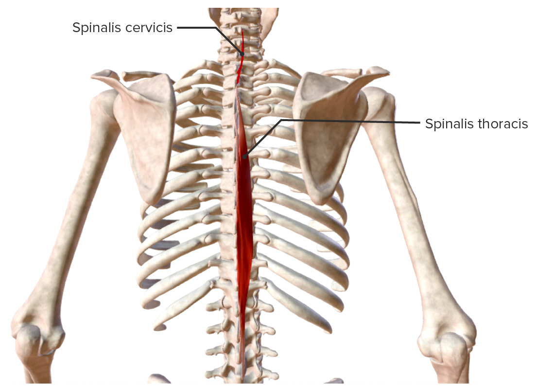
Intrinsic back muscles: spinalis muscle group
Image by BioDigital, edited by Lecturio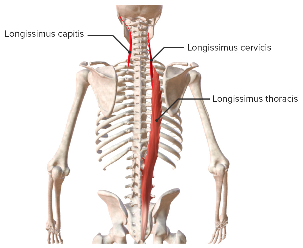
Intrinsic back muscles: longissimus muscle group
Image by BioDigital, edited by Lecturio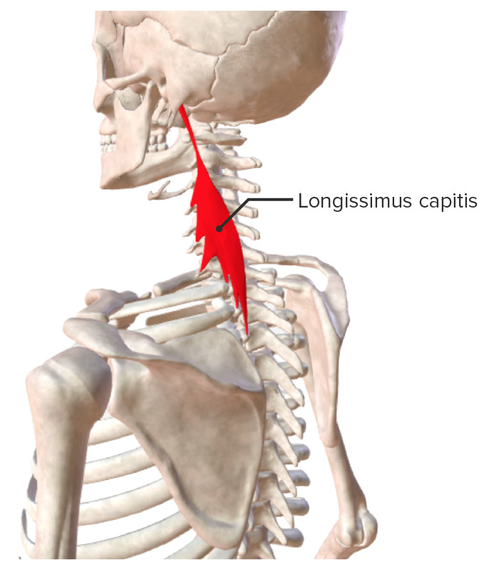
Intrinsic back muscles: longissimus capitis
Image by BioDigital, edited by Lecturio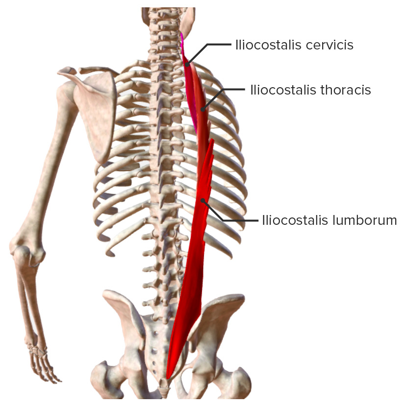
Intrinsic back muscles: iliocostalis muscle group
Image by BioDigital, edited by LecturioThe deepest layer of the deep back muscles is attached to the transverse and spinous processes of the vertebral column Vertebral column The human spine, or vertebral column, is the most important anatomical and functional axis of the human body. It consists of 7 cervical vertebrae, 12 thoracic vertebrae, and 5 lumbar vertebrae and is limited cranially by the skull and caudally by the sacrum. Vertebral Column: Anatomy and facilitates movement of the spine Spine The human spine, or vertebral column, is the most important anatomical and functional axis of the human body. It consists of 7 cervical vertebrae, 12 thoracic vertebrae, and 5 lumbar vertebrae and is limited cranially by the skull and caudally by the sacrum. Vertebral Column: Anatomy.
| Muscle | Origin | Insertion | Innervation | Action |
|---|---|---|---|---|
| Semispinalis | Capitis: articular processes of C4–C6 and the transverse processes of C7–T6 | Between the superior and inferior nuchal lines | Dorsal rami of the spinal nerves Spinal nerves The 31 paired peripheral nerves formed by the union of the dorsal and ventral spinal roots from each spinal cord segment. The spinal nerve plexuses and the spinal roots are also included. Spinal Cord: Anatomy | Extension Extension Examination of the Upper Limbs and contralateral rotation Rotation Motion of an object in which either one or more points on a line are fixed. It is also the motion of a particle about a fixed point. X-rays of the head and vertebral column Vertebral column The human spine, or vertebral column, is the most important anatomical and functional axis of the human body. It consists of 7 cervical vertebrae, 12 thoracic vertebrae, and 5 lumbar vertebrae and is limited cranially by the skull and caudally by the sacrum. Vertebral Column: Anatomy |
| Cervicis: posterior surfaces of the transverse processes of T1–T6 | Spines of C2–C5 | |||
| Thoracis: transverse processes of T6–T10 | Spines of C6– T4 T4 The major hormone derived from the thyroid gland. Thyroxine is synthesized via the iodination of tyrosines (monoiodotyrosine) and the coupling of iodotyrosines (diiodotyrosine) in the thyroglobulin. Thyroxine is released from thyroglobulin by proteolysis and secreted into the blood. Thyroxine is peripherally deiodinated to form triiodothyronine which exerts a broad spectrum of stimulatory effects on cell metabolism. Thyroid Hormones | |||
| Multifidus | Dorsal sacrum Sacrum Five fused vertebrae forming a triangle-shaped structure at the back of the pelvis. It articulates superiorly with the lumbar vertebrae, inferiorly with the coccyx, and anteriorly with the ilium of the pelvis. The sacrum strengthens and stabilizes the pelvis. Vertebral Column: Anatomy, iliac crest, posterior sacroiliac ligaments, mammillary processes, transverse processes of T1–T12, and the articular processes C4–C7 | Spinous processes of C2–L5 | Dorsal rami C3– S3 S3 Heart Sounds | Extend the complete vertebral column Vertebral column The human spine, or vertebral column, is the most important anatomical and functional axis of the human body. It consists of 7 cervical vertebrae, 12 thoracic vertebrae, and 5 lumbar vertebrae and is limited cranially by the skull and caudally by the sacrum. Vertebral Column: Anatomy |
| Rotatores | Transverse processes of the cervical, thoracic, and lumbar spine Spine The human spine, or vertebral column, is the most important anatomical and functional axis of the human body. It consists of 7 cervical vertebrae, 12 thoracic vertebrae, and 5 lumbar vertebrae and is limited cranially by the skull and caudally by the sacrum. Vertebral Column: Anatomy | Spinous processes of adjoining superior vertebra | Dorsal rami of the spinal nerves Spinal nerves The 31 paired peripheral nerves formed by the union of the dorsal and ventral spinal roots from each spinal cord segment. The spinal nerve plexuses and the spinal roots are also included. Spinal Cord: Anatomy | Stabilize the vertebral column Vertebral column The human spine, or vertebral column, is the most important anatomical and functional axis of the human body. It consists of 7 cervical vertebrae, 12 thoracic vertebrae, and 5 lumbar vertebrae and is limited cranially by the skull and caudally by the sacrum. Vertebral Column: Anatomy |
| Interspinales | Spinous processes of C3–T1, T2–L1, and L2– S1 S1 Heart Sounds | Spinous processes of C2–C7, T1–T12, and L1–L5 | Extend the complete vertebral column Vertebral column The human spine, or vertebral column, is the most important anatomical and functional axis of the human body. It consists of 7 cervical vertebrae, 12 thoracic vertebrae, and 5 lumbar vertebrae and is limited cranially by the skull and caudally by the sacrum. Vertebral Column: Anatomy | |
| Intertransversarii | Medial lumbar: mammillary and accessory processes of all lumbar vertebrae Lumbar vertebrae Vertebrae in the region of the lower back below the thoracic vertebrae and above the sacral vertebrae. Vertebral Column: Anatomy | Adjoining osseous structures of the lumbar spine Spine The human spine, or vertebral column, is the most important anatomical and functional axis of the human body. It consists of 7 cervical vertebrae, 12 thoracic vertebrae, and 5 lumbar vertebrae and is limited cranially by the skull and caudally by the sacrum. Vertebral Column: Anatomy | Lateral flexion Flexion Examination of the Upper Limbs of the spine Spine The human spine, or vertebral column, is the most important anatomical and functional axis of the human body. It consists of 7 cervical vertebrae, 12 thoracic vertebrae, and 5 lumbar vertebrae and is limited cranially by the skull and caudally by the sacrum. Vertebral Column: Anatomy | |
| Thoracic: transverse processes of T10–T12 | Transverse processes of T11–12 and at the accessory processes of L1 | |||
| Posterior cervical: C1–C7, posterior tubercle of the transverse processes | Posterior tubercles of the respective adjoining vertebra | |||
| Levatores costarum | Transverse processes of C7–T11 |
|
Extension Extension Examination of the Upper Limbs of the thoracic spine Spine The human spine, or vertebral column, is the most important anatomical and functional axis of the human body. It consists of 7 cervical vertebrae, 12 thoracic vertebrae, and 5 lumbar vertebrae and is limited cranially by the skull and caudally by the sacrum. Vertebral Column: Anatomy |
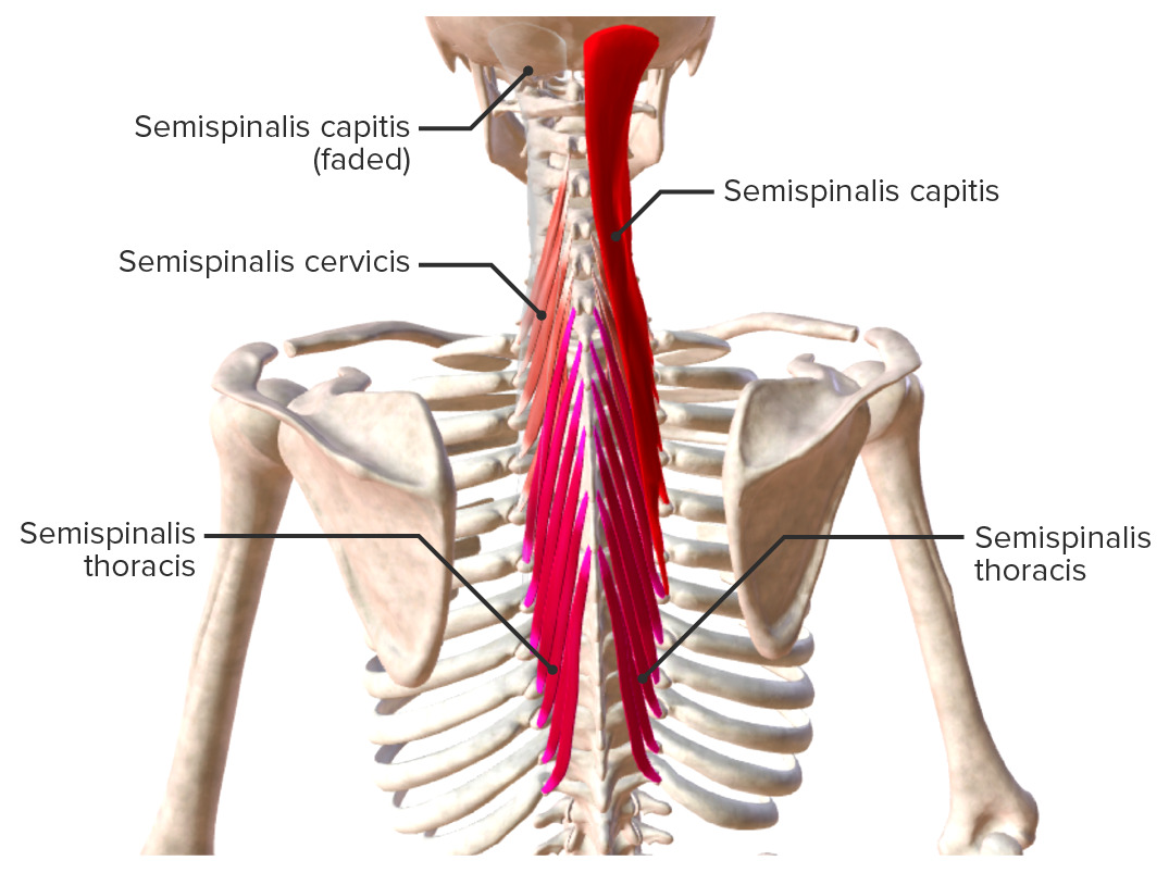
Intrinsic back muscles: semispinalis muscle group
Image by BioDigital, edited by Lecturio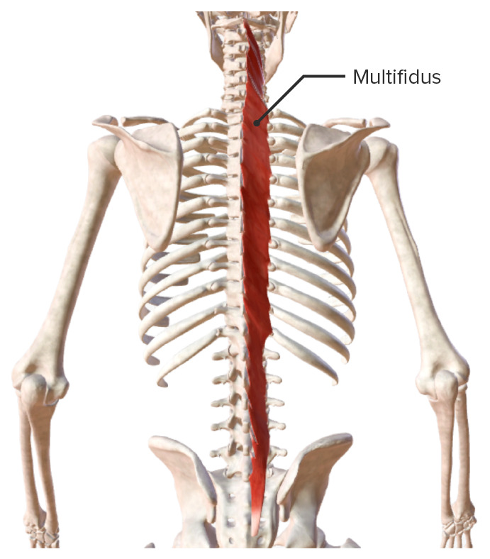
Intrinsic back muscles: multifidus muscle
Image by BioDigital, edited by Lecturio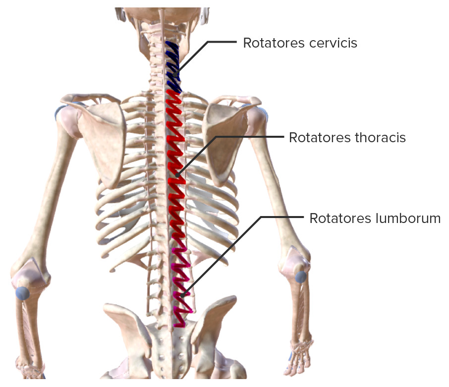
Intrinsic back muscles: rotatores muscle group
Image by BioDigital, edited by Lecturio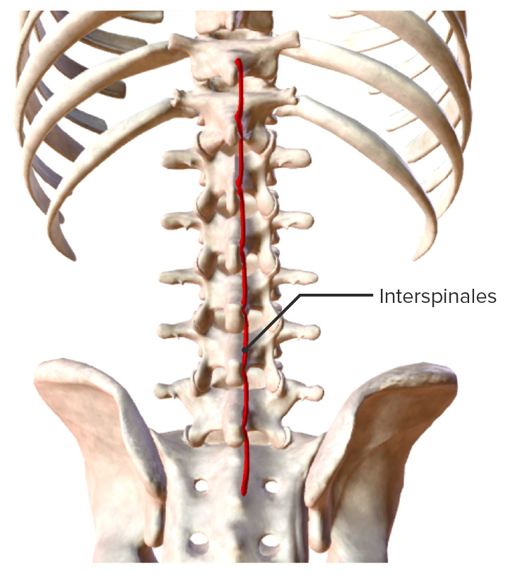
Intrinsic back muscles: interspinales muscle group
Image by BioDigital, edited by Lecturio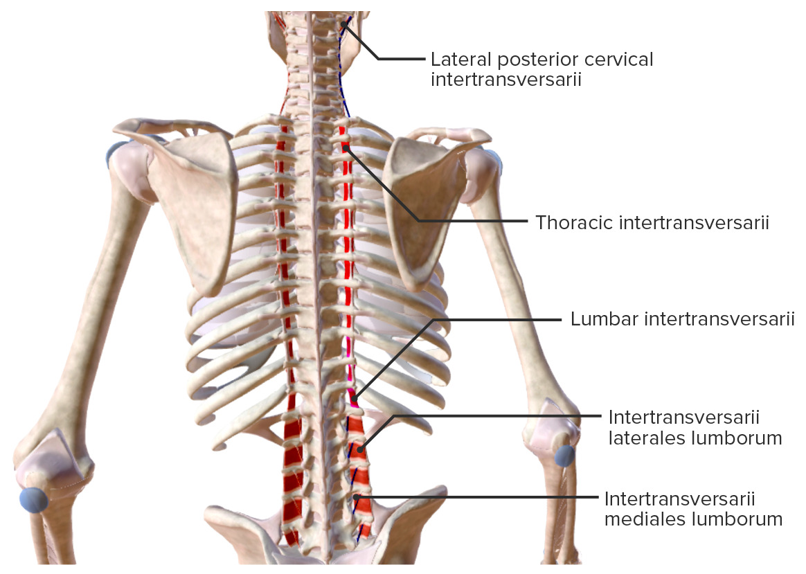
Intrinsic back muscles: intertransversarii muscle group
Image by BioDigital, edited by Lecturio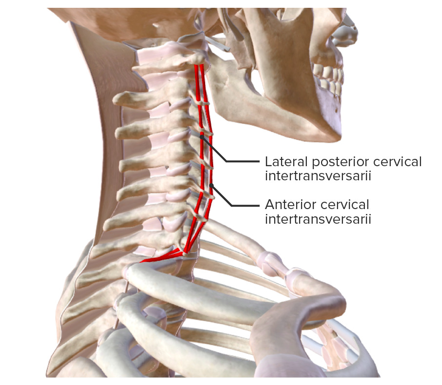
Intrinsic back muscles: cervical intertransversarii muscles
Image by BioDigital, edited by Lecturio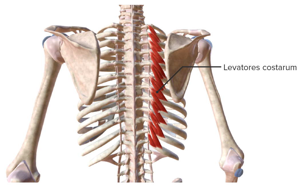
Intrinsic back muscles: levatores costarum muscle group
Image by BioDigital, edited by Lecturio