Advertisement

Advertisement

Advertisement

Advertisement

Molluscum Contagiosum (Clinical)

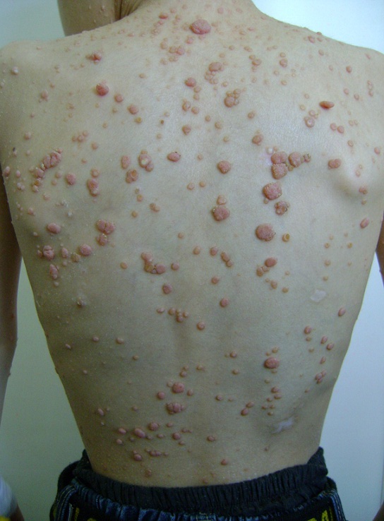
Molluscum contagiosum is a viral infection limited to the epidermis, typically without systemic manifestations. This infection is common in children below 5 years of age, though it can also be seen in healthy adolescents and adults, typically related to either contact sports or, with lesions on the genitals, as a sexually transmitted infection (STI). Lesions appear as grouped, flesh-colored, dome-shaped papules with central umbilication. Molluscum contagiosum is mild in immunocompetent patients and self resolves within months. Immunocompromised individuals present with extensive lesions which require treatment. Molluscum contagiosum is highly transmissible; therefore, patient education is key in its management. Cryotherapy with liquid nitrogen is the 1st-line treatment.

Last updated: Oct 31, 2022

Editorial responsibility: Stanley Oiseth, Lindsay Jones, Evelin Maza

Advertisement

Advertisement

Advertisement

Advertisement

Advertisement

Advertisement

Overview

Epidemiology[1,2,4,5]

  • 1% of all diagnosed skin Skin The skin, also referred to as the integumentary system, is the largest organ of the body. The skin is primarily composed of the epidermis (outer layer) and dermis (deep layer). The epidermis is primarily composed of keratinocytes that undergo rapid turnover, while the dermis contains dense layers of connective tissue. Skin: Structure and Functions disorders
  • Usually presents in children < 5 years of age or adolescents
  • Can be seen in adults as an STI STI Sexually transmitted infections (STIs) are infections that spread either by vaginal intercourse, anal sex, or oral sex. Symptoms and signs may include vaginal discharge, penile discharge, dysuria, skin lesions (e.g., warts, ulcers) on or around the genitals, and pelvic pain. Some infections can lead to infertility and chronic debilitating disease. Sexually Transmitted Infections (STIs)
  • Increased incidence Incidence The number of new cases of a given disease during a given period in a specified population. It also is used for the rate at which new events occur in a defined population. It is differentiated from prevalence, which refers to all cases in the population at a given time. Measures of Disease Frequency in hot, humid climates
  • Slight predominance in boys
  • More common in patients Patients Individuals participating in the health care system for the purpose of receiving therapeutic, diagnostic, or preventive procedures. Clinician–Patient Relationship with:[1,2]

Etiology and transmission[1‒5]

  • Molluscum contagiosum virus Molluscum contagiosum virus A species of molluscipoxvirus causing skin lesions in humans. It is transmitted by direct contact or from non-living reservoirs (fomites), such as books or clothing. Molluscum Contagiosum (MCV) is a double-stranded, linear, DNA DNA A deoxyribonucleotide polymer that is the primary genetic material of all cells. Eukaryotic and prokaryotic organisms normally contain DNA in a double-stranded state, yet several important biological processes transiently involve single-stranded regions. DNA, which consists of a polysugar-phosphate backbone possessing projections of purines (adenine and guanine) and pyrimidines (thymine and cytosine), forms a double helix that is held together by hydrogen bonds between these purines and pyrimidines (adenine to thymine and guanine to cytosine). DNA Types and Structure poxvirus.
  • Transmission:
    • Exposure to infected individuals (skin-to-skin contact):
      • Contact sports
      • Sexually transmitted
      • Peripartum
    • Fomites Fomites Inanimate objects that carry pathogenic microorganisms and thus can serve as the source of infection. Microorganisms typically survive on fomites for minutes or hours. Common fomites include clothing, tissue paper, hairbrushes, and cooking and eating utensils. Adenovirus (e.g., towels, toys, razors)
    • Autoinoculation Autoinoculation Molluscum Contagiosum (scraping)
  • Once the lesions are gone, the virus Virus Viruses are infectious, obligate intracellular parasites composed of a nucleic acid core surrounded by a protein capsid. Viruses can be either naked (non-enveloped) or enveloped. The classification of viruses is complex and based on many factors, including type and structure of the nucleoid and capsid, the presence of an envelope, the replication cycle, and the host range. Virology is gone → no recurrent outbreaks Outbreaks Sudden increase in the incidence of a disease. The concept includes epidemics and pandemics. Influenza Viruses/Influenza or asymptomatic viral shedding Viral shedding The expelling of virus particles from the body. Important routes include the respiratory tract, genital tract, and intestinal tract. Hepatitis A Virus (as with herpesviruses).[1]

Pathophysiology[2,4,5]

  • Incubation Incubation The amount time between exposure to an infectious agent and becoming symptomatic. Rabies Virus period from 2 weeks to 6 months (typically 2‒7 weeks)[2]
  • Virus Virus Viruses are infectious, obligate intracellular parasites composed of a nucleic acid core surrounded by a protein capsid. Viruses can be either naked (non-enveloped) or enveloped. The classification of viruses is complex and based on many factors, including type and structure of the nucleoid and capsid, the presence of an envelope, the replication cycle, and the host range. Virology infects only keratinocytes Keratinocytes Epidermal cells which synthesize keratin and undergo characteristic changes as they move upward from the basal layers of the epidermis to the cornified (horny) layer of the skin. Successive stages of differentiation of the keratinocytes forming the epidermal layers are basal cell, spinous or prickle cell, and the granular cell. Skin: Structure and Functions → localized to epidermis Epidermis The external, nonvascular layer of the skin. It is made up, from within outward, of five layers of epithelium: (1) basal layer (stratum basale epidermidis); (2) spinous layer (stratum spinosum epidermidis); (3) granular layer (stratum granulosum epidermidis); (4) clear layer (stratum lucidum epidermidis); and (5) horny layer (stratum corneum epidermidis). Skin: Structure and Functions
  • Inhibits innate immunity Innate immunity The capacity of a normal organism to remain unaffected by microorganisms and their toxins. It results from the presence of naturally occurring anti-infective agents, constitutional factors such as body temperature and immediate acting immune cells such as natural killer cells. Innate Immunity: Phagocytes and Antigen Presentation via antiviral Antiviral Antivirals for Hepatitis B protein production → persistent infection

Histopathology[3-5]

  • Hyperkeratosis Hyperkeratosis Ichthyosis Vulgaris (thickened epidermis Epidermis The external, nonvascular layer of the skin. It is made up, from within outward, of five layers of epithelium: (1) basal layer (stratum basale epidermidis); (2) spinous layer (stratum spinosum epidermidis); (3) granular layer (stratum granulosum epidermidis); (4) clear layer (stratum lucidum epidermidis); and (5) horny layer (stratum corneum epidermidis). Skin: Structure and Functions)
  • Epidermal proliferation into the dermis Dermis A layer of vascularized connective tissue underneath the epidermis. The surface of the dermis contains innervated papillae. Embedded in or beneath the dermis are sweat glands; hair follicles; and sebaceous glands. Skin: Structure and Functions
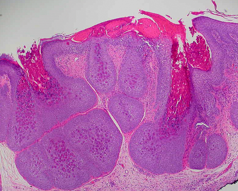
  • Henderson-Paterson bodies: inclusions visible in the keratinocytes Keratinocytes Epidermal cells which synthesize keratin and undergo characteristic changes as they move upward from the basal layers of the epidermis to the cornified (horny) layer of the skin. Successive stages of differentiation of the keratinocytes forming the epidermal layers are basal cell, spinous or prickle cell, and the granular cell. Skin: Structure and Functions of the basal, spinous, and granular layers of the epidermis Epidermis The external, nonvascular layer of the skin. It is made up, from within outward, of five layers of epithelium: (1) basal layer (stratum basale epidermidis); (2) spinous layer (stratum spinosum epidermidis); (3) granular layer (stratum granulosum epidermidis); (4) clear layer (stratum lucidum epidermidis); and (5) horny layer (stratum corneum epidermidis). Skin: Structure and Functions
Henderson-paterson bodies

Henderson-Paterson bodies:
Characteristic histological features of molluscum contagiosum. Henderson-Paterson bodies are inclusions that are visible in the keratinocytes of the basal, spinous, and granular layers of the epidermis.
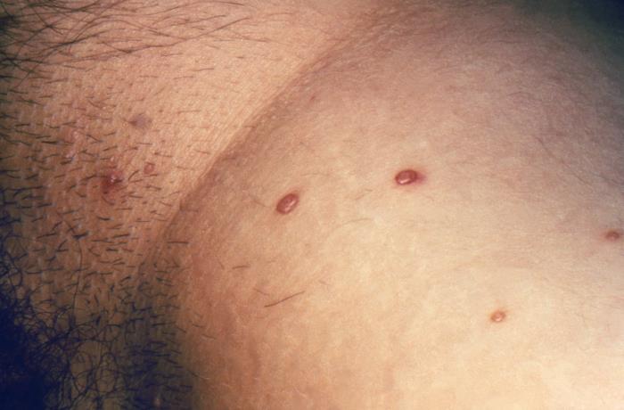
Image: “Molluscum Contagiosum 1” by Ed Uthman. License: CC BY 2.0

Clinical Presentation and Diagnosis

Clinical presentation of lesions[1-5]

  • Usually no systemic symptoms
  • Dermatological features:
    • Shape and color:[3,9]
      • Dome shaped
      • Pearly papules
      • Flesh colored
      • 2–6 mm MM Multiple myeloma (MM) is a malignant condition of plasma cells (activated B lymphocytes) primarily seen in the elderly. Monoclonal proliferation of plasma cells results in cytokine-driven osteoclastic activity and excessive secretion of IgG antibodies. Multiple Myeloma in diameter
      • Central umbilication (a key feature)
    • Can express a white, cheesy substance[1,3-5]
    • Usually multiple (most commonly between 1 and 20‒30 lesions) and occur in clusters[2,3]
    • Rarely associated with pruritus Pruritus An intense itching sensation that produces the urge to rub or scratch the skin to obtain relief. Atopic Dermatitis (Eczema) or tenderness
    • Distribution:[1-5]
      • Children: 
        • Face
        • Trunk
        • Axilla Axilla The axilla is a pyramid-shaped space located between the upper thorax and the arm. The axilla has a base, an apex, and 4 walls (anterior, medial, lateral, posterior). The base of the pyramid is made up of the axillary skin. The apex is the axillary inlet, located between the 1st rib, superior border of the scapula, and clavicle. Axilla and Brachial Plexus: Anatomy
        • Antecubital fossa Antecubital Fossa Molluscum Contagiosum
        • Popliteal fossa Popliteal fossa The popliteal fossa or the “knee pit” is a diamond-shaped, fat-filled, shallow depression on the posterior aspect of the knee joint. The popliteal fossa is located at the dorsal aspect of the knee and contains an increased number of lymph nodes as well as structures of the neurovascular system that travel from the thigh to the lower leg. Popliteal Fossa: Anatomy
      • Adults: 
        • Anogenital area
        • Lower abdomen
        • Proximal inner thighs
      • Palms and soles not involved
  • Immunocompromised immunocompromised A human or animal whose immunologic mechanism is deficient because of an immunodeficiency disorder or other disease or as the result of the administration of immunosuppressive drugs or radiation. Gastroenteritis individuals may have widespread/diffuse eruptions and/or extensive confluent or giant lesions.[3]
  • Atypical presentations can include:[3]
    • Cystic Cystic Fibrocystic Change lesions
    • Ulcers
    • Cellulitis-like lesions
    • Condyloma Condyloma Sexually transmitted form of anogenital warty growth caused by the human papillomaviruses. Male Genitourinary Examination acuminatum–like lesions
    • Conjunctivitis Conjunctivitis Conjunctivitis is a common inflammation of the bulbar and/or palpebral conjunctiva. It can be classified into infectious (mostly viral) and noninfectious conjunctivitis, which includes allergic causes. Patients commonly present with red eyes, increased tearing, burning, foreign body sensation, and photophobia. Conjunctivitis (if lesions are located on or near eyelids Eyelids Each of the upper and lower folds of skin which cover the eye when closed. Blepharitis)
  • Molluscum dermatitis Dermatitis Any inflammation of the skin. Atopic Dermatitis (Eczema):[2,3]
    • An eczematous reaction ( erythema Erythema Redness of the skin produced by congestion of the capillaries. This condition may result from a variety of disease processes. Chalazion, scales Scales Dry or greasy masses of keratin that represent thickened stratum corneum. Secondary Skin Lesions) encircling the lesion
    • Occurs in about 10% of patients Patients Individuals participating in the health care system for the purpose of receiving therapeutic, diagnostic, or preventive procedures. Clinician–Patient Relationship

Diagnosis

  • Clinical diagnosis is sufficient.[9]
  • Dermoscopy Dermoscopy A noninvasive technique that enables direct microscopic examination of the surface and architecture of the skin. Seborrheic Keratosis may aid in diagnosis, and may show:[3,4,9]
    • Central white‒yellow polylobular amorphous area
    • Peripheral linear or branched vessels in a crown pattern
  • Direct testing from lesion:[2]
    • Nucleic acid testing by PCR PCR Polymerase chain reaction (PCR) is a technique that amplifies DNA fragments exponentially for analysis. The process is highly specific, allowing for the targeting of specific genomic sequences, even with minuscule sample amounts. The PCR cycles multiple times through 3 phases: denaturation of the template DNA, annealing of a specific primer to the individual DNA strands, and synthesis/elongation of new DNA molecules. Polymerase Chain Reaction (PCR) may be available at reference centers.
    • Viral culture Viral culture West Nile Virus is not recommended, since virus Virus Viruses are infectious, obligate intracellular parasites composed of a nucleic acid core surrounded by a protein capsid. Viruses can be either naked (non-enveloped) or enveloped. The classification of viruses is complex and based on many factors, including type and structure of the nucleoid and capsid, the presence of an envelope, the replication cycle, and the host range. Virology does not grow readily in culture.
  • Histopathological analysis to confirm diagnosis[9]
  • Serologic testing is not typically available.[2]
  • Lesions in inguinal region Inguinal region Anterior Abdominal Wall: Anatomy → STD panel to rule out accompanying STIs STIs Sexually transmitted infections (STIs) or sexually transmitted diseases (STDs) are infections that spread either by vaginal intercourse, anal sex, or oral sex. Symptoms and signs may include vaginal discharge, penile discharge, dysuria, skin lesions (e.g., warts, ulcers) on or around the genitals, and pelvic pain. Some infections can lead to infertility and chronic debilitating disease. Sexually Transmitted Infections (STIs) (including HIV HIV Anti-HIV Drugs)

Management

General management principles

  • No treatment is necessary for healthy individuals as it is a self-limiting Self-Limiting Meningitis in Children disease.[1]
  • Patient education:[1,3,5]
    • Practice good hygiene, including regular Regular Insulin handwashing
    • Complete resolution can take up to 1 year.
    • Average duration for a single lesion is 2 months.
    • Avoid sharing towels and sheets.
    • Do not shave or wax across lesions.
    • Do not scratch or pick at lesions (increases risk of spread).
    • Cover lesions with clothing during regular Regular Insulin activity.
    • Cover lesions with a tight bandage when:
      • Swimming 
      • Participating in sports or other activities where direct contact with lesions is possible.
      • Note: Lesions should be uncovered when risk of direct contact with lesions is low → prevents skin Skin The skin, also referred to as the integumentary system, is the largest organ of the body. The skin is primarily composed of the epidermis (outer layer) and dermis (deep layer). The epidermis is primarily composed of keratinocytes that undergo rapid turnover, while the dermis contains dense layers of connective tissue. Skin: Structure and Functions breakdown under dressings
    • Practice safe sex Sex The totality of characteristics of reproductive structure, functions, phenotype, and genotype, differentiating the male from the female organism. Gender Dysphoria.
    • Children do not need to be removed from school or daycare.
  • Associated eczema Eczema Atopic dermatitis, also known as eczema, is a chronic, relapsing, pruritic, inflammatory skin disease that occurs more frequently in children, although adults can also be affected. The condition is often associated with elevated serum levels of IgE and a personal or family history of atopy. Skin dryness, erythema, oozing, crusting, and lichenification are present. Atopic Dermatitis (Eczema): treat to reduce risk of autoinoculation Autoinoculation Molluscum Contagiosum from scratching[3]
    • Emollients Emollients Oleaginous substances used topically to soothe, soften or protect skin or mucous membranes. They are used also as vehicles for other dermatologic agents. Pityriasis Rosea 
    • Potentially a topical corticosteroid
  • Indications for treatment:[2,3,5]
    • Patient preference
    • Adults/adolescents with STD (to prevent transmission)
    • Immunocompromised immunocompromised A human or animal whose immunologic mechanism is deficient because of an immunodeficiency disorder or other disease or as the result of the administration of immunosuppressive drugs or radiation. Gastroenteritis patients Patients Individuals participating in the health care system for the purpose of receiving therapeutic, diagnostic, or preventive procedures. Clinician–Patient Relationship (to prevent more severe disease)
    • To treat pruritus Pruritus An intense itching sensation that produces the urge to rub or scratch the skin to obtain relief. Atopic Dermatitis (Eczema) (if present), since scratching can increase risk of transmission
    • To prevent scarring Scarring Inflammation from lesions that are secondarily infected or otherwise inflamed
    • Desired for cosmetic reasons/reduction of psychological stress Psychological stress Stress wherein emotional factors predominate. Acute Stress Disorder
  • Note: Patients Patients Individuals participating in the health care system for the purpose of receiving therapeutic, diagnostic, or preventive procedures. Clinician–Patient Relationship with periocular lesions requiring treatment should be referred to an ophthalmologist for treatment.

Treatment

There is no consensus on optimal treatment.

Typical 1st-line therapies recommended by experts include:[1-5]

  • Cryotherapy Cryotherapy A form of therapy consisting in the local or general use of cold. The selective destruction of tissue by extreme cold or freezing is cryosurgery. Chondrosarcoma ( liquid nitrogen Liquid Nitrogen Molluscum Contagiosum):[3,5]
    • Freezes lesions, physically destroying them
    • Dip a cotton swab in liquid nitrogen Liquid Nitrogen Molluscum Contagiosum and apply to each individual lesion for 6‒10 seconds.
    • Painful; typically tolerated by adults and adolescents, but not by younger children
    • Potential adverse effects:
      • Scarring Scarring Inflammation
      • Hypopigmentation Hypopigmentation A condition caused by a deficiency or a loss of melanin pigmentation in the epidermis, also known as hypomelanosis. Hypopigmentation can be localized or generalized, and may result from genetic defects, trauma, inflammation, or infections. Malassezia Fungi (may be temporary or permanent)
  • Curettage Curettage A scraping, usually of the interior of a cavity or tract, for removal of new growth or other abnormal tissue, or to obtain material for tissue diagnosis. It is performed with a curet (curette), a spoon-shaped instrument designed for that purpose. Benign Bone Tumors:[3,5]
    • Physical removal of lesions using a curette
    • Use topical anesthetics Anesthetics Agents that are capable of inducing a total or partial loss of sensation, especially tactile sensation and pain. They may act to induce general anesthesia, in which an unconscious state is achieved, or may act locally to induce numbness or lack of sensation at a targeted site. Anesthesiology: History and Basic Concepts to help reduce pain Pain An unpleasant sensation induced by noxious stimuli which are detected by nerve endings of nociceptive neurons. Pain: Types and Pathways.
    • Advantage: may provide more definitive treatment
    • Disadvantages: 
      • Causes pain Pain An unpleasant sensation induced by noxious stimuli which are detected by nerve endings of nociceptive neurons. Pain: Types and Pathways and minor bleeding
      • Potential for small depressed scars
      • More time-consuming
  • Cautery/ electrodesiccation Electrodesiccation Seborrheic Keratosis:[2,3]
    • Burns Burns A burn is a type of injury to the skin and deeper tissues caused by exposure to heat, electricity, chemicals, friction, or radiation. Burns are classified according to their depth as superficial (1st-degree), partial-thickness (2nd-degree), full-thickness (3rd-degree), and 4th-degree burns. Burns lesions off
    • Lightly cauterize each lesion on its raised part.
    • Apply an anesthetic cream prior to cautery to reduce pain Pain An unpleasant sensation induced by noxious stimuli which are detected by nerve endings of nociceptive neurons. Pain: Types and Pathways.
  • Cantharidin Cantharidin A toxic compound, isolated from the spanish fly or blistering beetle (lytta (cantharis) vesicatoria) and other insects. It is a potent and specific inhibitor of protein phosphatases 1 (pp1) and 2a (pp2a). This compound can produce severe skin inflammation, and is extremely toxic if ingested orally. Molluscum Contagiosum:[5]
    • Topical blistering agent to be applied only by a trained clinician Clinician A physician, nurse practitioner, physician assistant, or another health professional who is directly involved in patient care and has a professional relationship with patients. Clinician–Patient Relationship
    • Avoid treatment on the face, genitals, or perianal areas.
    • Applied directly to lesions using the blunt wooden end of a cotton swab:
      • Cover site to avoid spread of vesicant Vesicant Osmotic Diuretics.
      • Wash off cantharidin Cantharidin A toxic compound, isolated from the spanish fly or blistering beetle (lytta (cantharis) vesicatoria) and other insects. It is a potent and specific inhibitor of protein phosphatases 1 (pp1) and 2a (pp2a). This compound can produce severe skin inflammation, and is extremely toxic if ingested orally. Molluscum Contagiosum 2‒6 hours after application (or at 1st signs of blistering).
    • Repeat treatment every 2‒4 weeks until lesions are resolved.
    • Potential adverse effects:
      • Burning/ pain Pain An unpleasant sensation induced by noxious stimuli which are detected by nerve endings of nociceptive neurons. Pain: Types and Pathways
      • Erythema Erythema Redness of the skin produced by congestion of the capillaries. This condition may result from a variety of disease processes. Chalazion
      • Pruritus Pruritus An intense itching sensation that produces the urge to rub or scratch the skin to obtain relief. Atopic Dermatitis (Eczema)
      • Scarring Scarring Inflammation is possible but uncommon.
  • Podophyllotoxin Podophyllotoxin A lignan (lignans) found in podophyllin resin from the roots of podophyllum plants. It is a potent spindle poison, toxic if taken internally, and has been used as a cathartic. It is very irritating to skin and mucous membranes, has keratolytic actions, has been used to treat warts and keratoses, and may have antineoplastic properties, as do some of its congeners and derivatives. Molluscum Contagiosum:[3]
    • An antimitotic drug causing lesion necrosis Necrosis The death of cells in an organ or tissue due to disease, injury or failure of the blood supply. Ischemic Cell Damage, also commonly used to treat genital warts Warts Benign epidermal proliferations or tumors; some are viral in origin. Female Genitourinary Examination
    • Safety and efficacy in young children has not been established.
    • Preparations:
      • 0.5% solution (apply with a cotton swab)
      • 0.5% gel (apply with fingers)
      • 0.15% cream (apply with fingers)
    • Regimen:
      • Apply to lesions twice daily for 3 days, then rest for 4 days. 
      • Repeat for 4‒5 weeks.
    • Notes:
      • Total treatment area should not exceed 10 cm2.
      • Avoid in pregnancy Pregnancy The status during which female mammals carry their developing young (embryos or fetuses) in utero before birth, beginning from fertilization to birth. Pregnancy: Diagnosis, Physiology, and Care.
    • Potential adverse effects:

Other potential treatment options include:[3,5]

  • Potassium Potassium An element in the alkali group of metals with an atomic symbol k, atomic number 19, and atomic weight 39. 10. It is the chief cation in the intracellular fluid of muscle and other cells. Potassium ion is a strong electrolyte that plays a significant role in the regulation of fluid volume and maintenance of the water-electrolyte balance. Hyperkalemia hydroxide: 5%‒15% solutions applied to lesions once daily
  • Salicylic acid
  • Benzoyl peroxide Benzoyl peroxide A peroxide derivative that has been used topically for burns and as a dermatologic agent in the treatment of acne and poison ivy dermatitis. It is used also as a bleach in the food industry. Molluscum Contagiosum
  • Hydrogen peroxide Hydrogen peroxide A strong oxidizing agent used in aqueous solution as a ripening agent, bleach, and topical anti-infective. It is relatively unstable and solutions deteriorate over time unless stabilized by the addition of acetanilide or similar organic materials. Myeloperoxidase Deficiency
  • Silver nitrate 
  • Iodine Iodine A nonmetallic element of the halogen group that is represented by the atomic symbol I, atomic number 53, and atomic weight of 126. 90. It is a nutritionally essential element, especially important in thyroid hormone synthesis. In solution, it has anti-infective properties and is used topically. Thyroid Hormones tinctures
  • 25%‒50% trichloroacetic acid (TCA): applied by trained providers in clinic
  • Topical retinoids Retinoids Retinol and derivatives of retinol that play an essential role in metabolic functioning of the retina, the growth of and differentiation of epithelial tissue, the growth of bone, reproduction, and the immune response. Dietary vitamin A is derived from a variety of carotenoids found in plants. It is enriched in the liver, egg yolks, and the fat component of dairy products. Fat-soluble Vitamins and their Deficiencies:
    • Options:
      • Tretinoin
      • Adapalene
    • Apply every other day to twice daily, as tolerated.
    • Causes irritation and abnormally dry skin Skin The skin, also referred to as the integumentary system, is the largest organ of the body. The skin is primarily composed of the epidermis (outer layer) and dermis (deep layer). The epidermis is primarily composed of keratinocytes that undergo rapid turnover, while the dermis contains dense layers of connective tissue. Skin: Structure and Functions ( xerosis Xerosis Sjögren’s Syndrome)
    • Avoid in pregnancy Pregnancy The status during which female mammals carry their developing young (embryos or fetuses) in utero before birth, beginning from fertilization to birth. Pregnancy: Diagnosis, Physiology, and Care
  • Oral cimetidine Cimetidine A histamine congener, it competitively inhibits histamine binding to histamine h2 receptors. Cimetidine has a range of pharmacological actions. It inhibits gastric acid secretion, as well as pepsin and gastrin output. Antihistamines

Immunocompromised immunocompromised A human or animal whose immunologic mechanism is deficient because of an immunodeficiency disorder or other disease or as the result of the administration of immunosuppressive drugs or radiation. Gastroenteritis patients Patients Individuals participating in the health care system for the purpose of receiving therapeutic, diagnostic, or preventive procedures. Clinician–Patient Relationship:[1,5,9]

  • Antiretroviral therapy Antiretroviral therapy Antiretroviral therapy (ART) targets the replication cycle of the human immunodeficiency virus (HIV) and is classified based on the viral enzyme or mechanism that is inhibited. The goal of therapy is to suppress viral replication to reach the outcome of undetected viral load. Anti-HIV Drugs for patients Patients Individuals participating in the health care system for the purpose of receiving therapeutic, diagnostic, or preventive procedures. Clinician–Patient Relationship with HIV HIV Anti-HIV Drugs
  • Intralesional interferon-alfa (significant side effects)
  • Cidofovir Cidofovir An acyclic nucleoside phosphonate that acts as a competitive inhibitor of viral DNA polymerases. It is used in the treatment of retinitis caused by cytomegalovirus infections and may also be useful for treating herpesvirus infections. Antivirals for Herpes Virus:
    • A cytosine Cytosine A pyrimidine base that is a fundamental unit of nucleic acids. Nucleic Acids nucleotide analogue with activity against MCV
    • May be given IV or topically
    • IV administration is associated with neutropenia Neutropenia Neutrophils are an important component of the immune system and play a significant role in the eradication of infections. Low numbers of circulating neutrophils, referred to as neutropenia, predispose the body to recurrent infections or sepsis, though patients can also be asymptomatic. Neutropenia and potentially permanent nephrotoxicity Nephrotoxicity Glycopeptides.

In pregnancy Pregnancy The status during which female mammals carry their developing young (embryos or fetuses) in utero before birth, beginning from fertilization to birth. Pregnancy: Diagnosis, Physiology, and Care:[3,5]

  • Physical treatments ( cryotherapy Cryotherapy A form of therapy consisting in the local or general use of cold. The selective destruction of tissue by extreme cold or freezing is cryosurgery. Chondrosarcoma, curettage Curettage A scraping, usually of the interior of a cavity or tract, for removal of new growth or other abnormal tissue, or to obtain material for tissue diagnosis. It is performed with a curet (curette), a spoon-shaped instrument designed for that purpose. Benign Bone Tumors) or observation are preferred.
  • For mothers with genital molluscum around the time of delivery:
    • Consider covering lesions with a dressing to reduce transmission to fetus.
    • Neonatal molluscum is extremely uncommon, so data are scant.

Prognosis Prognosis A prediction of the probable outcome of a disease based on a individual’s condition and the usual course of the disease as seen in similar situations. Non-Hodgkin Lymphomas[1,4,5]

  • Spontaneous resolution within 1 year in immunocompetent individuals (typically 6‒12 months)
  • Some cases may persist for 3‒5 years.
  • Because the virus Virus Viruses are infectious, obligate intracellular parasites composed of a nucleic acid core surrounded by a protein capsid. Viruses can be either naked (non-enveloped) or enveloped. The classification of viruses is complex and based on many factors, including type and structure of the nucleoid and capsid, the presence of an envelope, the replication cycle, and the host range. Virology remains confined to the epidermis Epidermis The external, nonvascular layer of the skin. It is made up, from within outward, of five layers of epithelium: (1) basal layer (stratum basale epidermidis); (2) spinous layer (stratum spinosum epidermidis); (3) granular layer (stratum granulosum epidermidis); (4) clear layer (stratum lucidum epidermidis); and (5) horny layer (stratum corneum epidermidis). Skin: Structure and Functions, once a lesion heals, the virus Virus Viruses are infectious, obligate intracellular parasites composed of a nucleic acid core surrounded by a protein capsid. Viruses can be either naked (non-enveloped) or enveloped. The classification of viruses is complex and based on many factors, including type and structure of the nucleoid and capsid, the presence of an envelope, the replication cycle, and the host range. Virology is gone from that location (no recurrent outbreaks Outbreaks Sudden increase in the incidence of a disease. The concept includes epidemics and pandemics. Influenza Viruses/Influenza, as with herpesvirus infections Infections Invasion of the host organism by microorganisms or their toxins or by parasites that can cause pathological conditions or diseases. Chronic Granulomatous Disease).
  • Scarring Scarring Inflammation is possible but uncommon.
  • Malignant transformation Transformation Change brought about to an organism’s genetic composition by unidirectional transfer (transfection; transduction, genetic; conjugation, genetic, etc.) and incorporation of foreign DNA into prokaryotic or eukaryotic cells by recombination of part or all of that DNA into the cell’s genome. Bacteriology has not been reported.

Complications[1,4]

  • Cellulitis Cellulitis Cellulitis is a common infection caused by bacteria that affects the dermis and subcutaneous tissue of the skin. It is frequently caused by Staphylococcus aureus and Streptococcus pyogenes. The skin infection presents as an erythematous and edematous area with warmth and tenderness. Cellulitis (secondary bacterial infections Infections Invasion of the host organism by microorganisms or their toxins or by parasites that can cause pathological conditions or diseases. Chronic Granulomatous Disease, especially in immunocompromised immunocompromised A human or animal whose immunologic mechanism is deficient because of an immunodeficiency disorder or other disease or as the result of the administration of immunosuppressive drugs or radiation. Gastroenteritis patients Patients Individuals participating in the health care system for the purpose of receiving therapeutic, diagnostic, or preventive procedures. Clinician–Patient Relationship)
  • Conjunctivitis Conjunctivitis Conjunctivitis is a common inflammation of the bulbar and/or palpebral conjunctiva. It can be classified into infectious (mostly viral) and noninfectious conjunctivitis, which includes allergic causes. Patients commonly present with red eyes, increased tearing, burning, foreign body sensation, and photophobia. Conjunctivitis (if present on the eyelids Eyelids Each of the upper and lower folds of skin which cover the eye when closed. Blepharitis)
  • Abscess Abscess Accumulation of purulent material in tissues, organs, or circumscribed spaces, usually associated with signs of infection. Chronic Granulomatous Disease
  • Scarring Scarring Inflammation/ hypopigmentation Hypopigmentation A condition caused by a deficiency or a loss of melanin pigmentation in the epidermis, also known as hypomelanosis. Hypopigmentation can be localized or generalized, and may result from genetic defects, trauma, inflammation, or infections. Malassezia Fungi after cryotherapy Cryotherapy A form of therapy consisting in the local or general use of cold. The selective destruction of tissue by extreme cold or freezing is cryosurgery. Chondrosarcoma

Differential Diagnosis

  • Basal cell Basal Cell Erythema Multiforme carcinoma: pearly skin Skin The skin, also referred to as the integumentary system, is the largest organ of the body. The skin is primarily composed of the epidermis (outer layer) and dermis (deep layer). The epidermis is primarily composed of keratinocytes that undergo rapid turnover, while the dermis contains dense layers of connective tissue. Skin: Structure and Functions lesions that may present as ulceration Ulceration Corneal Abrasions, Erosion, and Ulcers and mimic the lesions of molluscum contagiosum Molluscum contagiosum Molluscum contagiosum is a viral infection limited to the epidermis and is common in children below 5 years of age. Lesions appear as grouped, flesh-colored, dome-shaped papules with central umbilication. Molluscum Contagiosum. Basal cell Basal Cell Erythema Multiforme carcinoma is common in the elderly. Lesions are found in sun-exposed areas. A biopsy Biopsy Removal and pathologic examination of specimens from the living body. Ewing Sarcoma may be necessary to differentiate between basal cell Basal Cell Erythema Multiforme carcinoma and molluscum contagiosum Molluscum contagiosum Molluscum contagiosum is a viral infection limited to the epidermis and is common in children below 5 years of age. Lesions appear as grouped, flesh-colored, dome-shaped papules with central umbilication. Molluscum Contagiosum.
  • Condyloma Condyloma Sexually transmitted form of anogenital warty growth caused by the human papillomaviruses. Male Genitourinary Examination acuminatum: flesh-colored pedunculate lesions secondary to HPV HPV Human papillomavirus (HPV) is a nonenveloped, circular, double-stranded DNA virus belonging to the Papillomaviridae family. Humans are the only reservoir, and transmission occurs through close skin-to-skin or sexual contact. Human papillomaviruses infect basal epithelial cells and can affect cell-regulatory proteins to result in cell proliferation. Papillomavirus (HPV) infection. Lesions are mainly present in the anogenital region. Condyloma Condyloma Sexually transmitted form of anogenital warty growth caused by the human papillomaviruses. Male Genitourinary Examination acuminatum is usually sexually transmitted and can be prevented via vaccination Vaccination Vaccination is the administration of a substance to induce the immune system to develop protection against a disease. Unlike passive immunization, which involves the administration of pre-performed antibodies, active immunization constitutes the administration of a vaccine to stimulate the body to produce its own antibodies. Vaccination. Diagnosis for both condyloma Condyloma Sexually transmitted form of anogenital warty growth caused by the human papillomaviruses. Male Genitourinary Examination acuminatum and molluscum is usually clinical, since the classic appearance of the lesions are different; however, a biopsy Biopsy Removal and pathologic examination of specimens from the living body. Ewing Sarcoma can differentiate between them. Treatment of lesions is similar to the approach used to treat molluscum contagiosum Molluscum contagiosum Molluscum contagiosum is a viral infection limited to the epidermis and is common in children below 5 years of age. Lesions appear as grouped, flesh-colored, dome-shaped papules with central umbilication. Molluscum Contagiosum.
  • Condylomata lata Condylomata Lata Syphilis: smooth flesh-like lesions appearing gray or white, located in the vaginal, penile, or anal area, occurring secondary to a syphilis Syphilis Syphilis is a bacterial infection caused by the spirochete Treponema pallidum pallidum (T. p. pallidum), which is usually spread through sexual contact. Syphilis has 4 clinical stages: primary, secondary, latent, and tertiary. Syphilis infection. Condylomata lata Condylomata Lata Syphilis is an STI STI Sexually transmitted infections (STIs) are infections that spread either by vaginal intercourse, anal sex, or oral sex. Symptoms and signs may include vaginal discharge, penile discharge, dysuria, skin lesions (e.g., warts, ulcers) on or around the genitals, and pelvic pain. Some infections can lead to infertility and chronic debilitating disease. Sexually Transmitted Infections (STIs) and the treatment for primary syphilis Primary Syphilis Syphilis is with antibiotics. Treatment of lesions in condylomata lata Condylomata Lata Syphilis is similar to the approach used to treat the lesions in molluscum contagiosum Molluscum contagiosum Molluscum contagiosum is a viral infection limited to the epidermis and is common in children below 5 years of age. Lesions appear as grouped, flesh-colored, dome-shaped papules with central umbilication. Molluscum Contagiosum. Patients Patients Individuals participating in the health care system for the purpose of receiving therapeutic, diagnostic, or preventive procedures. Clinician–Patient Relationship should be examined for other STIs STIs Sexually transmitted infections (STIs) or sexually transmitted diseases (STDs) are infections that spread either by vaginal intercourse, anal sex, or oral sex. Symptoms and signs may include vaginal discharge, penile discharge, dysuria, skin lesions (e.g., warts, ulcers) on or around the genitals, and pelvic pain. Some infections can lead to infertility and chronic debilitating disease. Sexually Transmitted Infections (STIs).
  • Herpes: a painful condition with ulcerating lesions caused by HSV HSV Herpes simplex virus (HSV) is a double-stranded DNA virus belonging to the family Herpesviridae. Herpes simplex virus commonly causes recurrent infections involving the skin and mucosal surfaces, including the mouth, lips, eyes, and genitals. Herpes Simplex Virus 1 and 2 that presents with systemic symptoms. Lesions are more pustular, blistering, and erythematous than those occurring in molluscum contagiosum Molluscum contagiosum Molluscum contagiosum is a viral infection limited to the epidermis and is common in children below 5 years of age. Lesions appear as grouped, flesh-colored, dome-shaped papules with central umbilication. Molluscum Contagiosum. Treatment is supportive as infection cannot be completely eradicated. Outbreaks Outbreaks Sudden increase in the incidence of a disease. The concept includes epidemics and pandemics. Influenza Viruses/Influenza are treated with acyclovir Acyclovir A guanosine analog that acts as an antimetabolite. Viruses are especially susceptible. Used especially against herpes. Herpes Zoster (Shingles).
  • Varicella zoster: an infection caused by the Varicella zoster virus Virus Viruses are infectious, obligate intracellular parasites composed of a nucleic acid core surrounded by a protein capsid. Viruses can be either naked (non-enveloped) or enveloped. The classification of viruses is complex and based on many factors, including type and structure of the nucleoid and capsid, the presence of an envelope, the replication cycle, and the host range. Virology. Flesh-colored lesions with central umbilication present with systemic manifestations of fever Fever Fever is defined as a measured body temperature of at least 38°C (100.4°F). Fever is caused by circulating endogenous and/or exogenous pyrogens that increase levels of prostaglandin E2 in the hypothalamus. Fever is commonly associated with chills, rigors, sweating, and flushing of the skin. Fever and coryza Coryza Inflammation of the nasal mucosa, the mucous membrane lining the nasal cavities. Rhinitis. The lesions are extremely pruritic. Varicella-zoster infections Infections Invasion of the host organism by microorganisms or their toxins or by parasites that can cause pathological conditions or diseases. Chronic Granulomatous Disease are self limiting and of short duration, and are typically treated only with supportive care in immunocompetent individuals. Infections Infections Invasion of the host organism by microorganisms or their toxins or by parasites that can cause pathological conditions or diseases. Chronic Granulomatous Disease can be prevented by vaccination Vaccination Vaccination is the administration of a substance to induce the immune system to develop protection against a disease. Unlike passive immunization, which involves the administration of pre-performed antibodies, active immunization constitutes the administration of a vaccine to stimulate the body to produce its own antibodies. Vaccination.

References

  1. Centers for Disease Control and Prevention. (2015). Molluscum contagiosum Molluscum contagiosum Molluscum contagiosum is a viral infection limited to the epidermis and is common in children below 5 years of age. Lesions appear as grouped, flesh-colored, dome-shaped papules with central umbilication. Molluscum Contagiosum. Retrieved August 23, 2022, from https://www.cdc.gov/poxvirus/molluscum-contagiosum/ 
  2. American Academy of Pediatrics. (2018) Molluscum contagiosum Molluscum contagiosum Molluscum contagiosum is a viral infection limited to the epidermis and is common in children below 5 years of age. Lesions appear as grouped, flesh-colored, dome-shaped papules with central umbilication. Molluscum Contagiosum. In: Kimberlin DW, Brady MT, Jackson MA, Long SS SS Scleroderma (systemic sclerosis) is an autoimmune condition characterized by diffuse collagen deposition and fibrosis. The clinical presentation varies from limited skin involvement to diffuse involvement of internal organs. Scleroderma, eds EDS Ehlers-danlos syndrome (EDS) is a heterogeneous group of inherited connective tissue disorders that are characterized by hyperextensible skin, hypermobile joints, and fragility of the skin and connective tissue. Ehlers-Danlos Syndrome. Red Book: 2018 report of the committee on infectious diseases, 31st ed., pp. 565‒566.
  3. Edwards, S., Boffa, M.J., Calzavara-Pinton, P., et al AL Amyloidosis. (2021). 2020 European guideline on the management of genital molluscum contagiosum Molluscum contagiosum Molluscum contagiosum is a viral infection limited to the epidermis and is common in children below 5 years of age. Lesions appear as grouped, flesh-colored, dome-shaped papules with central umbilication. Molluscum Contagiosum. J Eur Acad Dermatol Venereol 35:17‒26. doi: 10.1111/jdv.16856
  4. Badri, T., Gandhi, G.R. (2020). Molluscum contagiosum Molluscum contagiosum Molluscum contagiosum is a viral infection limited to the epidermis and is common in children below 5 years of age. Lesions appear as grouped, flesh-colored, dome-shaped papules with central umbilication. Molluscum Contagiosum. StatPearls. Retrieved August 31, 2022, from https://www.ncbi.nlm.nih.gov/books/NBK441898/
  5. Isaacs, S. (2021). Molluscum contagiosum Molluscum contagiosum Molluscum contagiosum is a viral infection limited to the epidermis and is common in children below 5 years of age. Lesions appear as grouped, flesh-colored, dome-shaped papules with central umbilication. Molluscum Contagiosum. UpToDate. Retrieved February 15, 2021, from https://www.uptodate.com/contents/molluscum-contagiosum
  6. Coloe, J., Morrell, D.S. (2009). Cantharidin Cantharidin A toxic compound, isolated from the spanish fly or blistering beetle (lytta (cantharis) vesicatoria) and other insects. It is a potent and specific inhibitor of protein phosphatases 1 (pp1) and 2a (pp2a). This compound can produce severe skin inflammation, and is extremely toxic if ingested orally. Molluscum Contagiosum use among pediatric dermatologists in the treatment of molluscum contagiosum Molluscum contagiosum Molluscum contagiosum is a viral infection limited to the epidermis and is common in children below 5 years of age. Lesions appear as grouped, flesh-colored, dome-shaped papules with central umbilication. Molluscum Contagiosum. Pediatr Dermatol 26:405–408. https://pubmed.ncbi.nlm.nih.gov/19689514/ 
  7. Braue, A., Ross, G., Varigos, G., Kelly, H. (2005). Epidemiology and impact of childhood molluscum contagiosum Molluscum contagiosum Molluscum contagiosum is a viral infection limited to the epidermis and is common in children below 5 years of age. Lesions appear as grouped, flesh-colored, dome-shaped papules with central umbilication. Molluscum Contagiosum: a case series and critical review of the literature. Pediatr Dermatol 22:287–294. https://pubmed.ncbi.nlm.nih.gov/16060861/ 
  8. Brown, J., Janniger, C.K., Schwartz, R.A., Silverberg, N.B. (2006). Childhood molluscum contagiosum Molluscum contagiosum Molluscum contagiosum is a viral infection limited to the epidermis and is common in children below 5 years of age. Lesions appear as grouped, flesh-colored, dome-shaped papules with central umbilication. Molluscum Contagiosum. Int J Dermatol 45:93–99. https://pubmed.ncbi.nlm.nih.gov/16445494/
  9. Meza-Romero, R., Navarrete-Dechent, C., Downey, C. (2019). Molluscum contagiosum Molluscum contagiosum Molluscum contagiosum is a viral infection limited to the epidermis and is common in children below 5 years of age. Lesions appear as grouped, flesh-colored, dome-shaped papules with central umbilication. Molluscum Contagiosum: an update and review of new perspectives in etiology, diagnosis, and treatment. Clin Cosmet Investig Dermatol 12:373–381. https://www.ncbi.nlm.nih.gov/pmc/articles/PMC6553952/

User Reviews

Unwrap New Skills This Holiday 🎄 Save 30% on all plans now!

Details