Menstrual Cycle

The menstrual cycle is the cyclic pattern of hormonal and tissular activity that prepares a suitable uterine environment for the fertilization of an ovum and implantation of an embryo. The menstrual cycle involves both an endometrial and ovarian cycle that are dependent on one another for proper functioning. There are 2 phases of the ovarian cycle (follicular and luteal) and 3 phases of the endometrial cycle (desquamation or menses, proliferative, and secretory). The menstrual cycle is regulated by the hypothalamic-pituitary-ovarian axis via follicle-stimulating hormone (FSH) and luteinizing hormone (LH). A woman’s 1st menstrual cycle is referred to as menarche, and cycles continue until menopause.

Last updated: Dec 12, 2022

Editorial responsibility: Stanley Oiseth, Lindsay Jones, Evelin Maza

Overview

Terminology

  • Menarche: 1st menstrual period 
  • Menses: the time of menstruation
  • Perimenopause Perimenopause The transitional period before and after menopause. Perimenopausal symptoms are associated with irregular menstrual cycle and widely fluctuated hormone levels. They may appear 6 years before menopause and subside 2 to 5 years after menopause. Menopause: interval (months to years) of menstrual irregularities leading up to the total cessation of cycles
  • Menopause Menopause Menopause is a physiologic process in women characterized by the permanent cessation of menstruation that occurs after the loss of ovarian activity. Menopause can only be diagnosed retrospectively, after 12 months without menstrual bleeding. Menopause: cessation of menses for 12 months or more

Phases

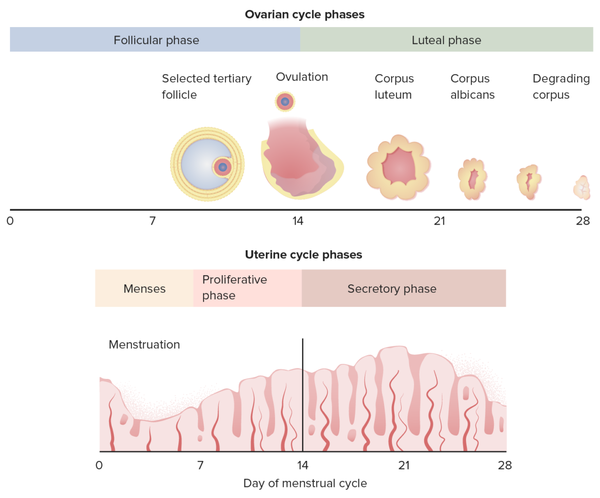
The menstrual cycle is divided into 2 components: ovarian cycle and endometrial cycle:

  • Average adult menstrual cycle is 28–35 days.
  • “Normal” cycle length is defined as 24–38 days.
  • Regular Regular Insulin” cycles are when variation in cycle length is ≤ 7‒9 days.
  • Intervals in cycles usually remain consistent until perimenopause Perimenopause The transitional period before and after menopause. Perimenopausal symptoms are associated with irregular menstrual cycle and widely fluctuated hormone levels. They may appear 6 years before menopause and subside 2 to 5 years after menopause. Menopause, when follicular phases become shorter and more frequent.
Note: This animation does not have sound.

Ovarian cycle phases:

  • Follicular phase:
    • Represents the time during which the follicle and its oocyte develop, leading up to ovulation
    • Spans from menses onset (day 1) to the day before the surge of luteinizing hormone (LH), leading to ovulation
    • Length: 14 to 21 days (may be shorter, especially in perimenopause Perimenopause The transitional period before and after menopause. Perimenopausal symptoms are associated with irregular menstrual cycle and widely fluctuated hormone levels. They may appear 6 years before menopause and subside 2 to 5 years after menopause. Menopause)
  • Luteal phase:
    • The time after ovulation when the ovary produces hormones Hormones Hormones are messenger molecules that are synthesized in one part of the body and move through the bloodstream to exert specific regulatory effects on another part of the body. Hormones play critical roles in coordinating cellular activities throughout the body in response to the constant changes in both the internal and external environments. Hormones: Overview and Types to support a potential pregnancy Pregnancy The status during which female mammals carry their developing young (embryos or fetuses) in utero before birth, beginning from fertilization to birth. Pregnancy: Diagnosis, Physiology, and Care and maintain a healthy endometrium Endometrium The mucous membrane lining of the uterine cavity that is hormonally responsive during the menstrual cycle and pregnancy. The endometrium undergoes cyclic changes that characterize menstruation. After successful fertilization, it serves to sustain the developing embryo. Embryoblast and Trophoblast Development.
    • Spans from the day of LH surge until the onset of the next menses
    • Length: 14 days

Endometrial cycle phases:

Correlation between the ovarian cycle and the endometrial cycle

A diagram showing the correlation between the ovarian cycle and the endometrial cycle

Image by Lecturio.

Regulation of the Menstrual Cycle

The menstrual cycle is regulated by the hypothalamic-pituitary-ovarian axis Hypothalamic-pituitary-ovarian axis Gonadal Hormones.

Hypothalamus Hypothalamus The hypothalamus is a collection of various nuclei within the diencephalon in the center of the brain. The hypothalamus plays a vital role in endocrine regulation as the primary regulator of the pituitary gland, and it is the major point of integration between the central nervous and endocrine systems. Hypothalamus:

  • Releases gonadotropin-releasing hormone Gonadotropin-releasing hormone A decapeptide that stimulates the synthesis and secretion of both pituitary gonadotropins, luteinizing hormone and follicle stimulating hormone. Gnrh is produced by neurons in the septum preoptic area of the hypothalamus and released into the pituitary portal blood, leading to stimulation of gonadotrophs in the anterior pituitary gland. Puberty (GnRH) → stimulates gonadotropes of the anterior pituitary Pituitary A small, unpaired gland situated in the sella turcica. It is connected to the hypothalamus by a short stalk which is called the infundibulum. Hormones: Overview and Types
  • Secreted from the preoptic neurons Neurons The basic cellular units of nervous tissue. Each neuron consists of a body, an axon, and dendrites. Their purpose is to receive, conduct, and transmit impulses in the nervous system. Nervous System: Histology of the hypothalamus Hypothalamus The hypothalamus is a collection of various nuclei within the diencephalon in the center of the brain. The hypothalamus plays a vital role in endocrine regulation as the primary regulator of the pituitary gland, and it is the major point of integration between the central nervous and endocrine systems. Hypothalamus in a pulsatile fashion
  • Regulated by biologic rhythms (and to a lesser extent by other physiologic factors, such as stress)

Anterior pituitary Pituitary A small, unpaired gland situated in the sella turcica. It is connected to the hypothalamus by a short stalk which is called the infundibulum. Hormones: Overview and Types:

  • Stimulated by GnRH → releases follicle-stimulating hormone (FSH) and luteinizing hormone (LH)
  • FSH and LH → stimulate the ovaries Ovaries Ovaries are the paired gonads of the female reproductive system that contain haploid gametes known as oocytes. The ovaries are located intraperitoneally in the pelvis, just posterior to the broad ligament, and are connected to the pelvic sidewall and to the uterus by ligaments. These organs function to secrete hormones (estrogen and progesterone) and to produce the female germ cells (oocytes). Ovaries: Anatomy
    • FSH:
      • Stimulates follicular development and egg maturation
      • Stimulates the granulosa cells within the ovary to produce estradiol Estradiol The 17-beta-isomer of estradiol, an aromatized C18 steroid with hydroxyl group at 3-beta- and 17-beta-position. Estradiol-17-beta is the most potent form of mammalian estrogenic steroids. Noncontraceptive Estrogen and Progestins
    • LH:
      • Stimulates theca cells Theca cells The flattened stroma cells forming a sheath or theca outside the basal lamina lining the mature ovarian follicle. Thecal interstitial or stromal cells are steroidogenic, and produce primarily androgens which serve as precursors of estrogens in the granulosa cells. Puberty within the ovary to produce testosterone Testosterone A potent androgenic steroid and major product secreted by the leydig cells of the testis. Its production is stimulated by luteinizing hormone from the pituitary gland. In turn, testosterone exerts feedback control of the pituitary LH and FSH secretion. Depending on the tissues, testosterone can be further converted to dihydrotestosterone or estradiol. Androgens and Antiandrogens (most of which is converted to estradiol Estradiol The 17-beta-isomer of estradiol, an aromatized C18 steroid with hydroxyl group at 3-beta- and 17-beta-position. Estradiol-17-beta is the most potent form of mammalian estrogenic steroids. Noncontraceptive Estrogen and Progestins in the granulosa cells)
      • A surge of LH midcycle triggers ovulation.

Ovaries Ovaries Ovaries are the paired gonads of the female reproductive system that contain haploid gametes known as oocytes. The ovaries are located intraperitoneally in the pelvis, just posterior to the broad ligament, and are connected to the pelvic sidewall and to the uterus by ligaments. These organs function to secrete hormones (estrogen and progesterone) and to produce the female germ cells (oocytes). Ovaries: Anatomy:
  • Estrogens:
    • Estradiol Estradiol The 17-beta-isomer of estradiol, an aromatized C18 steroid with hydroxyl group at 3-beta- and 17-beta-position. Estradiol-17-beta is the most potent form of mammalian estrogenic steroids. Noncontraceptive Estrogen and Progestins is the most notable.
    • Secreted by the granulosa cells of ovarian follicles → stimulated by FSH
    • Stimulates the endometrium Endometrium The mucous membrane lining of the uterine cavity that is hormonally responsive during the menstrual cycle and pregnancy. The endometrium undergoes cyclic changes that characterize menstruation. After successful fertilization, it serves to sustain the developing embryo. Embryoblast and Trophoblast Development to grow/develop
    • Makes the developing follicles more sensitive to FSH
    • Feedback/regulation:
      • Negative feedback Negative feedback Hypothalamic and Pituitary Hormones inhibition: During most of the menstrual cycle, estrogens inhibit further secretions of FSH, LH, and GnRH.
      • Positive feedback: For a short time midcycle, estradiol Estradiol The 17-beta-isomer of estradiol, an aromatized C18 steroid with hydroxyl group at 3-beta- and 17-beta-position. Estradiol-17-beta is the most potent form of mammalian estrogenic steroids. Noncontraceptive Estrogen and Progestins stimulates FSH and LH secretion Secretion Coagulation Studies from the pituitary Pituitary A small, unpaired gland situated in the sella turcica. It is connected to the hypothalamus by a short stalk which is called the infundibulum. Hormones: Overview and Types → results in ↑ estrogen Estrogen Compounds that interact with estrogen receptors in target tissues to bring about the effects similar to those of estradiol. Estrogens stimulate the female reproductive organs, and the development of secondary female sex characteristics. Estrogenic chemicals include natural, synthetic, steroidal, or non-steroidal compounds. Ovaries: Anatomy production in the ovaries Ovaries Ovaries are the paired gonads of the female reproductive system that contain haploid gametes known as oocytes. The ovaries are located intraperitoneally in the pelvis, just posterior to the broad ligament, and are connected to the pelvic sidewall and to the uterus by ligaments. These organs function to secrete hormones (estrogen and progesterone) and to produce the female germ cells (oocytes). Ovaries: Anatomy and causes the surge of LH, which triggers ovulation
  • Progestins Progestins Compounds that interact with progesterone receptors in target tissues to bring about the effects similar to those of progesterone. Primary actions of progestins, including natural and synthetic steroids, are on the uterus and the mammary gland in preparation for and in maintenance of pregnancy. Hormonal Contraceptives:
    • Progesterone Progesterone The major progestational steroid that is secreted primarily by the corpus luteum and the placenta. Progesterone acts on the uterus, the mammary glands and the brain. It is required in embryo implantation; pregnancy maintenance, and the development of mammary tissue for milk production. Progesterone, converted from pregnenolone, also serves as an intermediate in the biosynthesis of gonadal steroid hormones and adrenal corticosteroids. Gonadal Hormones is the most notable.
    • Secreted by the theca-lutein and granulosa lutein cells in the corpus luteum Corpus Luteum The yellow body derived from the ruptured ovarian follicle after ovulation. The process of corpus luteum formation, luteinization, is regulated by luteinizing hormone. Ovaries: Anatomy (stimulated by LH) after ovulation
    • Uterine effects:
      • ↓ Endometrial growth
      • Stabilizes and causes maturation of the endometrium Endometrium The mucous membrane lining of the uterine cavity that is hormonally responsive during the menstrual cycle and pregnancy. The endometrium undergoes cyclic changes that characterize menstruation. After successful fertilization, it serves to sustain the developing embryo. Embryoblast and Trophoblast Development → prepares the endometrium Endometrium The mucous membrane lining of the uterine cavity that is hormonally responsive during the menstrual cycle and pregnancy. The endometrium undergoes cyclic changes that characterize menstruation. After successful fertilization, it serves to sustain the developing embryo. Embryoblast and Trophoblast Development for implantation Implantation Endometrial implantation of embryo, mammalian at the blastocyst stage. Fertilization and First Week
      • ↑ Endometrial secretions (↑ secretion Secretion Coagulation Studies thickness)
      • Progestin withdrawal at the end of the luteal/secretory phases triggers menstrual bleeding.
    • Breast effects:
      • ↑ Lobular development
      • Inhibition of milk production
    • Body temperature Body Temperature The measure of the level of heat of a human or animal. Heatstroke → can be used to track ovulation
    • Required for the development of the placenta Placenta A highly vascularized mammalian fetal-maternal organ and major site of transport of oxygen, nutrients, and fetal waste products. It includes a fetal portion (chorionic villi) derived from trophoblasts and a maternal portion (decidua) derived from the uterine endometrium. The placenta produces an array of steroid, protein and peptide hormones (placental hormones). Placenta, Umbilical Cord, and Amniotic Cavity during pregnancy Pregnancy The status during which female mammals carry their developing young (embryos or fetuses) in utero before birth, beginning from fertilization to birth. Pregnancy: Diagnosis, Physiology, and Care
  • Activins:
    • Secreted by the granulosa cells of ovarian follicles (stimulated by FSH)
    • Provides positive feedback to gonadotropes → stimulates secretion Secretion Coagulation Studies of LH, especially midcycle
  • Inhibins:
    • Secreted by the granulosa cells of ovarian follicles (stimulated by FSH)
    • Provides negative feedback to gonadotropes → selectively inhibits further FSH secretion Secretion Coagulation Studies
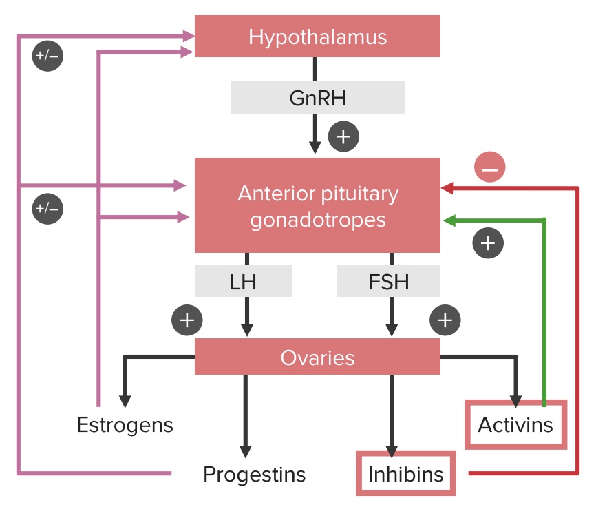
Hypothalamic pituitary ovarian axis

Positive and negative feedback loops of the hypothalamic-pituitary-ovarian axis:
Note that estrogens and progestins can have both a positive and a negative influence on the hypothalamus and pituitary gland, depending on the phase of the cycle. Estrogens provide negative feedback until the middle of the cycle. At this point, estrogen begins stimulating the gonadotropic cells in the pituitary, leading to the LH surge, which triggers ovulation.
LH: luteinizing hormone 
FSH: follicle-stimulating hormone
GnRH: gonadotropin-releasing hormone
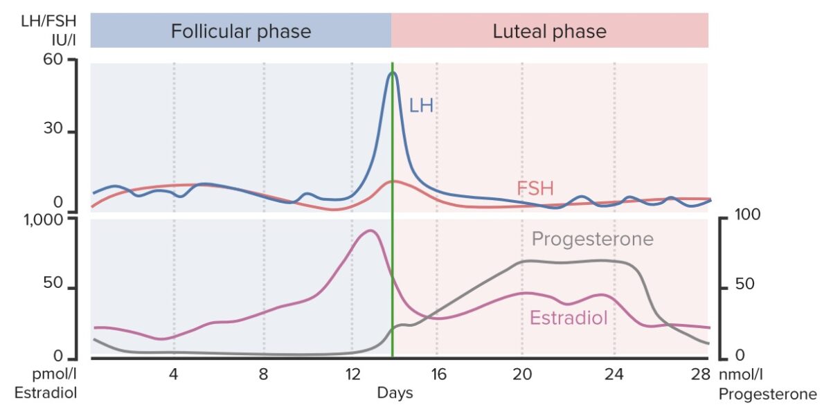
Image by Lecturio.

Ovarian Cycle

Ovarian cycle

A chart showing the changing concentrations of key hormones throughout the length of the menstrual cycle: Notice the sudden rise in estradiol, LH and FSH around day 14 (ovulation), and the rise in progesterone during the luteal phase in anticipation of fertilization and implantation of the ovum.

Image by Lecturio.

Follicular phase

The phase representing the time during which the follicle (and the oocyte it contains) develop, leading up to ovulation. The follicular phase of the ovaries Ovaries Ovaries are the paired gonads of the female reproductive system that contain haploid gametes known as oocytes. The ovaries are located intraperitoneally in the pelvis, just posterior to the broad ligament, and are connected to the pelvic sidewall and to the uterus by ligaments. These organs function to secrete hormones (estrogen and progesterone) and to produce the female germ cells (oocytes). Ovaries: Anatomy coincides with menses and the proliferative phase of the endometrium Endometrium The mucous membrane lining of the uterine cavity that is hormonally responsive during the menstrual cycle and pregnancy. The endometrium undergoes cyclic changes that characterize menstruation. After successful fertilization, it serves to sustain the developing embryo. Embryoblast and Trophoblast Development.
  • Takes place during days 1–14 (up to day 21) of the menstrual cycle
  • Primarily under the control of FSH
  • GnRH is released from the hypothalamus Hypothalamus The hypothalamus is a collection of various nuclei within the diencephalon in the center of the brain. The hypothalamus plays a vital role in endocrine regulation as the primary regulator of the pituitary gland, and it is the major point of integration between the central nervous and endocrine systems. Hypothalamus → stimulates the release of FSH from the anterior pituitary Pituitary A small, unpaired gland situated in the sella turcica. It is connected to the hypothalamus by a short stalk which is called the infundibulum. Hormones: Overview and Types → stimulates the primary follicles Primary follicles Secondary Lymphatic Organs of the ovary to begin developing/maturing
  • Maturing primary follicles Primary follicles Secondary Lymphatic Organs produce (via granulosa and theca cells Theca cells The flattened stroma cells forming a sheath or theca outside the basal lamina lining the mature ovarian follicle. Thecal interstitial or stromal cells are steroidogenic, and produce primarily androgens which serve as precursors of estrogens in the granulosa cells. Puberty):
    • Estradiol Estradiol The 17-beta-isomer of estradiol, an aromatized C18 steroid with hydroxyl group at 3-beta- and 17-beta-position. Estradiol-17-beta is the most potent form of mammalian estrogenic steroids. Noncontraceptive Estrogen and Progestins:
      • Stimulates development of the endometrium Endometrium The mucous membrane lining of the uterine cavity that is hormonally responsive during the menstrual cycle and pregnancy. The endometrium undergoes cyclic changes that characterize menstruation. After successful fertilization, it serves to sustain the developing embryo. Embryoblast and Trophoblast Development
      • Helps select a dominant follicle by inhibiting further release of FSH (“starving” other follicles) while simultaneously making the remaining follicles more sensitive to the dwindling FSH 
    • Inhibin A → inhibits the release of FSH from the anterior pituitary Pituitary A small, unpaired gland situated in the sella turcica. It is connected to the hypothalamus by a short stalk which is called the infundibulum. Hormones: Overview and Types ( negative feedback Negative feedback Hypothalamic and Pituitary Hormones)
  • The anterior pituitary Pituitary A small, unpaired gland situated in the sella turcica. It is connected to the hypothalamus by a short stalk which is called the infundibulum. Hormones: Overview and Types releases a luteinizing burst or “surge” → triggers ovulation approximately 12 hours later
    • During ovulation, a mature oocyte is released from the dominant follicle.
    • The LH surge ends the follicular phase.
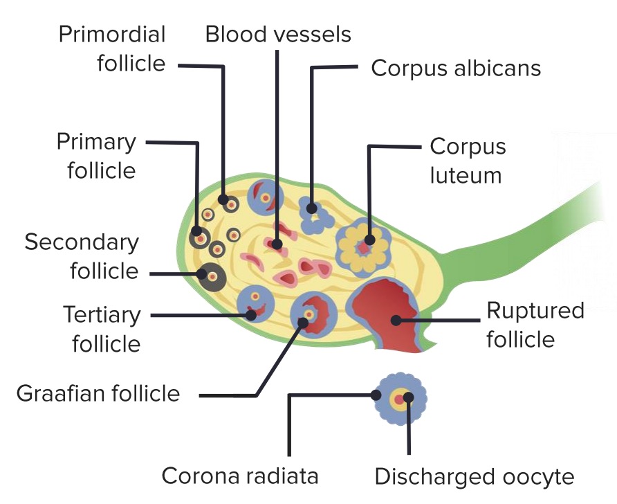
Stages of maturation of ovarian follicle

Stages of maturation of an ovarian follicle


During the follicular phase of the menstrual cycle, follicle-stimulating hormone (FSH) stimulates the primary follicles to mature. Typically, only a single follicle (the dominant follicle) matures all the way into a graafian follicle. The graafian follicle ruptures, releasing the oocyte (ovulation), and transforms into a corpus luteum. During the luteal phase of the menstrual cycle, the corpus luteum produces hormones (especially progesterone) necessary to cause maturation of the endometrium and potentially support an early pregnancy. Image by Lecturio.
Structure of primary ovarian follicle

Structure of a tertiary (antral) ovarian follicle

Image by Lecturio.

Luteal Phase

The luteal phase of the ovaries Ovaries Ovaries are the paired gonads of the female reproductive system that contain haploid gametes known as oocytes. The ovaries are located intraperitoneally in the pelvis, just posterior to the broad ligament, and are connected to the pelvic sidewall and to the uterus by ligaments. These organs function to secrete hormones (estrogen and progesterone) and to produce the female germ cells (oocytes). Ovaries: Anatomy coincides with the secretory phase of the endometrium Endometrium The mucous membrane lining of the uterine cavity that is hormonally responsive during the menstrual cycle and pregnancy. The endometrium undergoes cyclic changes that characterize menstruation. After successful fertilization, it serves to sustain the developing embryo. Embryoblast and Trophoblast Development.

  • Takes place typically during days 1528 of the menstrual cycle, after the oocyte is released. 
  • The oocyte migrates to the fimbria of the fallopian tube Fallopian Tube A pair of highly specialized canals extending from the uterus to its corresponding ovary. They provide the means for ovum transport from the ovaries and they are the site of the ovum’s final maturation and fertilization. The fallopian tube consists of an interstitium, an isthmus, an ampulla, an infundibulum, and fimbriae. Its wall consists of three layers: serous, muscular, and an internal mucosal layer lined with both ciliated and secretory cells. Uterus, Cervix, and Fallopian Tubes: Anatomy (can take 3 days).
  • Potential for fertilization Fertilization To undergo fertilization, the sperm enters the uterus, travels towards the ampulla of the fallopian tube, and encounters the oocyte. The zona pellucida (the outer layer of the oocyte) deteriorates along with the zygote, which travels towards the uterus and eventually forms a blastocyst, allowing for implantation to occur. Fertilization and First Week by sperm at this phase
  • The corpus luteum Corpus Luteum The yellow body derived from the ruptured ovarian follicle after ovulation. The process of corpus luteum formation, luteinization, is regulated by luteinizing hormone. Ovaries: Anatomy of the mature oocyte produces progesterone Progesterone The major progestational steroid that is secreted primarily by the corpus luteum and the placenta. Progesterone acts on the uterus, the mammary glands and the brain. It is required in embryo implantation; pregnancy maintenance, and the development of mammary tissue for milk production. Progesterone, converted from pregnenolone, also serves as an intermediate in the biosynthesis of gonadal steroid hormones and adrenal corticosteroids. Gonadal Hormones:
    • Causes maturation of the endometrium Endometrium The mucous membrane lining of the uterine cavity that is hormonally responsive during the menstrual cycle and pregnancy. The endometrium undergoes cyclic changes that characterize menstruation. After successful fertilization, it serves to sustain the developing embryo. Embryoblast and Trophoblast Development (proliferative → secretory endometrium Endometrium The mucous membrane lining of the uterine cavity that is hormonally responsive during the menstrual cycle and pregnancy. The endometrium undergoes cyclic changes that characterize menstruation. After successful fertilization, it serves to sustain the developing embryo. Embryoblast and Trophoblast Development)
    • Progesterone Progesterone The major progestational steroid that is secreted primarily by the corpus luteum and the placenta. Progesterone acts on the uterus, the mammary glands and the brain. It is required in embryo implantation; pregnancy maintenance, and the development of mammary tissue for milk production. Progesterone, converted from pregnenolone, also serves as an intermediate in the biosynthesis of gonadal steroid hormones and adrenal corticosteroids. Gonadal Hormones feedback inhibits LH secretion Secretion Coagulation Studies from the anterior pituitary Pituitary A small, unpaired gland situated in the sella turcica. It is connected to the hypothalamus by a short stalk which is called the infundibulum. Hormones: Overview and Types.
  • If conception does not occur:
    • LH levels continue to ↓ and the corpus luteum Corpus Luteum The yellow body derived from the ruptured ovarian follicle after ovulation. The process of corpus luteum formation, luteinization, is regulated by luteinizing hormone. Ovaries: Anatomy recedes into a corpus albicans.
    • As the corpus luteum Corpus Luteum The yellow body derived from the ruptured ovarian follicle after ovulation. The process of corpus luteum formation, luteinization, is regulated by luteinizing hormone. Ovaries: Anatomy recedes:
      • Progesterone Progesterone The major progestational steroid that is secreted primarily by the corpus luteum and the placenta. Progesterone acts on the uterus, the mammary glands and the brain. It is required in embryo implantation; pregnancy maintenance, and the development of mammary tissue for milk production. Progesterone, converted from pregnenolone, also serves as an intermediate in the biosynthesis of gonadal steroid hormones and adrenal corticosteroids. Gonadal Hormones ↓ → triggers menstruation (end of the luteal phase)
      • Estradiol Estradiol The 17-beta-isomer of estradiol, an aromatized C18 steroid with hydroxyl group at 3-beta- and 17-beta-position. Estradiol-17-beta is the most potent form of mammalian estrogenic steroids. Noncontraceptive Estrogen and Progestins ↓ → releases negative feedback Negative feedback Hypothalamic and Pituitary Hormones on hypothalamus Hypothalamus The hypothalamus is a collection of various nuclei within the diencephalon in the center of the brain. The hypothalamus plays a vital role in endocrine regulation as the primary regulator of the pituitary gland, and it is the major point of integration between the central nervous and endocrine systems. Hypothalamus and pituitary Pituitary A small, unpaired gland situated in the sella turcica. It is connected to the hypothalamus by a short stalk which is called the infundibulum. Hormones: Overview and Types → GnRH pulse begins again to start the next cycle 
  • If pregnancy Pregnancy The status during which female mammals carry their developing young (embryos or fetuses) in utero before birth, beginning from fertilization to birth. Pregnancy: Diagnosis, Physiology, and Care occurs, the secretion Secretion Coagulation Studies of human chorionic gonadotropin (hCG) saves the corpus luteum Corpus Luteum The yellow body derived from the ruptured ovarian follicle after ovulation. The process of corpus luteum formation, luteinization, is regulated by luteinizing hormone. Ovaries: Anatomy and allows it to continue its secretory function.

Endometrial Cycle

There are 3 phases of the endometrial cycle:

  1. Desquamation Desquamation Staphylococcal Scalded Skin Syndrome (SSSS) or menses 
  2. Proliferative phase
  3. Secretory phase

Menses

Day 1 of menstrual bleeding marks the beginning of the next cycle.

  • Coincides with day 1 of the follicular phase
  • If conception does not occur, the corpus luteum Corpus Luteum The yellow body derived from the ruptured ovarian follicle after ovulation. The process of corpus luteum formation, luteinization, is regulated by luteinizing hormone. Ovaries: Anatomy degenerates and progesterone Progesterone The major progestational steroid that is secreted primarily by the corpus luteum and the placenta. Progesterone acts on the uterus, the mammary glands and the brain. It is required in embryo implantation; pregnancy maintenance, and the development of mammary tissue for milk production. Progesterone, converted from pregnenolone, also serves as an intermediate in the biosynthesis of gonadal steroid hormones and adrenal corticosteroids. Gonadal Hormones levels decrease 
  • Progesterone Progesterone The major progestational steroid that is secreted primarily by the corpus luteum and the placenta. Progesterone acts on the uterus, the mammary glands and the brain. It is required in embryo implantation; pregnancy maintenance, and the development of mammary tissue for milk production. Progesterone, converted from pregnenolone, also serves as an intermediate in the biosynthesis of gonadal steroid hormones and adrenal corticosteroids. Gonadal Hormones withdrawal triggers desquamation Desquamation Staphylococcal Scalded Skin Syndrome (SSSS) of the functional layer of the endometrium Endometrium The mucous membrane lining of the uterine cavity that is hormonally responsive during the menstrual cycle and pregnancy. The endometrium undergoes cyclic changes that characterize menstruation. After successful fertilization, it serves to sustain the developing embryo. Embryoblast and Trophoblast Development (menses)
    • Spiral arteries Spiral arteries Placenta, Umbilical Cord, and Amniotic Cavity constrict.
    • Endometrium Endometrium The mucous membrane lining of the uterine cavity that is hormonally responsive during the menstrual cycle and pregnancy. The endometrium undergoes cyclic changes that characterize menstruation. After successful fertilization, it serves to sustain the developing embryo. Embryoblast and Trophoblast Development dies secondary to apoptosis Apoptosis A regulated cell death mechanism characterized by distinctive morphologic changes in the nucleus and cytoplasm, including the endonucleolytic cleavage of genomic DNA, at regularly spaced, internucleosomal sites, I.e., DNA fragmentation. It is genetically-programmed and serves as a balance to mitosis in regulating the size of animal tissues and in mediating pathologic processes associated with tumor growth. Ischemic Cell Damage.
    • Uterine contractions shed and expel the upper endometrial layer (stratum functionalis).
  • Normal menstruation:
    • Lasts 3‒8 days
    • Loss of < 80 ml of blood/tissue
  • The oocyte is lost in menstrual bleeding.

Proliferative phase

  • Proliferative phase (days 414) → new endometrium Endometrium The mucous membrane lining of the uterine cavity that is hormonally responsive during the menstrual cycle and pregnancy. The endometrium undergoes cyclic changes that characterize menstruation. After successful fertilization, it serves to sustain the developing embryo. Embryoblast and Trophoblast Development develops (stratum functionalis grows from the remaining deeper layer of endometrium Endometrium The mucous membrane lining of the uterine cavity that is hormonally responsive during the menstrual cycle and pregnancy. The endometrium undergoes cyclic changes that characterize menstruation. After successful fertilization, it serves to sustain the developing embryo. Embryoblast and Trophoblast Development (the stratum basalis) that does not shed during menses)
  • Stimulated by estrogen Estrogen Compounds that interact with estrogen receptors in target tissues to bring about the effects similar to those of estradiol. Estrogens stimulate the female reproductive organs, and the development of secondary female sex characteristics. Estrogenic chemicals include natural, synthetic, steroidal, or non-steroidal compounds. Ovaries: Anatomy produced by the growing follicles:
    • Theca cells Theca cells The flattened stroma cells forming a sheath or theca outside the basal lamina lining the mature ovarian follicle. Thecal interstitial or stromal cells are steroidogenic, and produce primarily androgens which serve as precursors of estrogens in the granulosa cells. Puberty in the developing follicles make androgens Androgens Androgens are naturally occurring steroid hormones responsible for development and maintenance of the male sex characteristics, including penile, scrotal, and clitoral growth, development of sexual hair, deepening of the voice, and musculoskeletal growth. Androgens and Antiandrogens.
    • Granulosa cells release aromatase Aromatase An enzyme that catalyzes the desaturation (aromatization) of the ring a of C19 androgens and converts them to C18 estrogens. In this process, the 19-methyl is removed. This enzyme is membrane-bound, located in the endoplasmic reticulum of estrogen-producing cells of ovaries, placenta, testes, adipose, and brain tissues. Aromatase is encoded by the cyp19 gene, and functions in complex with NADPH-ferrihemoprotein reductase in the cytochrome p450 system. Adipose Tissue: Histology → converts androgens Androgens Androgens are naturally occurring steroid hormones responsible for development and maintenance of the male sex characteristics, including penile, scrotal, and clitoral growth, development of sexual hair, deepening of the voice, and musculoskeletal growth. Androgens and Antiandrogens into estrogens, which act as a growth factor in endometrial tissue Endometrial tissue The mucous membrane lining of the uterine cavity that is hormonally responsive during the menstrual cycle and pregnancy. The endometrium undergoes cyclic changes that characterize menstruation. After successful fertilization, it serves to sustain the developing embryo. Endometriosis
  • Endometrial proliferation with straight, tubular glands

Secretory phase

  • Starts between days 13 and 15 of the menstrual cycle (later in some individuals) 
  • Preparation of the spiral arteries Spiral arteries Placenta, Umbilical Cord, and Amniotic Cavity and endometrial glands for potential implantation Implantation Endometrial implantation of embryo, mammalian at the blastocyst stage. Fertilization and First Week of an embryo Embryo The entity of a developing mammal, generally from the cleavage of a zygote to the end of embryonic differentiation of basic structures. For the human embryo, this represents the first two months of intrauterine development preceding the stages of the fetus. Fertilization and First Week:
    • Triggered by progesterone Progesterone The major progestational steroid that is secreted primarily by the corpus luteum and the placenta. Progesterone acts on the uterus, the mammary glands and the brain. It is required in embryo implantation; pregnancy maintenance, and the development of mammary tissue for milk production. Progesterone, converted from pregnenolone, also serves as an intermediate in the biosynthesis of gonadal steroid hormones and adrenal corticosteroids. Gonadal Hormones release 
    • Increased endometrial gland tortuosity
    • Glycogen-rich secretions
    • Edematous stromal cells
    • Uterine spiral arteries Spiral arteries Placenta, Umbilical Cord, and Amniotic Cavity extend the full length of the endometrium Endometrium The mucous membrane lining of the uterine cavity that is hormonally responsive during the menstrual cycle and pregnancy. The endometrium undergoes cyclic changes that characterize menstruation. After successful fertilization, it serves to sustain the developing embryo. Embryoblast and Trophoblast Development.
  • If no pregnancy Pregnancy The status during which female mammals carry their developing young (embryos or fetuses) in utero before birth, beginning from fertilization to birth. Pregnancy: Diagnosis, Physiology, and Care progesterone Progesterone The major progestational steroid that is secreted primarily by the corpus luteum and the placenta. Progesterone acts on the uterus, the mammary glands and the brain. It is required in embryo implantation; pregnancy maintenance, and the development of mammary tissue for milk production. Progesterone, converted from pregnenolone, also serves as an intermediate in the biosynthesis of gonadal steroid hormones and adrenal corticosteroids. Gonadal Hormones levels decrease → inducing apoptosis Apoptosis A regulated cell death mechanism characterized by distinctive morphologic changes in the nucleus and cytoplasm, including the endonucleolytic cleavage of genomic DNA, at regularly spaced, internucleosomal sites, I.e., DNA fragmentation. It is genetically-programmed and serves as a balance to mitosis in regulating the size of animal tissues and in mediating pathologic processes associated with tumor growth. Ischemic Cell Damage of the functional layer of the endometrium Endometrium The mucous membrane lining of the uterine cavity that is hormonally responsive during the menstrual cycle and pregnancy. The endometrium undergoes cyclic changes that characterize menstruation. After successful fertilization, it serves to sustain the developing embryo. Embryoblast and Trophoblast Development, leading to menses

Clinical Relevance

  • Pregnancy Pregnancy The status during which female mammals carry their developing young (embryos or fetuses) in utero before birth, beginning from fertilization to birth. Pregnancy: Diagnosis, Physiology, and Care: the period of time in which a fetus develops inside a uterus Uterus The uterus, cervix, and fallopian tubes are part of the internal female reproductive system. The uterus has a thick wall made of smooth muscle (the myometrium) and an inner mucosal layer (the endometrium). The most inferior portion of the uterus is the cervix, which connects the uterine cavity to the vagina. Uterus, Cervix, and Fallopian Tubes: Anatomy. When the oocyte is fertilized by a sperm cell and the developing embryo Embryo The entity of a developing mammal, generally from the cleavage of a zygote to the end of embryonic differentiation of basic structures. For the human embryo, this represents the first two months of intrauterine development preceding the stages of the fetus. Fertilization and First Week is implanted into the endometrium Endometrium The mucous membrane lining of the uterine cavity that is hormonally responsive during the menstrual cycle and pregnancy. The endometrium undergoes cyclic changes that characterize menstruation. After successful fertilization, it serves to sustain the developing embryo. Embryoblast and Trophoblast Development, the menstrual cycle is suppressed to prevent the evacuation of the uterine lining and embryo Embryo The entity of a developing mammal, generally from the cleavage of a zygote to the end of embryonic differentiation of basic structures. For the human embryo, this represents the first two months of intrauterine development preceding the stages of the fetus. Fertilization and First Week via uterine contractions. Pregnancy Pregnancy The status during which female mammals carry their developing young (embryos or fetuses) in utero before birth, beginning from fertilization to birth. Pregnancy: Diagnosis, Physiology, and Care lasts approximately 40 gestational weeks and creates a physiological state in the body to support a fetal gestation.
  • Dysmenorrhea: recurrent abdominal pain Abdominal Pain Acute Abdomen associated with menstruation; may be primary or secondary in nature
  • Endometrial hyperplasia Hyperplasia An increase in the number of cells in a tissue or organ without tumor formation. It differs from hypertrophy, which is an increase in bulk without an increase in the number of cells. Cellular Adaptation: occurs when the endometrium Endometrium The mucous membrane lining of the uterine cavity that is hormonally responsive during the menstrual cycle and pregnancy. The endometrium undergoes cyclic changes that characterize menstruation. After successful fertilization, it serves to sustain the developing embryo. Embryoblast and Trophoblast Development receives prolonged stimulation by estrogens to proliferate. Presents as abnormal uterine bleeding Abnormal Uterine Bleeding Abnormal uterine bleeding is the medical term for abnormalities in the frequency, volume, duration, and regularity of the menstrual cycle. Abnormal uterine bleeding is classified using the acronym PALM-COEIN, with PALM representing the structural causes and COEIN indicating the non-structural causes. Abnormal Uterine Bleeding. Women who suffer from endometrial hyperplasia Hyperplasia An increase in the number of cells in a tissue or organ without tumor formation. It differs from hypertrophy, which is an increase in bulk without an increase in the number of cells. Cellular Adaptation are at increased risk of developing dysplasia and endometrial cancer Endometrial Cancer Endometrial carcinoma (EC) is the most common gynecologic malignancy in the developed world, and it has several histologic types. Endometrioid carcinoma (known as type 1 EC) typically develops from atypical endometrial hyperplasia, is hormonally responsive, and carries a favorable prognosis. Endometrial Hyperplasia and Endometrial Cancer.

The following conditions are related to abnormalities of the menstrual cycle, known as abnormal uterine bleeding Abnormal Uterine Bleeding Abnormal uterine bleeding is the medical term for abnormalities in the frequency, volume, duration, and regularity of the menstrual cycle. Abnormal uterine bleeding is classified using the acronym PALM-COEIN, with PALM representing the structural causes and COEIN indicating the non-structural causes. Abnormal Uterine Bleeding ( AUB AUB Abnormal uterine bleeding is the medical term for abnormalities in the frequency, volume, duration, and regularity of the menstrual cycle. Abnormal uterine bleeding is classified using the acronym palm-coein, with palm representing the structural causes and coein indicating the non-structural causes. Abnormal Uterine Bleeding):

References:
  1. Welt, C.K. (2021). Physiology of the normal menstrual cycle. UpToDate. Retrieved May 17, 2022 from https://www.uptodate.com/contents/physiology-of-the-normal-menstrual-cycle 
  2. Saladin, K.S., Miller, L. (2004). Anatomy and Physiology, 3rd ed., McGraw-Hill Education, pp. 1050‒1055.
  3. Munro, M.G., Critchley, H.O.D., Fraser, I.S. (2018). The two FIGO systems for normal and abnormal uterine bleeding Abnormal Uterine Bleeding Abnormal uterine bleeding is the medical term for abnormalities in the frequency, volume, duration, and regularity of the menstrual cycle. Abnormal uterine bleeding is classified using the acronym PALM-COEIN, with PALM representing the structural causes and COEIN indicating the non-structural causes. Abnormal Uterine Bleeding symptoms and classification of causes of abnormal uterine bleeding Abnormal Uterine Bleeding Abnormal uterine bleeding is the medical term for abnormalities in the frequency, volume, duration, and regularity of the menstrual cycle. Abnormal uterine bleeding is classified using the acronym PALM-COEIN, with PALM representing the structural causes and COEIN indicating the non-structural causes. Abnormal Uterine Bleeding in the reproductive years: 2018 revisions. Int J Gynecol Obstet; 143: 393‒408.
  4. Moore, K.L., Dalley, A.F. (2006). Clinically Oriented Anatomy, 5th ed., Lippincott Williams and Wilkins, pp. 415-429.

Create your free account or log in to continue reading!

Sign up now and get free access to Lecturio with concept pages, medical videos, and questions for your medical education.

User Reviews

Details