Meckel’s Diverticulum

A Meckel’s diverticulum Diverticulum A pouch or sac opening from the colon. Diverticular Disease is a persistent remnant of the omphalomesenteric (vitelline) duct. A Meckel’s diverticulum Diverticulum A pouch or sac opening from the colon. Diverticular Disease is usually located in the antimesenteric border of the ileum Ileum The distal and narrowest portion of the small intestine, between the jejunum and the ileocecal valve of the large intestine. Small Intestine: Anatomy. The mucosal lining of the diverticulum Diverticulum A pouch or sac opening from the colon. Diverticular Disease may contain heterotopic Heterotopic Transplantation of tissue typical of one area to a different recipient site. The tissue may be autologous, heterologous, or homologous. Organ Transplantation mucosa (most commonly gastric). Though frequently asymptomatic, a Meckel’s diverticulum Diverticulum A pouch or sac opening from the colon. Diverticular Disease can cause ulceration Ulceration Corneal Abrasions, Erosion, and Ulcers and present with lower gastrointestinal (GI) bleeding. Other complications include diverticulitis Diverticulitis Inflammation of a diverticulum or diverticula. Diverticular Disease or small bowel obstruction Bowel obstruction Any impairment, arrest, or reversal of the normal flow of intestinal contents toward the anal canal. Ascaris/Ascariasis ( SBO SBO Small bowel obstruction (SBO) is an interruption of the flow of the intraluminal contents through the small intestine, and is classified as mechanical (due to physical blockage) or functional (due to disruption of normal motility). The most common cause of SBO in the Western countries is post-surgical adhesions. Small bowel obstruction typically presents with nausea, vomiting, abdominal pain, distention, constipation, and/or obstipation. Small Bowel Obstruction). A Meckel’s scan can detect the diverticulum Diverticulum A pouch or sac opening from the colon. Diverticular Disease in hemodynamically stable patients Hemodynamically Stable Patients Blunt Chest Trauma. For those with active bleeding, arteriography is the diagnostic option. The treatment for a symptomatic Meckel’s diverticulum Diverticulum A pouch or sac opening from the colon. Diverticular Disease is surgery.

Last updated: Jan 18, 2024

Editorial responsibility: Stanley Oiseth, Lindsay Jones, Evelin Maza

Overview

Definition

A Meckel’s diverticulum Diverticulum A pouch or sac opening from the colon. Diverticular Disease is a persistent remnant of the omphalomesenteric duct. A Meckel’s diverticulum Diverticulum A pouch or sac opening from the colon. Diverticular Disease is a true diverticulum Diverticulum A pouch or sac opening from the colon. Diverticular Disease (contains all layers of the bowel wall), arising from the antimesenteric surface of the middle-to-distal ileum Ileum The distal and narrowest portion of the small intestine, between the jejunum and the ileocecal valve of the large intestine. Small Intestine: Anatomy.
Epidemiology

  • The most common congenital Congenital Chorioretinitis gastrointestinal (GI) tract anomaly 
  • Prevalence Prevalence The total number of cases of a given disease in a specified population at a designated time. It is differentiated from incidence, which refers to the number of new cases in the population at a given time. Measures of Disease Frequency: approximately 2% of the population
  • Sex Sex The totality of characteristics of reproductive structure, functions, phenotype, and genotype, differentiating the male from the female organism. Gender Dysphoria: males > females (2:1)
  • Most commonly presents with symptoms at 2–4 years of age
  • Increased incidence Incidence The number of new cases of a given disease during a given period in a specified population. It also is used for the rate at which new events occur in a defined population. It is differentiated from prevalence, which refers to all cases in the population at a given time. Measures of Disease Frequency in children with major malformations involving the:
    • Umbilicus
    • GI tract
    • Nervous system Nervous system The nervous system is a small and complex system that consists of an intricate network of neural cells (or neurons) and even more glial cells (for support and insulation). It is divided according to its anatomical components as well as its functional characteristics. The brain and spinal cord are referred to as the central nervous system, and the branches of nerves from these structures are referred to as the peripheral nervous system. Nervous System: Anatomy, Structure, and Classification
    • Heart

Rule of 2(s)

  • 2% of population
  • 2 times more common in males
  • Frequently presents by 2 years of age
  • 2% develop symptoms/complications
  • 2 inches long
  • 2 feet from the ileocecal valve Ileocecal valve The valve, at the junction of the cecum with the colon, that guards the opening where the ileum enters the large intestine. Small Intestine: Anatomy

Etiology

  • Omphalomesenteric duct:
    • Connects the midgut Midgut Development of the Abdominal Organs to the yolk sac Yolk Sac The first of four extra-embryonic membranes to form during embryogenesis. In reptiles and birds, it arises from endoderm and mesoderm to incorporate the egg yolk into the digestive tract for nourishing the embryo. In placental mammals, its nutritional function is vestigial; however, it is the source of intestinal mucosa; blood cells; and germ cells. It is sometimes called the vitelline sac, which should not be confused with the vitelline membrane of the egg. Embryoblast and Trophoblast Development in utero
    • Normally involutes between the 5th and 6th weeks of gestation
  • Omphalomesenteric duct that does not fully involute can give rise to:
    • A Meckel’s diverticulum Diverticulum A pouch or sac opening from the colon. Diverticular Disease (most common persistent remnant)
    • Omphalomesenteric cysts Cysts Any fluid-filled closed cavity or sac that is lined by an epithelium. Cysts can be of normal, abnormal, non-neoplastic, or neoplastic tissues. Fibrocystic Change
    • Omphalomesenteric fistula Fistula Abnormal communication most commonly seen between two internal organs, or between an internal organ and the surface of the body. Anal Fistula (drain through umbilicus)
    • Fibrous Fibrous Fibrocystic Change bands (can cause bowel obstruction Bowel obstruction Any impairment, arrest, or reversal of the normal flow of intestinal contents toward the anal canal. Ascaris/Ascariasis)
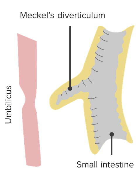
Meckel's diverticulum

A Meckel’s diverticulum

Image by Lecturio.
Related videos

Pathophysiology and Clinical Presentation

Anatomy

  • True diverticulum Diverticulum A pouch or sac opening from the colon. Diverticular Disease: contains all histologic layers of the intestinal tract
  • Located in the middle to distal ileum Ileum The distal and narrowest portion of the small intestine, between the jejunum and the ileocecal valve of the large intestine. Small Intestine: Anatomy (2 feet from the ileocecal valve Ileocecal valve The valve, at the junction of the cecum with the colon, that guards the opening where the ileum enters the large intestine. Small Intestine: Anatomy)  
  • Usually arises from the antimesenteric surface
  • Mucosal lining:
    • Resembles a normal small intestine Small intestine The small intestine is the longest part of the GI tract, extending from the pyloric orifice of the stomach to the ileocecal junction. The small intestine is the major organ responsible for chemical digestion and absorption of nutrients. It is divided into 3 segments: the duodenum, the jejunum, and the ileum. Small Intestine: Anatomy
    • But may contain ectopic ( heterotopic Heterotopic Transplantation of tissue typical of one area to a different recipient site. The tissue may be autologous, heterologous, or homologous. Organ Transplantation) mucosa (12%–44%):
      • Gastric (most common)
      • Pancreatic
      • Colonic
  • Blood supply: vitelline artery (a branch of the superior mesenteric artery Superior mesenteric artery A large vessel supplying the whole length of the small intestine except the superior part of the duodenum. It also supplies the cecum and the ascending part of the colon and about half the transverse part of the colon. It arises from the anterior surface of the aorta below the celiac artery at the level of the first lumbar vertebra. Small Intestine: Anatomy)
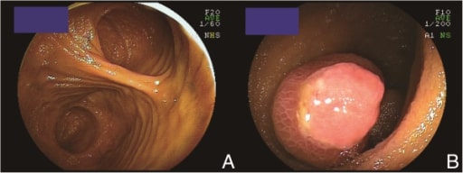
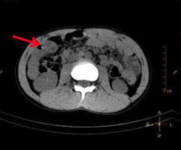
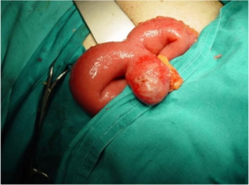
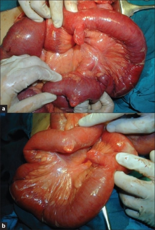
Double meckel's diverticulum

Images (a) and (b) showing a double Meckel’s diverticulum

Image: “Photograph showing double Meckel’s diverticulum” by the Department of General Surgery, Fr. Muller Medical College Hospital, Kankanady, Mangalore (D.K.), Karnataka, India. License: CC BY 2.0.
Pathophysiology

  • GI bleeding:
    • Occurs if a Meckel’s diverticulum Diverticulum A pouch or sac opening from the colon. Diverticular Disease contains ectopic gastric mucosa Gastric mucosa Lining of the stomach, consisting of an inner epithelium, a middle lamina propria, and an outer muscularis mucosae. The surface cells produce mucus that protects the stomach from attack by digestive acid and enzymes. When the epithelium invaginates into the lamina propria at various region of the stomach (cardia; gastric fundus; and pylorus), different tubular gastric glands are formed. These glands consist of cells that secrete mucus, enzymes, hydrochloric acid, or hormones. Stomach: Anatomy
    • Gastric mucosa Gastric mucosa Lining of the stomach, consisting of an inner epithelium, a middle lamina propria, and an outer muscularis mucosae. The surface cells produce mucus that protects the stomach from attack by digestive acid and enzymes. When the epithelium invaginates into the lamina propria at various region of the stomach (cardia; gastric fundus; and pylorus), different tubular gastric glands are formed. These glands consist of cells that secrete mucus, enzymes, hydrochloric acid, or hormones. Stomach: Anatomy secretes acid and ulceration Ulceration Corneal Abrasions, Erosion, and Ulcers occurs → ulceration Ulceration Corneal Abrasions, Erosion, and Ulcers is downstream or in the adjacent small bowel Small bowel The small intestine is the longest part of the GI tract, extending from the pyloric orifice of the stomach to the ileocecal junction. The small intestine is the major organ responsible for chemical digestion and absorption of nutrients. It is divided into 3 segments: the duodenum, the jejunum, and the ileum. Small Intestine: Anatomy mucosa → bleeding
  • Meckel’s diverticulitis Diverticulitis Inflammation of a diverticulum or diverticula. Diverticular Disease:
    • Acute inflammation Acute Inflammation Inflammation of a Meckel’s diverticulum Diverticulum A pouch or sac opening from the colon. Diverticular Disease (similar to appendicitis Appendicitis Appendicitis is the acute inflammation of the vermiform appendix and the most common abdominal surgical emergency globally. The condition has a lifetime risk of 8%. Characteristic features include periumbilical abdominal pain that migrates to the right lower quadrant, fever, anorexia, nausea, and vomiting. Appendicitis)
    • Diverticular opening becomes obstructed ( fecalith Fecalith Imaging of the Intestines, food, foreign body Foreign Body Foreign Body Aspiration, tumor Tumor Inflammation), leading to bacterial overgrowth Bacterial overgrowth Lactose Intolerance and inflammation Inflammation Inflammation is a complex set of responses to infection and injury involving leukocytes as the principal cellular mediators in the body’s defense against pathogenic organisms. Inflammation is also seen as a response to tissue injury in the process of wound healing. The 5 cardinal signs of inflammation are pain, heat, redness, swelling, and loss of function. Inflammation.
    • May result in ischemia Ischemia A hypoperfusion of the blood through an organ or tissue caused by a pathologic constriction or obstruction of its blood vessels, or an absence of blood circulation. Ischemic Cell Damage and perforation Perforation A pathological hole in an organ, blood vessel or other soft part of the body, occurring in the absence of external force. Esophagitis in severe cases
  • Small bowel obstruction Bowel obstruction Any impairment, arrest, or reversal of the normal flow of intestinal contents toward the anal canal. Ascaris/Ascariasis ( SBO SBO Small bowel obstruction (SBO) is an interruption of the flow of the intraluminal contents through the small intestine, and is classified as mechanical (due to physical blockage) or functional (due to disruption of normal motility). The most common cause of SBO in the Western countries is post-surgical adhesions. Small bowel obstruction typically presents with nausea, vomiting, abdominal pain, distention, constipation, and/or obstipation. Small Bowel Obstruction) may result from several mechanisms:
    • Intussusception Intussusception Intussusception occurs when a part of the intestine (intussusceptum) telescopes into another part (intussuscipiens) of the intestine. The condition can cause obstruction and, if untreated, progress to bowel ischemia. Intussusception is most common in the pediatric population, but is occasionally encountered in adults. Intussusception:
      • Part of the bowel telescopes into itself.
      • A Meckel’s diverticulum Diverticulum A pouch or sac opening from the colon. Diverticular Disease acts as a lead point Lead point A lesion that gets trapped during peristalsis, dragging that segment into the distal part of the intestine Intussusception.
    • Volvulus Volvulus A volvulus is the twisting or axial rotation of a portion of the bowel around its mesentery. The most common site of volvulus in adults is the colon; most frequently the sigmoid volvulus. Patients typically present with symptoms of bowel obstruction such as abdominal pain, distension, vomiting, and constipation/obstipation. Volvulus:
      • A Meckel’s diverticulum Diverticulum A pouch or sac opening from the colon. Diverticular Disease sometimes has fibrous Fibrous Fibrocystic Change bands connecting to the peritoneum Peritoneum The peritoneum is a serous membrane lining the abdominopelvic cavity. This lining is formed by connective tissue and originates from the mesoderm. The membrane lines both the abdominal walls (as parietal peritoneum) and all of the visceral organs (as visceral peritoneum). Peritoneum: Anatomy/ abdominal wall Abdominal wall The outer margins of the abdomen, extending from the osteocartilaginous thoracic cage to the pelvis. Though its major part is muscular, the abdominal wall consists of at least seven layers: the skin, subcutaneous fat, deep fascia; abdominal muscles, transversalis fascia, extraperitoneal fat, and the parietal peritoneum. Surgical Anatomy of the Abdomen.
      • Intestines can twist around the fibrous Fibrous Fibrocystic Change bands.
    • Torsion: twisting of the diverticulum Diverticulum A pouch or sac opening from the colon. Diverticular Disease itself
    • Meckel’s diverticulitis Diverticulitis Inflammation of a diverticulum or diverticula. Diverticular Disease: Inflammation Inflammation Inflammation is a complex set of responses to infection and injury involving leukocytes as the principal cellular mediators in the body’s defense against pathogenic organisms. Inflammation is also seen as a response to tissue injury in the process of wound healing. The 5 cardinal signs of inflammation are pain, heat, redness, swelling, and loss of function. Inflammation can narrow the lumen of the adjacent small bowel Small bowel The small intestine is the longest part of the GI tract, extending from the pyloric orifice of the stomach to the ileocecal junction. The small intestine is the major organ responsible for chemical digestion and absorption of nutrients. It is divided into 3 segments: the duodenum, the jejunum, and the ileum. Small Intestine: Anatomy.
    • Littre’s hernia Hernia Protrusion of tissue, structure, or part of an organ through the bone, muscular tissue, or the membrane by which it is normally contained. Hernia may involve tissues such as the abdominal wall or the respiratory diaphragm. Hernias may be internal, external, congenital, or acquired. Abdominal Hernias: The diverticulum Diverticulum A pouch or sac opening from the colon. Diverticular Disease becomes incarcerated into a hernia Hernia Protrusion of tissue, structure, or part of an organ through the bone, muscular tissue, or the membrane by which it is normally contained. Hernia may involve tissues such as the abdominal wall or the respiratory diaphragm. Hernias may be internal, external, congenital, or acquired. Abdominal Hernias.
    • Inversion: The diverticulum Diverticulum A pouch or sac opening from the colon. Diverticular Disease inverts into a bowel lumen and causes intermittent obstruction.

Clinical presentation

  • Frequently clinically silent
  • 25%–50% of symptomatic patients Patients Individuals participating in the health care system for the purpose of receiving therapeutic, diagnostic, or preventive procedures. Clinician–Patient Relationship are < 10 years of age.
  • In adults (especially < 40 years of age): A Meckel’s diverticulum Diverticulum A pouch or sac opening from the colon. Diverticular Disease is suspected if there is no identifiable source of GI bleeding.
  • Presentation similar in children and adults:
    • Painless lower GI bleeding (most common):
    • SBO SBO Small bowel obstruction (SBO) is an interruption of the flow of the intraluminal contents through the small intestine, and is classified as mechanical (due to physical blockage) or functional (due to disruption of normal motility). The most common cause of SBO in the Western countries is post-surgical adhesions. Small bowel obstruction typically presents with nausea, vomiting, abdominal pain, distention, constipation, and/or obstipation. Small Bowel Obstruction:
      • Nausea Nausea An unpleasant sensation in the stomach usually accompanied by the urge to vomit. Common causes are early pregnancy, sea and motion sickness, emotional stress, intense pain, food poisoning, and various enteroviruses. Antiemetics/ vomiting Vomiting The forcible expulsion of the contents of the stomach through the mouth. Hypokalemia
      • Cramping abdominal pain Abdominal Pain Acute Abdomen
      • Abdominal distention Abdominal distention Megacolon
      • In children: most commonly in the form of recurrent intussusception Intussusception Intussusception occurs when a part of the intestine (intussusceptum) telescopes into another part (intussuscipiens) of the intestine. The condition can cause obstruction and, if untreated, progress to bowel ischemia. Intussusception is most common in the pediatric population, but is occasionally encountered in adults. Intussusception
    • Meckel’s diverticulitis Diverticulitis Inflammation of a diverticulum or diverticula. Diverticular Disease:
      • Symptoms similar to acute appendicitis Appendicitis Appendicitis is the acute inflammation of the vermiform appendix and the most common abdominal surgical emergency globally. The condition has a lifetime risk of 8%. Characteristic features include periumbilical abdominal pain that migrates to the right lower quadrant, fever, anorexia, nausea, and vomiting. Appendicitis
      • With signs of peritoneal irritation if perforated
      • Abdominal tenderness is usually more midline.

Diagnosis

Tests for GI bleeding

  • Radiology:
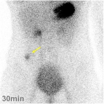
    • Meckel’s scan:
      • Used as a 1st-line test in hemodynamically stable patients Hemodynamically Stable Patients Blunt Chest Trauma and if suspicion is high (usually children)
      • Nuclear medicine Nuclear medicine A specialty field of radiology concerned with diagnostic, therapeutic, and investigative use of radioactive compounds. Nuclear Imaging scan utilizing radioactively labeled technetium that binds to gastric mucosa Gastric mucosa Lining of the stomach, consisting of an inner epithelium, a middle lamina propria, and an outer muscularis mucosae. The surface cells produce mucus that protects the stomach from attack by digestive acid and enzymes. When the epithelium invaginates into the lamina propria at various region of the stomach (cardia; gastric fundus; and pylorus), different tubular gastric glands are formed. These glands consist of cells that secrete mucus, enzymes, hydrochloric acid, or hormones. Stomach: Anatomy
      • Ectopic gastric mucosa Gastric mucosa Lining of the stomach, consisting of an inner epithelium, a middle lamina propria, and an outer muscularis mucosae. The surface cells produce mucus that protects the stomach from attack by digestive acid and enzymes. When the epithelium invaginates into the lamina propria at various region of the stomach (cardia; gastric fundus; and pylorus), different tubular gastric glands are formed. These glands consist of cells that secrete mucus, enzymes, hydrochloric acid, or hormones. Stomach: Anatomy is identified on scintigraphy Scintigraphy Sjögren’s Syndrome.
    • Arteriography:
      • If the bleeding is brisk enough to necessitate blood transfusion
      • An anomalous branch of the superior mesenteric artery Superior mesenteric artery A large vessel supplying the whole length of the small intestine except the superior part of the duodenum. It also supplies the cecum and the ascending part of the colon and about half the transverse part of the colon. It arises from the anterior surface of the aorta below the celiac artery at the level of the first lumbar vertebra. Small Intestine: Anatomy feeding the Meckel’s diverticulum Diverticulum A pouch or sac opening from the colon. Diverticular Disease can be identified.
      • Invasive test
    • Computed tomography (CT) angiography Angiography Radiography of blood vessels after injection of a contrast medium. Cardiac Surgery:
      • More sensitive than arteriography for less-brisk hemorrhage
      • Detects slow bleeding (0.3 mL/min)
  • Endoscopy Endoscopy Procedures of applying endoscopes for disease diagnosis and treatment. Endoscopy involves passing an optical instrument through a small incision in the skin i.e., percutaneous; or through a natural orifice and along natural body pathways such as the digestive tract; and/or through an incision in the wall of a tubular structure or organ, i.e. Transluminal, to examine or perform surgery on the interior parts of the body. Gastroesophageal Reflux Disease (GERD):
    • Capsule Capsule An envelope of loose gel surrounding a bacterial cell which is associated with the virulence of pathogenic bacteria. Some capsules have a well-defined border, whereas others form a slime layer that trails off into the medium. Most capsules consist of relatively simple polysaccharides but there are some bacteria whose capsules are made of polypeptides. Bacteroides endoscopy Endoscopy Procedures of applying endoscopes for disease diagnosis and treatment. Endoscopy involves passing an optical instrument through a small incision in the skin i.e., percutaneous; or through a natural orifice and along natural body pathways such as the digestive tract; and/or through an incision in the wall of a tubular structure or organ, i.e. Transluminal, to examine or perform surgery on the interior parts of the body. Gastroesophageal Reflux Disease (GERD): can visualize a normal, bleeding, inverted, and ulcerated Meckel’s diverticulum Diverticulum A pouch or sac opening from the colon. Diverticular Disease
    • Double-balloon enteroscopy:
      • Confirms diagnosis and complications by visualization
      • Enteroscope can be passed via the mouth into the small bowel Small bowel The small intestine is the longest part of the GI tract, extending from the pyloric orifice of the stomach to the ileocecal junction. The small intestine is the major organ responsible for chemical digestion and absorption of nutrients. It is divided into 3 segments: the duodenum, the jejunum, and the ileum. Small Intestine: Anatomy, or by retrograde fashion, through the colon Colon The large intestines constitute the last portion of the digestive system. The large intestine consists of the cecum, appendix, colon (with ascending, transverse, descending, and sigmoid segments), rectum, and anal canal. The primary function of the colon is to remove water and compact the stool prior to expulsion from the body via the rectum and anal canal. Colon, Cecum, and Appendix: Anatomy.
      • Requires specialized skills and longer time

Tests for SBO SBO Small bowel obstruction (SBO) is an interruption of the flow of the intraluminal contents through the small intestine, and is classified as mechanical (due to physical blockage) or functional (due to disruption of normal motility). The most common cause of SBO in the Western countries is post-surgical adhesions. Small bowel obstruction typically presents with nausea, vomiting, abdominal pain, distention, constipation, and/or obstipation. Small Bowel Obstruction and diverticulitis Diverticulitis Inflammation of a diverticulum or diverticula. Diverticular Disease

  • CT scan:
    • Will identify SBO SBO Small bowel obstruction (SBO) is an interruption of the flow of the intraluminal contents through the small intestine, and is classified as mechanical (due to physical blockage) or functional (due to disruption of normal motility). The most common cause of SBO in the Western countries is post-surgical adhesions. Small bowel obstruction typically presents with nausea, vomiting, abdominal pain, distention, constipation, and/or obstipation. Small Bowel Obstruction, inflammatory changes, perforation Perforation A pathological hole in an organ, blood vessel or other soft part of the body, occurring in the absence of external force. Esophagitis
    • A Meckel’s diverticulum Diverticulum A pouch or sac opening from the colon. Diverticular Disease itself is diagnosed correctly in about 50%.
  • Diagnostic laparoscopy Diagnostic Laparoscopy Laparotomy and Laparoscopy:
    • Performed if imaging studies are equivocal (difficult to distinguish an inflamed diverticulum Diverticulum A pouch or sac opening from the colon. Diverticular Disease from appendicitis Appendicitis Appendicitis is the acute inflammation of the vermiform appendix and the most common abdominal surgical emergency globally. The condition has a lifetime risk of 8%. Characteristic features include periumbilical abdominal pain that migrates to the right lower quadrant, fever, anorexia, nausea, and vomiting. Appendicitis on imaging)
    • Can be therapeutic as well as diagnostic

Management

Asymptomatic

  • A Meckel’s diverticulum Diverticulum A pouch or sac opening from the colon. Diverticular Disease incidentally found on imaging: no treatment necessary
  • A Meckel’s diverticulum Diverticulum A pouch or sac opening from the colon. Diverticular Disease found during surgery (for another condition):
    • Considerations:
      • Clinical status (higher perioperative risk in older patients Patients Individuals participating in the health care system for the purpose of receiving therapeutic, diagnostic, or preventive procedures. Clinician–Patient Relationship)
      • Life-long risk of complications (higher in children)
      • Diverticular abnormalities or features (that increase the risk of complications)
    • Resection recommended:
      • In children 
      • In healthy (< 50 years of age) adults: if a Meckel’s diverticulum Diverticulum A pouch or sac opening from the colon. Diverticular Disease is > 2 cm long or palpable abnormalities/ fibrous Fibrous Fibrocystic Change bands are noted
      • In patients Patients Individuals participating in the health care system for the purpose of receiving therapeutic, diagnostic, or preventive procedures. Clinician–Patient Relationship > 50 years of age or with comorbidities Comorbidities The presence of co-existing or additional diseases with reference to an initial diagnosis or with reference to the index condition that is the subject of study. Comorbidity may affect the ability of affected individuals to function and also their survival; it may be used as a prognostic indicator for length of hospital stay, cost factors, and outcome or survival. St. Louis Encephalitis Virus: if there is a palpable abnormality ( heterotopic Heterotopic Transplantation of tissue typical of one area to a different recipient site. The tissue may be autologous, heterologous, or homologous. Organ Transplantation mucosa, possible tumor Tumor Inflammation, fibrous Fibrous Fibrocystic Change bands)
    • No resection recommended: if patient is > 50 years of age and there is no palpable abnormality
  • Counseling should be provided regarding possible future symptoms.

Symptomatic

  • Supportive:
    • Intravenous hydration and resuscitation Resuscitation The restoration to life or consciousness of one apparently dead. . Neonatal Respiratory Distress Syndrome
    • Blood transfusion if necessary (for GI bleeding)
    • Bowel rest, nasogastric decompression (for SBO SBO Small bowel obstruction (SBO) is an interruption of the flow of the intraluminal contents through the small intestine, and is classified as mechanical (due to physical blockage) or functional (due to disruption of normal motility). The most common cause of SBO in the Western countries is post-surgical adhesions. Small bowel obstruction typically presents with nausea, vomiting, abdominal pain, distention, constipation, and/or obstipation. Small Bowel Obstruction)
    • Intravenous antibiotics (for Meckel’s diverticulitis Diverticulitis Inflammation of a diverticulum or diverticula. Diverticular Disease)
  • Surgery (definitive treatment):
    • Emergent if signs of sepsis Sepsis Systemic inflammatory response syndrome with a proven or suspected infectious etiology. When sepsis is associated with organ dysfunction distant from the site of infection, it is called severe sepsis. When sepsis is accompanied by hypotension despite adequate fluid infusion, it is called septic shock. Sepsis and Septic Shock, peritonitis Peritonitis Inflammation of the peritoneum lining the abdominal cavity as the result of infectious, autoimmune, or chemical processes. Primary peritonitis is due to infection of the peritoneal cavity via hematogenous or lymphatic spread and without intra-abdominal source. Secondary peritonitis arises from the abdominal cavity itself through rupture or abscess of intra-abdominal organs. Penetrating Abdominal Injury, perforation Perforation A pathological hole in an organ, blood vessel or other soft part of the body, occurring in the absence of external force. Esophagitis
    • Resection of a Meckel’s diverticulum Diverticulum A pouch or sac opening from the colon. Diverticular Disease
    • Segmental small bowel Small bowel The small intestine is the longest part of the GI tract, extending from the pyloric orifice of the stomach to the ileocecal junction. The small intestine is the major organ responsible for chemical digestion and absorption of nutrients. It is divided into 3 segments: the duodenum, the jejunum, and the ileum. Small Intestine: Anatomy resection (including a Meckel’s diverticulum Diverticulum A pouch or sac opening from the colon. Diverticular Disease):
      • To include ulcerated bleeding mucosa
      • If the adjacent small bowel Small bowel The small intestine is the longest part of the GI tract, extending from the pyloric orifice of the stomach to the ileocecal junction. The small intestine is the major organ responsible for chemical digestion and absorption of nutrients. It is divided into 3 segments: the duodenum, the jejunum, and the ileum. Small Intestine: Anatomy is severely inflamed/ischemic

Differential Diagnosis

  • Acute appendicitis Appendicitis Appendicitis is the acute inflammation of the vermiform appendix and the most common abdominal surgical emergency globally. The condition has a lifetime risk of 8%. Characteristic features include periumbilical abdominal pain that migrates to the right lower quadrant, fever, anorexia, nausea, and vomiting. Appendicitis: presents with abdominal pain Abdominal Pain Acute Abdomen and tenderness mostly in the right lower quadrant Right lower quadrant Anterior Abdominal Wall: Anatomy. A CT scan can help differentiate between a Meckel’s diverticulum Diverticulum A pouch or sac opening from the colon. Diverticular Disease and appendicitis Appendicitis Appendicitis is the acute inflammation of the vermiform appendix and the most common abdominal surgical emergency globally. The condition has a lifetime risk of 8%. Characteristic features include periumbilical abdominal pain that migrates to the right lower quadrant, fever, anorexia, nausea, and vomiting. Appendicitis, but not with 100% accuracy. Sometimes the definitive diagnosis is only made at surgery.
  • Intussusception Intussusception Intussusception occurs when a part of the intestine (intussusceptum) telescopes into another part (intussuscipiens) of the intestine. The condition can cause obstruction and, if untreated, progress to bowel ischemia. Intussusception is most common in the pediatric population, but is occasionally encountered in adults. Intussusception due to other causes: in infants and toddlers, classically presents as intermittent colicky abdominal pain Abdominal Pain Acute Abdomen with currant jelly stools. Adults and older children present with symptoms of bowel obstruction Bowel obstruction Any impairment, arrest, or reversal of the normal flow of intestinal contents toward the anal canal. Ascaris/Ascariasis. Intussusception Intussusception Intussusception occurs when a part of the intestine (intussusceptum) telescopes into another part (intussuscipiens) of the intestine. The condition can cause obstruction and, if untreated, progress to bowel ischemia. Intussusception is most common in the pediatric population, but is occasionally encountered in adults. Intussusception in young children is often idiopathic Idiopathic Dermatomyositis, but in adults, intussusception Intussusception Intussusception occurs when a part of the intestine (intussusceptum) telescopes into another part (intussuscipiens) of the intestine. The condition can cause obstruction and, if untreated, progress to bowel ischemia. Intussusception is most common in the pediatric population, but is occasionally encountered in adults. Intussusception is frequently associated with tumors.
  • Volvulus Volvulus A volvulus is the twisting or axial rotation of a portion of the bowel around its mesentery. The most common site of volvulus in adults is the colon; most frequently the sigmoid volvulus. Patients typically present with symptoms of bowel obstruction such as abdominal pain, distension, vomiting, and constipation/obstipation. Volvulus (due to malrotation Malrotation Pediatric Gastrointestinal Abnormalities): congenital Congenital Chorioretinitis condition that usually manifests in infancy, but sometimes not until adulthood. Twisting of the mesentery Mesentery A layer of the peritoneum which attaches the abdominal viscera to the abdominal wall and conveys their blood vessels and nerves. Peritoneum: Anatomy results in intestinal ischemia Ischemia A hypoperfusion of the blood through an organ or tissue caused by a pathologic constriction or obstruction of its blood vessels, or an absence of blood circulation. Ischemic Cell Damage. A volvulus Volvulus A volvulus is the twisting or axial rotation of a portion of the bowel around its mesentery. The most common site of volvulus in adults is the colon; most frequently the sigmoid volvulus. Patients typically present with symptoms of bowel obstruction such as abdominal pain, distension, vomiting, and constipation/obstipation. Volvulus presents with severe abdominal pain Abdominal Pain Acute Abdomen and bloody stools in advanced cases. A volvulus Volvulus A volvulus is the twisting or axial rotation of a portion of the bowel around its mesentery. The most common site of volvulus in adults is the colon; most frequently the sigmoid volvulus. Patients typically present with symptoms of bowel obstruction such as abdominal pain, distension, vomiting, and constipation/obstipation. Volvulus requires emergent surgery.
  • Colonic diverticulosis Diverticulosis A pathological condition characterized by the presence of a number of colonic diverticula in the colon. Its pathogenesis is multifactorial, including colon aging, motor dysfunction, increases in intraluminal pressure, and lack of dietary fibers. Diverticular Disease and arteriovenous (AV) malformations: most common causes of painless lower GI bleeding in older adults. Colonic diverticulosis Diverticulosis A pathological condition characterized by the presence of a number of colonic diverticula in the colon. Its pathogenesis is multifactorial, including colon aging, motor dysfunction, increases in intraluminal pressure, and lack of dietary fibers. Diverticular Disease and AV malformations can present with acute ( hematochezia Hematochezia Gastrointestinal Bleeding) or chronic slow bleeding ( melena Melena The black, tarry, foul-smelling feces that contain degraded blood. Gastrointestinal Bleeding). Diagnosis is usually established by colonoscopy Colonoscopy Endoscopic examination, therapy or surgery of the luminal surface of the colon. Colorectal Cancer Screening or angiography Angiography Radiography of blood vessels after injection of a contrast medium. Cardiac Surgery (in actively bleeding AV malformations).

References

  1. An, J., & Zabbo, C.P. (2020). Meckel diverticulum. https://www.ncbi.nlm.nih.gov/books/NBK499960/
  2. Javid, P.J., & Pauli, E.M. (2020). Meckel’s diverticulum. Retrieved 01 December 2020, from https://www.uptodate.com/contents/meckels-diverticulum?search=meckel%20diverticulum&source=search_result&selectedTitle=1~43&usage_type=default&display_rank=1#H3152275

Create your free account or log in to continue reading!

Sign up now and get free access to Lecturio with concept pages, medical videos, and questions for your medical education.

User Reviews

Details