Macrolides and Ketolides

Macrolides and ketolides are antibiotics that inhibit bacterial protein synthesis Synthesis Polymerase Chain Reaction (PCR) by binding to the 50S ribosomal subunit and blocking transpeptidation Transpeptidation Lincosamides. These antibiotics have a broad spectrum of antimicrobial activity but are best known for their coverage of atypical microorganisms. Common macrolides are erythromycin, clarithromycin, azithromycin, and fidaxomicin. Side effects include GI upset, QT prolongation, and hepatotoxicity Hepatotoxicity Acetaminophen. Resistance Resistance Physiologically, the opposition to flow of air caused by the forces of friction. As a part of pulmonary function testing, it is the ratio of driving pressure to the rate of air flow. Ventilation: Mechanics of Breathing mechanisms include ribosomal methylation Methylation Addition of methyl groups. In histo-chemistry methylation is used to esterify carboxyl groups and remove sulfate groups by treating tissue sections with hot methanol in the presence of hydrochloric acid. . Glucocorticoids or mutation Mutation Genetic mutations are errors in DNA that can cause protein misfolding and dysfunction. There are various types of mutations, including chromosomal, point, frameshift, and expansion mutations. Types of Mutations, inactivation of the drug through enzyme production, or removal of the drug through efflux pumps Efflux Pumps Lincosamides.

Last updated: Oct 14, 2022

Editorial responsibility: Stanley Oiseth, Lindsay Jones, Evelin Maza

Chemistry and Pharmacodynamics

Chemical structure

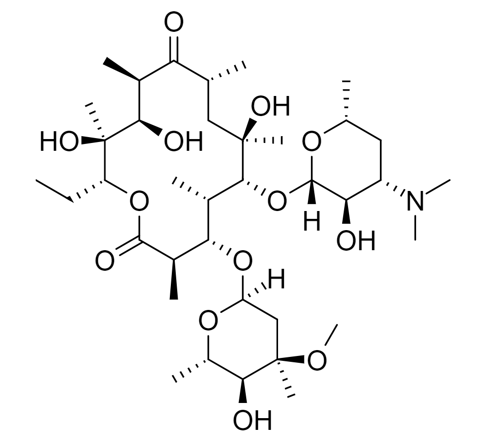
  • Macrolides: characterized by a macrocyclic lactone ring Lactone Ring Polyenes
    • Ring contains 14‒16 atoms
    • 1 or more sugars are attached via glycosidic bonds
    • Erythromycin is the prototype drug of the class.
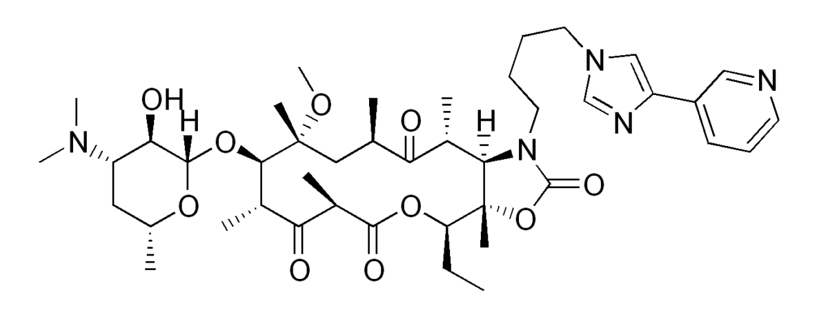
  • Ketolides (e.g., telithromycin): similar structure to macrolides 
    • 14-atom ring
    • 1 of the sugars is substituted with a keto group
    • Derived from erythromycin

Mechanism of action

General:

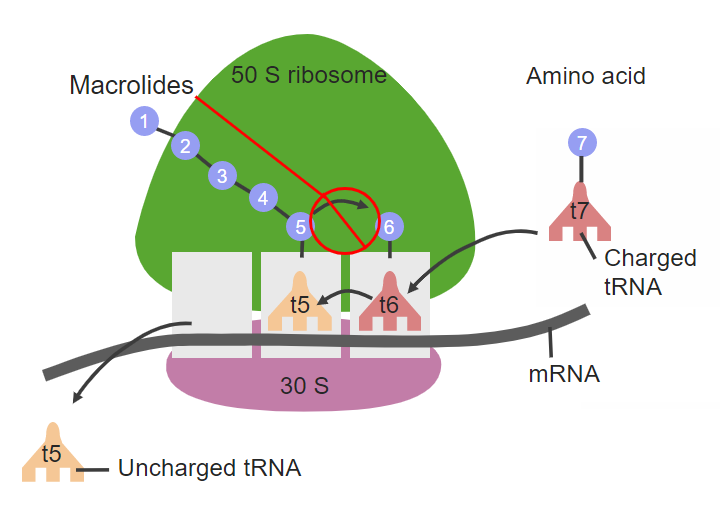
  • Inhibits bacterial protein synthesis Synthesis Polymerase Chain Reaction (PCR) by binding reversibly to the 50S ribosomal subunit   
  • Limits bacterial growth → bacteriostatic Bacteriostatic Sulfonamides and Trimethoprim
  • Additional effects:
    • Antiinflammatory effect by ↓ interleukins Interleukins Interleukins are a type of cytokines (signaling proteins) that communicate messages between different parts of the immune system. The majority of interleukins are synthesized by helper CD4 T lymphocytes along with other cells such as monocytes, macrophages, and endothelial cells. Interleukins and tumor Tumor Inflammation necrosis Necrosis The death of cells in an organ or tissue due to disease, injury or failure of the blood supply. Ischemic Cell Damage factor-alpha
    • Erythromycin is a motilin Motilin A peptide of about 22-amino acids isolated from the duodenum. At low ph it inhibits gastric motor activity, whereas at high ph it has a stimulating effect. Gastrointestinal Secretions receptor Receptor Receptors are proteins located either on the surface of or within a cell that can bind to signaling molecules known as ligands (e.g., hormones) and cause some type of response within the cell. Receptors agonist at duodenal enterochromaffin cells → has prokinetic properties

Fidaxomicin is an exception:

  • Binds to RNA RNA A polynucleotide consisting essentially of chains with a repeating backbone of phosphate and ribose units to which nitrogenous bases are attached. RNA is unique among biological macromolecules in that it can encode genetic information, serve as an abundant structural component of cells, and also possesses catalytic activity. RNA Types and Structure polymerase → prevents RNA RNA A polynucleotide consisting essentially of chains with a repeating backbone of phosphate and ribose units to which nitrogenous bases are attached. RNA is unique among biological macromolecules in that it can encode genetic information, serve as an abundant structural component of cells, and also possesses catalytic activity. RNA Types and Structure transcription Transcription Transcription of genetic information is the first step in gene expression. Transcription is the process by which DNA is used as a template to make mRNA. This process is divided into 3 stages: initiation, elongation, and termination. Stages of Transcription
  • Bactericidal Bactericidal Penicillins
The site of action for macrolides

The site of action for macrolides on the 50S ribosomal subunit
tRNA: transfer RNA
mRNA: messenger RNA

Image by Lecturio. License: CC BY-NC-SA 4.0

Pharmacokinetics

Table: Pharmacokinetics Pharmacokinetics Pharmacokinetics is the science that analyzes how the human body interacts with a drug. Pharmacokinetics examines how the drug is absorbed, distributed, metabolized, and excreted by the body. Pharmacokinetics and Pharmacodynamics of macrolides and ketolides
Macrolides Ketolides
Absorption Absorption Absorption involves the uptake of nutrient molecules and their transfer from the lumen of the GI tract across the enterocytes and into the interstitial space, where they can be taken up in the venous or lymphatic circulation. Digestion and Absorption Erythromycin:
  • Destroyed by stomach Stomach The stomach is a muscular sac in the upper left portion of the abdomen that plays a critical role in digestion. The stomach develops from the foregut and connects the esophagus with the duodenum. Structurally, the stomach is C-shaped and forms a greater and lesser curvature and is divided grossly into regions: the cardia, fundus, body, and pylorus. Stomach: Anatomy acid → requires enteric coating
  • Relatively poor absorption Absorption Absorption involves the uptake of nutrient molecules and their transfer from the lumen of the GI tract across the enterocytes and into the interstitial space, where they can be taken up in the venous or lymphatic circulation. Digestion and Absorption
  • Food interferes with absorption Absorption Absorption involves the uptake of nutrient molecules and their transfer from the lumen of the GI tract across the enterocytes and into the interstitial space, where they can be taken up in the venous or lymphatic circulation. Digestion and Absorption
Azithromycin and clarithromycin:
  • Stable in the stomach Stomach The stomach is a muscular sac in the upper left portion of the abdomen that plays a critical role in digestion. The stomach develops from the foregut and connects the esophagus with the duodenum. Structurally, the stomach is C-shaped and forms a greater and lesser curvature and is divided grossly into regions: the cardia, fundus, body, and pylorus. Stomach: Anatomy
  • Better absorption Absorption Absorption involves the uptake of nutrient molecules and their transfer from the lumen of the GI tract across the enterocytes and into the interstitial space, where they can be taken up in the venous or lymphatic circulation. Digestion and Absorption
Fidaxomicin:
  • Minimal systemic absorption Absorption Absorption involves the uptake of nutrient molecules and their transfer from the lumen of the GI tract across the enterocytes and into the interstitial space, where they can be taken up in the venous or lymphatic circulation. Digestion and Absorption
  • Reaches high fecal concentrations
Rapid absorption Absorption Absorption involves the uptake of nutrient molecules and their transfer from the lumen of the GI tract across the enterocytes and into the interstitial space, where they can be taken up in the venous or lymphatic circulation. Digestion and Absorption
Distribution
  • Lipophilic
  • Widely distributed in body fluids and tissues
  • Uptake by macrophages Macrophages The relatively long-lived phagocytic cell of mammalian tissues that are derived from blood monocytes. Main types are peritoneal macrophages; alveolar macrophages; histiocytes; kupffer cells of the liver; and osteoclasts. They may further differentiate within chronic inflammatory lesions to epithelioid cells or may fuse to form foreign body giant cells or langhans giant cells. Innate Immunity: Phagocytes and Antigen Presentation:
    • Most notable in azithromycin
    • Contributes to a long half-life Half-Life The time it takes for a substance (drug, radioactive nuclide, or other) to lose half of its pharmacologic, physiologic, or radiologic activity. Pharmacokinetics and Pharmacodynamics
  • Cross the placenta Placenta A highly vascularized mammalian fetal-maternal organ and major site of transport of oxygen, nutrients, and fetal waste products. It includes a fetal portion (chorionic villi) derived from trophoblasts and a maternal portion (decidua) derived from the uterine endometrium. The placenta produces an array of steroid, protein and peptide hormones (placental hormones). Placenta, Umbilical Cord, and Amniotic Cavity
  • Do not cross the blood-brain barrier Blood-brain barrier Specialized non-fenestrated tightly-joined endothelial cells with tight junctions that form a transport barrier for certain substances between the cerebral capillaries and the brain tissue. Systemic and Special Circulations
  • Exception: fidaxomicin acts locally in the GI lumen
  • Widely distributed in tissues
  • 70% protein bound ( albumin Albumin Serum albumin from humans. It is an essential carrier of both endogenous substances, such as fatty acids and bilirubin, and of xenobiotics in the blood. Liver Function Tests)
Metabolism
Excretion
  • Hepatic ( bile Bile An emulsifying agent produced in the liver and secreted into the duodenum. Its composition includes bile acids and salts; cholesterol; and electrolytes. It aids digestion of fats in the duodenum. Gallbladder and Biliary Tract: Anatomy → feces):
    • Azithromycin
    • Erythromycin
  • Renal: clarithromycin
  • Fecal: fidaxomicin
  • Renal
  • Feces

Indications

Antimicrobial coverage

  • Gram positive Gram positive Bacteria which retain the crystal violet stain when treated by gram’s method. Nocardia/Nocardiosis:
    • Streptococcus Streptococcus Streptococcus is one of the two medically important genera of gram-positive cocci, the other being Staphylococcus. Streptococci are identified as different species on blood agar on the basis of their hemolytic pattern and sensitivity to optochin and bacitracin. There are many pathogenic species of streptococci, including S. pyogenes, S. agalactiae, S. pneumoniae, and the viridans streptococci. Streptococcus
    • Staphylococcus Staphylococcus Staphylococcus is a medically important genera of Gram-positive, aerobic cocci. These bacteria form clusters resembling grapes on culture plates. Staphylococci are ubiquitous for humans, and many strains compose the normal skin flora. Staphylococcus
    • Clostridioides difficile (fidaxomicin)
  • Gram negative Gram negative Bacteria which lose crystal violet stain but are stained pink when treated by gram’s method. Yersinia spp./Yersiniosis:
    • Escherichia coli Escherichia coli The gram-negative bacterium Escherichia coli is a key component of the human gut microbiota. Most strains of E. coli are avirulent, but occasionally they escape the GI tract, infecting the urinary tract and other sites. Less common strains of E. coli are able to cause disease within the GI tract, most commonly presenting as abdominal pain and diarrhea. Escherichia coli
    • Haemophilus influenzae Haemophilus Influenzae A species of Haemophilus found on the mucous membranes of humans and a variety of animals. The species is further divided into biotypes I through viii. Haemophilus and H. ducreyi
    • Moraxella catarrhalis Moraxella catarrhalis Gram-negative aerobic cocci of low virulence that colonize the nasopharynx and occasionally cause meningitis; bacteremia; empyema; pericarditis; and pneumonia. Moraxella
    • Salmonella Salmonella Salmonellae are gram-negative bacilli of the family Enterobacteriaceae. Salmonellae are flagellated, non-lactose-fermenting, and hydrogen sulfide-producing microbes. Salmonella enterica, the most common disease-causing species in humans, is further classified based on serotype as typhoidal (S. typhi and paratyphi) and nontyphoidal (S. enteritidis and typhimurium). Salmonella
    • Yersinia enterocolitica Yersinia enterocolitica A species of the genus yersinia, isolated from both man and animal. It is a frequent cause of bacterial gastroenteritis in children. Yersinia spp./Yersiniosis
    • Shigella Shigella Shigella is a genus of gram-negative, non-lactose-fermenting facultative intracellular bacilli. Infection spreads most commonly via person-to-person contact or through contaminated food and water. Humans are the only known reservoir. Shigella
    • Campylobacter jejuni Campylobacter jejuni A species of bacteria that resemble small tightly coiled spirals. Its organisms are known to cause abortion in sheep and fever and enteritis in man and may be associated with enteric diseases of calves, lambs, and other animals. Campylobacter
    • Vibrio cholerae Vibrio cholerae The etiologic agent of cholera. Vibrio
    • Neisseria gonorrhoeae Neisseria gonorrhoeae A species of gram-negative, aerobic bacteria primarily found in purulent venereal discharges. It is the causative agent of gonorrhea. Neisseria
    • Helicobacter pylori Helicobacter pylori A spiral bacterium active as a human gastric pathogen. It is a gram-negative, urease-positive, curved or slightly spiral organism initially isolated in 1982 from patients with lesions of gastritis or peptic ulcers in Western Australia. Helicobacter pylori was originally classified in the genus campylobacter, but RNA sequencing, cellular fatty acid profiles, growth patterns, and other taxonomic characteristics indicate that the micro-organism should be included in the genus Helicobacter. It has been officially transferred to Helicobacter gen. Helicobacter
    • Bordetella pertussis Bordetella pertussis A species of gram-negative, aerobic bacteria that is the causative agent of whooping cough. Its cells are minute coccobacilli that are surrounded by a slime sheath. Bordetella
  • Atypical:
    • Mycoplasma pneumoniae Mycoplasma pneumoniae Short filamentous organism of the genus mycoplasma, which binds firmly to the cells of the respiratory epithelium. It is one of the etiologic agents of non-viral primary atypical pneumonia in man. Mycoplasma
    • Chlamydia pneumoniae Chlamydia pneumoniae A species of chlamydophila that causes acute respiratory infection, especially atypical pneumonia, in humans, horses, and koalas. Chlamydia
    • Legionella pneumophila Legionella pneumophila A species of gram-negative, aerobic bacteria that is the causative agent of legionnaires’ disease. It has been isolated from numerous environmental sites as well as from human lung tissue, respiratory secretions, and blood. Legionella/Legionellosis
    • Treponema pallidum Treponema pallidum The causative agent of venereal and non-venereal syphilis as well as yaws. Treponema
    • Babesia Babesia Babesiosis is an infection caused by a protozoa belonging to the genus, Babesia. The most common Babesia seen in the United States is B. microti, which is transmitted by the Ixodes tick. The protozoa thrive and replicate within host erythrocytes. Lysis of erythrocytes and the body’s immune response result in clinical symptoms. Babesia/Babesiosis microti
    • Ureaplasma
  • Mycobacterium avium Mycobacterium avium A bacterium causing tuberculosis in domestic fowl and other birds. In pigs, it may cause localized and sometimes disseminated disease. The organism occurs occasionally in sheep and cattle. It should be distinguished from the m. avium complex, which infects primarily humans. Mycobacterium complex (MAC)

Types of infection

  • Respiratory infections Infections Invasion of the host organism by microorganisms or their toxins or by parasites that can cause pathological conditions or diseases. Chronic Granulomatous Disease:
  • Sexually transmitted infections Infections Invasion of the host organism by microorganisms or their toxins or by parasites that can cause pathological conditions or diseases. Chronic Granulomatous Disease:
    • Gonorrhea Gonorrhea Gonorrhea is a sexually transmitted infection (STI) caused by the gram-negative bacteria Neisseria gonorrhoeae (N. gonorrhoeae). Gonorrhea may be asymptomatic but commonly manifests as cervicitis or urethritis with less common presentations such as proctitis, conjunctivitis, or pharyngitis. Gonorrhea
    • Chlamydia Chlamydia Chlamydiae are obligate intracellular gram-negative bacteria. They lack a peptidoglycan layer and are best visualized using Giemsa stain. The family of Chlamydiaceae comprises 3 pathogens that can infect humans: Chlamydia trachomatis, Chlamydia psittaci, and Chlamydia pneumoniae. Chlamydia
    • Chancroid Chancroid Chancroid is a highly transmissible STD caused by Haemophilus ducreyi. The disease presents with painful ulcer(s) on the genital tract (termed chancroid or “soft chancre”). Up to 50% of patients will develop painful inguinal lymphadenopathy. Chancroid
  • H. pylori H. pylori A spiral bacterium active as a human gastric pathogen. It is a gram-negative, urease-positive, curved or slightly spiral organism initially isolated in 1982 from patients with lesions of gastritis or peptic ulcers in Western Australia. Helicobacter pylori was originally classified in the genus campylobacter, but RNA sequencing, cellular fatty acid profiles, growth patterns, and other taxonomic characteristics indicate that the micro-organism should be included in the genus Helicobacter. It has been officially transferred to Helicobacter gen. Helicobacter infection (clarithromycin is part of triple therapy)
  • C. difficile-associated diarrhea Diarrhea Diarrhea is defined as ≥ 3 watery or loose stools in a 24-hour period. There are a multitude of etiologies, which can be classified based on the underlying mechanism of disease. The duration of symptoms (acute or chronic) and characteristics of the stools (e.g., watery, bloody, steatorrheic, mucoid) can help guide further diagnostic evaluation. Diarrhea (fidaxomicin is monotherapy)
  • Skin Skin The skin, also referred to as the integumentary system, is the largest organ of the body. The skin is primarily composed of the epidermis (outer layer) and dermis (deep layer). The epidermis is primarily composed of keratinocytes that undergo rapid turnover, while the dermis contains dense layers of connective tissue. Skin: Structure and Functions and soft tissue Soft Tissue Soft Tissue Abscess infections Infections Invasion of the host organism by microorganisms or their toxins or by parasites that can cause pathological conditions or diseases. Chronic Granulomatous Disease: acne

Other indications

Ketolide indications

Adverse Effects and Contraindications

Macrolides

  • Adverse effects:
    • GI upset (especially with erythromycin and fidaxomicin)
    • Dizziness Dizziness An imprecise term which may refer to a sense of spatial disorientation, motion of the environment, or lightheadedness. Lateral Medullary Syndrome (Wallenberg Syndrome)
    • Hepatotoxicity Hepatotoxicity Acetaminophen:
      • Abnormal liver function tests Liver function tests Liver function tests, also known as hepatic function panels, are one of the most commonly performed screening blood tests. Such tests are also used to detect, evaluate, and monitor acute and chronic liver diseases. Liver Function Tests
      • Hepatitis
      • Cholestatic jaundice Jaundice Jaundice is the abnormal yellowing of the skin and/or sclera caused by the accumulation of bilirubin. Hyperbilirubinemia is caused by either an increase in bilirubin production or a decrease in the hepatic uptake, conjugation, or excretion of bilirubin. Jaundice
      • Hepatic necrosis Necrosis The death of cells in an organ or tissue due to disease, injury or failure of the blood supply. Ischemic Cell Damage
      • Hepatic failure Hepatic failure Severe inability of the liver to perform its normal metabolic functions, as evidenced by severe jaundice and abnormal serum levels of ammonia; bilirubin; alkaline phosphatase; aspartate aminotransferase; lactate dehydrogenases; and albumin/globulin ratio. Autoimmune Hepatitis
    • QT prolongation
  • Contraindications Contraindications A condition or factor associated with a recipient that makes the use of a drug, procedure, or physical agent improper or inadvisable. Contraindications may be absolute (life threatening) or relative (higher risk of complications in which benefits may outweigh risks). Noninvasive Ventilation: history of hepatic impairment or cholestatic jaundice Jaundice Jaundice is the abnormal yellowing of the skin and/or sclera caused by the accumulation of bilirubin. Hyperbilirubinemia is caused by either an increase in bilirubin production or a decrease in the hepatic uptake, conjugation, or excretion of bilirubin. Jaundice
  • Warnings:
    • Altered cardiac conduction (especially those with QT prolongation and electrolyte abnormalities) 
    • Hepatotoxicity Hepatotoxicity Acetaminophen 
    • Myasthenia gravis Myasthenia Gravis Myasthenia gravis (MG) is an autoimmune neuromuscular disorder characterized by weakness and fatigability of skeletal muscles caused by dysfunction/destruction of acetylcholine receptors at the neuromuscular junction. MG presents with fatigue, ptosis, diplopia, dysphagia, respiratory difficulties, and progressive weakness in the limbs, leading to difficulty in movement. Myasthenia Gravis exacerbation
    • Clarithromycin: potential ↑ mortality Mortality All deaths reported in a given population. Measures of Health Status in patients Patients Individuals participating in the health care system for the purpose of receiving therapeutic, diagnostic, or preventive procedures. Clinician–Patient Relationship with coronary artery Coronary Artery Truncus Arteriosus disease  
  • Drug interactions (particularly with erythromycin and clarithromycin due to CYP3A4 CYP3A4 Class 3 Antiarrhythmic Drugs (Potassium Channel Blockers) inhibition):
    • Warfarin Warfarin An anticoagulant that acts by inhibiting the synthesis of vitamin K-dependent coagulation factors. Warfarin is indicated for the prophylaxis and/or treatment of venous thrombosis and its extension, pulmonary embolism, and atrial fibrillation with embolization. It is also used as an adjunct in the prophylaxis of systemic embolism after myocardial infarction. Warfarin is also used as a rodenticide. Anticoagulants: ↑ INR
    • Simvastatin Simvastatin A derivative of lovastatin and potent competitive inhibitor of 3-hydroxy-3-methylglutaryl coenzyme a reductase, which is the rate-limiting enzyme in cholesterol biosynthesis. It may also interfere with steroid hormone production. Due to the induction of hepatic ldl receptors, it increases breakdown of ldl cholesterol. Statins, lovastatin Lovastatin A fungal metabolite isolated from cultures of aspergillus terreus. The compound is a potent anticholesteremic agent. It inhibits 3-hydroxy-3-methylglutaryl coenzyme a reductase, which is the rate-limiting enzyme in cholesterol biosynthesis. It also stimulates the production of low-density lipoprotein receptors in the liver. Statins: ↑ risk of myalgia Myalgia Painful sensation in the muscles. Ion Channel Myopathy and/or rhabdomyolysis Rhabdomyolysis Rhabdomyolysis is characterized by muscle necrosis and the release of toxic intracellular contents, especially myoglobin, into the circulation. Rhabdomyolysis
    • Midazolam Midazolam A short-acting hypnotic-sedative drug with anxiolytic and amnestic properties. It is used in dentistry, cardiac surgery, endoscopic procedures, as preanesthetic medication, and as an adjunct to local anesthesia. The short duration and cardiorespiratory stability makes it useful in poor-risk, elderly, and cardiac patients. It is water-soluble at ph less than 4 and lipid-soluble at physiological pH. Benzodiazepines: somnolence
    • Theophylline Theophylline A methyl xanthine derivative from tea with diuretic, smooth muscle relaxant, bronchial dilation, cardiac and central nervous system stimulant activities. Theophylline inhibits the 3. Asthma Drugs: seizures Seizures A seizure is abnormal electrical activity of the neurons in the cerebral cortex that can manifest in numerous ways depending on the region of the brain affected. Seizures consist of a sudden imbalance that occurs between the excitatory and inhibitory signals in cortical neurons, creating a net excitation. The 2 major classes of seizures are focal and generalized. Seizures
    • ↑ Serum concentrations of:
      • Tacrolimus Tacrolimus A macrolide isolated from the culture broth of a strain of streptomyces tsukubaensis that has strong immunosuppressive activity in vivo and prevents the activation of T-lymphocytes in response to antigenic or mitogenic stimulation in vitro. Immunosuppressants 
      • Cyclosporine Cyclosporine A cyclic undecapeptide from an extract of soil fungi. It is a powerful immunosupressant with a specific action on T-lymphocytes. It is used for the prophylaxis of graft rejection in organ and tissue transplantation. Immunosuppressants
      • Ergot alkaloids Ergot alkaloids Alkaloids originally isolated from the ergot fungus claviceps purpurea (hypocreaceae). They include compounds that are structurally related to ergoline (ergolines) and ergotamine (ergotamines). Many of the ergot alkaloids act as alpha-adrenergic antagonists. Triptans and Ergot Alkaloids 
      • Colchicine Colchicine A major alkaloid from colchicum autumnale l. And found also in other colchicum species. Its primary therapeutic use is in the treatment of gout. Gout Drugs

Ketolides

  • Adverse effects:
    • GI upset
    • Visual disturbance
    • Hepatotoxicity Hepatotoxicity Acetaminophen
    • Syncope Syncope Syncope is a short-term loss of consciousness and loss of postural stability followed by spontaneous return of consciousness to the previous neurologic baseline without the need for resuscitation. The condition is caused by transient interruption of cerebral blood flow that may be benign or related to a underlying life-threatening condition. Syncope
  • Contraindications Contraindications A condition or factor associated with a recipient that makes the use of a drug, procedure, or physical agent improper or inadvisable. Contraindications may be absolute (life threatening) or relative (higher risk of complications in which benefits may outweigh risks). Noninvasive Ventilation:
    • Hypersensitivity to macrolide antibiotics
    • Myasthenia gravis Myasthenia Gravis Myasthenia gravis (MG) is an autoimmune neuromuscular disorder characterized by weakness and fatigability of skeletal muscles caused by dysfunction/destruction of acetylcholine receptors at the neuromuscular junction. MG presents with fatigue, ptosis, diplopia, dysphagia, respiratory difficulties, and progressive weakness in the limbs, leading to difficulty in movement. Myasthenia Gravis
    • Hepatitis or jaundice Jaundice Jaundice is the abnormal yellowing of the skin and/or sclera caused by the accumulation of bilirubin. Hyperbilirubinemia is caused by either an increase in bilirubin production or a decrease in the hepatic uptake, conjugation, or excretion of bilirubin. Jaundice
  • Warnings:
    • Hepatotoxicity Hepatotoxicity Acetaminophen
    • Myasthenia gravis Myasthenia Gravis Myasthenia gravis (MG) is an autoimmune neuromuscular disorder characterized by weakness and fatigability of skeletal muscles caused by dysfunction/destruction of acetylcholine receptors at the neuromuscular junction. MG presents with fatigue, ptosis, diplopia, dysphagia, respiratory difficulties, and progressive weakness in the limbs, leading to difficulty in movement. Myasthenia Gravis exacerbation
    • QT prolongation

Mechanism of Resistance

There are 3 methods of resistance Resistance Physiologically, the opposition to flow of air caused by the forces of friction. As a part of pulmonary function testing, it is the ratio of driving pressure to the rate of air flow. Ventilation: Mechanics of Breathing to macrolides:

  • Ribosomal methylation Methylation Addition of methyl groups. In histo-chemistry methylation is used to esterify carboxyl groups and remove sulfate groups by treating tissue sections with hot methanol in the presence of hydrochloric acid. . Glucocorticoids or mutation Mutation Genetic mutations are errors in DNA that can cause protein misfolding and dysfunction. There are various types of mutations, including chromosomal, point, frameshift, and expansion mutations. Types of Mutations:
    • Prevents macrolide binding
    • Can be either plasmid mediated or chromosomal
  • Production of drug-inactivating enzymes Enzymes Enzymes are complex protein biocatalysts that accelerate chemical reactions without being consumed by them. Due to the body’s constant metabolic needs, the absence of enzymes would make life unsustainable, as reactions would occur too slowly without these molecules. Basics of Enzymes:
    • Esterases
    • Kinases 
  • Production of active ATP-dependent efflux proteins Proteins Linear polypeptides that are synthesized on ribosomes and may be further modified, crosslinked, cleaved, or assembled into complex proteins with several subunits. The specific sequence of amino acids determines the shape the polypeptide will take, during protein folding, and the function of the protein. Energy Homeostasis
    • Transport the drug outside the cell
    • Ketolides are not affected by this resistance Resistance Physiologically, the opposition to flow of air caused by the forces of friction. As a part of pulmonary function testing, it is the ratio of driving pressure to the rate of air flow. Ventilation: Mechanics of Breathing mechanism → allows for their use in some macrolide-resistant strains

Comparison of Medications

Table: Comparison of several classes of bacterial protein synthesis Synthesis Polymerase Chain Reaction (PCR) inhibitor antibiotics
Drug class Mechanism of action Coverage Adverse effects
Amphenicols
  • Gram positives
  • Gram negatives
  • Atypicals
  • GI upset
  • Optic neuritis Optic neuritis Inflammation of the optic nerve. Commonly associated conditions include autoimmune disorders such as multiple sclerosis, infections, and granulomatous diseases. Clinical features include retro-orbital pain that is aggravated by eye movement, loss of color vision, and contrast sensitivity that may progress to severe visual loss, an afferent pupillary defect (Marcus-Gunn pupil), and in some instances optic disc hyperemia and swelling. Inflammation may occur in the portion of the nerve within the globe (neuropapillitis or anterior optic neuritis) or the portion behind the globe (retrobulbar neuritis or posterior optic neuritis). Cranial Nerve Palsies
  • Aplastic anemia Aplastic Anemia Aplastic anemia (AA) is a rare, life-threatening condition characterized by pancytopenia and hypocellularity of the bone marrow (in the absence of any abnormal cells) reflecting damage to hematopoietic stem cells. Aplastic anemia can be acquired or inherited, however, most cases of AA are acquired and caused by autoimmune damage to hematopoietic stem cells. Aplastic Anemia
  • Gray baby syndrome Gray Baby Syndrome Chloramphenicol
Lincosamides Lincosamides The lincosamides, lincomycin and clindamycin, are inhibitors of bacterial protein synthesis. Drugs in this class share the same binding site as that of macrolides and amphenicols; however, they differ in chemical structure. Lincosamides target the 50S ribosomal subunit and interfere with transpeptidation. Lincosamides
  • Gram-positive Gram-Positive Penicillins cocci Cocci Bacteriology:
    • MSSA
    • MRSA MRSA A strain of Staphylococcus aureus that is non-susceptible to the action of methicillin. The mechanism of resistance usually involves modification of normal or the presence of acquired penicillin binding proteins. Staphylococcus
    • Streptococcus Streptococcus Streptococcus is one of the two medically important genera of gram-positive cocci, the other being Staphylococcus. Streptococci are identified as different species on blood agar on the basis of their hemolytic pattern and sensitivity to optochin and bacitracin. There are many pathogenic species of streptococci, including S. pyogenes, S. agalactiae, S. pneumoniae, and the viridans streptococci. Streptococcus
  • Anaerobes Anaerobes Lincosamides
  • GI upset
  • Allergic reactions Allergic Reactions Type I hypersensitivity reaction against plasma proteins in donor blood Transfusion Reactions
  • Pseudomembranous colitis Colitis Inflammation of the colon section of the large intestine, usually with symptoms such as diarrhea (often with blood and mucus), abdominal pain, and fever. Pseudomembranous Colitis
Macrolides
  • Gram positives
  • Gram negatives
  • Atypicals
  • Mycobacterium avium Mycobacterium avium A bacterium causing tuberculosis in domestic fowl and other birds. In pigs, it may cause localized and sometimes disseminated disease. The organism occurs occasionally in sheep and cattle. It should be distinguished from the m. avium complex, which infects primarily humans. Mycobacterium complex
  • GI upset
  • QT prolongation
  • Hepatotoxicity Hepatotoxicity Acetaminophen
  • Myasthenia gravis Myasthenia Gravis Myasthenia gravis (MG) is an autoimmune neuromuscular disorder characterized by weakness and fatigability of skeletal muscles caused by dysfunction/destruction of acetylcholine receptors at the neuromuscular junction. MG presents with fatigue, ptosis, diplopia, dysphagia, respiratory difficulties, and progressive weakness in the limbs, leading to difficulty in movement. Myasthenia Gravis exacerbation
Oxazolidinones Oxazolidinones The oxazolidinones (linezolid and tedizolid) are bacterial protein synthesis inhibitors. Their unique binding site on the 23S ribosomal RNA of the 50S ribosome gives them zero cross-resistance with other antibiotics. Oxazolidinones
  • Bind BIND Hyperbilirubinemia of the Newborn to the 23S rRNA rRNA The most abundant form of RNA. Together with proteins, it forms the ribosomes, playing a structural role and also a role in ribosomal binding of mRNA and tRNAs. Individual chains are conventionally designated by their sedimentation coefficients. In eukaryotes, four large chains exist, synthesized in the nucleolus and constituting about 50% of the ribosome. RNA Types and Structure of the 50S subunit
  • Prevent initiation complex formation Initiation Complex Formation Oxazolidinones
Gram-positive Gram-Positive Penicillins cocci Cocci Bacteriology:
  • MSSA
  • MRSA MRSA A strain of Staphylococcus aureus that is non-susceptible to the action of methicillin. The mechanism of resistance usually involves modification of normal or the presence of acquired penicillin binding proteins. Staphylococcus
  • VRE
  • Streptococcus Streptococcus Streptococcus is one of the two medically important genera of gram-positive cocci, the other being Staphylococcus. Streptococci are identified as different species on blood agar on the basis of their hemolytic pattern and sensitivity to optochin and bacitracin. There are many pathogenic species of streptococci, including S. pyogenes, S. agalactiae, S. pneumoniae, and the viridans streptococci. Streptococcus
rRNA: ribosomal RNA
VRE: vancomycin-resistant Enterococcus
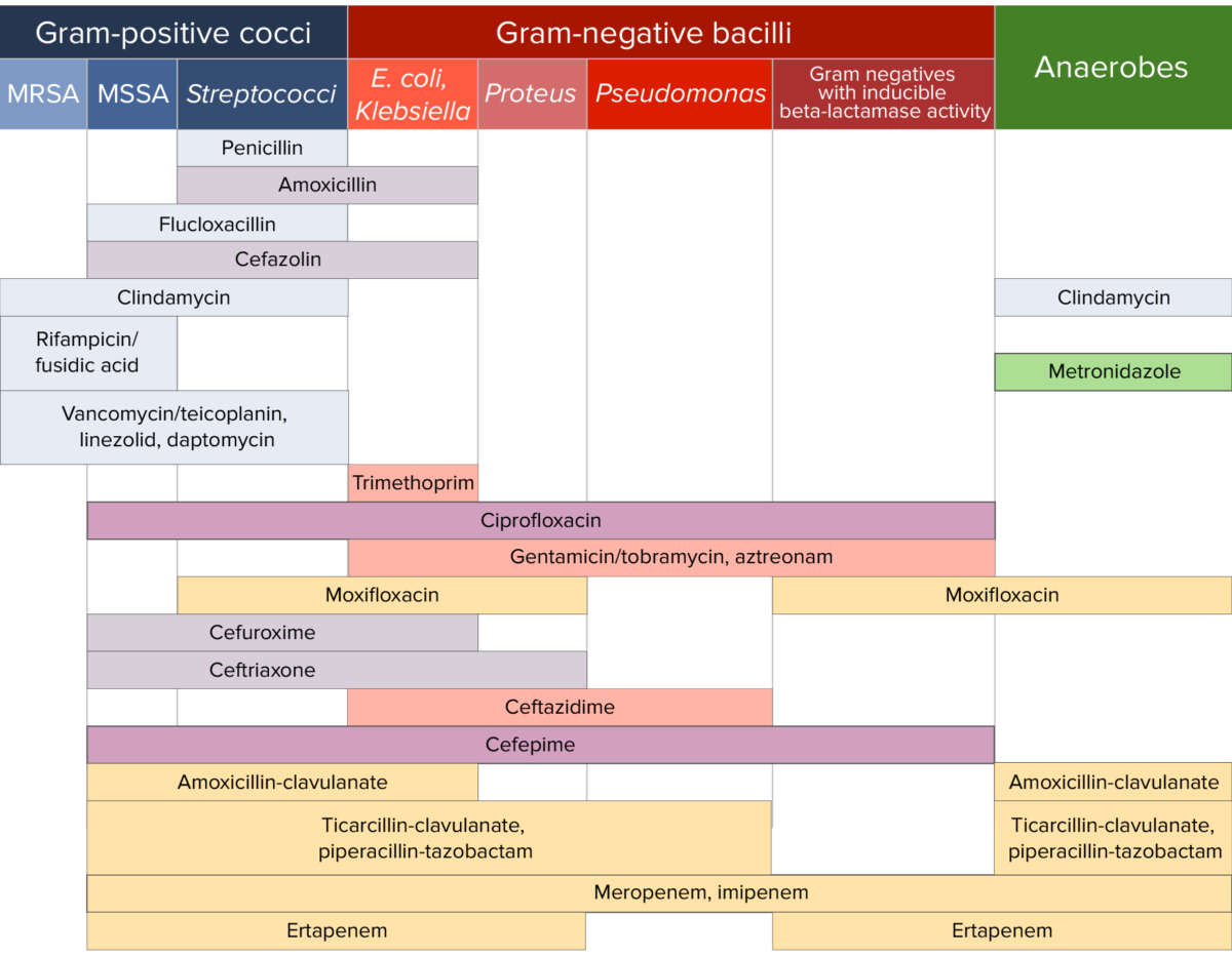
Antibiotic sensitivity chart

Antibiotic sensitivity:
Chart comparing the microbial coverage of different antibiotics for gram-positive cocci, gram-negative bacilli, and anaerobes.

Image by Lecturio. License: CC BY-NC-SA 4.0

References

  1. Bertram G. (2007). Macrolide Antibiotics Comparison: Erythromycin, Clarithromycin, Azithromycin. Basic and Clinical Pharmacology. Retrieved March 5, 2021, from https://www.emedexpert.com/compare/macrolides.shtml.
  2. Tenson T, Lovmar M, Ehrenberg M (2003). The mechanism of action of macrolides, lincosamides and streptogramin B reveals the nascent peptide exit path in the ribosome. Journal of Molecular Biology. 330 (5): 1005–14. https://www.sciencedirect.com/science/article/abs/pii/S0022283603006624?via%3Dihub
  3. Leclercq, R. (2002). Mechanisms of resistance to macrolides and lincosamides: Nature of the resistance elements and their clinical implications. Clinical Infectious Diseases, 34(4):482-492. https://academic.oup.com/cid/article/34/4/482/412492
  4. Werth BJ. (2020). Macrolides. MSD Manual Professional Version. Retrieved June 29, 2021, from https://www.msdmanuals.com/professional/infectious-diseases/bacteria-and-antibacterial-drugs/macrolides
  5. Patel PH, Hashmi MF. (2021). Macrolides. StatPearls. Retrieved June 29, 2021, from https://www.ncbi.nlm.nih.gov/books/NBK551495/
  6. Graziani, A.L. (2021). Azithromycin and clarithromycin. In Bond, S. (Ed.), UpToDate. Retrieved June 29, 2021, from https://www.uptodate.com/contents/azithromycin-and-clarithromycin
  7. Deck DH, Winston LG. (2012). Tetracyclines, macrolides, clindamycin, chloramphenicol, streptogramins, & oxazolidinones. In Katzung BG, Masters SB, Trevor AJ. (Eds.), Basic & Clinical Pharmacology (12th edition, pp. 809-819). https://pharmacomedicale.org/images/cnpm/CNPM_2016/katzung-pharmacology.pdf
  8. Vedantam G, Clark A, Chu M, McQuade R, Mallozzi M, Viswanathan VK. Clostridium difficile infection: toxins and non-toxin virulence factors, and their contributions to disease establishment and host response. Gut Microbes. 2012 Mar-Apr;3(2):121-34. doi: 10.4161/gmic.19399. Epub 2012 Mar 1. PMID: 22555464; PMCID: PMC3370945.
  9. Sears P, Crook DW, Louie TJ, Miller MA, Weiss K. Fidaxomicin attains high fecal concentrations with minimal plasma concentrations following oral administration in patients with Clostridium difficile infection. Clin Infect Dis. 2012 Aug;55 Suppl 2(Suppl 2):S116-20. doi: 10.1093/cid/cis337. PMID: 22752859; PMCID: PMC3388019.

Create your free account or log in to continue reading!

Sign up now and get free access to Lecturio with concept pages, medical videos, and questions for your medical education.

User Reviews

Details