Lymphatic Filariasis (Elephantiasis)

Lymphatic filariasis, also known as elephantiasis, is a chronic mosquito-borne infection caused by Wuchereria bancrofti, Brugia malayi, and B. timori. The majority of causes are due to W. bancrofti. Mosquitos are the vectors, and humans are the primary reservoir Reservoir Animate or inanimate sources which normally harbor disease-causing organisms and thus serve as potential sources of disease outbreaks. Reservoirs are distinguished from vectors (disease vectors) and carriers, which are agents of disease transmission rather than continuing sources of potential disease outbreaks. Humans may serve both as disease reservoirs and carriers. Escherichia coli. Patients Patients Individuals participating in the health care system for the purpose of receiving therapeutic, diagnostic, or preventive procedures. Clinician–Patient Relationship with acute infection can present with fever Fever Fever is defined as a measured body temperature of at least 38°C (100.4°F). Fever is caused by circulating endogenous and/or exogenous pyrogens that increase levels of prostaglandin E2 in the hypothalamus. Fever is commonly associated with chills, rigors, sweating, and flushing of the skin. Fever, adenolymphangitis, dermatolymphangioadenitis, and tropical pulmonary eosinophilia Eosinophilia Abnormal increase of eosinophils in the blood, tissues or organs. Autosomal Dominant Hyperimmunoglobulin E Syndrome. Patients Patients Individuals participating in the health care system for the purpose of receiving therapeutic, diagnostic, or preventive procedures. Clinician–Patient Relationship with chronic infection present with lymphedema, which commonly affects the lower extremities (but can cause testicular swelling Swelling Inflammation or hydrocele Hydrocele Accumulation of serous fluid between the layers of membrane (tunica vaginalis) covering the testis in the scrotum. Varicocele, Hydrocele, and Spermatocele). Long-term effects also include renal manifestations. Thick and thin peripheral blood smears are the mainstay of diagnosis. Lymphatic filariasis without co-infection is generally managed with diethylcarbamazine Diethylcarbamazine An anthelmintic used primarily as the citrate in the treatment of filariasis, particularly infestations with wucheria bancrofti or loa loa. Anthelmintic Drugs. Prognosis Prognosis A prediction of the probable outcome of a disease based on a individual's condition and the usual course of the disease as seen in similar situations. Non-Hodgkin Lymphomas is good with early diagnosis and intervention. Elephantiasis, or late-stage lymphedema, is associated with significant disability Disability Determination of the degree of a physical, mental, or emotional handicap. The diagnosis is applied to legal qualification for benefits and income under disability insurance and to eligibility for social security and workman's compensation benefits. ABCDE Assessment and would require different methods (including surgery) to reduce swelling Swelling Inflammation and complications.

Last updated: Sep 19, 2022

Editorial responsibility: Stanley Oiseth, Lindsay Jones, Evelin Maza

Epidemiology and Etiology

Epidemiology

  • 120 million patients Patients Individuals participating in the health care system for the purpose of receiving therapeutic, diagnostic, or preventive procedures. Clinician–Patient Relationship infected worldwide as of 2019
  • Geographic distribution:
    • Wuchereria bancrofti
      • Etiology for 90% of lymphatic filariasis
      • Most prevalent in sub-Saharan Africa, Southeast Asia ASIA Spinal Cord Injuries, Indian subcontinent, Pacific islands, and Caribbean and South American tropical and subtropical climates
    • Brugia malayi: Southeast Asia ASIA Spinal Cord Injuries, China, India, and Pacific islands
    • B. timori: Timor island of Indonesia
  • Most patients Patients Individuals participating in the health care system for the purpose of receiving therapeutic, diagnostic, or preventive procedures. Clinician–Patient Relationship in endemic areas were exposed in their 30s or 40s.
  • Lymphatic filariasis has a 10:1 predilection for men over women.

Etiology

Lymphatic filariasis is caused by nematodes.

Causative species:

  • W. bancrofti
  • B. malayi
  • B. timori

General characteristics:

  • Nematode Nematode A phylum of unsegmented helminths with fundamental bilateral symmetry and secondary triradiate symmetry of the oral and esophageal structures. Many species are parasites. Toxocariasis (roundworm)
  • Eukaryotic Eukaryotic Eukaryotes can be single-celled or multicellular organisms and include plants, animals, fungi, and protozoa. Eukaryotic cells contain a well-organized nucleus contained by a membrane, along with other membrane-bound organelles. Cell Types: Eukaryotic versus Prokaryotic
  • Thread-like, nuclei do not appear at the end of the tail
  • Anaerobic
  • Reproduce by sexual reproduction

Stages of life:

  • Microfilariae:
    • Found in the peripheral blood of the human host
    • Produced by adult nematodes
    • Ingested by a mosquito during a blood meal
  • Larvae Larvae Wormlike or grublike stage, following the egg in the life cycle of insects, worms, and other metamorphosing animals. Ascaris/Ascariasis:
    • Microfilariae that lose their sheaths
    • In the mosquito, microfilariae mature into larvae Larvae Wormlike or grublike stage, following the egg in the life cycle of insects, worms, and other metamorphosing animals. Ascaris/Ascariasis inside the mosquito: L1 (1st stage) larvae Larvae Wormlike or grublike stage, following the egg in the life cycle of insects, worms, and other metamorphosing animals. Ascaris/Ascariasis to L3 (3rd stage) larvae Larvae Wormlike or grublike stage, following the egg in the life cycle of insects, worms, and other metamorphosing animals. Ascaris/Ascariasis
    • L3 larvae Larvae Wormlike or grublike stage, following the egg in the life cycle of insects, worms, and other metamorphosing animals. Ascaris/Ascariasis travel to the mosquito’s proboscis, and human infection occurs during a blood meal.
    • In the infected human, larvae Larvae Wormlike or grublike stage, following the egg in the life cycle of insects, worms, and other metamorphosing animals. Ascaris/Ascariasis migrate to the bloodstream and lymphatics.
    • Maturity reached in 6–9 months
  • Adults:
    • Mature from L3 larvae Larvae Wormlike or grublike stage, following the egg in the life cycle of insects, worms, and other metamorphosing animals. Ascaris/Ascariasis in the regional lymphatics 
    • Adult female Wuchereria worms: 80–100 mm MM Multiple myeloma (MM) is a malignant condition of plasma cells (activated B lymphocytes) primarily seen in the elderly. Monoclonal proliferation of plasma cells results in cytokine-driven osteoclastic activity and excessive secretion of IgG antibodies. Multiple Myeloma in length and 0.24–0.30 mm MM Multiple myeloma (MM) is a malignant condition of plasma cells (activated B lymphocytes) primarily seen in the elderly. Monoclonal proliferation of plasma cells results in cytokine-driven osteoclastic activity and excessive secretion of IgG antibodies. Multiple Myeloma in diameter
    • Adult male Wuchereria worms: about 40 mm MM Multiple myeloma (MM) is a malignant condition of plasma cells (activated B lymphocytes) primarily seen in the elderly. Monoclonal proliferation of plasma cells results in cytokine-driven osteoclastic activity and excessive secretion of IgG antibodies. Multiple Myeloma by 0.1 mm MM Multiple myeloma (MM) is a malignant condition of plasma cells (activated B lymphocytes) primarily seen in the elderly. Monoclonal proliferation of plasma cells results in cytokine-driven osteoclastic activity and excessive secretion of IgG antibodies. Multiple Myeloma
    • Adult female Brugia worms: 43–55 mm MM Multiple myeloma (MM) is a malignant condition of plasma cells (activated B lymphocytes) primarily seen in the elderly. Monoclonal proliferation of plasma cells results in cytokine-driven osteoclastic activity and excessive secretion of IgG antibodies. Multiple Myeloma in length
    • Adult male Brugia worms: 13–23 mm MM Multiple myeloma (MM) is a malignant condition of plasma cells (activated B lymphocytes) primarily seen in the elderly. Monoclonal proliferation of plasma cells results in cytokine-driven osteoclastic activity and excessive secretion of IgG antibodies. Multiple Myeloma in length

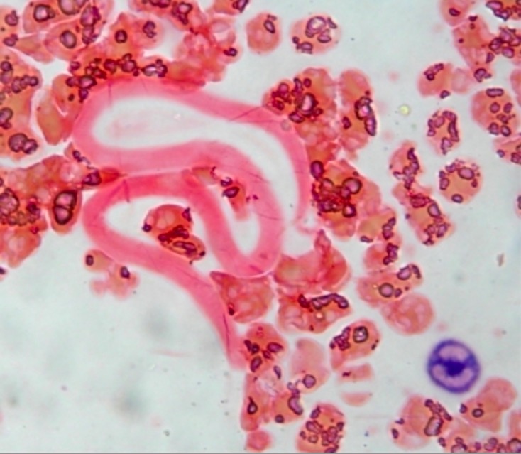
Transmitted to mosquitoes:

Wuchereria bancrofti

Wuchereria bancrofti microfilariae

Image: “Neutrophil Alkaline Phosphatase stained peripheral smear” by Department of Hematology, All India Institute of Medical Sciences, New Delhi. License: CC BY 2.0
Pathophysiology

Transmission

  • The primary reservoir Reservoir Animate or inanimate sources which normally harbor disease-causing organisms and thus serve as potential sources of disease outbreaks. Reservoirs are distinguished from vectors (disease vectors) and carriers, which are agents of disease transmission rather than continuing sources of potential disease outbreaks. Humans may serve both as disease reservoirs and carriers. Escherichia coli for filariasis is humans, and the vector is mosquitos. 
  • Transmission from human to human occurs via mosquito bites.

Life cycle Cycle The type of signal that ends the inspiratory phase delivered by the ventilator Invasive Mechanical Ventilation

  1. Mosquitos deposit L3 larvae Larvae Wormlike or grublike stage, following the egg in the life cycle of insects, worms, and other metamorphosing animals. Ascaris/Ascariasis into a human’s skin Skin The skin, also referred to as the integumentary system, is the largest organ of the body. The skin is primarily composed of the epidermis (outer layer) and dermis (deep layer). The epidermis is primarily composed of keratinocytes that undergo rapid turnover, while the dermis contains dense layers of connective tissue. Skin: Structure and Functions, which burrow through the bite wound to the bloodstream.
  2. L3 larvae Larvae Wormlike or grublike stage, following the egg in the life cycle of insects, worms, and other metamorphosing animals. Ascaris/Ascariasis migrate to the lymph nodes Lymph Nodes They are oval or bean shaped bodies (1 – 30 mm in diameter) located along the lymphatic system. Lymphatic Drainage System: Anatomy and regional lymphatics and mature into adults, which have a predilection for inguinal lymph nodes Lymph Nodes They are oval or bean shaped bodies (1 – 30 mm in diameter) located along the lymphatic system. Lymphatic Drainage System: Anatomy.
  3. Adult worms undergo sexual reproduction, with females birthing microfilariae that migrate actively through lymph Lymph The interstitial fluid that is in the lymphatic system. Secondary Lymphatic Organs and blood.
  4. A mosquito ingests the microfilariae during a blood meal. 
  5. After ingestion, the microfilariae work their way through the wall of the mosquito’s midgut Midgut Development of the Abdominal Organs and reach the thoracic muscles. 
  6. Within the mosquito, the microfilariae develop into L1 larvae Larvae Wormlike or grublike stage, following the egg in the life cycle of insects, worms, and other metamorphosing animals. Ascaris/Ascariasis.
  7. L1 larvae Larvae Wormlike or grublike stage, following the egg in the life cycle of insects, worms, and other metamorphosing animals. Ascaris/Ascariasis subsequently develop into L2 then L3 infective larvae Larvae Wormlike or grublike stage, following the egg in the life cycle of insects, worms, and other metamorphosing animals. Ascaris/Ascariasis.
  8. The L3 larvae Larvae Wormlike or grublike stage, following the egg in the life cycle of insects, worms, and other metamorphosing animals. Ascaris/Ascariasis migrate to the mosquito’s proboscis, where they can infect a human during the mosquito’s next blood meal.
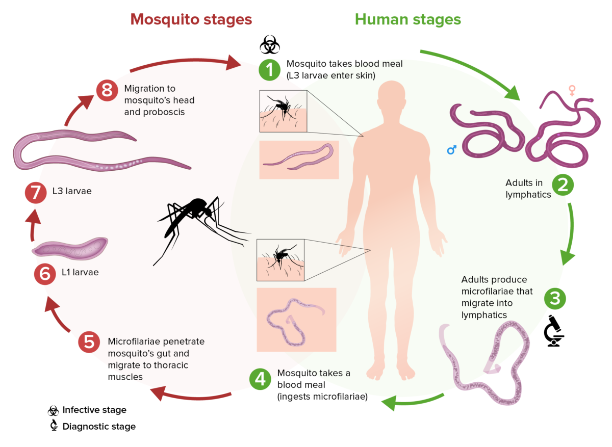
Filarial life cycle

Schematic of the filarial life cycle of Wuchereria bancrofti:

(1) During a blood meal, an infected mosquito introduces L3 larvae into the skin of the human host.
(2) These larvae develop into adults that commonly reside in the lymphatics.
(3) Adults produce microfilariae that migrate actively through lymph and blood.
(4) A mosquito ingests the microfilariae during a blood meal.
(5) After ingestion, the microfilariae work their way through the wall of the mosquito’s midgut and reach the thoracic muscles.
(6) In the thoracic muscles, the microfilariae develop into L1 larvae.
(7) The L1 larvae subsequently develop into L3 infective larvae.
(8) The L3 larvae migrate to the mosquito’s proboscis.
(9) The L3 larvae can infect another human when the mosquito takes another blood meal.

Image by Lecturio.

Disease process

  • After mating, the adult female lays thousands of microfilariae daily:
    • In most areas, W. bancrofti microfilariae are in circulation Circulation The movement of the blood as it is pumped through the cardiovascular system. ABCDE Assessment (found in the bloodstream) during nocturnal hours.
    • In the South Pacific, the microfilariae are found in the circulation Circulation The movement of the blood as it is pumped through the cardiovascular system. ABCDE Assessment all day.
  • Acute infection:
    • Filarial antigens trigger Trigger The type of signal that initiates the inspiratory phase by the ventilator Invasive Mechanical Ventilation increased cytokines Cytokines Non-antibody proteins secreted by inflammatory leukocytes and some non-leukocytic cells, that act as intercellular mediators. They differ from classical hormones in that they are produced by a number of tissue or cell types rather than by specialized glands. They generally act locally in a paracrine or autocrine rather than endocrine manner. Adaptive Immune Response and immunoglobulins Immunoglobulins Immunoglobulins (Igs), also known as antibodies, are glycoprotein molecules produced by plasma cells that act in immune responses by recognizing and binding particular antigens. The various Ig classes are IgG (the most abundant), IgM, IgE, IgD, and IgA, which differ in their biologic features, structure, target specificity, and distribution. Immunoglobulins: Types and Functions ( IgE IgE An immunoglobulin associated with mast cells. Overexpression has been associated with allergic hypersensitivity. Immunoglobulins: Types and Functions and IgG4).
    • Molting, dying, or dead adult worms produce pathologic changes:
      • Dilatation of lymphatics
      • Hyperplasia Hyperplasia An increase in the number of cells in a tissue or organ without tumor formation. It differs from hypertrophy, which is an increase in bulk without an increase in the number of cells. Cellular Adaptation of vascular endothelium Endothelium A layer of epithelium that lines the heart, blood vessels (vascular endothelium), lymph vessels (lymphatic endothelium), and the serous cavities of the body. Arteries: Histology
      • Lymphangitis Lymphangitis A lymphatic disease characterized by inflammation of lymphatic vessels. Erysipelas (infiltration of eosinophils Eosinophils Granular leukocytes with a nucleus that usually has two lobes connected by a slender thread of chromatin, and cytoplasm containing coarse, round granules that are uniform in size and stainable by eosin. Innate Immunity: Phagocytes and Antigen Presentation, lymphocytes Lymphocytes Lymphocytes are heterogeneous WBCs involved in immune response. Lymphocytes develop from the bone marrow, starting from hematopoietic stem cells (HSCs) and progressing to common lymphoid progenitors (CLPs). B and T lymphocytes and natural killer (NK) cells arise from the lineage. Lymphocytes: Histology in the lymphatics, and thrombi formation)
  • Chronic and repeated infections Infections Invasion of the host organism by microorganisms or their toxins or by parasites that can cause pathological conditions or diseases. Chronic Granulomatous Disease lead to:
    • Granuloma formation
    • Fibrosis Fibrosis Any pathological condition where fibrous connective tissue invades any organ, usually as a consequence of inflammation or other injury. Bronchiolitis Obliterans of lymphatic vessels Lymphatic Vessels Tubular vessels that are involved in the transport of lymph and lymphocytes. Lymphatic Drainage System: Anatomy and the surrounding connective tissue Connective tissue Connective tissues originate from embryonic mesenchyme and are present throughout the body except inside the brain and spinal cord. The main function of connective tissues is to provide structural support to organs. Connective tissues consist of cells and an extracellular matrix. Connective Tissue: Histology
  • Effects:
    • Contractile dysfunction and lymphatic occlusion, causing lymphedema and prohibiting lymphatic drainage
    • Susceptibility of the human host to bacterial and fungal infections Infections Invasion of the host organism by microorganisms or their toxins or by parasites that can cause pathological conditions or diseases. Chronic Granulomatous Disease, which further contribute to tissue damage
  • Adult filaria can survive inside humans for up to 9 years. 
  • Adult worms also carry Wolbachia (endosymbiotic bacteria Bacteria Bacteria are prokaryotic single-celled microorganisms that are metabolically active and divide by binary fission. Some of these organisms play a significant role in the pathogenesis of diseases. Bacteriology) in their gut, which appear to benefit the worms.

Clinical Presentation

Symptoms may take 9 months up to 1 year to manifest after the initial infection. Children or individuals in endemic areas often remain asymptomatic ( subclinical infection Subclinical infection Toxoplasma/Toxoplasmosis), while others show acute and/or chronic signs and symptoms.
Acute manifestations

  • Filarial fever Fever Fever is defined as a measured body temperature of at least 38°C (100.4°F). Fever is caused by circulating endogenous and/or exogenous pyrogens that increase levels of prostaglandin E2 in the hypothalamus. Fever is commonly associated with chills, rigors, sweating, and flushing of the skin. Fever:
  • Acute adenolymphangitis (ADL):
    • Fever Fever Fever is defined as a measured body temperature of at least 38°C (100.4°F). Fever is caused by circulating endogenous and/or exogenous pyrogens that increase levels of prostaglandin E2 in the hypothalamus. Fever is commonly associated with chills, rigors, sweating, and flushing of the skin. Fever and lymphadenopathy Lymphadenopathy Lymphadenopathy is lymph node enlargement (> 1 cm) and is benign and self-limited in most patients. Etiologies include malignancy, infection, and autoimmune disorders, as well as iatrogenic causes such as the use of certain medications. Generalized lymphadenopathy often indicates underlying systemic disease. Lymphadenopathy
    • Lymphangitis Lymphangitis A lymphatic disease characterized by inflammation of lymphatic vessels. Erysipelas ( inflammation Inflammation Inflammation is a complex set of responses to infection and injury involving leukocytes as the principal cellular mediators in the body’s defense against pathogenic organisms. Inflammation is also seen as a response to tissue injury in the process of wound healing. The 5 cardinal signs of inflammation are pain, heat, redness, swelling, and loss of function. Inflammation spread distally to the lymph Lymph The interstitial fluid that is in the lymphatic system. Secondary Lymphatic Organs node), with lymphatic vessels Lymphatic Vessels Tubular vessels that are involved in the transport of lymph and lymphocytes. Lymphatic Drainage System: Anatomy in the leg Leg The lower leg, or just “leg” in anatomical terms, is the part of the lower limb between the knee and the ankle joint. The bony structure is composed of the tibia and fibula bones, and the muscles of the leg are grouped into the anterior, lateral, and posterior compartments by extensions of fascia. Leg: Anatomy(s) becoming warm, enlarged, red, and tender
    • Commonly seen in inguinal lymph nodes Lymph Nodes They are oval or bean shaped bodies (1 – 30 mm in diameter) located along the lymphatic system. Lymphatic Drainage System: Anatomy but can also affect the genitalia ( epididymitis Epididymitis Epididymitis and orchitis are characterized by acute inflammation of the epididymis and the testicle, respectively, due to viral or bacterial infections. Patients typically present with gradually worsening testicular pain and scrotal swelling along with systemic symptoms such as fever, depending on severity. Epididymitis and Orchitis in males)
    • Lasts a few days then resolves, but recurs periodically
  • Acute dermatolymphangioadenitis (DLA):
    • Fever Fever Fever is defined as a measured body temperature of at least 38°C (100.4°F). Fever is caused by circulating endogenous and/or exogenous pyrogens that increase levels of prostaglandin E2 in the hypothalamus. Fever is commonly associated with chills, rigors, sweating, and flushing of the skin. Fever, chills Chills The sudden sensation of being cold. It may be accompanied by shivering. Fever
    • Edematous plaques (commonly interdigital) that are believed to be entry lesions and caused by bacteria Bacteria Bacteria are prokaryotic single-celled microorganisms that are metabolically active and divide by binary fission. Some of these organisms play a significant role in the pathogenesis of diseases. Bacteriology
    • Lymphedema (worsens with recurrent episodes)
    • Seen in endemic areas
  • Tropical pulmonary eosinophilia Eosinophilia Abnormal increase of eosinophils in the blood, tissues or organs. Autosomal Dominant Hyperimmunoglobulin E Syndrome (TPE):
    • Immune reaction to microfilariae trapped in the lungs Lungs Lungs are the main organs of the respiratory system. Lungs are paired viscera located in the thoracic cavity and are composed of spongy tissue. The primary function of the lungs is to oxygenate blood and eliminate CO2. Lungs: Anatomy
    • Restrictive lung disease, which can progress to interstitial pulmonary fibrosis Fibrosis Any pathological condition where fibrous connective tissue invades any organ, usually as a consequence of inflammation or other injury. Bronchiolitis Obliterans
    • Wheezing Wheezing Wheezing is an abnormal breath sound characterized by a whistling noise that can be relatively high-pitched and shrill (more common) or coarse. Wheezing is produced by the movement of air through narrowed or compressed small (intrathoracic) airways. Wheezing
    • Shortness of breath Shortness of breath Dyspnea is the subjective sensation of breathing discomfort. Dyspnea is a normal manifestation of heavy physical or psychological exertion, but also may be caused by underlying conditions (both pulmonary and extrapulmonary). Dyspnea
    • Bloody sputum
    • Eosinophilia Eosinophilia Abnormal increase of eosinophils in the blood, tissues or organs. Autosomal Dominant Hyperimmunoglobulin E Syndrome

Chronic manifestations

  • Lymphedema:
    • Chronic swelling Swelling Inflammation of the limb from chronic inflammation Chronic Inflammation Inflammation of the lymphatic vessels Lymphatic Vessels Tubular vessels that are involved in the transport of lymph and lymphocytes. Lymphatic Drainage System: Anatomy
    • Graded based on the extent and progression of the symptoms:
      • Grade 0: subclinical
      • Grade I: pitting edema Pitting edema Edema caused by excess fluid without excess colloid. Leaves “pits” due to fluid displacement when pressure is applied to the area Edema of the extremities, reversible (↓ with limb elevation)
      • Grade II: pitting or nonpitting edema Edema Edema is a condition in which excess serous fluid accumulates in the body cavity or interstitial space of connective tissues. Edema is a symptom observed in several medical conditions. It can be categorized into 2 types, namely, peripheral (in the extremities) and internal (in an organ or body cavity). Edema of the extremities, nonreversible
      • Grade III: nonpitting edema Edema Edema is a condition in which excess serous fluid accumulates in the body cavity or interstitial space of connective tissues. Edema is a symptom observed in several medical conditions. It can be categorized into 2 types, namely, peripheral (in the extremities) and internal (in an organ or body cavity). Edema with skin Skin The skin, also referred to as the integumentary system, is the largest organ of the body. The skin is primarily composed of the epidermis (outer layer) and dermis (deep layer). The epidermis is primarily composed of keratinocytes that undergo rapid turnover, while the dermis contains dense layers of connective tissue. Skin: Structure and Functions thickening and overgrowths, nonreversible  (elephantiasis)
  • Hydrocele Hydrocele Accumulation of serous fluid between the layers of membrane (tunica vaginalis) covering the testis in the scrotum. Varicocele, Hydrocele, and Spermatocele:
    • Lymphatic disease involving the scrotum Scrotum A cutaneous pouch of skin containing the testicles and spermatic cords. Testicles: Anatomy
    • Unilateral or bilateral
  • Renal involvement:
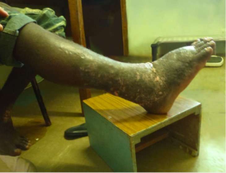
Elephantiasis

Filariasis:
A patient with chronic lymphedema of the right lower extremity manifesting as elephantiasis

Image: “Elephantiasis” by Humpress Harrington et al. License: CC BY 2.0

Diagnosis

Laboratory tests

  • Peripheral blood smear Peripheral Blood Smear Anemia: Overview and Types:
    • Thick and thin smears
    • Venipuncture or finger/heel stick
    • Taken between 10 pm and 2 am (microfilaria have nocturnal periodicity)
    • Giemsa or Wright stains
  • Circulating filarial antigen Antigen Substances that are recognized by the immune system and induce an immune reaction. Vaccination (W. bancrofti): 
    • Detects antigens of adult filarial worms
    • May be positive even in those without microfilariae
  • Antifilarial antibody tests:
    • Elevated levels of antifilarial IgG4 in the blood 
    • Used mostly for travelers (who are not from endemic areas)
  • PCR PCR Polymerase chain reaction (PCR) is a technique that amplifies DNA fragments exponentially for analysis. The process is highly specific, allowing for the targeting of specific genomic sequences, even with minuscule sample amounts. The PCR cycles multiple times through 3 phases: denaturation of the template DNA, annealing of a specific primer to the individual DNA strands, and synthesis/elongation of new DNA molecules. Polymerase Chain Reaction (PCR) for antigen detection Antigen detection Respiratory Syncytial Virus
    • Used in research Research Critical and exhaustive investigation or experimentation, having for its aim the discovery of new facts and their correct interpretation, the revision of accepted conclusions, theories, or laws in the light of newly discovered facts, or the practical application of such new or revised conclusions, theories, or laws. Conflict of Interest
    • Assay not approved for commercial use
  • Biopsy Biopsy Removal and pathologic examination of specimens from the living body. Ewing Sarcoma: tissue from cutaneous lesions
  • Additional laboratory tests:
    • In areas endemic for onchocerciasis (Onchocerca volvulus Volvulus A volvulus is the twisting or axial rotation of a portion of the bowel around its mesentery. The most common site of volvulus in adults is the colon; most frequently the sigmoid volvulus. Patients typically present with symptoms of bowel obstruction such as abdominal pain, distension, vomiting, and constipation/obstipation. Volvulus) or loiasis (Loa loa), co-infection has to be determined, as management will differ.
    • CBC showing eosinophilia Eosinophilia Abnormal increase of eosinophils in the blood, tissues or organs. Autosomal Dominant Hyperimmunoglobulin E Syndrome and elevated IgE IgE An immunoglobulin associated with mast cells. Overexpression has been associated with allergic hypersensitivity. Immunoglobulins: Types and Functions noted in TPE

Imaging

  • Ultrasonography:
    • Reveals adult worms moving in lymphatic vessels Lymphatic Vessels Tubular vessels that are involved in the transport of lymph and lymphocytes. Lymphatic Drainage System: Anatomy
    • The “filarial dance sign”—an irregular worm movement pattern—may be detected on Doppler Doppler Ultrasonography applying the doppler effect, with frequency-shifted ultrasound reflections produced by moving targets (usually red blood cells) in the bloodstream along the ultrasound axis in direct proportion to the velocity of movement of the targets, to determine both direction and velocity of blood flow. Ultrasound (Sonography).
  • Lymphoscintigraphy:
    • Assess lymphatic drainage
    • Can detect preclinical lymphedema
  • Chest X-ray X-ray Penetrating electromagnetic radiation emitted when the inner orbital electrons of an atom are excited and release radiant energy. X-ray wavelengths range from 1 pm to 10 nm. Hard x-rays are the higher energy, shorter wavelength x-rays. Soft x-rays or grenz rays are less energetic and longer in wavelength. The short wavelength end of the x-ray spectrum overlaps the gamma rays wavelength range. The distinction between gamma rays and x-rays is based on their radiation source. Pulmonary Function Tests:
    • To evaluate for TPE
    • Diffuse interstitial lesions and pronounced bronchoalveolar markings

Management

Treatment

Filariasis without co-infection:

  • Diethylcarbamazine Diethylcarbamazine An anthelmintic used primarily as the citrate in the treatment of filariasis, particularly infestations with wucheria bancrofti or loa loa. Anthelmintic Drugs (DEC): 
    • 1st-line therapy
    • Single dose
    • 2–3 weeks of treatment for TPE due to W. bancrofti
  • Doxycycline: 
    • In addition to DEC or an alternative therapy
    • For nonpregnant adults and children > 8 years of age
    • Effective against Wolbachia

Filariasis with loiasis:

  • DEC intake increases the risk of life-threatening encephalopathy Encephalopathy Hyper-IgM Syndrome in co-infection as microfilarial load increases.
  • Medication will vary depending on the load:
    • < 2500 L. loa microfilariae/mL: DEC
    • > 2500–8000 microfilariae/mL: ivermectin Ivermectin A mixture of mostly avermectin h2b1a (rn 71827-03-7) with some avermectin h2b1b (rn 70209-81-3), which are macrolides from streptomyces avermitilis. It binds glutamate-gated chloride channel to cause increased permeability and hyperpolarization of nerve and muscle cells. It also interacts with other chloride channels. It is a broad spectrum antiparasitic that is active against microfilariae of onchocerca volvulus but not the adult form. Anthelmintic Drugs to decrease the microfilarial load, then DEC
    • > 8000 microfilariae/mL: doxycycline for 4–6 weeks

Filariasis with onchocerciasis:

  • DEC intake worsens eye disease (↑ risk of blindness Blindness The inability to see or the loss or absence of perception of visual stimuli. This condition may be the result of eye diseases; optic nerve diseases; optic chiasm diseases; or brain diseases affecting the visual pathways or occipital lobe. Retinopathy of Prematurity).
  • Treatment options:
    • Treat onchocerciasis 1st with ivermectin Ivermectin A mixture of mostly avermectin h2b1a (rn 71827-03-7) with some avermectin h2b1b (rn 70209-81-3), which are macrolides from streptomyces avermitilis. It binds glutamate-gated chloride channel to cause increased permeability and hyperpolarization of nerve and muscle cells. It also interacts with other chloride channels. It is a broad spectrum antiparasitic that is active against microfilariae of onchocerca volvulus but not the adult form. Anthelmintic Drugs, followed by DEC. 
    • For those with eye disease, give doxycycline followed by ivermectin Ivermectin A mixture of mostly avermectin h2b1a (rn 71827-03-7) with some avermectin h2b1b (rn 70209-81-3), which are macrolides from streptomyces avermitilis. It binds glutamate-gated chloride channel to cause increased permeability and hyperpolarization of nerve and muscle cells. It also interacts with other chloride channels. It is a broad spectrum antiparasitic that is active against microfilariae of onchocerca volvulus but not the adult form. Anthelmintic Drugs.

Surgical treatment:

  • Skin Skin The skin, also referred to as the integumentary system, is the largest organ of the body. The skin is primarily composed of the epidermis (outer layer) and dermis (deep layer). The epidermis is primarily composed of keratinocytes that undergo rapid turnover, while the dermis contains dense layers of connective tissue. Skin: Structure and Functions debulking and lymphovenous anastomosis for drainage improvement
  • Surgical excision of hydrocele Hydrocele Accumulation of serous fluid between the layers of membrane (tunica vaginalis) covering the testis in the scrotum. Varicocele, Hydrocele, and Spermatocele

Long-term management to reduce lymphedema progression:

  • Skin Skin The skin, also referred to as the integumentary system, is the largest organ of the body. The skin is primarily composed of the epidermis (outer layer) and dermis (deep layer). The epidermis is primarily composed of keratinocytes that undergo rapid turnover, while the dermis contains dense layers of connective tissue. Skin: Structure and Functions hygiene
  • Wearing comfortable shoes
  • Compressive bandages
  • Limb elevation
  • Cold and heat Heat Inflammation therapy
  • Antibiotic and antifungals to prevent flares

Prevention

  • Avoid mosquito bites:
    • Sleep Sleep A readily reversible suspension of sensorimotor interaction with the environment, usually associated with recumbency and immobility. Physiology of Sleep in an air-conditioned room or under mosquito nets.
    • Use mosquito repellent.
    • Wear long sleeves and trousers.
  • Mosquito control with insecticide spraying
  • Global Program for the Elimination Elimination The initial damage and destruction of tumor cells by innate and adaptive immunity. Completion of the phase means no cancer growth. Cancer Immunotherapy of Lymphatic Filariasis:
    • Launched to eliminate spread and reduce morbidity Morbidity The proportion of patients with a particular disease during a given year per given unit of population. Measures of Health Status
    • Consists of an annual mass Mass Three-dimensional lesion that occupies a space within the breast Imaging of the Breast drug administration (for at least 5 years) implemented using various regimens with:
      • DEC
      • Ivermectin Ivermectin A mixture of mostly avermectin h2b1a (rn 71827-03-7) with some avermectin h2b1b (rn 70209-81-3), which are macrolides from streptomyces avermitilis. It binds glutamate-gated chloride channel to cause increased permeability and hyperpolarization of nerve and muscle cells. It also interacts with other chloride channels. It is a broad spectrum antiparasitic that is active against microfilariae of onchocerca volvulus but not the adult form. Anthelmintic Drugs
      • Albendazole Albendazole A benzimidazole broad-spectrum anthelmintic structurally related to mebendazole that is effective against many diseases. Anthelmintic Drugs

Complications and prognosis Prognosis A prediction of the probable outcome of a disease based on a individual’s condition and the usual course of the disease as seen in similar situations. Non-Hodgkin Lymphomas

Occult filariasis is a filarial infection that extends into tissues, without any evidence in the blood. This leads to chronic complications, such as:

  • TPE
  • Filarial arthritis Arthritis Acute or chronic inflammation of joints. Osteoarthritis
  • Filaria-associated immune complex glomerulonephritis
  • Filarial breast abscesses

Prognosis Prognosis A prediction of the probable outcome of a disease based on a individual’s condition and the usual course of the disease as seen in similar situations. Non-Hodgkin Lymphomas:

  • When diseases is diagnosed and treated early, the prognosis Prognosis A prediction of the probable outcome of a disease based on a individual’s condition and the usual course of the disease as seen in similar situations. Non-Hodgkin Lymphomas is good. 
  • As symptoms may present later in adulthood, lymphedema and, subsequently, elephantiasis are associated with disability Disability Determination of the degree of a physical, mental, or emotional handicap. The diagnosis is applied to legal qualification for benefits and income under disability insurance and to eligibility for social security and workman’s compensation benefits. ABCDE Assessment and morbidity Morbidity The proportion of patients with a particular disease during a given year per given unit of population. Measures of Health Status.

Differential Diagnosis

  • Onchocerciasis: infection caused by the filarial nematode Nematode A phylum of unsegmented helminths with fundamental bilateral symmetry and secondary triradiate symmetry of the oral and esophageal structures. Many species are parasites. Toxocariasis Onchocerca volvulus Volvulus A volvulus is the twisting or axial rotation of a portion of the bowel around its mesentery. The most common site of volvulus in adults is the colon; most frequently the sigmoid volvulus. Patients typically present with symptoms of bowel obstruction such as abdominal pain, distension, vomiting, and constipation/obstipation. Volvulus. also called river blindness Blindness The inability to see or the loss or absence of perception of visual stimuli. This condition may be the result of eye diseases; optic nerve diseases; optic chiasm diseases; or brain diseases affecting the visual pathways or occipital lobe. Retinopathy of Prematurity: The vector for O. volvulus Volvulus A volvulus is the twisting or axial rotation of a portion of the bowel around its mesentery. The most common site of volvulus in adults is the colon; most frequently the sigmoid volvulus. Patients typically present with symptoms of bowel obstruction such as abdominal pain, distension, vomiting, and constipation/obstipation. Volvulus is the black fly from the Simulium genus. Manifestations involve the eye ( keratitis Keratitis Inflammation of the cornea. Herpes Simplex Virus 1 and 2, uveitis Uveitis Uveitis is the inflammation of the uvea, the pigmented middle layer of the eye, which comprises the iris, ciliary body, and choroid. The condition is categorized based on the site of disease; anterior uveitis is the most common. Diseases of the Uvea, chorioretinitis Chorioretinitis Chorioretinitis is the inflammation of the posterior segment of the eye, including the choroid and the retina. The condition is usually caused by infections, the most common of which is toxoplasmosis. Some of these infections can affect the fetus in utero and present as congenital abnormalities. Chorioretinitis, optic atrophy Atrophy Decrease in the size of a cell, tissue, organ, or multiple organs, associated with a variety of pathological conditions such as abnormal cellular changes, ischemia, malnutrition, or hormonal changes. Cellular Adaptation), skin Skin The skin, also referred to as the integumentary system, is the largest organ of the body. The skin is primarily composed of the epidermis (outer layer) and dermis (deep layer). The epidermis is primarily composed of keratinocytes that undergo rapid turnover, while the dermis contains dense layers of connective tissue. Skin: Structure and Functions (subcutaneous nodules, dermatitis Dermatitis Any inflammation of the skin. Atopic Dermatitis (Eczema)), and nervous system Nervous system The nervous system is a small and complex system that consists of an intricate network of neural cells (or neurons) and even more glial cells (for support and insulation). It is divided according to its anatomical components as well as its functional characteristics. The brain and spinal cord are referred to as the central nervous system, and the branches of nerves from these structures are referred to as the peripheral nervous system. Nervous System: Anatomy, Structure, and Classification ( seizures Seizures A seizure is abnormal electrical activity of the neurons in the cerebral cortex that can manifest in numerous ways depending on the region of the brain affected. Seizures consist of a sudden imbalance that occurs between the excitatory and inhibitory signals in cortical neurons, creating a net excitation. The 2 major classes of seizures are focal and generalized. Seizures). Diagnosis is by skin Skin The skin, also referred to as the integumentary system, is the largest organ of the body. The skin is primarily composed of the epidermis (outer layer) and dermis (deep layer). The epidermis is primarily composed of keratinocytes that undergo rapid turnover, while the dermis contains dense layers of connective tissue. Skin: Structure and Functions snip biopsy Biopsy Removal and pathologic examination of specimens from the living body. Ewing Sarcoma. Treatment is with ivermectin Ivermectin A mixture of mostly avermectin h2b1a (rn 71827-03-7) with some avermectin h2b1b (rn 70209-81-3), which are macrolides from streptomyces avermitilis. It binds glutamate-gated chloride channel to cause increased permeability and hyperpolarization of nerve and muscle cells. It also interacts with other chloride channels. It is a broad spectrum antiparasitic that is active against microfilariae of onchocerca volvulus but not the adult form. Anthelmintic Drugs.
  • Loiasis: infection caused by the filarial nematode Nematode A phylum of unsegmented helminths with fundamental bilateral symmetry and secondary triradiate symmetry of the oral and esophageal structures. Many species are parasites. Toxocariasis Loa loa. Loiasis is also known as the African eye worm. Transmission is via a bite of the Chrysops fly. Infected individuals can be asymptomatic, but the disease can manifest with subcutaneous swellings (Calabar swelling Swelling Inflammation) and the subconjunctival migration of the adult worm. Diagnosis is through peripheral blood smear Peripheral Blood Smear Anemia: Overview and Types (microfilariae seen) or identification Identification Defense Mechanisms of a migrating worm in the eye or subcutaneous tissue Subcutaneous tissue Loose connective tissue lying under the dermis, which binds skin loosely to subjacent tissues. It may contain a pad of adipocytes, which vary in number according to the area of the body and vary in size according to the nutritional state. Soft Tissue Abscess. Treatment is with DEC if microfilarial load is low. With elevated levels of microfilariae, reduction of the load is required before treatment with DEC.
  • Other testicular conditions: Other testicular conditions may include varicocele Varicocele A condition characterized by the dilated tortuous veins of the spermatic cord with a marked left-sided predominance. Adverse effect on male fertility occurs when varicocele leads to an increased scrotal (and testicular) temperature and reduced testicular volume. Varicocele, Hydrocele, and Spermatocele or dilatation of the pampiniform venous plexus, which is connected to the internal spermatic or gonadal vein. Varicoceles present with a painless “bag of worms” appearance upon exam. Hydrocele Hydrocele Accumulation of serous fluid between the layers of membrane (tunica vaginalis) covering the testis in the scrotum. Varicocele, Hydrocele, and Spermatocele or extra fluid in the tunica vaginalis Tunica vaginalis Testicles: Anatomy leading to a swollen scrotum Scrotum A cutaneous pouch of skin containing the testicles and spermatic cords. Testicles: Anatomy can be congenital Congenital Chorioretinitis or can be from trauma. Spermatocele Spermatocele A cystic dilation of the epididymis, usually in the head portion (caput epididymis). The cyst fluid contains dead spermatozoa and can be easily differentiated from testicular hydrocele and other testicular lesions. Varicocele, Hydrocele, and Spermatocele, or epididymal cyst, commonly arises from the head of the epididymis Epididymis The convoluted cordlike structure attached to the posterior of the testis. Epididymis consists of the head (caput), the body (corpus), and the tail (cauda). A network of ducts leaving the testis joins into a common epididymal tubule proper which provides the transport, storage, and maturation of spermatozoa. Testicles: Anatomy and usually presents as a painless, incidental scrotal mass Mass Three-dimensional lesion that occupies a space within the breast Imaging of the Breast on exam.
  • Other etiology of lymphedema: abnormal interstitial fluid Interstitial fluid Body Fluid Compartments accumulation from lymphatic obstruction. Primary lymphedema is from impaired lymphatic function or anomalous development due to genetic disorder(s). Secondary lymphedema has an underlying disease or treatment that led to the swelling Swelling Inflammation. Causes include malignancy Malignancy Hemothorax (and treatment), infection, trauma, or inflammatory conditions. The diagnostic approach involves a thorough history and examination, with workup dependent on clinical suspicion.

References

  1. Bjerum, C.M., Ouattara, A.F., Aboulaye, M., Kouadio, O., Marius, V.K., Andersen, B.J., Weil, G.J., Koudou, B.G., King, C.L. (2020). Efficacy and safety of a single dose of ivermectin, diethylcarbamazine, and albendazole for treatment of lymphatic filariasis in Côte d’Ivoire: an open-label randomized controlled trial. Clin Infect Dis 71:e68–e75.
  2. Centers for Disease Control and Prevention. (2019). Lymphatic filariasis. Retrieved May 10, 2021, from http://www.cdc.gov/parasites/lymphaticfilariasis/ 
  3. Chandy, A., Thakur, A.S., Singh, M.P., Manigauha, A. (2011). A review of neglected tropical diseases: filariasis. Asian Pac J Trop Med 4:581–586. 
  4. Kalyanasundaram, R., Khatri, V., Chauhan, N. (2020). Advances in vaccine development for human lymphatic filariasis. Trends Parasitol 36:195–205. 
  5. Klion, A.D. (2021). Lymphatic filariasis: epidemiology, clinical manifestations, and diagnosis. UpToDate. Retrieved April 27, 2021, from: https://www.uptodate.com/contents/lymphatic-filariasis-epidemiology-clinical-manifestations-and-diagnosis
  6. Klion, A.D. (2021). Lymphatic filariasis: treatment and prevention. UpToDate. Retrieved May 10, 2021, from https://www.uptodate.com/contents/lymphatic-filariasis-treatment-and-prevention
  7. Lich, B. (2018). Filariasis. Medscape. Retrieved May 10, 2021, from https://emedicine.medscape.com/article/217776-overview
  8. Mehrara, B. (2021) Clinical staging and conservative management of peripheral lymphedema. UpToDate. Retrieved May 10, 2021, from https://www.uptodate.com/contents/clinical-staging-and-conservative-management-of-peripheral-lymphedema
  9. Newman, T.E., Juergens, A.L. (2020). Filariasis. StatPearls. https://www.ncbi.nlm.nih.gov/books/NBK556012/ 
  10. Rebollo, M.P., Bockarie, M.J. (2017). Can lymphatic filariasis be eliminated by 2020? Trends Parasitol 33(2):83–92.
  11. Ryan K.J. (Ed.), (2017). Tissue nematodes. Chapter 55 of Sherris Medical Microbiology, 7th ed. McGraw-Hill. https://accessmedicine.mhmedical.com/content.aspx?bookid=2268&sectionid=176089905
  12. Shenoy, R.K. (2008). Clinical and pathological aspects of filarial lymphedema and its management. Korean J Parasitol 46(3):119–125.
  13. Shukla, S.K., Kusum, A., Sharma, S., Kandari, D. (2019). Filariasis presenting as a solitary testicular mass. Trop Parasitol 9(2):124–126.

Create your free account or log in to continue reading!

Sign up now and get free access to Lecturio with concept pages, medical videos, and questions for your medical education.

User Reviews

Details