Advertisement

Advertisement

Advertisement

Advertisement

Lyme Disease

Lyme disease is a tick-borne infection caused by the gram-negative spirochete Spirochete Treponema is a gram-negative, microaerophilic spirochete. Owing to its very thin structure, it is not easily seen on Gram stain, but can be visualized using dark-field microscopy. This spirochete contains endoflagella, which allow for a characteristic corkscrew movement. Treponema Borrelia burgdorferi Borrelia burgdorferi A specific species of bacteria, part of the borrelia burgdorferi group, whose common name is lyme disease spirochete. Borrelia. Lyme disease is transmitted by the black-legged Ixodes tick (commonly known as a deer tick Deer tick Borrelia), which is only found in specific geographic regions. Patient presentation can vary depending on the stage of the disease and may include a characteristic erythema Erythema Redness of the skin produced by congestion of the capillaries. This condition may result from a variety of disease processes. Chalazion migrans rash Rash Rocky Mountain Spotted Fever. Neurologic, cardiac, ocular, and joint manifestations are also common in later stages. Diagnosis relies on clinical findings and tick exposure, and is supported by serological testing. Antibiotics are used for treatment. Avoidance of tick exposure is key to prevention in endemic areas.

Last updated: Nov 7, 2022

Editorial responsibility: Stanley Oiseth, Lindsay Jones, Evelin Maza

Advertisement

Advertisement

Advertisement

Advertisement

Advertisement

Advertisement

Overview

Epidemiology

  • First recognized in 1977 as “Lyme arthritis Arthritis Acute or chronic inflammation of joints. Osteoarthritis” in Connecticut
  • Most common tick-borne infection in North America and Europe
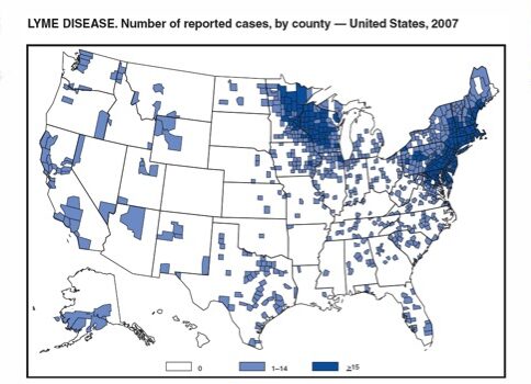
    • United States endemic areas: Northeast and Midwest regions
    • European endemic areas: central and eastern European countries
    • Geographic distribution appears to be increasing and may be related to:
      • Climate change
      • Land-use practices
      • Increasing virulent strains
  • > 30,000 cases reported in the United States each year
  • 5%–35% of Ixodes ticks Ticks Blood-sucking acarid parasites of the order ixodida comprising two families: the softbacked ticks (argasidae) and hardbacked ticks (ixodidae). Ticks are larger than their relatives, the mites. They penetrate the skin of their host by means of highly specialized, hooked mouth parts and feed on its blood. Ticks attack all groups of terrestrial vertebrates. In humans they are responsible for many tick-borne diseases, including the transmission of rocky mountain spotted fever; tularemia; babesiosis; african swine fever; and relapsing fever. Coxiella/Q Fever are infected with Borrelia Borrelia Borrelia are gram-negative microaerophilic spirochetes. Owing to their small size, they are not easily seen on Gram stain but can be visualized using dark-field microscopy, Giemsa, or Wright stain. Spirochetes are motile and move in a characteristic spinning fashion due to axial filaments in the periplasmic space. Borrelia.
  • Infection can occur from March to October and peaks around June–July.

Etiology

  • Causative organism: 
    • Spirochete Spirochete Treponema is a gram-negative, microaerophilic spirochete. Owing to its very thin structure, it is not easily seen on Gram stain, but can be visualized using dark-field microscopy. This spirochete contains endoflagella, which allow for a characteristic corkscrew movement. Treponema (gram-negative bacterium)
    • Most common species in North America: Borrelia burgdorferi Borrelia burgdorferi A specific species of bacteria, part of the borrelia burgdorferi group, whose common name is lyme disease spirochete. Borrelia 
  • Reservoir Reservoir Animate or inanimate sources which normally harbor disease-causing organisms and thus serve as potential sources of disease outbreaks. Reservoirs are distinguished from vectors (disease vectors) and carriers, which are agents of disease transmission rather than continuing sources of potential disease outbreaks. Humans may serve both as disease reservoirs and carriers. Escherichia coli hosts:
    • Small mammals (white-footed mouse)
    • White-tailed deer 
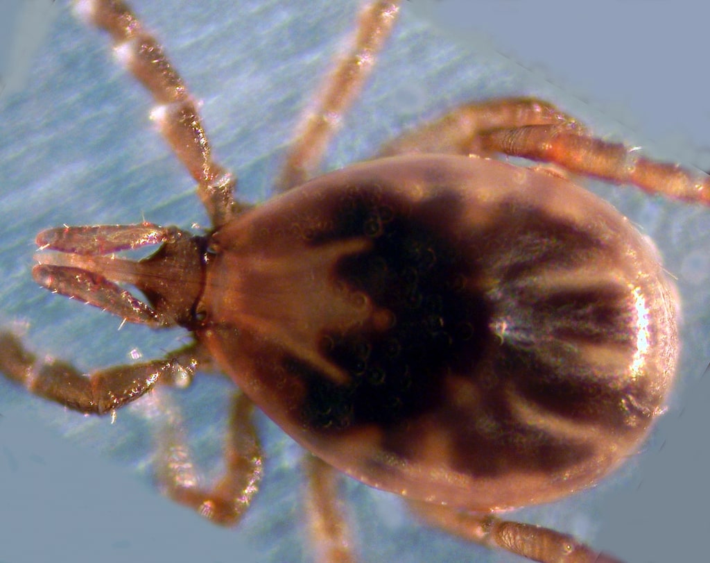
  • Vector for transmission: Ixodes tick
    • Transmission occurs through saliva Saliva The clear, viscous fluid secreted by the salivary glands and mucous glands of the mouth. It contains mucins, water, organic salts, and ptyalin. Salivary Glands: Anatomy injection during feeding
    • Longer attachment is associated with a higher risk of transmission (typically 36–72 hours)
  • At-risk groups:
    • Hikers
    • Woodworkers
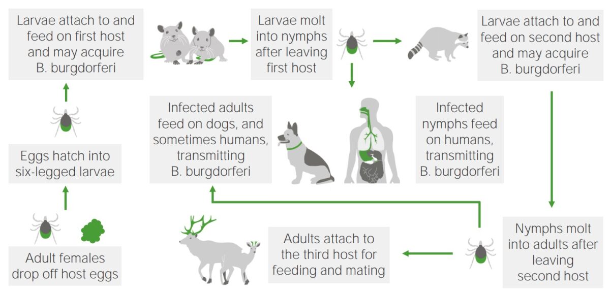
Life cycle and reservoirs of b. Burgdorferi

Lifecycle, reservoirs, and vector transmission of B. burgdorferi

Image by Lecturio.
Pathophysiology

  • Tick attachment and feeding → Borrelia Borrelia Borrelia are gram-negative microaerophilic spirochetes. Owing to their small size, they are not easily seen on Gram stain but can be visualized using dark-field microscopy, Giemsa, or Wright stain. Spirochetes are motile and move in a characteristic spinning fashion due to axial filaments in the periplasmic space. Borrelia (found in saliva Saliva The clear, viscous fluid secreted by the salivary glands and mucous glands of the mouth. It contains mucins, water, organic salts, and ptyalin. Salivary Glands: Anatomy) is injected into the skin Skin The skin, also referred to as the integumentary system, is the largest organ of the body. The skin is primarily composed of the epidermis (outer layer) and dermis (deep layer). The epidermis is primarily composed of keratinocytes that undergo rapid turnover, while the dermis contains dense layers of connective tissue. Skin: Structure and Functions:
    • May be eliminated by the immune system Immune system The body’s defense mechanism against foreign organisms or substances and deviant native cells. It includes the humoral immune response and the cell-mediated response and consists of a complex of interrelated cellular, molecular, and genetic components. Primary Lymphatic Organs
    • Otherwise, will produce an erythema Erythema Redness of the skin produced by congestion of the capillaries. This condition may result from a variety of disease processes. Chalazion migrans skin Skin The skin, also referred to as the integumentary system, is the largest organ of the body. The skin is primarily composed of the epidermis (outer layer) and dermis (deep layer). The epidermis is primarily composed of keratinocytes that undergo rapid turnover, while the dermis contains dense layers of connective tissue. Skin: Structure and Functions lesion
  • Spirochetes Spirochetes An order of slender, flexuous, helically coiled bacteria, with one or more complete turns in the helix. Treponema then spread to the blood and regional lymph nodes Lymph Nodes They are oval or bean shaped bodies (1 – 30 mm in diameter) located along the lymphatic system. Lymphatic Drainage System: Anatomy within days to weeks.
  • Eventual spread to multiple organs (e.g., heart, joints, central nervous system Central nervous system The main information-processing organs of the nervous system, consisting of the brain, spinal cord, and meninges. Nervous System: Anatomy, Structure, and Classification, eyes)
  • Manifestations are secondary to the host’s immune response:
    • Induction of autoantibodies Autoantibodies Antibodies that react with self-antigens (autoantigens) of the organism that produced them. Blotting Techniques against neuronal and glial antigens
    • Cross-reactivity of Borrelia Borrelia Borrelia are gram-negative microaerophilic spirochetes. Owing to their small size, they are not easily seen on Gram stain but can be visualized using dark-field microscopy, Giemsa, or Wright stain. Spirochetes are motile and move in a characteristic spinning fashion due to axial filaments in the periplasmic space. Borrelia antibodies Antibodies Immunoglobulins (Igs), also known as antibodies, are glycoprotein molecules produced by plasma cells that act in immune responses by recognizing and binding particular antigens. The various Ig classes are IgG (the most abundant), IgM, IgE, IgD, and IgA, which differ in their biologic features, structure, target specificity, and distribution. Immunoglobulins: Types and Functions with neural and connective tissue Connective tissue Connective tissues originate from embryonic mesenchyme and are present throughout the body except inside the brain and spinal cord. The main function of connective tissues is to provide structural support to organs. Connective tissues consist of cells and an extracellular matrix. Connective Tissue: Histology
    • T-cell–mediated immune response
  • There may be some weak endotoxin-like activity.
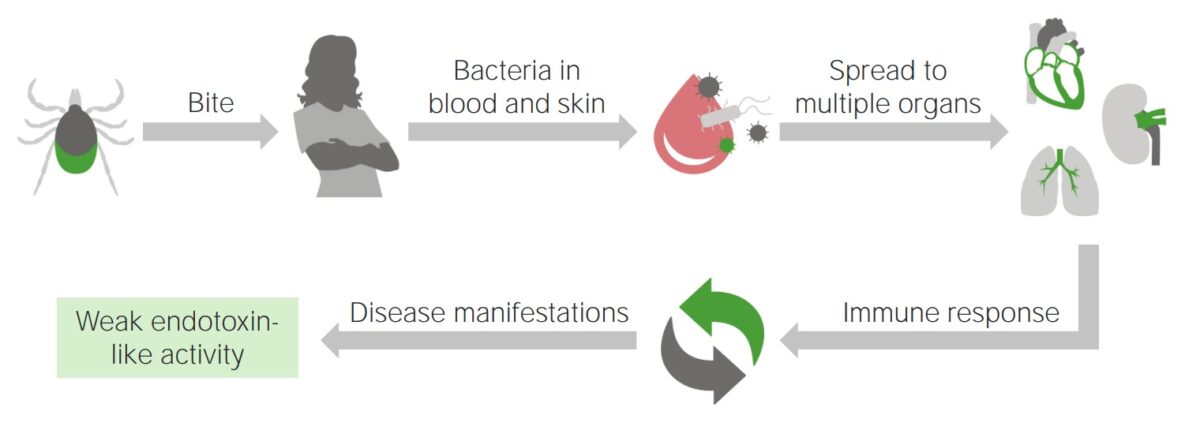
Pathophysiology of lyme disease

Progression of Lyme disease after a tick bite

Image by Lecturio.

Related videos

Clinical Presentation

The incubation Incubation The amount time between exposure to an infectious agent and becoming symptomatic. Rabies Virus period for Lyme disease is 3–30 days (mean of 7 days). The clinical manifestations of Lyme disease are broken down into 3 stages: early localized disease, early disseminated disease, and late disease.

Early localized disease

Symptoms appear in 1–5 weeks and resolve in approximately 30 days.
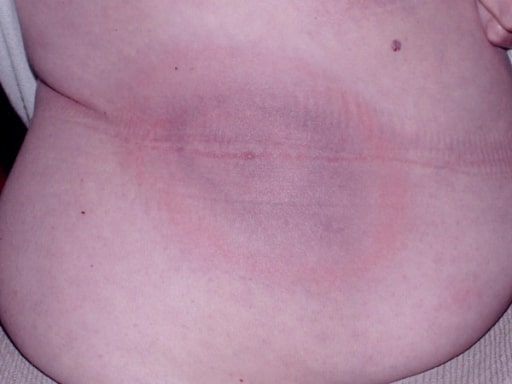
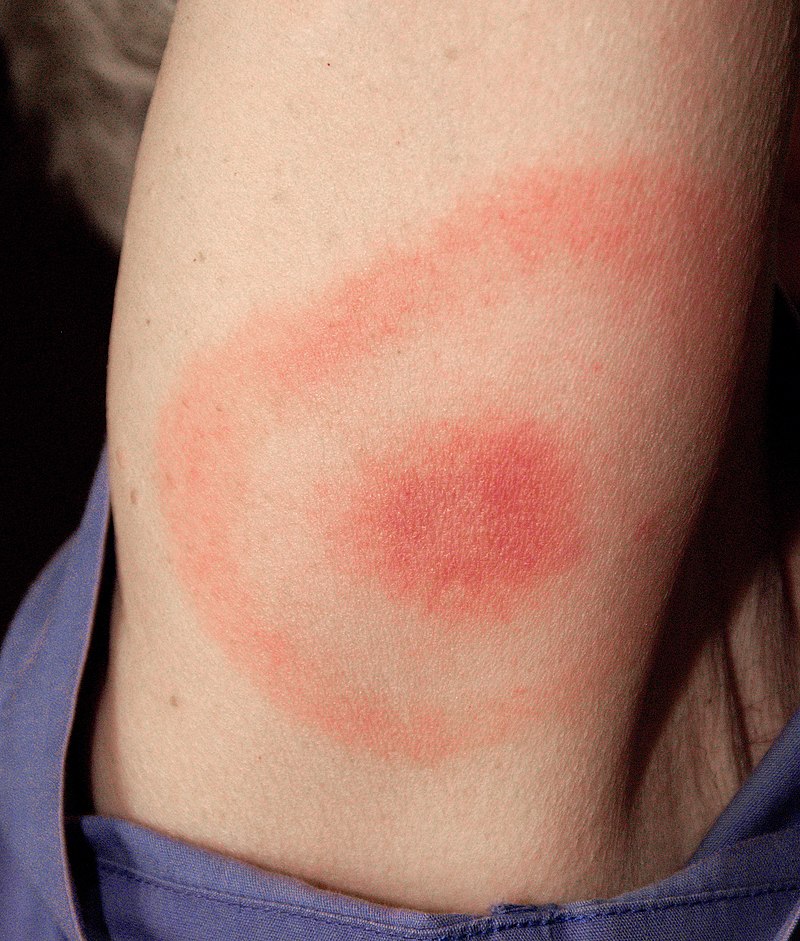
  • Erythema Erythema Redness of the skin produced by congestion of the capillaries. This condition may result from a variety of disease processes. Chalazion chronicum migrans (EM)
    • Occurs in approximately 80% of patients Patients Individuals participating in the health care system for the purpose of receiving therapeutic, diagnostic, or preventive procedures. Clinician–Patient Relationship
    • Classic “bull’s-eye” rash Rash Rocky Mountain Spotted Fever with slowly expanding red ring and central clearing
      • Central clearing usually occurs after a few days of illness.
      • The lesion can be homogeneous Homogeneous Imaging of the Spleen (have no central clearing) in up to 59% of cases.
    • Appears at the site of the tick bite
  • Fever Fever Fever is defined as a measured body temperature of at least 38°C (100.4°F). Fever is caused by circulating endogenous and/or exogenous pyrogens that increase levels of prostaglandin E2 in the hypothalamus. Fever is commonly associated with chills, rigors, sweating, and flushing of the skin. Fever
  • Myalgia Myalgia Painful sensation in the muscles. Ion Channel Myopathy
  • Arthralgia Arthralgia Pain in the joint. Rheumatic Fever
  • Fatigue Fatigue The state of weariness following a period of exertion, mental or physical, characterized by a decreased capacity for work and reduced efficiency to respond to stimuli. Fibromyalgia
  • Lymphadenopathy Lymphadenopathy Lymphadenopathy is lymph node enlargement (> 1 cm) and is benign and self-limited in most patients. Etiologies include malignancy, infection, and autoimmune disorders, as well as iatrogenic causes such as the use of certain medications. Generalized lymphadenopathy often indicates underlying systemic disease. Lymphadenopathy

Early disseminated disease

Symptoms develop in weeks to months in untreated patients Patients Individuals participating in the health care system for the purpose of receiving therapeutic, diagnostic, or preventive procedures. Clinician–Patient Relationship.

  • Musculoskeletal:
  • Cardiac (approximately 8% of patients Patients Individuals participating in the health care system for the purpose of receiving therapeutic, diagnostic, or preventive procedures. Clinician–Patient Relationship): 
    • Myocarditis Myocarditis Myocarditis is an inflammatory disease of the myocardium, which may occur alone or in association with a systemic process. There are numerous etiologies of myocarditis, but all lead to inflammation and myocyte injury, most often leading to signs and symptoms of heart failure. Myocarditis
      • Inflammation Inflammation Inflammation is a complex set of responses to infection and injury involving leukocytes as the principal cellular mediators in the body’s defense against pathogenic organisms. Inflammation is also seen as a response to tissue injury in the process of wound healing. The 5 cardinal signs of inflammation are pain, heat, redness, swelling, and loss of function. Inflammation to the myocardium Myocardium The muscle tissue of the heart. It is composed of striated, involuntary muscle cells connected to form the contractile pump to generate blood flow. Heart: Anatomy, which affects the muscle and electrical system of the heart
      • Manifests as chest pain Pain An unpleasant sensation induced by noxious stimuli which are detected by nerve endings of nociceptive neurons. Pain: Types and Pathways and shortness of breath Shortness of breath Dyspnea is the subjective sensation of breathing discomfort. Dyspnea is a normal manifestation of heavy physical or psychological exertion, but also may be caused by underlying conditions (both pulmonary and extrapulmonary). Dyspnea
      • Can lead to congestive heart failure Heart Failure A heterogeneous condition in which the heart is unable to pump out sufficient blood to meet the metabolic need of the body. Heart failure can be caused by structural defects, functional abnormalities (ventricular dysfunction), or a sudden overload beyond its capacity. Chronic heart failure is more common than acute heart failure which results from sudden insult to cardiac function, such as myocardial infarction. Total Anomalous Pulmonary Venous Return (TAPVR)
    • Pericarditis Pericarditis Pericarditis is an inflammation of the pericardium, often with fluid accumulation. It can be caused by infection (often viral), myocardial infarction, drugs, malignancies, metabolic disorders, autoimmune disorders, or trauma. Acute, subacute, and chronic forms exist. Pericarditis
      • Inflammation Inflammation Inflammation is a complex set of responses to infection and injury involving leukocytes as the principal cellular mediators in the body’s defense against pathogenic organisms. Inflammation is also seen as a response to tissue injury in the process of wound healing. The 5 cardinal signs of inflammation are pain, heat, redness, swelling, and loss of function. Inflammation of the pericardium Pericardium A conical fibroserous sac surrounding the heart and the roots of the great vessels (aorta; venae cavae; pulmonary artery). Pericardium consists of two sacs: the outer fibrous pericardium and the inner serous pericardium. The latter consists of an outer parietal layer facing the fibrous pericardium, and an inner visceral layer (epicardium) resting next to the heart, and a pericardial cavity between these two layers. Heart: Anatomy
      • Manifests as sudden onset of sharp chest pain Sharp Chest Pain Chest Pain
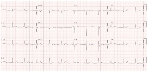
    • Atrioventricular (AV) block 
      • Secondary to myocarditis Myocarditis Myocarditis is an inflammatory disease of the myocardium, which may occur alone or in association with a systemic process. There are numerous etiologies of myocarditis, but all lead to inflammation and myocyte injury, most often leading to signs and symptoms of heart failure. Myocarditis
      • Most common cardiac manifestation
  • Neurologic (approximately 15% of patients Patients Individuals participating in the health care system for the purpose of receiving therapeutic, diagnostic, or preventive procedures. Clinician–Patient Relationship):
    • Facial nerve Facial nerve The 7th cranial nerve. The facial nerve has two parts, the larger motor root which may be called the facial nerve proper, and the smaller intermediate or sensory root. Together they provide efferent innervation to the muscles of facial expression and to the lacrimal and salivary glands, and convey afferent information for taste from the anterior two-thirds of the tongue and for touch from the external ear. The 12 Cranial Nerves: Overview and Functions palsy Palsy paralysis of an area of the body, thus incapable of voluntary movement Cranial Nerve Palsies (Bell’s palsy Palsy paralysis of an area of the body, thus incapable of voluntary movement Cranial Nerve Palsies)
      • Typically bilateral
      • Most common neurologic manifestation
    • Other cranial nerve palsy Palsy paralysis of an area of the body, thus incapable of voluntary movement Cranial Nerve Palsies (e.g., abducens nerve Abducens nerve The 6th cranial nerve which originates in the abducens nucleus of the pons and sends motor fibers to the lateral rectus muscles of the eye. Damage to the nerve or its nucleus disrupts horizontal eye movement control. The 12 Cranial Nerves: Overview and Functions)
    • Meningoradiculitis (Bannwarth syndrome):
      • Radiculopathy Radiculopathy Disease involving a spinal nerve root which may result from compression related to intervertebral disk displacement; spinal cord injuries; spinal diseases; and other conditions. Clinical manifestations include radicular pain, weakness, and sensory loss referable to structures innervated by the involved nerve root. Rheumatoid Arthritis and paresthesia
      • Follows a dermatome Dermatome Spinal Disk Herniation distribution
    • Polyneuropathy Polyneuropathy Polyneuropathy is any disease process affecting the function of or causing damage to multiple nerves of the peripheral nervous system. There are numerous etiologies of polyneuropathy, most of which are systemic and the most common of which is diabetic neuropathy. Polyneuropathy
    • Lymphocytic meningitis Meningitis Meningitis is inflammation of the meninges, the protective membranes of the brain, and spinal cord. The causes of meningitis are varied, with the most common being bacterial or viral infection. The classic presentation of meningitis is a triad of fever, altered mental status, and nuchal rigidity. Meningitis
    • Encephalitis Encephalitis Encephalitis is inflammation of the brain parenchyma caused by an infection, usually viral. Encephalitis may present with mild symptoms such as headache, fever, fatigue, and muscle and joint pain or with severe symptoms such as seizures, altered consciousness, and paralysis. Encephalitis
  • Cutaneous:
    • Multiple secondary EM lesions
      • Due to spirochetemia (presence of Borrelia Borrelia Borrelia are gram-negative microaerophilic spirochetes. Owing to their small size, they are not easily seen on Gram stain but can be visualized using dark-field microscopy, Giemsa, or Wright stain. Spirochetes are motile and move in a characteristic spinning fashion due to axial filaments in the periplasmic space. Borrelia in the blood)
      • Not from multiple tick bites
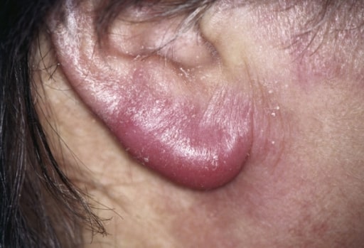
    • Lymphadenosis cutis benigna (also known as pseudolymphoma Pseudolymphoma Extranodal marginal zone lymphoma (EMZL) of mucosa-associated lymphoid tissue (also called maltoma, malt lymphoma, and pseudolymphoma) is a group of non-hodgkin’s lymphomas that have historically been grouped together because they appear to arise from postgerminal center marginal zone B cells and share a similar immunophenotype. MALT Lymphoma or borrelial lymphocytoma)
  • Ocular:
    • Conjunctivitis Conjunctivitis Conjunctivitis is a common inflammation of the bulbar and/or palpebral conjunctiva. It can be classified into infectious (mostly viral) and noninfectious conjunctivitis, which includes allergic causes. Patients commonly present with red eyes, increased tearing, burning, foreign body sensation, and photophobia. Conjunctivitis (10% of patients Patients Individuals participating in the health care system for the purpose of receiving therapeutic, diagnostic, or preventive procedures. Clinician–Patient Relationship)
    • Rare manifestations:
      • Keratitis Keratitis Inflammation of the cornea. Herpes Simplex Virus 1 and 2 ( inflammation Inflammation Inflammation is a complex set of responses to infection and injury involving leukocytes as the principal cellular mediators in the body’s defense against pathogenic organisms. Inflammation is also seen as a response to tissue injury in the process of wound healing. The 5 cardinal signs of inflammation are pain, heat, redness, swelling, and loss of function. Inflammation of the cornea Cornea The transparent anterior portion of the fibrous coat of the eye consisting of five layers: stratified squamous corneal epithelium; bowman membrane; corneal stroma; descemet membrane; and mesenchymal corneal endothelium. It serves as the first refracting medium of the eye. Eye: Anatomy)
      • Retinal vasculitis Vasculitis Inflammation of any one of the blood vessels, including the arteries; veins; and rest of the vasculature system in the body. Systemic Lupus Erythematosus
      • Optic neuropathy Neuropathy Leprosy
      • Uveitis Uveitis Uveitis is the inflammation of the uvea, the pigmented middle layer of the eye, which comprises the iris, ciliary body, and choroid. The condition is categorized based on the site of disease; anterior uveitis is the most common. Diseases of the Uvea ( inflammation Inflammation Inflammation is a complex set of responses to infection and injury involving leukocytes as the principal cellular mediators in the body’s defense against pathogenic organisms. Inflammation is also seen as a response to tissue injury in the process of wound healing. The 5 cardinal signs of inflammation are pain, heat, redness, swelling, and loss of function. Inflammation of the uvea Uvea The pigmented vascular coat of the eyeball, consisting of the choroid; ciliary body; and iris, which are continuous with each other. Eye: Anatomy)

Late disease

Symptoms develop in months to years in untreated patients Patients Individuals participating in the health care system for the purpose of receiving therapeutic, diagnostic, or preventive procedures. Clinician–Patient Relationship.
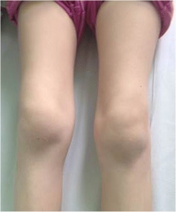
  • Migratory polyarthritis Migratory Polyarthritis Rheumatic Fever (60% of patients Patients Individuals participating in the health care system for the purpose of receiving therapeutic, diagnostic, or preventive procedures. Clinician–Patient Relationship)
    • Large joints, particularly the knee
    • Intermittent joint swelling Swelling Inflammation and pain Pain An unpleasant sensation induced by noxious stimuli which are detected by nerve endings of nociceptive neurons. Pain: Types and Pathways
    • Baker cysts Cysts Any fluid-filled closed cavity or sac that is lined by an epithelium. Cysts can be of normal, abnormal, non-neoplastic, or neoplastic tissues. Fibrocystic Change can form.
  • Neurologic symptoms:
    • Rare because of appropriate antibiotic treatment during the early stages
    • Lyme encephalopathy Encephalopathy Hyper-IgM Syndrome: subtle cognitive, mood, and sleep Sleep A readily reversible suspension of sensorimotor interaction with the environment, usually associated with recumbency and immobility. Physiology of Sleep disturbances
    • Chronic axonal polyneuropathy Polyneuropathy Polyneuropathy is any disease process affecting the function of or causing damage to multiple nerves of the peripheral nervous system. There are numerous etiologies of polyneuropathy, most of which are systemic and the most common of which is diabetic neuropathy. Polyneuropathy
    • Chronic encephalomyelitis
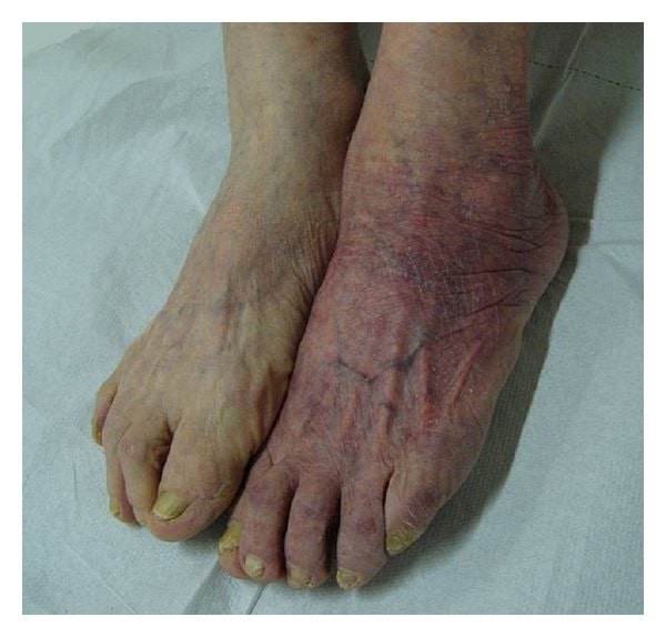
  • Acrodermatitis chronica atrophicans 
    • More common in Europe due to B. afzelii B. afzelii Borrelia
    • Starts with unilateral bluish-red discoloration and swelling Swelling Inflammation
    • Atrophy Atrophy Decrease in the size of a cell, tissue, organ, or multiple organs, associated with a variety of pathological conditions such as abnormal cellular changes, ischemia, malnutrition, or hormonal changes. Cellular Adaptation eventually develops and vasculature appears more prominent.

Mnemonic

Common symptoms of Lyme disease can be remembered by the mnemonic phrase “a key Lyme pie to the FACE.”
  • Facial nerve palsy Palsy paralysis of an area of the body, thus incapable of voluntary movement Cranial Nerve Palsies
  • Arthritis
  • Cardiac block
  • Erythema migrans

Diagnosis

Diagnostic algorithm

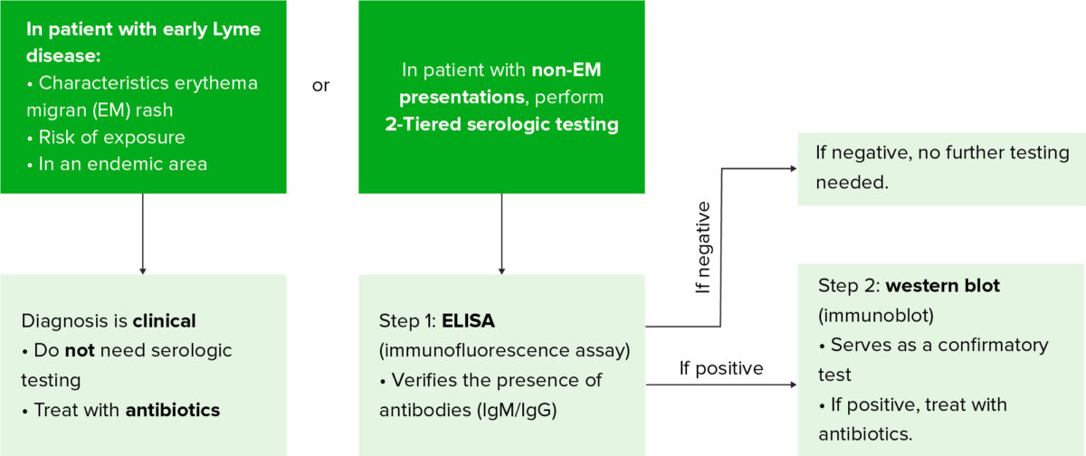
Lyme disease diagnosis

Diagnostic algorithm for Lyme disease

ELISA: enzyme-linked immunosorbent assay

Image by Lecturio.
  • A Lyme disease diagnosis is based on the clinical picture and supported by serologic testing.
  • Clinical symptoms largely depend on the stage of disease.
  • Lyme testing is not warranted for:

Laboratory testing

Laboratory tests are only significant in conjunction with the clinical history due to a high false-positive and false-negative rate.
  • Antibody testing
    • Enzyme-linked immunosorbent assay ( ELISA ELISA An immunoassay utilizing an antibody labeled with an enzyme marker such as horseradish peroxidase. While either the enzyme or the antibody is bound to an immunosorbent substrate, they both retain their biologic activity; the change in enzyme activity as a result of the enzyme-antibody-antigen reaction is proportional to the concentration of the antigen and can be measured spectrophotometrically or with the naked eye. Many variations of the method have been developed. St. Louis Encephalitis Virus)
      • Initial test
      • Detects antibodies Antibodies Immunoglobulins (Igs), also known as antibodies, are glycoprotein molecules produced by plasma cells that act in immune responses by recognizing and binding particular antigens. The various Ig classes are IgG (the most abundant), IgM, IgE, IgD, and IgA, which differ in their biologic features, structure, target specificity, and distribution. Immunoglobulins: Types and Functions through a coupled enzyme-antibody complex, which produces a color change
      • False-positives can occur due to other infections Infections Invasion of the host organism by microorganisms or their toxins or by parasites that can cause pathological conditions or diseases. Chronic Granulomatous Disease (e.g., syphilis Syphilis Syphilis is a bacterial infection caused by the spirochete Treponema pallidum pallidum (T. p. pallidum), which is usually spread through sexual contact. Syphilis has 4 clinical stages: primary, secondary, latent, and tertiary. Syphilis) or autoimmune disease.
      • False-negatives can occur in the early stage; thus, ELISA ELISA An immunoassay utilizing an antibody labeled with an enzyme marker such as horseradish peroxidase. While either the enzyme or the antibody is bound to an immunosorbent substrate, they both retain their biologic activity; the change in enzyme activity as a result of the enzyme-antibody-antigen reaction is proportional to the concentration of the antigen and can be measured spectrophotometrically or with the naked eye. Many variations of the method have been developed. St. Louis Encephalitis Virus is not done if the characteristic EM rash Rash Rocky Mountain Spotted Fever is seen.
    • Western blot
      • Confirmation test if the ELISA ELISA An immunoassay utilizing an antibody labeled with an enzyme marker such as horseradish peroxidase. While either the enzyme or the antibody is bound to an immunosorbent substrate, they both retain their biologic activity; the change in enzyme activity as a result of the enzyme-antibody-antigen reaction is proportional to the concentration of the antigen and can be measured spectrophotometrically or with the naked eye. Many variations of the method have been developed. St. Louis Encephalitis Virus is positive
      • Detects antibodies Antibodies Immunoglobulins (Igs), also known as antibodies, are glycoprotein molecules produced by plasma cells that act in immune responses by recognizing and binding particular antigens. The various Ig classes are IgG (the most abundant), IgM, IgE, IgD, and IgA, which differ in their biologic features, structure, target specificity, and distribution. Immunoglobulins: Types and Functions to individual protein components of B. burgdorferi using electrophoresis Electrophoresis An electrochemical process in which macromolecules or colloidal particles with a net electric charge migrate in a solution under the influence of an electric current. Blotting Techniques
  • Polymerase chain reaction Polymerase chain reaction Polymerase chain reaction (PCR) is a technique that amplifies DNA fragments exponentially for analysis. The process is highly specific, allowing for the targeting of specific genomic sequences, even with minuscule sample amounts. The PCR cycles multiple times through 3 phases: denaturation of the template DNA, annealing of a specific primer to the individual DNA strands, and synthesis/elongation of new DNA molecules. Polymerase Chain Reaction (PCR) ( PCR PCR Polymerase chain reaction (PCR) is a technique that amplifies DNA fragments exponentially for analysis. The process is highly specific, allowing for the targeting of specific genomic sequences, even with minuscule sample amounts. The PCR cycles multiple times through 3 phases: denaturation of the template DNA, annealing of a specific primer to the individual DNA strands, and synthesis/elongation of new DNA molecules. Polymerase Chain Reaction (PCR))
    • May be performed on cerebrospinal fluid Cerebrospinal Fluid A watery fluid that is continuously produced in the choroid plexus and circulates around the surface of the brain; spinal cord; and in the cerebral ventricles. Ventricular System: Anatomy and synovial fluid for those who present with meningitis Meningitis Meningitis is inflammation of the meninges, the protective membranes of the brain, and spinal cord. The causes of meningitis are varied, with the most common being bacterial or viral infection. The classic presentation of meningitis is a triad of fever, altered mental status, and nuchal rigidity. Meningitis or arthritis Arthritis Acute or chronic inflammation of joints. Osteoarthritis. (Note: Although PCR PCR Polymerase chain reaction (PCR) is a technique that amplifies DNA fragments exponentially for analysis. The process is highly specific, allowing for the targeting of specific genomic sequences, even with minuscule sample amounts. The PCR cycles multiple times through 3 phases: denaturation of the template DNA, annealing of a specific primer to the individual DNA strands, and synthesis/elongation of new DNA molecules. Polymerase Chain Reaction (PCR) is an option, serum antibody testing is still preferred.)
    • High false-negative rate
    • Can remain positive for years after treatment
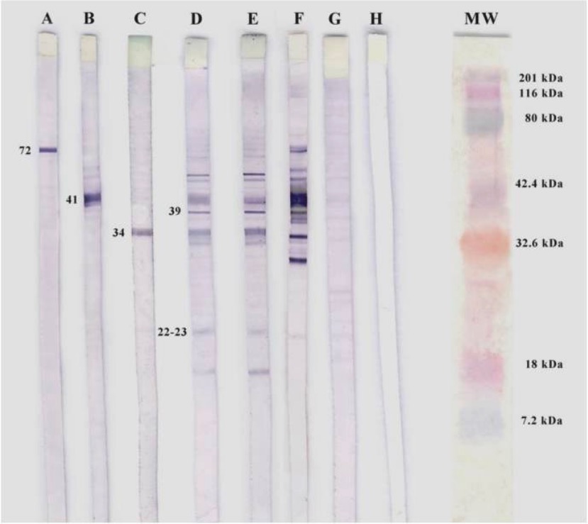
Western blotting of b. Burgdorferi

Western blotting of B. burgdorferi.
A, B, and C: positive controls for specific monoclonal antibodies
D and E: seropositive patients for IgM
F: positive control
G: negative control
H: white control
MW: molecular weight

Image: “Western blotting of B. burgdorferi” by Department of Biomedicine and Biotecnology, Alcalá University, 28871 Alcalá de Henares, Spain. License: CC BY 3.0

Management

Treatment

Oral antibiotics:

  • Doxycycline
  • Amoxicillin Amoxicillin A broad-spectrum semisynthetic antibiotic similar to ampicillin except that its resistance to gastric acid permits higher serum levels with oral administration. Penicillins
  • Cefuroxime
  • Used in all stages of Lyme disease

Intravenous (IV) antibiotics:

  • Ceftriaxone Ceftriaxone A broad-spectrum cephalosporin antibiotic and cefotaxime derivative with a very long half-life and high penetrability to meninges, eyes and inner ears. Cephalosporins
  • Cefotaxime Cefotaxime Semisynthetic broad-spectrum cephalosporin. Cephalosporins
  • Penicillin Penicillin Rheumatic Fever G
  • Used for:
    • Those who cannot tolerate oral antibiotics
    • Early disseminated disease with meningitis Meningitis Meningitis is inflammation of the meninges, the protective membranes of the brain, and spinal cord. The causes of meningitis are varied, with the most common being bacterial or viral infection. The classic presentation of meningitis is a triad of fever, altered mental status, and nuchal rigidity. Meningitis, encephalomyelitis, or severe cardiac manifestations (3rd-degree AV block AV block Atrioventricular (AV) block is a bradyarrhythmia caused by delay, or interruption, in the electrical conduction between the atria and the ventricles. Atrioventricular block occurs due to either anatomic or functional impairment, and is classified into 3 types. Atrioventricular block (AV block))
    • Late disease with neurologic manifestations
    • Recurrent arthritis Arthritis Acute or chronic inflammation of joints. Osteoarthritis despite oral therapy

Additional considerations:

  • 3rd-degree AV block AV block Atrioventricular (AV) block is a bradyarrhythmia caused by delay, or interruption, in the electrical conduction between the atria and the ventricles. Atrioventricular block occurs due to either anatomic or functional impairment, and is classified into 3 types. Atrioventricular block (AV block)
    • May require temporary pacing
    • AV block AV block Atrioventricular (AV) block is a bradyarrhythmia caused by delay, or interruption, in the electrical conduction between the atria and the ventricles. Atrioventricular block occurs due to either anatomic or functional impairment, and is classified into 3 types. Atrioventricular block (AV block) is usually short-lived with treatment.
  • Jarisch-Herxheimer reaction
    • Seen in 15% of patients Patients Individuals participating in the health care system for the purpose of receiving therapeutic, diagnostic, or preventive procedures. Clinician–Patient Relationship with early Lyme disease
    • Transient worsening of symptoms during the initial 24 hours of treatment
    • Due to an immune response to the antigens released by dying spirochetes Spirochetes An order of slender, flexuous, helically coiled bacteria, with one or more complete turns in the helix. Treponema
Table: Summary of the management of the different clinical manifestations of Lyme disease
Clinical manifestations Management
Asymptomatic patient with a tick bite requires no prophylactic treatment unless: If meets criteria, then must receive prophylactic doxycycline within 72 hours of bite
Erythema Erythema Redness of the skin produced by congestion of the capillaries. This condition may result from a variety of disease processes. Chalazion migrans (localized disease)
  • Doxycycline
  • Amoxicillin Amoxicillin A broad-spectrum semisynthetic antibiotic similar to ampicillin except that its resistance to gastric acid permits higher serum levels with oral administration. Penicillins
  • Cefuroxime
Early disseminated disease:
  • Erythema Erythema Redness of the skin produced by congestion of the capillaries. This condition may result from a variety of disease processes. Chalazion migrans lesions (multiple)
  • Doxycycline
  • Amoxicillin Amoxicillin A broad-spectrum semisynthetic antibiotic similar to ampicillin except that its resistance to gastric acid permits higher serum levels with oral administration. Penicillins
  • Cefuroxime
  • Facial nerve Facial nerve The 7th cranial nerve. The facial nerve has two parts, the larger motor root which may be called the facial nerve proper, and the smaller intermediate or sensory root. Together they provide efferent innervation to the muscles of facial expression and to the lacrimal and salivary glands, and convey afferent information for taste from the anterior two-thirds of the tongue and for touch from the external ear. The 12 Cranial Nerves: Overview and Functions palsy Palsy paralysis of an area of the body, thus incapable of voluntary movement Cranial Nerve Palsies
  • Meningitis Meningitis Meningitis is inflammation of the meninges, the protective membranes of the brain, and spinal cord. The causes of meningitis are varied, with the most common being bacterial or viral infection. The classic presentation of meningitis is a triad of fever, altered mental status, and nuchal rigidity. Meningitis
  • Radiculoneuropathy
  • Doxycycline
  • Amoxicillin Amoxicillin A broad-spectrum semisynthetic antibiotic similar to ampicillin except that its resistance to gastric acid permits higher serum levels with oral administration. Penicillins
  • Cefuroxime
Cardiac and severe neurologic manifestations Intravenous ceftriaxone Ceftriaxone A broad-spectrum cephalosporin antibiotic and cefotaxime derivative with a very long half-life and high penetrability to meninges, eyes and inner ears. Cephalosporins
Late disease:
  • Doxycycline
  • Amoxicillin Amoxicillin A broad-spectrum semisynthetic antibiotic similar to ampicillin except that its resistance to gastric acid permits higher serum levels with oral administration. Penicillins
  • Cefuroxime
Reinfection Same antibiotic as recommended for a primary infection Primary infection Herpes Simplex Virus 1 and 2

Post-Lyme disease syndrome (PLDS) and chronic Lyme disease

  • Description:
    • Previous history of Lyme disease (early or late)
    • Manifestations of Lyme disease resolved or stabilized after being treated with an accepted regimen.
    • Subjective symptoms (e.g., fatigue Fatigue The state of weariness following a period of exertion, mental or physical, characterized by a decreased capacity for work and reduced efficiency to respond to stimuli. Fibromyalgia, widespread musculoskeletal pain Pain An unpleasant sensation induced by noxious stimuli which are detected by nerve endings of nociceptive neurons. Pain: Types and Pathways, reports of cognitive difficulties): 
      • Started within 6 months after the diagnosis of Lyme disease
      • Persist (continuously or relapsing) for ≥ 6 months after completing treatment
  • Symptoms are severe enough to cause substantial reduction of activities.
  • Chronic subjective symptoms are not thought to be due to persistent infection.
  • Patients Patients Individuals participating in the health care system for the purpose of receiving therapeutic, diagnostic, or preventive procedures. Clinician–Patient Relationship should be reevaluated since this may be due to:
    • Incorrect Lyme diagnosis
    • Coinfection with another tick-borne disease ( Babesia Babesia Babesiosis is an infection caused by a protozoa belonging to the genus, Babesia. The most common Babesia seen in the United States is B. microti, which is transmitted by the Ixodes tick. The protozoa thrive and replicate within host erythrocytes. Lysis of erythrocytes and the body’s immune response result in clinical symptoms. Babesia/Babesiosis, Anaplasma)
    • Another concurrent condition ( fibromyalgia Fibromyalgia Fibromyalgia is a chronic pain syndrome characterized by widespread body pain, chronic fatigue, mood disturbance, and cognitive disturbance. It also presents with other comorbid symptoms such as migraine headaches, depression, sleep disturbance, and irritable bowel syndrome. Fibromyalgia, depression)
    • Permanent tissue damage (particularly from neurologic involvement)
  • No proven benefit to additional antibiotics
  • No proven treatment

Prevention and Prophylaxis

  • If the tick is removed within the first 12 hours, the infection risk is very low.
  • Do not assume immunity is developed after a previous infection.
  • For persons from non-endemic areas, there is no need for prophylactic antibiotics, but the tick site should be observed.

Precautions against ticks Ticks Blood-sucking acarid parasites of the order ixodida comprising two families: the softbacked ticks (argasidae) and hardbacked ticks (ixodidae). Ticks are larger than their relatives, the mites. They penetrate the skin of their host by means of highly specialized, hooked mouth parts and feed on its blood. Ticks attack all groups of terrestrial vertebrates. In humans they are responsible for many tick-borne diseases, including the transmission of rocky mountain spotted fever; tularemia; babesiosis; african swine fever; and relapsing fever. Coxiella/Q Fever

  • Protective clothing
  • Repellents
  • Scanning the body for ticks Ticks Blood-sucking acarid parasites of the order ixodida comprising two families: the softbacked ticks (argasidae) and hardbacked ticks (ixodidae). Ticks are larger than their relatives, the mites. They penetrate the skin of their host by means of highly specialized, hooked mouth parts and feed on its blood. Ticks attack all groups of terrestrial vertebrates. In humans they are responsible for many tick-borne diseases, including the transmission of rocky mountain spotted fever; tularemia; babesiosis; african swine fever; and relapsing fever. Coxiella/Q Fever after being outside
  • Removing ticks Ticks Blood-sucking acarid parasites of the order ixodida comprising two families: the softbacked ticks (argasidae) and hardbacked ticks (ixodidae). Ticks are larger than their relatives, the mites. They penetrate the skin of their host by means of highly specialized, hooked mouth parts and feed on its blood. Ticks attack all groups of terrestrial vertebrates. In humans they are responsible for many tick-borne diseases, including the transmission of rocky mountain spotted fever; tularemia; babesiosis; african swine fever; and relapsing fever. Coxiella/Q Fever from bite sites as soon as possible with a tweezer

Doxycycline prophylaxis Prophylaxis Cephalosporins

Doxycycline prophylaxis Prophylaxis Cephalosporins is indicated if all the following criteria are met MET Preoperative Care:

  • Tick is identified as Ixodes scapularis Ixodes scapularis Borrelia.
  • Tick has been attached for ≥ 36 hours.
  • Prophylaxis Prophylaxis Cephalosporins will start within 72 hours of tick removal.
  • Rate of B. burgdorferi infection of ticks Ticks Blood-sucking acarid parasites of the order ixodida comprising two families: the softbacked ticks (argasidae) and hardbacked ticks (ixodidae). Ticks are larger than their relatives, the mites. They penetrate the skin of their host by means of highly specialized, hooked mouth parts and feed on its blood. Ticks attack all groups of terrestrial vertebrates. In humans they are responsible for many tick-borne diseases, including the transmission of rocky mountain spotted fever; tularemia; babesiosis; african swine fever; and relapsing fever. Coxiella/Q Fever in the area is ≥ 20%.
  • Doxycycline is not contraindicated.

Differential Diagnosis

  • Babesiosis Babesiosis Babesiosis is an infection caused by a protozoa belonging to the genus, Babesia. The most common Babesia seen in the United States is B. microti, which is transmitted by the Ixodes tick. The protozoa thrive and replicate within host erythrocytes. Lysis of erythrocytes and the body’s immune response result in clinical symptoms. Babesia/Babesiosis: a tick-borne infection caused by Babesia Babesia Babesiosis is an infection caused by a protozoa belonging to the genus, Babesia. The most common Babesia seen in the United States is B. microti, which is transmitted by the Ixodes tick. The protozoa thrive and replicate within host erythrocytes. Lysis of erythrocytes and the body’s immune response result in clinical symptoms. Babesia/Babesiosis. Patients Patients Individuals participating in the health care system for the purpose of receiving therapeutic, diagnostic, or preventive procedures. Clinician–Patient Relationship can be asymptomatic or develop fever Fever Fever is defined as a measured body temperature of at least 38°C (100.4°F). Fever is caused by circulating endogenous and/or exogenous pyrogens that increase levels of prostaglandin E2 in the hypothalamus. Fever is commonly associated with chills, rigors, sweating, and flushing of the skin. Fever, fatigue Fatigue The state of weariness following a period of exertion, mental or physical, characterized by a decreased capacity for work and reduced efficiency to respond to stimuli. Fibromyalgia, malaise Malaise Tick-borne Encephalitis Virus, and arthralgias. Asplenic, immunocompromised immunocompromised A human or animal whose immunologic mechanism is deficient because of an immunodeficiency disorder or other disease or as the result of the administration of immunosuppressive drugs or radiation. Gastroenteritis, and elderly patients Patients Individuals participating in the health care system for the purpose of receiving therapeutic, diagnostic, or preventive procedures. Clinician–Patient Relationship are at risk for severe disease, causing hemolytic anemia Hemolytic Anemia Hemolytic anemia (HA) is the term given to a large group of anemias that are caused by the premature destruction/hemolysis of circulating red blood cells (RBCs). Hemolysis can occur within (intravascular hemolysis) or outside the blood vessels (extravascular hemolysis). Hemolytic Anemia, thrombocytopenia Thrombocytopenia Thrombocytopenia occurs when the platelet count is < 150,000 per microliter. The normal range for platelets is usually 150,000-450,000/µL of whole blood. Thrombocytopenia can be a result of decreased production, increased destruction, or splenic sequestration of platelets. Patients are often asymptomatic until platelet counts are < 50,000/µL. Thrombocytopenia, hepatosplenomegaly Hepatosplenomegaly Cytomegalovirus, renal failure Renal failure Conditions in which the kidneys perform below the normal level in the ability to remove wastes, concentrate urine, and maintain electrolyte balance; blood pressure; and calcium metabolism. Renal insufficiency can be classified by the degree of kidney damage (as measured by the level of proteinuria) and reduction in glomerular filtration rate. Crush Syndrome, and death. Diagnosis is confirmed with a peripheral blood smear Peripheral Blood Smear Anemia: Overview and Types, serologic testing, and PCR PCR Polymerase chain reaction (PCR) is a technique that amplifies DNA fragments exponentially for analysis. The process is highly specific, allowing for the targeting of specific genomic sequences, even with minuscule sample amounts. The PCR cycles multiple times through 3 phases: denaturation of the template DNA, annealing of a specific primer to the individual DNA strands, and synthesis/elongation of new DNA molecules. Polymerase Chain Reaction (PCR). Treatment includes antibiotics, such as atovaquone Atovaquone A hydroxynaphthoquinone that has antimicrobial activity and is being used in antimalarial protocols. Antimalarial Drugs plus azithromycin Azithromycin A semi-synthetic macrolide antibiotic structurally related to erythromycin. It has been used in the treatment of Mycobacterium avium intracellulare infections, toxoplasmosis, and cryptosporidiosis. Macrolides and Ketolides.
  • Ehrlichiosis Ehrlichiosis Ehrlichiosis is a tick-borne bacterial infection. The most common causative species include Ehrlichia chaffeensis, which infect and multiply within monocytes. The clinical presentation can vary widely, but often includes fever, malaise, headache, myalgia, and arthralgias. Ehrlichiosis and Anaplasmosis and anaplasmosis Anaplasmosis Anaplasmosis is a tick-borne bacterial infection. The most common causative species include Anaplasma phagocytophilum, which infect and multiply within granulocytes. The clinical presentation can vary widely, but often includes fever, malaise, headache, myalgia, and arthralgias. Ehrlichiosis and Anaplasmosis: tick-borne infections Infections Invasion of the host organism by microorganisms or their toxins or by parasites that can cause pathological conditions or diseases. Chronic Granulomatous Disease caused by Ehrlichia chaffeensis Ehrlichia chaffeensis A species of gram-negative bacteria that is the causative agent of human ehrlichiosis. This organism was first discovered at fort chaffee, arkansas, when blood samples from suspected human ehrlichiosis patients were studied. Ehrlichiosis and Anaplasmosis and Anaplasmosis Anaplasmosis Anaplasmosis is a tick-borne bacterial infection. The most common causative species include Anaplasma phagocytophilum, which infect and multiply within granulocytes. The clinical presentation can vary widely, but often includes fever, malaise, headache, myalgia, and arthralgias. Ehrlichiosis and Anaplasmosis phagocytophilum, respectively. Symptoms include fever Fever Fever is defined as a measured body temperature of at least 38°C (100.4°F). Fever is caused by circulating endogenous and/or exogenous pyrogens that increase levels of prostaglandin E2 in the hypothalamus. Fever is commonly associated with chills, rigors, sweating, and flushing of the skin. Fever, headache Headache The symptom of pain in the cranial region. It may be an isolated benign occurrence or manifestation of a wide variety of headache disorders. Brain Abscess, and malaise Malaise Tick-borne Encephalitis Virus. A rash Rash Rocky Mountain Spotted Fever is uncommon, but can appear petechial or maculopapular Maculopapular Dermatologic Examination. The diagnosis is made with a PCR PCR Polymerase chain reaction (PCR) is a technique that amplifies DNA fragments exponentially for analysis. The process is highly specific, allowing for the targeting of specific genomic sequences, even with minuscule sample amounts. The PCR cycles multiple times through 3 phases: denaturation of the template DNA, annealing of a specific primer to the individual DNA strands, and synthesis/elongation of new DNA molecules. Polymerase Chain Reaction (PCR) test. Treatment of both diseases is with doxycycline.
  • Rocky Mountain spotted fever Fever Fever is defined as a measured body temperature of at least 38°C (100.4°F). Fever is caused by circulating endogenous and/or exogenous pyrogens that increase levels of prostaglandin E2 in the hypothalamus. Fever is commonly associated with chills, rigors, sweating, and flushing of the skin. Fever: a disease caused by Rickettsia rickettsii Rickettsia rickettsii A species of gram-negative, aerobic bacteria that is the etiologic agent of rocky mountain spotted fever. Its cells are slightly smaller and more uniform in size than those of rickettsia prowazekii. Rickettsia that presents with fever Fever Fever is defined as a measured body temperature of at least 38°C (100.4°F). Fever is caused by circulating endogenous and/or exogenous pyrogens that increase levels of prostaglandin E2 in the hypothalamus. Fever is commonly associated with chills, rigors, sweating, and flushing of the skin. Fever, fatigue Fatigue The state of weariness following a period of exertion, mental or physical, characterized by a decreased capacity for work and reduced efficiency to respond to stimuli. Fibromyalgia, headache Headache The symptom of pain in the cranial region. It may be an isolated benign occurrence or manifestation of a wide variety of headache disorders. Brain Abscess, and a rash Rash Rocky Mountain Spotted Fever following a tick bite. However, this disease is associated with the Dermacentor Dermacentor A widely distributed genus of ticks, in the family ixodidae, including a number that infest humans and other mammals. Several are vectors of diseases such as tularemia; rocky mountain spotted fever; colorado tick fever; and anaplasmosis. Rickettsia tick, and the rash Rash Rocky Mountain Spotted Fever begins on the distal extremities and spreads centrally. Diagnosis is made based on the clinical features, biopsy Biopsy Removal and pathologic examination of specimens from the living body. Ewing Sarcoma of the rash Rash Rocky Mountain Spotted Fever, and serologic testing. Treatment involves antibiotics, including doxycycline.
  • Epidemic typhus Epidemic Typhus Epidemic typhus is a febrile illness caused by the obligate intracellular gram-negative bacterium, Rickettsia prowazekii. Epidemic typhus is also known as louse-borne typhus or jail fever, and its symptoms include high fever, headache, myalgias, dry cough, delirium, stupor, and rash. Epidemic Typhus: a disease caused by Rickettsia prowazekii Rickettsia prowazekii A species of gram-negative, aerobic bacteria that is the etiologic agent of epidemic typhus fever acquired through contact with lice as well as brill’s disease. Rickettsia that presents with myalgia Myalgia Painful sensation in the muscles. Ion Channel Myopathy, arthralgia Arthralgia Pain in the joint. Rheumatic Fever, rash Rash Rocky Mountain Spotted Fever, and encephalitis Encephalitis Encephalitis is inflammation of the brain parenchyma caused by an infection, usually viral. Encephalitis may present with mild symptoms such as headache, fever, fatigue, and muscle and joint pain or with severe symptoms such as seizures, altered consciousness, and paralysis. Encephalitis. A rash Rash Rocky Mountain Spotted Fever starts on the trunk and spreads outward to the extremities. Epidemic typhus Epidemic Typhus Epidemic typhus is a febrile illness caused by the obligate intracellular gram-negative bacterium, Rickettsia prowazekii. Epidemic typhus is also known as louse-borne typhus or jail fever, and its symptoms include high fever, headache, myalgias, dry cough, delirium, stupor, and rash. Epidemic Typhus is transmitted to humans by lice. Diagnosis is based on the clinical picture, biopsy Biopsy Removal and pathologic examination of specimens from the living body. Ewing Sarcoma of the rash Rash Rocky Mountain Spotted Fever, and serologic testing. Treatment involves antibiotics, including doxycycline. 
  • Infectious mononucleosis Mononucleosis Infectious mononucleosis (IM), also known as “the kissing disease,” is a highly contagious viral infection caused by the Epstein-Barr virus. Its common name is derived from its main method of transmission: the spread of infected saliva via kissing. Clinical manifestations of IM include fever, tonsillar pharyngitis, and lymphadenopathy. Mononucleosis: a disease caused by the Epstein-Barr virus Epstein-Barr Virus Epstein-Barr virus (EBV) is a linear, double-stranded DNA virus belonging to the Herpesviridae family. This highly prevalent virus is mostly transmitted through contact with oropharyngeal secretions from an infected individual. The virus can infect epithelial cells and B lymphocytes, where it can undergo lytic replication or latency. Epstein-Barr Virus that is characterized by fever Fever Fever is defined as a measured body temperature of at least 38°C (100.4°F). Fever is caused by circulating endogenous and/or exogenous pyrogens that increase levels of prostaglandin E2 in the hypothalamus. Fever is commonly associated with chills, rigors, sweating, and flushing of the skin. Fever, fatigue Fatigue The state of weariness following a period of exertion, mental or physical, characterized by a decreased capacity for work and reduced efficiency to respond to stimuli. Fibromyalgia, lymphadenopathy Lymphadenopathy Lymphadenopathy is lymph node enlargement (> 1 cm) and is benign and self-limited in most patients. Etiologies include malignancy, infection, and autoimmune disorders, as well as iatrogenic causes such as the use of certain medications. Generalized lymphadenopathy often indicates underlying systemic disease. Lymphadenopathy, and pharyngitis Pharyngitis Pharyngitis is an inflammation of the back of the throat (pharynx). Pharyngitis is usually caused by an upper respiratory tract infection, which is viral in most cases. It typically results in a sore throat and fever. Other symptoms may include a runny nose, cough, headache, and hoarseness. Pharyngitis. Diagnosis is based on clinical features and testing, such as a heterophile antibody test or serology Serology The study of serum, especially of antigen-antibody reactions in vitro. Yellow Fever Virus. Treatment is supportive. The use of amoxicillin Amoxicillin A broad-spectrum semisynthetic antibiotic similar to ampicillin except that its resistance to gastric acid permits higher serum levels with oral administration. Penicillins can cause a characteristic maculopapular Maculopapular Dermatologic Examination rash Rash Rocky Mountain Spotted Fever
  • Juvenile idiopathic Idiopathic Dermatomyositis arthritis Arthritis Acute or chronic inflammation of joints. Osteoarthritis: a group of rheumatic diseases that cause arthritis Arthritis Acute or chronic inflammation of joints. Osteoarthritis, fever Fever Fever is defined as a measured body temperature of at least 38°C (100.4°F). Fever is caused by circulating endogenous and/or exogenous pyrogens that increase levels of prostaglandin E2 in the hypothalamus. Fever is commonly associated with chills, rigors, sweating, and flushing of the skin. Fever, rash Rash Rocky Mountain Spotted Fever, adenopathy, splenomegaly Splenomegaly Splenomegaly is pathologic enlargement of the spleen that is attributable to numerous causes, including infections, hemoglobinopathies, infiltrative processes, and outflow obstruction of the portal vein. Splenomegaly, and iridocyclitis Iridocyclitis Acute or chronic inflammation of the iris and ciliary body characterized by exudates into the anterior chamber, discoloration of the iris, and constricted, sluggish pupil. Symptoms include radiating pain, photophobia, lacrimation, and interference with vision. Relapsing Fever in children under 16 years of age. The rash Rash Rocky Mountain Spotted Fever may vary, but can appear macular with a central clearing. Diagnosis is based on the clinical picture and an autoimmune workup, including rheumatoid factor Rheumatoid factor Antibodies found in adult rheumatoid arthritis patients that are directed against gamma-chain immunoglobulins. Autoimmune Hepatitis ( RF RF Rheumatoid Arthritis), antinuclear antibodies Antibodies Immunoglobulins (Igs), also known as antibodies, are glycoprotein molecules produced by plasma cells that act in immune responses by recognizing and binding particular antigens. The various Ig classes are IgG (the most abundant), IgM, IgE, IgD, and IgA, which differ in their biologic features, structure, target specificity, and distribution. Immunoglobulins: Types and Functions (ANA), anticyclic citrullinated peptide antibodies Antibodies Immunoglobulins (Igs), also known as antibodies, are glycoprotein molecules produced by plasma cells that act in immune responses by recognizing and binding particular antigens. The various Ig classes are IgG (the most abundant), IgM, IgE, IgD, and IgA, which differ in their biologic features, structure, target specificity, and distribution. Immunoglobulins: Types and Functions (anti-CCP), and human leukocyte antigen Antigen Substances that are recognized by the immune system and induce an immune reaction. Vaccination (HLA)-B27. Treatment includes corticosteroids Corticosteroids Chorioretinitis and disease-modifying antirheumatic drugs Disease-modifying antirheumatic drugs Disease-modifying antirheumatic drugs are antiinflammatory medications used to manage rheumatoid arthritis. The medications slow, but do not cure, the progression of the disease. The medications are classified as either synthetic or biologic agents and each has unique mechanisms of action and side effects. Disease-Modifying Antirheumatic Drugs (DMARDs) ( DMARDs DMARDs Disease-modifying antirheumatic drugs are antiinflammatory medications used to manage rheumatoid arthritis. The medications slow, but do not cure, the progression of the disease. The medications are classified as either synthetic or biologic agents and each has unique mechanisms of action and side effects. Disease-Modifying Antirheumatic Drugs (DMARDs)).
  • Rheumatoid arthritis Arthritis Acute or chronic inflammation of joints. Osteoarthritis: an autoimmune disease of the joints, causing an inflammatory and destructive arthritis Destructive Arthritis Juvenile Idiopathic Arthritis. Patients Patients Individuals participating in the health care system for the purpose of receiving therapeutic, diagnostic, or preventive procedures. Clinician–Patient Relationship typically have swelling Swelling Inflammation and pain Pain An unpleasant sensation induced by noxious stimuli which are detected by nerve endings of nociceptive neurons. Pain: Types and Pathways of the peripheral joints (e.g., hands, wrists, knees, ankles). Associated extra-articular manifestations include pericarditis Pericarditis Pericarditis is an inflammation of the pericardium, often with fluid accumulation. It can be caused by infection (often viral), myocardial infarction, drugs, malignancies, metabolic disorders, autoimmune disorders, or trauma. Acute, subacute, and chronic forms exist. Pericarditis, lymphadenopathy Lymphadenopathy Lymphadenopathy is lymph node enlargement (> 1 cm) and is benign and self-limited in most patients. Etiologies include malignancy, infection, and autoimmune disorders, as well as iatrogenic causes such as the use of certain medications. Generalized lymphadenopathy often indicates underlying systemic disease. Lymphadenopathy, episcleritis, or mononeuritis multiplex Mononeuritis Multiplex Chronic Diabetic Complications. Diagnosis is based on the clinical picture, inflammatory markers, RF RF Rheumatoid Arthritis, and anti-CCP. Treatment usually starts with glucocorticoids Glucocorticoids Glucocorticoids are a class within the corticosteroid family. Glucocorticoids are chemically and functionally similar to endogenous cortisol. There are a wide array of indications, which primarily benefit from the antiinflammatory and immunosuppressive effects of this class of drugs. Glucocorticoids, DMARDs DMARDs Disease-modifying antirheumatic drugs are antiinflammatory medications used to manage rheumatoid arthritis. The medications slow, but do not cure, the progression of the disease. The medications are classified as either synthetic or biologic agents and each has unique mechanisms of action and side effects. Disease-Modifying Antirheumatic Drugs (DMARDs), and nonsteroidal anti-inflammatory drugs ( NSAIDs NSAIDS Primary vs Secondary Headaches).
  • Reactive arthritis Arthritis Acute or chronic inflammation of joints. Osteoarthritis: a spondyloarthropathy Spondyloarthropathy Ankylosing Spondylitis that is often precipitated by a gastrointestinal or genitourinary infection. Patients Patients Individuals participating in the health care system for the purpose of receiving therapeutic, diagnostic, or preventive procedures. Clinician–Patient Relationship may present with an asymmetric arthritis Arthritis Acute or chronic inflammation of joints. Osteoarthritis, typically of the lower extremities. This arthritis Arthritis Acute or chronic inflammation of joints. Osteoarthritis can be associated with fever Fever Fever is defined as a measured body temperature of at least 38°C (100.4°F). Fever is caused by circulating endogenous and/or exogenous pyrogens that increase levels of prostaglandin E2 in the hypothalamus. Fever is commonly associated with chills, rigors, sweating, and flushing of the skin. Fever, tendinitis Tendinitis Ankylosing Spondylitis, enthesitis Enthesitis Ankylosing Spondylitis, mucocutaneous ulcers, and conjunctivitis Conjunctivitis Conjunctivitis is a common inflammation of the bulbar and/or palpebral conjunctiva. It can be classified into infectious (mostly viral) and noninfectious conjunctivitis, which includes allergic causes. Patients commonly present with red eyes, increased tearing, burning, foreign body sensation, and photophobia. Conjunctivitis. Diagnosis is clinical. Treatment includes NSAIDs NSAIDS Primary vs Secondary Headaches, DMARDs DMARDs Disease-modifying antirheumatic drugs are antiinflammatory medications used to manage rheumatoid arthritis. The medications slow, but do not cure, the progression of the disease. The medications are classified as either synthetic or biologic agents and each has unique mechanisms of action and side effects. Disease-Modifying Antirheumatic Drugs (DMARDs), and treatment of the infection.

References

  1. Bush, L.M., and Vazquez-Pertejo, M.T. (2020). Lyme disease. [online] MSD Manual Professional Version. Retrieved December 5, 2020, from https://www.msdmanuals.com/professional/infectious-diseases/spirochetes/lyme-disease
  2. Mead, P., and Schwartz, A. (2020). Epidemiology of Lyme disease. In Mitty, J. (Ed.), UpToDate. Retrieved December 5, 2020, from https://www.uptodate.com/contents/epidemiology-of-lyme-disease
  3. Hu, L. (2020). Clinical manifestations of Lyme disease in adults. In Mitty, J. (Ed.), UpToDate. Retrieved December 5, 2020, from https://www.uptodate.com/contents/clinical-manifestations-of-lyme-disease-in-adults
  4. Hu, L. (2019). Diagnosis of Lyme disease. In Mitty, J. (Ed.), update. Retrieved December 5, 2020, from https://www.uptodate.com/contents/diagnosis-of-lyme-disease
  5. Hu, L. (2020). Treatment of Lyme disease. In Mitty, J. (Ed.), Uptodate. Retrieved December 5, 2020, from https://www.uptodate.com/contents/treatment-of-lyme-disease
  6. Meyerhoff, J. O., Steele, R. W., Zaidman, G.W. (2021). Lyme disease. Medscape. Retrieved June 18, 2022, from https://emedicine.medscape.com/article/330178-overview

Create your free account or log in to continue reading!

Sign up now and get free access to Lecturio with concept pages, medical videos, and questions for your medical education.

User Reviews

Details