Lung Cancer

Lung cancer is the malignant transformation Transformation Change brought about to an organism's genetic composition by unidirectional transfer (transfection; transduction, genetic; conjugation, genetic, etc.) and incorporation of foreign DNA into prokaryotic or eukaryotic cells by recombination of part or all of that DNA into the cell's genome. Bacteriology of lung tissue and the leading cause of cancer-related deaths. The majority of cases are associated with long-term smoking Smoking Willful or deliberate act of inhaling and exhaling smoke from burning substances or agents held by hand. Interstitial Lung Diseases. The disease is generally classified histologically as either small cell lung cancer or non-small cell lung cancer. Molecular profiling of the cancer provides further distinction of the tumor Tumor Inflammation's biological behavior, prognosis Prognosis A prediction of the probable outcome of a disease based on a individual's condition and the usual course of the disease as seen in similar situations. Non-Hodgkin Lymphomas, and treatment options. Symptoms include cough, dyspnea Dyspnea Dyspnea is the subjective sensation of breathing discomfort. Dyspnea is a normal manifestation of heavy physical or psychological exertion, but also may be caused by underlying conditions (both pulmonary and extrapulmonary). Dyspnea, weight loss Weight loss Decrease in existing body weight. Bariatric Surgery, and chest discomfort. Regional and metastatic spread cause additional symptoms and complications depending on the location and organ(s) affected. Related paraneoplastic syndromes Paraneoplastic syndromes Paraneoplastic syndromes are a heterogeneous group of disorders caused by an abnormal immune response to a neoplasm. The substances produced are not due to the direct effect of the tumor, such as metastasis, mass effect, or invasion. Antibodies, hormones, cytokines, and other substances are generated and affect multiple organ systems. Paraneoplastic Syndromes include hypercalcemia Hypercalcemia Hypercalcemia (serum calcium > 10.5 mg/dL) can result from various conditions, the majority of which are due to hyperparathyroidism and malignancy. Other causes include disorders leading to vitamin D elevation, granulomatous diseases, and the use of certain pharmacological agents. Symptoms vary depending on calcium levels and the onset of hypercalcemia. Hypercalcemia, hyponatremia Hyponatremia Hyponatremia is defined as a decreased serum sodium (sNa+) concentration less than 135 mmol/L. Serum sodium is the greatest contributor to plasma osmolality, which is very tightly controlled via antidiuretic hormone (ADH) release from the hypothalamus and by the thirst mechanism. Hyponatremia, Lambert-Eaton syndrome, Cushing's syndrome, polydermatomyositis, and dermatomyositis Dermatomyositis A subacute or chronic inflammatory disease of muscle and skin, marked by proximal muscle weakness and a characteristic skin rash. The illness occurs with approximately equal frequency in children and adults. The skin lesions usually take the form of a purplish rash (or less often an exfoliative dermatitis) involving the nose, cheeks, forehead, upper trunk, and arms. The disease is associated with a complement mediated intramuscular microangiopathy, leading to loss of capillaries, muscle ischemia, muscle-fiber necrosis, and perifascicular atrophy. The childhood form of this disease tends to evolve into a systemic vasculitis. Dermatomyositis may occur in association with malignant neoplasms. Paraneoplastic Syndromes. Definitive diagnosis and staging Staging Methods which attempt to express in replicable terms the extent of the neoplasm in the patient. Grading, Staging, and Metastasis are made by biopsy Biopsy Removal and pathologic examination of specimens from the living body. Ewing Sarcoma, genetic mutation Mutation Genetic mutations are errors in DNA that can cause protein misfolding and dysfunction. There are various types of mutations, including chromosomal, point, frameshift, and expansion mutations. Types of Mutations with biomarker testing, and imaging. Management is guided by the cancer stage and associated molecular profile. Lung cancer carries an overall poor prognosis Prognosis A prediction of the probable outcome of a disease based on a individual's condition and the usual course of the disease as seen in similar situations. Non-Hodgkin Lymphomas.

Last updated: Nov 27, 2024

Editorial responsibility: Stanley Oiseth, Lindsay Jones, Evelin Maza

Overview

Epidemiology

  • Lung cancer is the leading cause of cancer-related death in the United States:
    • 90% of cases are fatal.
    • 5-year survival in approximately 18% of cases (even with advances in treatment)
    • Annual deaths:
      • 160,000 in the United States
      • 1.6 million worldwide
  • 230,000 new cases are reported annually in the United States (accounts for 13% of all cancer cases).
  • Incidence Incidence The number of new cases of a given disease during a given period in a specified population. It also is used for the rate at which new events occur in a defined population. It is differentiated from prevalence, which refers to all cases in the population at a given time. Measures of Disease Frequency increases with age:
    • Rare under 50 years old
    • Incidence Incidence The number of new cases of a given disease during a given period in a specified population. It also is used for the rate at which new events occur in a defined population. It is differentiated from prevalence, which refers to all cases in the population at a given time. Measures of Disease Frequency peaks at 75–79 years old.
  • 85%–90% of lung cancer cases are attributed to smoking Smoking Willful or deliberate act of inhaling and exhaling smoke from burning substances or agents held by hand. Interstitial Lung Diseases.

Risk factors

  • Smoking Smoking Willful or deliberate act of inhaling and exhaling smoke from burning substances or agents held by hand. Interstitial Lung Diseases (most common):
    • Increased risk related to the number of cigarettes smoked per pack-year (pack-year = number of packs of cigarettes smoked per day multiplied by the years the person has smoked, assuming 20 cigarettes in 1 pack)
    • Lung cancer is more likely to develop by smoking Smoking Willful or deliberate act of inhaling and exhaling smoke from burning substances or agents held by hand. Interstitial Lung Diseases at a lower intensity for a long time than smoking Smoking Willful or deliberate act of inhaling and exhaling smoke from burning substances or agents held by hand. Interstitial Lung Diseases at a higher intensity for a short time.
    • The association with electronic cigarettes is not yet clear.
  • Environmental exposures:
    • Secondhand smoke
    • Air pollution Pollution The presence of contaminants or pollutant substances in the air (air pollutants) that interfere with human health or welfare, or produce other harmful environmental effects. The substances may include gases; particulate matter; or volatile organic chemicals. Asthma
    • Asbestos
    • Radon Radon A naturally radioactive element with atomic symbol Rn, and atomic number 86. It is a member of the noble gas family found in soil, and is released during the decay of radium. Squamous Cell Carcinoma (SCC)
    • Chromium Chromium A trace element that plays a role in glucose metabolism. It has the atomic symbol cr, atomic number 24, and atomic weight 52. According to the fourth annual report on carcinogens, chromium and some of its compounds have been listed as known carcinogens. Trace Elements
    • Nickel
    • Arsenic Arsenic A shiny gray element with atomic symbol as, atomic number 33, and atomic weight 75. It occurs throughout the universe, mostly in the form of metallic arsenides. Most forms are toxic. According to the fourth annual report on carcinogens, arsenic and certain arsenic compounds have been listed as known carcinogens. Metal Poisoning (Lead, Arsenic, Iron)
    • Polycyclic aromatic hydrocarbons
  • Radiation Radiation Emission or propagation of acoustic waves (sound), electromagnetic energy waves (such as light; radio waves; gamma rays; or x-rays), or a stream of subatomic particles (such as electrons; neutrons; protons; or alpha particles). Osteosarcoma treatment
  • Lung disease:
  • HIV HIV Anti-HIV Drugs infection
  • Family history Family History Adult Health Maintenance
  • Alcohol consumption

Pathophysiology and Classification

Pathophysiology

  • Incompletely understood
  • Cigarette smoking Smoking Willful or deliberate act of inhaling and exhaling smoke from burning substances or agents held by hand. Interstitial Lung Diseases exposes patients Patients Individuals participating in the health care system for the purpose of receiving therapeutic, diagnostic, or preventive procedures. Clinician–Patient Relationship to carcinogens Carcinogens Substances that increase the risk of neoplasms in humans or animals. Both genotoxic chemicals, which affect DNA directly, and nongenotoxic chemicals, which induce neoplasms by other mechanism, are included. Carcinogenesis:
  • Nonsmokers:
    • Approximately 10–20% of lung cancers (more often women)
    • In the United States, approximately 20% of women with lung cancer have never smoked vs ~10% of men with lung cancer
    • 50–60% of cases of lung cancer in nonsmokers are adenocarcinomas
    • 10–20% of cases of lung cancer in nonsmokers are squamous cell carcinomas
    • Associated targetable mutations are found (likely epidermal growth factor (EGF) receptor Receptor Receptors are proteins located either on the surface of or within a cell that can bind to signaling molecules known as ligands (e.g., hormones) and cause some type of response within the cell. Receptors mutations).
  • Precursor lesions:
    • Atypical adenomatous hyperplasia Hyperplasia An increase in the number of cells in a tissue or organ without tumor formation. It differs from hypertrophy, which is an increase in bulk without an increase in the number of cells. Cellular Adaptation
    • Adenocarcinoma in situ
    • Squamous dysplasia and carcinoma in situ
    • Diffuse idiopathic Idiopathic Dermatomyositis pulmonary neuroendocrine cell hyperplasia Hyperplasia An increase in the number of cells in a tissue or organ without tumor formation. It differs from hypertrophy, which is an increase in bulk without an increase in the number of cells. Cellular Adaptation

Classification scheme

  • Major histologic types of lung cancer:
    • Non-small cell lung carcinoma (NSCLC) makes up 85% of all lung cancers and generally subcategorized into:
      • Adenocarcinoma
      • SCC
      • Large cell carcinoma
    • Small cell lung carcinoma (SCLC) makes up 15% of all lung cancers.
  • World Health Organization 2015 classification:
    • Recognizes heterogeneous histological and molecular profiles of lung cancers
    • Classification utilizes immunohistochemistry Immunohistochemistry Histochemical localization of immunoreactive substances using labeled antibodies as reagents. Myeloperoxidase Deficiency and histologic findings to characterize tumors.
    • Guides treatment approach to personalize treatment strategies (molecular-targeted therapies)

2015 classification

  • Lists the most common NSCLCs, and groups SCLCs with other tumors exhibiting neuroendocrine differentiation
  • Epithelial tumors:
    • Adenocarcinoma
    • SCC
    • Large cell carcinoma
    • Neuroendocrine carcinoma: 
      • Small cell carcinoma 
      • Large cell neuroendocrine carcinoma
      • Carcinoid (typical or atypical)
    • Other rare epithelial tumors:
      • Adenosquamous carcinoma 
      • Pleomorphic Pleomorphic Bacteroides carcinoma
      • Spindle cell carcinoma
      • Giant cell carcinoma
      • Carcinosarcoma
      • Pulmonary blastoma
      • Other and unclassified carcinomas
      • Salivary gland Salivary gland Glands that secrete saliva in the mouth. There are three pairs of salivary glands (parotid gland; sublingual gland; submandibular gland). Diseases of the Salivary Glands–type carcinomas
  • Mesenchymal tumors
  • Lymphohistiocytic tumors
  • Tumors of ectopic origin
  • Metastatic tumors

Pathology

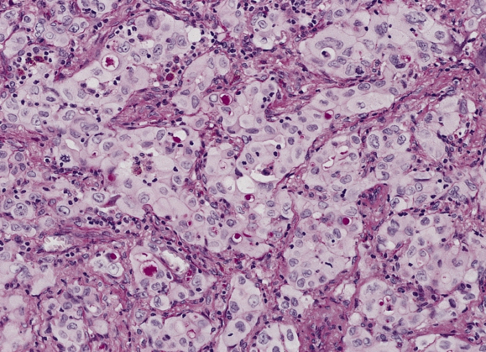
Adenocarcinoma

  • Most common (about 40%–50% of lung cancers)
  • Location: peripheral
  • Associated mutations:
    • EGF receptor Receptor Receptors are proteins located either on the surface of or within a cell that can bind to signaling molecules known as ligands (e.g., hormones) and cause some type of response within the cell. Receptors
    • Anaplastic lymphoma Lymphoma A general term for various neoplastic diseases of the lymphoid tissue. Imaging of the Mediastinum kinase (ALK)
    • KRAS
    • BRAF
    • RET
    • ROS1
    • MET MET Preoperative Care
  • Pathology:
    • Neoplastic gland formation
    • Intracytoplasmic mucin
    • Pneumocyte/immunohistochemical markers:
      • Thyroid Thyroid The thyroid gland is one of the largest endocrine glands in the human body. The thyroid gland is a highly vascular, brownish-red gland located in the visceral compartment of the anterior region of the neck. Thyroid Gland: Anatomy transcription factor Transcription factor Generic term for proteins necessary for transcription Regulation of Transcription 1 (TTF-1)
      • Napsin A

Subtypes:

  • Preinvasive:
    • Atypical adenomatous hyperplasia Hyperplasia An increase in the number of cells in a tissue or organ without tumor formation. It differs from hypertrophy, which is an increase in bulk without an increase in the number of cells. Cellular Adaptation (≤ 5 mm growth)
    • Adenocarcinoma in situ (≤ 3 cm, lepidic growth or noninvasive growth along alveolar structures)
  • Invasive (based on predominant pattern):

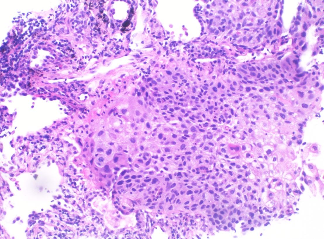
Squamous cell carcinoma Squamous cell carcinoma Cutaneous squamous cell carcinoma (cSCC) is caused by malignant proliferation of atypical keratinocytes. This condition is the 2nd most common skin malignancy and usually affects sun-exposed areas of fair-skinned patients. The cancer presents as a firm, erythematous, keratotic plaque or papule. Squamous Cell Carcinoma (SCC)

  • Approximately 20% of lung cancers 
  • Location:
    • Central 
    • Arise mostly in the proximal tracheobronchial tree
  • Associated mutations/ chromosome Chromosome In a prokaryotic cell or in the nucleus of a eukaryotic cell, a structure consisting of or containing DNA which carries the genetic information essential to the cell. Basic Terms of Genetics deletions in:
    • 3p
    • 9p (site of CDKN2A gene Gene A category of nucleic acid sequences that function as units of heredity and which code for the basic instructions for the development, reproduction, and maintenance of organisms. Basic Terms of Genetics)
    • 17p (site of TP53 gene Gene A category of nucleic acid sequences that function as units of heredity and which code for the basic instructions for the development, reproduction, and maintenance of organisms. Basic Terms of Genetics)
  • Pathology:
    • Intercellular bridges/ desmosomes Desmosomes A type of junction that attaches one cell to its neighbor. One of a number of differentiated regions which occur, for example, where the cytoplasmic membranes of adjacent epithelial cells are closely apposed. It consists of a circular region of each membrane together with associated intracellular microfilaments and an intercellular material which may include, for example, mucopolysaccharides. Bullous Pemphigoid and Pemphigus Vulgaris
    • Keratinization: squamous pearls or cells with eosinophilic cytoplasm
    • Infiltrating nest of tumor Tumor Inflammation cells shows central necrosis Necrosis The death of cells in an organ or tissue due to disease, injury or failure of the blood supply. Ischemic Cell Damage, which results in cavitation Cavitation Imaging of the Lungs and Pleura.
    • Immunohistochemical markers:
  • Associated with parathyroid Parathyroid The parathyroid glands are 2 pairs of small endocrine glands found in close proximity to the thyroid gland. The superior parathyroid glands are lodged within the parenchyma of the upper poles of the right and left thyroid lobes; the inferior parathyroid glands are close to the inferior tips or poles of the lobes. Parathyroid Glands: Anatomy hormone-related peptide secretion Secretion Coagulation Studies

Subtypes:

  • Keratinizing
  • Nonkeratinizing
  • Basaloid

Large cell carcinoma

  • 2% of lung cancers
  • Location: peripheral
  • Pathology:
    • Undifferentiated
    • Large tumor Tumor Inflammation cells (large nuclei, prominent nucleoli, moderate cytoplasm); possibly arranged in sheets
    • Does not express any immunohistochemical markers of adenocarcinoma or SCC
  • Can secrete beta-human chorionic gonadotropin and cause gynecomastia Gynecomastia Gynecomastia is a benign proliferation of male breast glandular ductal tissue, usually bilateral, caused by increased estrogen activity, decreased testosterone activity, or medications. The condition is common and physiological in neonates, adolescent boys, and elderly men. Gynecomastia

Small cell lung carcinoma

  • About 15% of all lung cancers
  • Highly aggressive: rapid presentation and > 60% with metastatic disease at diagnosis
  • Almost always seen in smokers
  • Location: central
  • Associated gene Gene A category of nucleic acid sequences that function as units of heredity and which code for the basic instructions for the development, reproduction, and maintenance of organisms. Basic Terms of Genetics mutations:
    • TP53
    • MYC
    • RB1
    • BLC2
    • Deletion in 3p
  • Pathology:
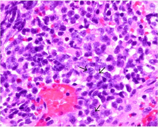
    • Sheets of small blue cells, rosette formation, necrosis Necrosis The death of cells in an organ or tissue due to disease, injury or failure of the blood supply. Ischemic Cell Damage
    • High mitotic rate (> 11 mitoses/2 mm²)
    • Neuroendocrine Kulchitsky cells: 
      • Small cells with scant cytoplasm Scant Cytoplasm Ewing Sarcoma
      • Hyperchromatic nuclei with finely granular chromatin Chromatin The material of chromosomes. It is a complex of dna; histones; and nonhistone proteins found within the nucleus of a cell. DNA Types and Structure (salt-and-pepper pattern)
    • Neuroendocrine markers:
      • CD56 (detected in 90%–100% of cases)
      • Chromogranin A Chromogranin A A type of chromogranin which was first isolated from chromaffin cells of the adrenal medulla but is also found in other tissues and in many species including human, bovine, rat, mouse, and others. It is an acidic protein with 431 to 445 amino acid residues. It contains fragments that inhibit vasoconstriction or release of hormones and neurotransmitter, while other fragments exert antimicrobial actions. Pancreatic Neuroendocrine Tumors (PanNETs)
      • Synaptophysin Synaptophysin A marvel domain-containing protein found in the presynaptic vesicles of neurons and neuroendocrine cells. It is commonly used as an immunocytochemical marker for neuroendocrine differentiation. Gastrinoma
      • Neurospecific enolase Enolase A hydro-lyase that catalyzes the dehydration of 2-phosphoglycerate to form phosphoenolpyruvate. Several different isoforms of this enzyme exist, each with its own tissue specificity. Glycolysis
    • Can be positive for TTF-1
  • May produce peptide hormones Hormones Hormones are messenger molecules that are synthesized in one part of the body and move through the bloodstream to exert specific regulatory effects on another part of the body. Hormones play critical roles in coordinating cellular activities throughout the body in response to the constant changes in both the internal and external environments. Hormones: Overview and Types:
    • Adrenocorticotropic hormone Adrenocorticotropic hormone An anterior pituitary hormone that stimulates the adrenal cortex and its production of corticosteroids. Acth is a 39-amino acid polypeptide of which the n-terminal 24-amino acid segment is identical in all species and contains the adrenocorticotropic activity. Upon further tissue-specific processing, acth can yield alpha-msh and corticotropin-like intermediate lobe peptide (clip). Adrenal Hormones (ACTH)
    • Arginine Arginine An essential amino acid that is physiologically active in the l-form. Urea Cycle vasopressin or antidiuretic hormone Antidiuretic hormone Antidiuretic hormones released by the neurohypophysis of all vertebrates (structure varies with species) to regulate water balance and osmolarity. In general, vasopressin is a nonapeptide consisting of a six-amino-acid ring with a cysteine 1 to cysteine 6 disulfide bridge or an octapeptide containing a cystine. All mammals have arginine vasopressin except the pig with a lysine at position 8. Vasopressin, a vasoconstrictor, acts on the kidney collecting ducts to increase water reabsorption, increase blood volume and blood pressure. Hypernatremia (ADH)
    • Atrial natriuretic peptide Atrial natriuretic peptide A potent natriuretic and vasodilatory peptide or mixture of different-sized low molecular weight peptides derived from a common precursor and secreted mainly by the heart atrium. All these peptides share a sequence of about 20 amino acids. Renal Sodium and Water Regulation (ANP) 
    • Gastrin-releasing peptide gastrin-releasing peptide Neuropeptide and gut hormone that helps regulate gastric acid secretion and motor function. Once released from nerves in the antrum of the stomach, the neuropeptide stimulates release of gastrin from the gastrin-secreting cells. Gastrointestinal Neural and Hormonal Signaling

Large cell neuroendocrine carcinoma

  • Rare but aggressive lung cancer 
  • Associated with smoking Smoking Willful or deliberate act of inhaling and exhaling smoke from burning substances or agents held by hand. Interstitial Lung Diseases
  • Location: peripheral
  • Pathology:
    • Cellular arrangement: nests, rosette formation, necrosis Necrosis The death of cells in an organ or tissue due to disease, injury or failure of the blood supply. Ischemic Cell Damage 
    • Cells are large with abundant eosinophilic cytoplasm.
    • High mitotic rate (> 10 mitoses/2 mm²)
    • Neuroendocrine markers:
      • Chromogranin
      • Synaptophysin Synaptophysin A marvel domain-containing protein found in the presynaptic vesicles of neurons and neuroendocrine cells. It is commonly used as an immunocytochemical marker for neuroendocrine differentiation. Gastrinoma
      • CD56

Carcinoids

  • 1%–2% of all lung cancers
  • Well-differentiated neuroendocrine tumors Neuroendocrine tumors Tumors whose cells possess secretory granules and originate from the neuroectoderm, i.e., the cells of the ectoblast or epiblast that program the neuroendocrine system. Common properties across most neuroendocrine tumors include ectopic hormone production (often via apud cells), the presence of tumor-associated antigens, and isozyme composition. Gastrinoma (generally slow growth)
  • Younger, nonsmoking patients Patients Individuals participating in the health care system for the purpose of receiving therapeutic, diagnostic, or preventive procedures. Clinician–Patient Relationship
  • Location: central > peripheral
  • Pathology:

Clinical Presentation

General symptoms

  • Asymptomatic in approximately 25% of patients Patients Individuals participating in the health care system for the purpose of receiving therapeutic, diagnostic, or preventive procedures. Clinician–Patient Relationship
  • Cough (most common):
    • New-onset cough in a smoker or ex-smoker
    • Frequent symptom in squamous and small cell cancers due to central location
    • May have hemoptysis Hemoptysis Hemoptysis is defined as the expectoration of blood originating in the lower respiratory tract. Hemoptysis is a consequence of another disease process and can be classified as either life threatening or non-life threatening. Hemoptysis can result in significant morbidity and mortality due to both drowning (reduced gas exchange as the lungs fill with blood) and hemorrhagic shock. Hemoptysis
  • Dyspnea Dyspnea Dyspnea is the subjective sensation of breathing discomfort. Dyspnea is a normal manifestation of heavy physical or psychological exertion, but also may be caused by underlying conditions (both pulmonary and extrapulmonary). Dyspnea due to malignancy-related:
    • Airway Airway ABCDE Assessment obstruction
    • Atelectasis Atelectasis Atelectasis is the partial or complete collapse of a part of the lung. Atelectasis is almost always a secondary phenomenon from conditions causing bronchial obstruction, external compression, surfactant deficiency, or scarring. Atelectasis
    • Pleural effusion Pleural Effusion Pleural effusion refers to the accumulation of fluid between the layers of the parietal and visceral pleura. Common causes of this condition include infection, malignancy, autoimmune disorders, or volume overload. Clinical manifestations include chest pain, cough, and dyspnea. Pleural Effusion
    • Pneumothorax Pneumothorax A pneumothorax is a life-threatening condition in which air collects in the pleural space, causing partial or full collapse of the lung. A pneumothorax can be traumatic or spontaneous. Patients present with a sudden onset of sharp chest pain, dyspnea, and diminished breath sounds on exam. Pneumothorax
    • Anemia Anemia Anemia is a condition in which individuals have low Hb levels, which can arise from various causes. Anemia is accompanied by a reduced number of RBCs and may manifest with fatigue, shortness of breath, pallor, and weakness. Subtypes are classified by the size of RBCs, chronicity, and etiology. Anemia: Overview and Types
  • Chest pain Pain An unpleasant sensation induced by noxious stimuli which are detected by nerve endings of nociceptive neurons. Pain: Types and Pathways:
    • Vague
    • Localized
    • Pleuritic
  • Fatigue Fatigue The state of weariness following a period of exertion, mental or physical, characterized by a decreased capacity for work and reduced efficiency to respond to stimuli. Fibromyalgia
  • Cancer cachexia:
    • Loss of muscle mass Mass Three-dimensional lesion that occupies a space within the breast Imaging of the Breast and fat, weight loss Weight loss Decrease in existing body weight. Bariatric Surgery, anorexia Anorexia The lack or loss of appetite accompanied by an aversion to food and the inability to eat. It is the defining characteristic of the disorder anorexia nervosa. Anorexia Nervosa
    • Inflammatory mediators play a role (lead to skeletal muscle degradation)

Regional spread

  • Recurrent laryngeal nerve encroachment: hoarseness Hoarseness An unnaturally deep or rough quality of voice. Parapharyngeal Abscess
  • Phrenic nerve Phrenic nerve The motor nerve of the diaphragm. The phrenic nerve fibers originate in the cervical spinal column (mostly C4) and travel through the cervical plexus to the diaphragm. Diaphragm: Anatomy encroachment:
    • Diaphragmatic paralysis (elevated diaphragm Diaphragm The diaphragm is a large, dome-shaped muscle that separates the thoracic cavity from the abdominal cavity. The diaphragm consists of muscle fibers and a large central tendon, which is divided into right and left parts. As the primary muscle of inspiration, the diaphragm contributes 75% of the total inspiratory muscle force. Diaphragm: Anatomy)
    • Dyspnea Dyspnea Dyspnea is the subjective sensation of breathing discomfort. Dyspnea is a normal manifestation of heavy physical or psychological exertion, but also may be caused by underlying conditions (both pulmonary and extrapulmonary). Dyspnea
    • Hypoxia Hypoxia Sub-optimal oxygen levels in the ambient air of living organisms. Ischemic Cell Damage
  • Superior vena cava Superior vena cava The venous trunk which returns blood from the head, neck, upper extremities and chest. Mediastinum and Great Vessels: Anatomy (SVC) syndrome:
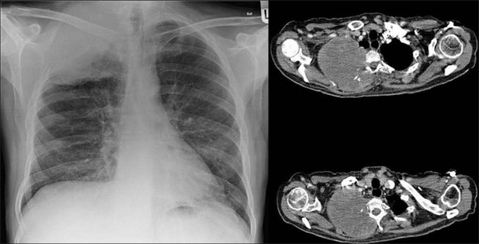
  • Pancoast syndrome:
    • Mostly due to NSCLC 
    • Apical tumors invade the brachial plexus Brachial Plexus The large network of nerve fibers which distributes the innervation of the upper extremity. The brachial plexus extends from the neck into the axilla. In humans, the nerves of the plexus usually originate from the lower cervical and the first thoracic spinal cord segments (c5-c8 and T1), but variations are not uncommon. Peripheral Nerve Injuries in the Cervicothoracic Region, pleura Pleura The pleura is a serous membrane that lines the walls of the thoracic cavity and the surface of the lungs. This structure of mesodermal origin covers both lungs, the mediastinum, the thoracic surface of the diaphragm, and the inner part of the thoracic cage. The pleura is divided into a visceral pleura and parietal pleura. Pleura: Anatomy, or ribs Ribs A set of twelve curved bones which connect to the vertebral column posteriorly, and terminate anteriorly as costal cartilage. Together, they form a protective cage around the internal thoracic organs. Chest Wall: Anatomy resulting in:
      • Shoulder and upper extremity pain Pain An unpleasant sensation induced by noxious stimuli which are detected by nerve endings of nociceptive neurons. Pain: Types and Pathways
      • Upper extremity weakness
      • Atrophy Atrophy Decrease in the size of a cell, tissue, organ, or multiple organs, associated with a variety of pathological conditions such as abnormal cellular changes, ischemia, malnutrition, or hormonal changes. Cellular Adaptation of the ipsilateral hand Hand The hand constitutes the distal part of the upper limb and provides the fine, precise movements needed in activities of daily living. It consists of 5 metacarpal bones and 14 phalanges, as well as numerous muscles innervated by the median and ulnar nerves. Hand: Anatomy
      • Horner’s syndrome: ptosis Ptosis Cranial Nerve Palsies, miosis Miosis Pupil: Physiology and Abnormalities, anhidrosis
  • Pericardial spread:
    • Pericarditis Pericarditis Pericarditis is an inflammation of the pericardium, often with fluid accumulation. It can be caused by infection (often viral), myocardial infarction, drugs, malignancies, metabolic disorders, autoimmune disorders, or trauma. Acute, subacute, and chronic forms exist. Pericarditis → chest pain Pain An unpleasant sensation induced by noxious stimuli which are detected by nerve endings of nociceptive neurons. Pain: Types and Pathways 
    • Pericardial effusion Pericardial effusion Fluid accumulation within the pericardium. Serous effusions are associated with pericardial diseases. Hemopericardium is associated with trauma. Lipid-containing effusion (chylopericardium) results from leakage of thoracic duct. Severe cases can lead to cardiac tamponade. Pericardial Effusion and Cardiac Tamponade (can lead to cardiac tamponade Tamponade Pericardial effusion, usually of rapid onset, exceeding ventricular filling pressures and causing collapse of the heart with a markedly reduced cardiac output. Pericarditis) → dyspnea Dyspnea Dyspnea is the subjective sensation of breathing discomfort. Dyspnea is a normal manifestation of heavy physical or psychological exertion, but also may be caused by underlying conditions (both pulmonary and extrapulmonary). Dyspnea
  • Esophageal compression Compression Blunt Chest Trauma: dysphagia Dysphagia Dysphagia is the subjective sensation of difficulty swallowing. Symptoms can range from a complete inability to swallow, to the sensation of solids or liquids becoming “stuck.” Dysphagia is classified as either oropharyngeal or esophageal, with esophageal dysphagia having 2 sub-types: functional and mechanical. Dysphagia 

Metastasis Metastasis The transfer of a neoplasm from one organ or part of the body to another remote from the primary site. Grading, Staging, and Metastasis

  • Liver Liver The liver is the largest gland in the human body. The liver is found in the superior right quadrant of the abdomen and weighs approximately 1.5 kilograms. Its main functions are detoxification, metabolism, nutrient storage (e.g., iron and vitamins), synthesis of coagulation factors, formation of bile, filtration, and storage of blood. Liver: Anatomy:
  • Brain Brain The part of central nervous system that is contained within the skull (cranium). Arising from the neural tube, the embryonic brain is comprised of three major parts including prosencephalon (the forebrain); mesencephalon (the midbrain); and rhombencephalon (the hindbrain). The developed brain consists of cerebrum; cerebellum; and other structures in the brain stem. Nervous System: Anatomy, Structure, and Classification:
    • Behavioral changes and confusion
    • Aphasia Aphasia A cognitive disorder marked by an impaired ability to comprehend or express language in its written or spoken form. This condition is caused by diseases which affect the language areas of the dominant hemisphere. Clinical features are used to classify the various subtypes of this condition. General categories include receptive, expressive, and mixed forms of aphasia. Ischemic Stroke
    • Focal neurologic deficits Neurologic Deficits High-Risk Headaches
    • Nausea Nausea An unpleasant sensation in the stomach usually accompanied by the urge to vomit. Common causes are early pregnancy, sea and motion sickness, emotional stress, intense pain, food poisoning, and various enteroviruses. Antiemetics and vomiting Vomiting The forcible expulsion of the contents of the stomach through the mouth. Hypokalemia
    • Seizures Seizures A seizure is abnormal electrical activity of the neurons in the cerebral cortex that can manifest in numerous ways depending on the region of the brain affected. Seizures consist of a sudden imbalance that occurs between the excitatory and inhibitory signals in cortical neurons, creating a net excitation. The 2 major classes of seizures are focal and generalized. Seizures
  • Bone Bone Bone is a compact type of hardened connective tissue composed of bone cells, membranes, an extracellular mineralized matrix, and central bone marrow. The 2 primary types of bone are compact and spongy. Bones: Structure and Types:
    • Pain Pain An unpleasant sensation induced by noxious stimuli which are detected by nerve endings of nociceptive neurons. Pain: Types and Pathways (back, chest, extremities)
    • Pathologic fractures
  • Adrenal glands Adrenal Glands The adrenal glands are a pair of retroperitoneal endocrine glands located above the kidneys. The outer parenchyma is called the adrenal cortex and has 3 distinct zones, each with its own secretory products. Beneath the cortex lies the adrenal medulla, which secretes catecholamines involved in the fight-or-flight response. Adrenal Glands: Anatomy:
    • Usually asymptomatic
    • Adrenal insufficiency Adrenal Insufficiency Conditions in which the production of adrenal corticosteroids falls below the requirement of the body. Adrenal insufficiency can be caused by defects in the adrenal glands, the pituitary gland, or the hypothalamus. Adrenal Insufficiency and Addison Disease (if both glands are affected by metastatic cancer):
      • Weakness
      • Nausea Nausea An unpleasant sensation in the stomach usually accompanied by the urge to vomit. Common causes are early pregnancy, sea and motion sickness, emotional stress, intense pain, food poisoning, and various enteroviruses. Antiemetics
      • Anorexia Anorexia The lack or loss of appetite accompanied by an aversion to food and the inability to eat. It is the defining characteristic of the disorder anorexia nervosa. Anorexia Nervosa
      • Electrolyte imbalances

Paraneoplastic syndrome

  • Hypertrophic pulmonary osteoarthropathy:
  • Hypercalcemia Hypercalcemia Hypercalcemia (serum calcium > 10.5 mg/dL) can result from various conditions, the majority of which are due to hyperparathyroidism and malignancy. Other causes include disorders leading to vitamin D elevation, granulomatous diseases, and the use of certain pharmacological agents. Symptoms vary depending on calcium levels and the onset of hypercalcemia. Hypercalcemia:
    • Sources:
      • SCC produces parathyroid Parathyroid The parathyroid glands are 2 pairs of small endocrine glands found in close proximity to the thyroid gland. The superior parathyroid glands are lodged within the parenchyma of the upper poles of the right and left thyroid lobes; the inferior parathyroid glands are close to the inferior tips or poles of the lobes. Parathyroid Glands: Anatomy hormone-related protein.
      • Extensive bone Bone Bone is a compact type of hardened connective tissue composed of bone cells, membranes, an extracellular mineralized matrix, and central bone marrow. The 2 primary types of bone are compact and spongy. Bones: Structure and Types metastases
    • Signs and symptoms:
      • Anorexia Anorexia The lack or loss of appetite accompanied by an aversion to food and the inability to eat. It is the defining characteristic of the disorder anorexia nervosa. Anorexia Nervosa
      • Nausea Nausea An unpleasant sensation in the stomach usually accompanied by the urge to vomit. Common causes are early pregnancy, sea and motion sickness, emotional stress, intense pain, food poisoning, and various enteroviruses. Antiemetics
      • Constipation Constipation Constipation is common and may be due to a variety of causes. Constipation is generally defined as bowel movement frequency < 3 times per week. Patients who are constipated often strain to pass hard stools. The condition is classified as primary (also known as idiopathic or functional constipation) or secondary, and as acute or chronic. Constipation
      • Lethargy Lethargy A general state of sluggishness, listless, or uninterested, with being tired, and having difficulty concentrating and doing simple tasks. It may be related to depression or drug addiction. Hyponatremia
      • Polyuria Polyuria Urination of a large volume of urine with an increase in urinary frequency, commonly seen in diabetes. Renal Potassium Regulation and polydipsia Polydipsia Excessive thirst manifested by excessive fluid intake. It is characteristic of many diseases such as diabetes mellitus; diabetes insipidus; and nephrogenic diabetes insipidus. The condition may be psychogenic in origin. Diabetes Insipidus
      • Confusion
      • Coma Coma Coma is defined as a deep state of unarousable unresponsiveness, characterized by a score of 3 points on the GCS. A comatose state can be caused by a multitude of conditions, making the precise epidemiology and prognosis of coma difficult to determine. Coma
  • Syndrome of inappropriate antidiuretic hormone Antidiuretic hormone Antidiuretic hormones released by the neurohypophysis of all vertebrates (structure varies with species) to regulate water balance and osmolarity. In general, vasopressin is a nonapeptide consisting of a six-amino-acid ring with a cysteine 1 to cysteine 6 disulfide bridge or an octapeptide containing a cystine. All mammals have arginine vasopressin except the pig with a lysine at position 8. Vasopressin, a vasoconstrictor, acts on the kidney collecting ducts to increase water reabsorption, increase blood volume and blood pressure. Hypernatremia secretion Secretion Coagulation Studies ( SIADH SIADH Syndrome of inappropriate antidiuretic hormone secretion (SIADH) is a disorder of impaired water excretion due to the inability to suppress the secretion of antidiuretic hormone (ADH). SIADH is characterized by impaired water excretion leading to dilutional hyponatremia, which is mainly asymptomatic but may cause neurologic symptoms. S Syndrome of Inappropriate Antidiuretic Hormone Secretion (SIADH))/Schwartz-Bartter syndrome:
    • Associated with SCLC
    • Signs and symptoms:
      • Anorexia Anorexia The lack or loss of appetite accompanied by an aversion to food and the inability to eat. It is the defining characteristic of the disorder anorexia nervosa. Anorexia Nervosa
      • Nausea Nausea An unpleasant sensation in the stomach usually accompanied by the urge to vomit. Common causes are early pregnancy, sea and motion sickness, emotional stress, intense pain, food poisoning, and various enteroviruses. Antiemetics, vomiting Vomiting The forcible expulsion of the contents of the stomach through the mouth. Hypokalemia
      • Confusion, coma Coma Coma is defined as a deep state of unarousable unresponsiveness, characterized by a score of 3 points on the GCS. A comatose state can be caused by a multitude of conditions, making the precise epidemiology and prognosis of coma difficult to determine. Coma
      • Seizures Seizures A seizure is abnormal electrical activity of the neurons in the cerebral cortex that can manifest in numerous ways depending on the region of the brain affected. Seizures consist of a sudden imbalance that occurs between the excitatory and inhibitory signals in cortical neurons, creating a net excitation. The 2 major classes of seizures are focal and generalized. Seizures
      • Hyponatremia Hyponatremia Hyponatremia is defined as a decreased serum sodium (sNa+) concentration less than 135 mmol/L. Serum sodium is the greatest contributor to plasma osmolality, which is very tightly controlled via antidiuretic hormone (ADH) release from the hypothalamus and by the thirst mechanism. Hyponatremia
  • Lambert-Eaton syndrome:
  • Cushing’s syndrome:
    • SCLC can produce adrenocorticotropic hormone Adrenocorticotropic hormone An anterior pituitary hormone that stimulates the adrenal cortex and its production of corticosteroids. Acth is a 39-amino acid polypeptide of which the n-terminal 24-amino acid segment is identical in all species and contains the adrenocorticotropic activity. Upon further tissue-specific processing, acth can yield alpha-msh and corticotropin-like intermediate lobe peptide (clip). Adrenal Hormones (ACTH).
    • Also seen in carcinoid tumors Carcinoid tumors Carcinoid tumors are small, well-differentiated, slow-growing neuroendocrine tumors (NET). Carcinoid syndrome describes the signs and symptoms associated with unregulated vasoactive hormone production by neuroendocrine tumors. Carcinoid tumors are most commonly found in the GI and bronchopulmonary tracts. Carcinoid Tumors and Syndrome
    • Signs and symptoms:
      • Muscle weakness
      • Weight loss Weight loss Decrease in existing body weight. Bariatric Surgery
      • Hypertension Hypertension Hypertension, or high blood pressure, is a common disease that manifests as elevated systemic arterial pressures. Hypertension is most often asymptomatic and is found incidentally as part of a routine physical examination or during triage for an unrelated medical encounter. Hypertension
      • Hirsutism Hirsutism A condition observed in women and children when there is excess coarse body hair of an adult male distribution pattern, such as facial and chest areas. It is the result of elevated androgens from the ovaries, the adrenal glands, or exogenous sources. The concept does not include hypertrichosis, which is an androgen-independent excessive hair growth. Polycystic Ovarian Syndrome
      • Osteoporosis Osteoporosis Osteoporosis refers to a decrease in bone mass and density leading to an increased number of fractures. There are 2 forms of osteoporosis: primary, which is commonly postmenopausal or senile; and secondary, which is a manifestation of immobilization, underlying medical disorders, or long-term use of certain medications. Osteoporosis
  • Hypercoagulability Hypercoagulability Hypercoagulable States:
  • Dermatomyositis Dermatomyositis A subacute or chronic inflammatory disease of muscle and skin, marked by proximal muscle weakness and a characteristic skin rash. The illness occurs with approximately equal frequency in children and adults. The skin lesions usually take the form of a purplish rash (or less often an exfoliative dermatitis) involving the nose, cheeks, forehead, upper trunk, and arms. The disease is associated with a complement mediated intramuscular microangiopathy, leading to loss of capillaries, muscle ischemia, muscle-fiber necrosis, and perifascicular atrophy. The childhood form of this disease tends to evolve into a systemic vasculitis. Dermatomyositis may occur in association with malignant neoplasms. Paraneoplastic Syndromes and polymyositis Polymyositis Polymyositis (PM) is an autoimmune inflammatory myopathy caused by T cell-mediated muscle injury. The etiology of PM is unclear, but there are several genetic and environmental associations. Polymyositis is most common in middle-aged women and rarely affects children. Polymyositis:
    • Associated with lung cancer as well as malignancies of the ovaries Ovaries Ovaries are the paired gonads of the female reproductive system that contain haploid gametes known as oocytes. The ovaries are located intraperitoneally in the pelvis, just posterior to the broad ligament, and are connected to the pelvic sidewall and to the uterus by ligaments. These organs function to secrete hormones (estrogen and progesterone) and to produce the female germ cells (oocytes). Ovaries: Anatomy, pancreas Pancreas The pancreas lies mostly posterior to the stomach and extends across the posterior abdominal wall from the duodenum on the right to the spleen on the left. This organ has both exocrine and endocrine tissue. Pancreas: Anatomy, bladder Bladder A musculomembranous sac along the urinary tract. Urine flows from the kidneys into the bladder via the ureters, and is held there until urination. Pyelonephritis and Perinephric Abscess, and stomach Stomach The stomach is a muscular sac in the upper left portion of the abdomen that plays a critical role in digestion. The stomach develops from the foregut and connects the esophagus with the duodenum. Structurally, the stomach is C-shaped and forms a greater and lesser curvature and is divided grossly into regions: the cardia, fundus, body, and pylorus. Stomach: Anatomy
    • Signs and symptoms:

Diagnosis

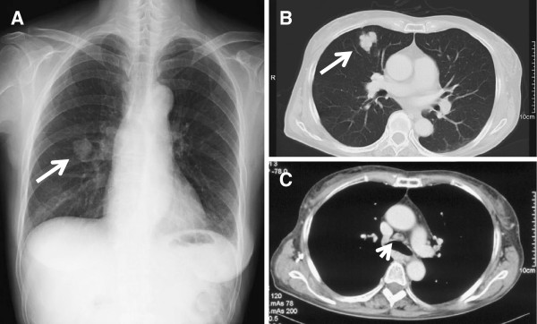
Chest radiography

  • Not diagnostic, but often performed in patients Patients Individuals participating in the health care system for the purpose of receiving therapeutic, diagnostic, or preventive procedures. Clinician–Patient Relationship with respiratory symptoms
  • Findings that should raise suspicion of malignancy Malignancy Hemothorax:
    • New or enlarging focal lesion
    • Pleural effusion Pleural Effusion Pleural effusion refers to the accumulation of fluid between the layers of the parietal and visceral pleura. Common causes of this condition include infection, malignancy, autoimmune disorders, or volume overload. Clinical manifestations include chest pain, cough, and dyspnea. Pleural Effusion
    • Pleural nodularity
    • Enlarged hilar and paratracheal lymph nodes Lymph Nodes They are oval or bean shaped bodies (1 – 30 mm in diameter) located along the lymphatic system. Lymphatic Drainage System: Anatomy
    • Endobronchial lesion
    • Postobstructive pneumonia Pneumonia Pneumonia or pulmonary inflammation is an acute or chronic inflammation of lung tissue. Causes include infection with bacteria, viruses, or fungi. In more rare cases, pneumonia can also be caused through toxic triggers through inhalation of toxic substances, immunological processes, or in the course of radiotherapy. Pneumonia
    • Segmental or lobar atelectasis Atelectasis Atelectasis is the partial or complete collapse of a part of the lung. Atelectasis is almost always a secondary phenomenon from conditions causing bronchial obstruction, external compression, surfactant deficiency, or scarring. Atelectasis

Computed tomography

  • Used as a screening Screening Preoperative Care tool (low dose)
  • Low-dose CT produces high-resolution images with less radiation Radiation Emission or propagation of acoustic waves (sound), electromagnetic energy waves (such as light; radio waves; gamma rays; or x-rays), or a stream of subatomic particles (such as electrons; neutrons; protons; or alpha particles). Osteosarcoma:
    • Concerning findings in a patient with a solitary pulmonary nodule Nodule Chalazion:
      • Size > 15 mm
      • Irregular or spiculated borders
      • Location in an upper lobe
      • Cavitation Cavitation Imaging of the Lungs and Pleura
      • A solid component within a ground glass lesion
      • Growth is noted in follow-up imaging.
    • Multiple nodules should be concerning for pulmonary metastasis Metastasis The transfer of a neoplasm from one organ or part of the body to another remote from the primary site. Grading, Staging, and Metastasis.
  • Abnormal findings should be followed up with diagnostic CT.
  • Performed in all patients Patients Individuals participating in the health care system for the purpose of receiving therapeutic, diagnostic, or preventive procedures. Clinician–Patient Relationship with suspected lung cancer to assess the location of:
  • Aids in biopsy Biopsy Removal and pathologic examination of specimens from the living body. Ewing Sarcoma planning

Follow-up imaging

In addition to CT, the following modalities are used to determine the extent of disease for staging Staging Methods which attempt to express in replicable terms the extent of the neoplasm in the patient. Grading, Staging, and Metastasis purposes:
  • PET PET An imaging technique that combines a positron-emission tomography (PET) scanner and a ct X ray scanner. This establishes a precise anatomic localization in the same session. Nuclear Imaging:
    • Evaluates for distant metastases
    • More accurate in evaluating mediastinal disease than CT
    • Assesses the metabolic activity of the primary tumor Tumor Inflammation
  • MRI:
    • Preferred imaging for brain Brain The part of central nervous system that is contained within the skull (cranium). Arising from the neural tube, the embryonic brain is comprised of three major parts including prosencephalon (the forebrain); mesencephalon (the midbrain); and rhombencephalon (the hindbrain). The developed brain consists of cerebrum; cerebellum; and other structures in the brain stem. Nervous System: Anatomy, Structure, and Classification and spine Spine The human spine, or vertebral column, is the most important anatomical and functional axis of the human body. It consists of 7 cervical vertebrae, 12 thoracic vertebrae, and 5 lumbar vertebrae and is limited cranially by the skull and caudally by the sacrum. Vertebral Column: Anatomy metastases
    • Used to assess nerve involvement (e.g., brachial plexus Brachial Plexus The large network of nerve fibers which distributes the innervation of the upper extremity. The brachial plexus extends from the neck into the axilla. In humans, the nerves of the plexus usually originate from the lower cervical and the first thoracic spinal cord segments (c5-c8 and T1), but variations are not uncommon. Peripheral Nerve Injuries in the Cervicothoracic Region)

Biopsy Biopsy Removal and pathologic examination of specimens from the living body. Ewing Sarcoma

  • Definitive pathology is required for diagnosis (tissue biopsy Biopsy Removal and pathologic examination of specimens from the living body. Ewing Sarcoma is preferred over cytologic specimen).
  • Tissue samples may be obtained by way of:
  • Cytologic specimen can be obtained from sputum, bronchial washings, and needle aspirates.
  • Histopathology: differentiates NSCLC from SCLC and classifies lung cancers 
  • Additional molecular testing (mutations and immune targets):
    • Techniques used for determining mutations (allows for targeted therapies in NSCLC):
      • DNA DNA A deoxyribonucleotide polymer that is the primary genetic material of all cells. Eukaryotic and prokaryotic organisms normally contain DNA in a double-stranded state, yet several important biological processes transiently involve single-stranded regions. DNA, which consists of a polysugar-phosphate backbone possessing projections of purines (adenine and guanine) and pyrimidines (thymine and cytosine), forms a double helix that is held together by hydrogen bonds between these purines and pyrimidines (adenine to thymine and guanine to cytosine). DNA Types and Structure sequencing
      • FISH FISH A type of in situ hybridization in which target sequences are stained with fluorescent dye so their location and size can be determined using fluorescence microscopy. This staining is sufficiently distinct that the hybridization signal can be seen both in metaphase spreads and in interphase nuclei. Chromosome Testing
      • Immunohistochemistry Immunohistochemistry Histochemical localization of immunoreactive substances using labeled antibodies as reagents. Myeloperoxidase Deficiency
    • Key driver mutations detected:
      • EGF receptor Receptor Receptors are proteins located either on the surface of or within a cell that can bind to signaling molecules known as ligands (e.g., hormones) and cause some type of response within the cell. Receptors mutation Mutation Genetic mutations are errors in DNA that can cause protein misfolding and dysfunction. There are various types of mutations, including chromosomal, point, frameshift, and expansion mutations. Types of Mutations
      • ALK rearrangements
      • ROS1 rearrangements
      • RET fusion
      • BRAF
      • MET MET Preoperative Care mutation Mutation Genetic mutations are errors in DNA that can cause protein misfolding and dysfunction. There are various types of mutations, including chromosomal, point, frameshift, and expansion mutations. Types of Mutations
    • Expression of programmed cell death Cell death Injurious stimuli trigger the process of cellular adaptation, whereby cells respond to withstand the harmful changes in their environment. Overwhelmed adaptive mechanisms lead to cell injury. Mild stimuli produce reversible injury. If the stimulus is severe or persistent, injury becomes irreversible. Apoptosis is programmed cell death, a mechanism with both physiologic and pathologic effects. Cell Injury and Death ligand 1 (PD-L1) on immunohistochemistry Immunohistochemistry Histochemical localization of immunoreactive substances using labeled antibodies as reagents. Myeloperoxidase Deficiency helps determine use of checkpoint inhibitors in cancer therapies.
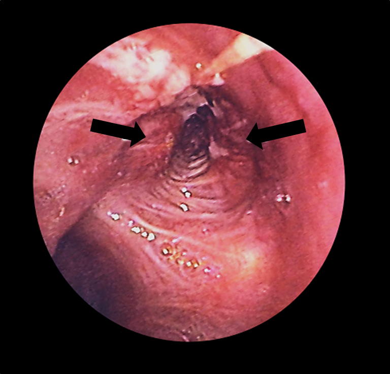
Bronchoscopy lung cancer

Bronchoscopy:
Lung cancer (arrows) in the left bronchus as seen with a bronchoscope

Image: “Lung cancer in L. Bronchus – bronchoscopic view” by JHeuser. License: CC BY 2.5

Laboratory evaluation

The following tests are possible findings for lung cancer:

  • CBC:
    • ↓ Hemoglobin
    • Platelets Platelets Platelets are small cell fragments involved in hemostasis. Thrombopoiesis takes place primarily in the bone marrow through a series of cell differentiation and is influenced by several cytokines. Platelets are formed after fragmentation of the megakaryocyte cytoplasm. Platelets: Histology
  • Electrolytes Electrolytes Electrolytes are mineral salts that dissolve in water and dissociate into charged particles called ions, which can be either be positively (cations) or negatively (anions) charged. Electrolytes are distributed in the extracellular and intracellular compartments in different concentrations. Electrolytes are essential for various basic life-sustaining functions. Electrolytes:
    • Sodium Sodium A member of the alkali group of metals. It has the atomic symbol na, atomic number 11, and atomic weight 23. Hyponatremia SIADH SIADH Syndrome of inappropriate antidiuretic hormone secretion (SIADH) is a disorder of impaired water excretion due to the inability to suppress the secretion of antidiuretic hormone (ADH). SIADH is characterized by impaired water excretion leading to dilutional hyponatremia, which is mainly asymptomatic but may cause neurologic symptoms. S Syndrome of Inappropriate Antidiuretic Hormone Secretion (SIADH)
    • Calcium Calcium A basic element found in nearly all tissues. It is a member of the alkaline earth family of metals with the atomic symbol ca, atomic number 20, and atomic weight 40. Calcium is the most abundant mineral in the body and combines with phosphorus to form calcium phosphate in the bones and teeth. It is essential for the normal functioning of nerves and muscles and plays a role in blood coagulation (as factor IV) and in many enzymatic processes. Electrolytes → paraneoplastic manifestation, bone Bone Bone is a compact type of hardened connective tissue composed of bone cells, membranes, an extracellular mineralized matrix, and central bone marrow. The 2 primary types of bone are compact and spongy. Bones: Structure and Types metastasis Metastasis The transfer of a neoplasm from one organ or part of the body to another remote from the primary site. Grading, Staging, and Metastasis
  • Liver Liver The liver is the largest gland in the human body. The liver is found in the superior right quadrant of the abdomen and weighs approximately 1.5 kilograms. Its main functions are detoxification, metabolism, nutrient storage (e.g., iron and vitamins), synthesis of coagulation factors, formation of bile, filtration, and storage of blood. Liver: Anatomy function test → liver Liver The liver is the largest gland in the human body. The liver is found in the superior right quadrant of the abdomen and weighs approximately 1.5 kilograms. Its main functions are detoxification, metabolism, nutrient storage (e.g., iron and vitamins), synthesis of coagulation factors, formation of bile, filtration, and storage of blood. Liver: Anatomy metastasis Metastasis The transfer of a neoplasm from one organ or part of the body to another remote from the primary site. Grading, Staging, and Metastasis:
    • Alkaline phosphatase Alkaline Phosphatase An enzyme that catalyzes the conversion of an orthophosphoric monoester and water to an alcohol and orthophosphate. Osteosarcoma may also be due to bone Bone Bone is a compact type of hardened connective tissue composed of bone cells, membranes, an extracellular mineralized matrix, and central bone marrow. The 2 primary types of bone are compact and spongy. Bones: Structure and Types metastasis Metastasis The transfer of a neoplasm from one organ or part of the body to another remote from the primary site. Grading, Staging, and Metastasis.

Staging

Tumor-nodes-metastases (TNM) staging Staging Methods which attempt to express in replicable terms the extent of the neoplasm in the patient. Grading, Staging, and Metastasis

The following are the 8th-edition staging Staging Methods which attempt to express in replicable terms the extent of the neoplasm in the patient. Grading, Staging, and Metastasis guidelines from the International Association for the Study of Lung Cancer:

Table: The primary tumor Tumor Inflammation (T category)
Stage Description
Tx Primary tumor Tumor Inflammation cannot be assessed.
T0 No evidence of a primary tumor Tumor Inflammation
Tis Carcinoma in situ
T1 Tumor Tumor Inflammation ≤ 3 cm, surrounded by lung or visceral pleura Visceral pleura Pleura: Anatomy, without invasion of the main bronchus:
  • T1a: ≤ 1 cm
  • T1b: > 1 cm but ≤ 2 cm
  • T1c: > 2 cm but ≤ 3 cm
T2 Tumor Tumor Inflammation > 3 cm but ≤ 5 cm, or:
  • Involves the main bronchus (without involvement of the carina)
  • Invades the visceral pleura Visceral pleura Pleura: Anatomy
  • Associated with atelectasis Atelectasis Atelectasis is the partial or complete collapse of a part of the lung. Atelectasis is almost always a secondary phenomenon from conditions causing bronchial obstruction, external compression, surfactant deficiency, or scarring. Atelectasis or obstructive pneumonitis Pneumonitis Human Herpesvirus 6 and 7 extending to the hilar region:
    • T2a: > 3 cm but ≤ 4 cm
    • T2b: > 4 cm but ≤ 5 cm
T3 T3 A T3 thyroid hormone normally synthesized and secreted by the thyroid gland in much smaller quantities than thyroxine (T4). Most T3 is derived from peripheral monodeiodination of T4 at the 5′ position of the outer ring of the iodothyronine nucleus. The hormone finally delivered and used by the tissues is mainly t3. Thyroid Hormones Tumor Tumor Inflammation > 5 cm but ≤ 7 cm, or associated tumor Tumor Inflammation nodules in the same lobe as the primary tumor Tumor Inflammation, or direct invasion of the chest wall Chest wall The chest wall consists of skin, fat, muscles, bones, and cartilage. The bony structure of the chest wall is composed of the ribs, sternum, and thoracic vertebrae. The chest wall serves as armor for the vital intrathoracic organs and provides the stability necessary for the movement of the shoulders and arms. Chest Wall: Anatomy, phrenic nerve Phrenic nerve The motor nerve of the diaphragm. The phrenic nerve fibers originate in the cervical spinal column (mostly C4) and travel through the cervical plexus to the diaphragm. Diaphragm: Anatomy, or parietal Parietal One of a pair of irregularly shaped quadrilateral bones situated between the frontal bone and occipital bone, which together form the sides of the cranium. Skull: Anatomy pericardium Pericardium A conical fibroserous sac surrounding the heart and the roots of the great vessels (aorta; venae cavae; pulmonary artery). Pericardium consists of two sacs: the outer fibrous pericardium and the inner serous pericardium. The latter consists of an outer parietal layer facing the fibrous pericardium, and an inner visceral layer (epicardium) resting next to the heart, and a pericardial cavity between these two layers. Heart: Anatomy
T4 T4 The major hormone derived from the thyroid gland. Thyroxine is synthesized via the iodination of tyrosines (monoiodotyrosine) and the coupling of iodotyrosines (diiodotyrosine) in the thyroglobulin. Thyroxine is released from thyroglobulin by proteolysis and secreted into the blood. Thyroxine is peripherally deiodinated to form triiodothyronine which exerts a broad spectrum of stimulatory effects on cell metabolism. Thyroid Hormones Tumor Tumor Inflammation > 7 cm, or associated tumor Tumor Inflammation nodules in a different ipsilateral lobe from the primary tumor Tumor Inflammation, or invasion into the diaphragm Diaphragm The diaphragm is a large, dome-shaped muscle that separates the thoracic cavity from the abdominal cavity. The diaphragm consists of muscle fibers and a large central tendon, which is divided into right and left parts. As the primary muscle of inspiration, the diaphragm contributes 75% of the total inspiratory muscle force. Diaphragm: Anatomy, mediastinum Mediastinum The mediastinum is the thoracic area between the 2 pleural cavities. The mediastinum contains vital structures of the circulatory, respiratory, digestive, and nervous systems including the heart and esophagus, and major thoracic vessels. Mediastinum and Great Vessels: Anatomy, heart, great vessels, trachea Trachea The trachea is a tubular structure that forms part of the lower respiratory tract. The trachea is continuous superiorly with the larynx and inferiorly becomes the bronchial tree within the lungs. The trachea consists of a support frame of semicircular, or C-shaped, rings made out of hyaline cartilage and reinforced by collagenous connective tissue. Trachea: Anatomy, esophagus Esophagus The esophagus is a muscular tube-shaped organ of around 25 centimeters in length that connects the pharynx to the stomach. The organ extends from approximately the 6th cervical vertebra to the 11th thoracic vertebra and can be divided grossly into 3 parts: the cervical part, the thoracic part, and the abdominal part. Esophagus: Anatomy, recurrent laryngeal nerve, vertebral body Vertebral body Main portion of the vertebra which bears majority of the weight. Vertebral Column: Anatomy, or carina
Table: The lymph nodes Lymph Nodes They are oval or bean shaped bodies (1 – 30 mm in diameter) located along the lymphatic system. Lymphatic Drainage System: Anatomy (N category)
Stage Description
Nx Unable to assess regional lymph nodes Lymph Nodes They are oval or bean shaped bodies (1 – 30 mm in diameter) located along the lymphatic system. Lymphatic Drainage System: Anatomy
N0 No regional lymph Lymph The interstitial fluid that is in the lymphatic system. Secondary Lymphatic Organs node metastasis Metastasis The transfer of a neoplasm from one organ or part of the body to another remote from the primary site. Grading, Staging, and Metastasis
N1 Ipsilateral peribronchial and/or hilar lymph nodes Lymph Nodes They are oval or bean shaped bodies (1 – 30 mm in diameter) located along the lymphatic system. Lymphatic Drainage System: Anatomy and intrapulmonary node involvement
N2 Ipsilateral mediastinal and/or subcarinal lymph Lymph The interstitial fluid that is in the lymphatic system. Secondary Lymphatic Organs node involvement
N3 Contralateral mediastinal, contralateral hilar, ipsilateral or contralateral scalene, or supraclavicular lymph Lymph The interstitial fluid that is in the lymphatic system. Secondary Lymphatic Organs node involvement
Table: Metastasis Metastasis The transfer of a neoplasm from one organ or part of the body to another remote from the primary site. Grading, Staging, and Metastasis (M category)
Stage Description
M0 No distant metastasis Metastasis The transfer of a neoplasm from one organ or part of the body to another remote from the primary site. Grading, Staging, and Metastasis
M1 Distant metastasis Metastasis The transfer of a neoplasm from one organ or part of the body to another remote from the primary site. Grading, Staging, and Metastasis:
  • M1a: separate tumor Tumor Inflammation nodules in the contralateral lobe, pleural or pericardial nodules, or malignant pleural or pericardial effusion Pericardial effusion Fluid accumulation within the pericardium. Serous effusions are associated with pericardial diseases. Hemopericardium is associated with trauma. Lipid-containing effusion (chylopericardium) results from leakage of thoracic duct. Severe cases can lead to cardiac tamponade. Pericardial Effusion and Cardiac Tamponade
  • M1b: single extrathoracic metastasis Metastasis The transfer of a neoplasm from one organ or part of the body to another remote from the primary site. Grading, Staging, and Metastasis
  • M1c: multiple extrathoracic metastases

Non-small cell lung cancer staging Staging Methods which attempt to express in replicable terms the extent of the neoplasm in the patient. Grading, Staging, and Metastasis

Stage grouping TNM stage
Occult
  • TXN0M0
0
  • TisN0M0
I
  • IA1: T1aN0M0
  • IA2: T1bN0M0
  • IA3: T1cN0M0
  • IB: T2aN0M0
II
  • IIA: T2bN0M0
  • IIB: 
    • T1a‒cN1M0
    • T2a‒bN1M0
    • T3N0M0
III
  • IIIA:
    • T1a‒cN2M0
    • T2a‒bN2M0
    • T3N1M0
    • T4N0‒1M0
  • IIIB:
    • T1a‒cN3M0
    • T2a‒bN3M0
    • T3N2M0
    • T4N2M0
  • IIIC:
    • T3N3M0
    • T4N3M0
IV
  • IVA: any T and N plus M1a or M1b
  • IVB: any T and N plus M1c

Small cell lung cancer staging Staging Methods which attempt to express in replicable terms the extent of the neoplasm in the patient. Grading, Staging, and Metastasis

  • Staging Staging Methods which attempt to express in replicable terms the extent of the neoplasm in the patient. Grading, Staging, and Metastasis is divided into limited and extensive disease (differs from NSCLC).
  • A simplified system is utilized as the majority of SCLCs present with locally advanced or metastatic (> 60%) disease.
Stage (SCLC) Description
Limited disease
  • Confined to the ipsilateral hemithorax
  • Involvement of regional lymph nodes Lymph Nodes They are oval or bean shaped bodies (1 – 30 mm in diameter) located along the lymphatic system. Lymphatic Drainage System: Anatomy
  • Corresponds to TNM stages I‒III
Extensive disease
  • Tumor Tumor Inflammation beyond the ipsilateral hemithorax
  • Involvement of contralateral supraclavicular and hilar nodes
  • Distant metastases
  • Pericardial or pleural effusions
  • Corresponds to TNM stage IV
NSCLC: non-small cell lung cancer
SCLC: small cell lung cancer
TNM: tumor-nodes-metastases

Management

Available treatment options

Surgery:

  • Assess performance status ( comorbidities Comorbidities The presence of co-existing or additional diseases with reference to an initial diagnosis or with reference to the index condition that is the subject of study. Comorbidity may affect the ability of affected individuals to function and also their survival; it may be used as a prognostic indicator for length of hospital stay, cost factors, and outcome or survival. St. Louis Encephalitis Virus and pulmonary function) to determine if the patient is an appropriate surgical candidate.
  • Surgical lobectomy for those with excellent pulmonary reserve (preferred)
  • Sublobar resection or wedge resection for those who cannot tolerate a full lobectomy

Chemotherapy Chemotherapy Osteosarcoma:

  • Combination therapy: cisplatin-containing regimens ( cisplatin Cisplatin An inorganic and water-soluble platinum complex. After undergoing hydrolysis, it reacts with DNA to produce both intra and interstrand crosslinks. These cross links appear to impair replication and transcription of DNA. The cytotoxicity of cisplatin correlates with cellular arrest in the g2 phase of the cell cycle. Alkylating Agents and Platinum plus vinorelbine Vinorelbine A vinca alkaloid related to vinblastine that is used as a first-line treatment for non-small cell lung cancer, or for advanced or metastatic breast cancer refractory to treatment with anthracyclines. Microtubule and Topoisomerase Inhibitors, docetaxel Docetaxel A semisynthetic analog of paclitaxel used in the treatment of locally advanced or metastatic breast neoplasms and non-small cell lung cancer. Microtubule and Topoisomerase Inhibitors, or paclitaxel Paclitaxel A cyclodecane isolated from the bark of the pacific yew tree, taxus brevifolia. It stabilizes microtubules in their polymerized form leading to cell death. Microtubule and Topoisomerase Inhibitors)
  • Carboplatin Carboplatin An organoplatinum compound that possesses antineoplastic activity. Alkylating Agents and Platinum: alternative to cisplatin Cisplatin An inorganic and water-soluble platinum complex. After undergoing hydrolysis, it reacts with DNA to produce both intra and interstrand crosslinks. These cross links appear to impair replication and transcription of DNA. The cytotoxicity of cisplatin correlates with cellular arrest in the g2 phase of the cell cycle. Alkylating Agents and Platinum

Radiation Radiation Emission or propagation of acoustic waves (sound), electromagnetic energy waves (such as light; radio waves; gamma rays; or x-rays), or a stream of subatomic particles (such as electrons; neutrons; protons; or alpha particles). Osteosarcoma therapy:

  • Stereotactic body radiotherapy:
    • Radiation Radiation Emission or propagation of acoustic waves (sound), electromagnetic energy waves (such as light; radio waves; gamma rays; or x-rays), or a stream of subatomic particles (such as electrons; neutrons; protons; or alpha particles). Osteosarcoma to a small, well-defined target
    • Preferred for tumors < 5 cm
  • Definitive radiotherapy with conventional fractionation for larger lesions

Targeted therapies:

Immunotherapy:

  • Utilized in tumors with PD-L1 expression (≥ 50%)
  • Programmed cell death Cell death Injurious stimuli trigger the process of cellular adaptation, whereby cells respond to withstand the harmful changes in their environment. Overwhelmed adaptive mechanisms lead to cell injury. Mild stimuli produce reversible injury. If the stimulus is severe or persistent, injury becomes irreversible. Apoptosis is programmed cell death, a mechanism with both physiologic and pathologic effects. Cell Injury and Death receptor Receptor Receptors are proteins located either on the surface of or within a cell that can bind to signaling molecules known as ligands (e.g., hormones) and cause some type of response within the cell. Receptors 1 ( PD-1 PD-1 An inhibitory t-lymphocyte receptor that has specificity for CD274 antigen and programmed cell death 1 ligand 2 protein. Signaling by the receptor limits T cell proliferation and interferon gamma synthesis. The receptor also may play an essential role in the regulatory pathway that induces peripheral tolerance. T cells: Types and Functions) and PD-L1 binding is targeted because:
    • PD-1 PD-1 An inhibitory t-lymphocyte receptor that has specificity for CD274 antigen and programmed cell death 1 ligand 2 protein. Signaling by the receptor limits T cell proliferation and interferon gamma synthesis. The receptor also may play an essential role in the regulatory pathway that induces peripheral tolerance. T cells: Types and Functions:PD-L1 interaction inhibits tumor Tumor Inflammation cell apoptosis Apoptosis A regulated cell death mechanism characterized by distinctive morphologic changes in the nucleus and cytoplasm, including the endonucleolytic cleavage of genomic DNA, at regularly spaced, internucleosomal sites, I.e., DNA fragmentation. It is genetically-programmed and serves as a balance to mitosis in regulating the size of animal tissues and in mediating pathologic processes associated with tumor growth. Ischemic Cell Damage.
    • Upregulation of PD-L1 allows cancer cells to evade the immune system Immune system The body’s defense mechanism against foreign organisms or substances and deviant native cells. It includes the humoral immune response and the cell-mediated response and consists of a complex of interrelated cellular, molecular, and genetic components. Primary Lymphatic Organs ( T cells T cells Lymphocytes responsible for cell-mediated immunity. Two types have been identified – cytotoxic (t-lymphocytes, cytotoxic) and helper T-lymphocytes (t-lymphocytes, helper-inducer). They are formed when lymphocytes circulate through the thymus gland and differentiate to thymocytes. When exposed to an antigen, they divide rapidly and produce large numbers of new T cells sensitized to that antigen. T cells: Types and Functions are inactivated).
  • Used if there is tumor Tumor Inflammation progression despite chemotherapy Chemotherapy Osteosarcoma
  • Available medications:
    • Pembrolizumab Pembrolizumab Squamous Cell Carcinoma (SCC)
    • Atezolizumab
    • Nivolumab Nivolumab A genetically engineered, fully humanized immunoglobulin g4 monoclonal antibody that binds to the pd-1 receptor, activating an immune response to tumor cells. It is used as monotherapy or in combination with ipilimumab for the treatment of advanced malignant melanoma. It is also used in the treatment of advanced or recurring non-small cell lung cancer; renal cell carcinoma; and Hodgkin’s lymphoma. Melanoma

Non-small cell lung cancer

Stage (NSCLC) Treatment approach
Stage I–II
  • Surgical lobectomy: treatment of choice if lesion is resectable
  • Radiation Radiation Emission or propagation of acoustic waves (sound), electromagnetic energy waves (such as light; radio waves; gamma rays; or x-rays), or a stream of subatomic particles (such as electrons; neutrons; protons; or alpha particles). Osteosarcoma therapy:
    • Alternative for poor surgical candidates
    • Recommended after surgery if positive surgical resection margins
  • Adjuvant Adjuvant Substances that augment, stimulate, activate, potentiate, or modulate the immune response at either the cellular or humoral level. The classical agents (freund’s adjuvant, bcg, corynebacterium parvum, et al.) contain bacterial antigens. Some are endogenous (e.g., histamine, interferon, transfer factor, tuftsin, interleukin-1). Their mode of action is either non-specific, resulting in increased immune responsiveness to a wide variety of antigens, or antigen-specific, i.e., affecting a restricted type of immune response to a narrow group of antigens. The therapeutic efficacy of many biological response modifiers is related to their antigen-specific immunoadjuvanticity. Vaccination chemotherapy Chemotherapy Osteosarcoma:
    • Not for stage IA
    • For stage II and some cases of stage IB
  • Chemoradiotherapy followed by surgical resection (for Pancoast tumor Pancoast Tumor Thoracic Outlet Syndrome)
Stage III
  • Surgical resection, if feasible, followed by adjuvant Adjuvant Substances that augment, stimulate, activate, potentiate, or modulate the immune response at either the cellular or humoral level. The classical agents (freund’s adjuvant, bcg, corynebacterium parvum, et al.) contain bacterial antigens. Some are endogenous (e.g., histamine, interferon, transfer factor, tuftsin, interleukin-1). Their mode of action is either non-specific, resulting in increased immune responsiveness to a wide variety of antigens, or antigen-specific, i.e., affecting a restricted type of immune response to a narrow group of antigens. The therapeutic efficacy of many biological response modifiers is related to their antigen-specific immunoadjuvanticity. Vaccination chemotherapy Chemotherapy Osteosarcoma
  • If resection is not feasible:
    • Chemotherapy Chemotherapy Osteosarcoma with radiation Radiation Emission or propagation of acoustic waves (sound), electromagnetic energy waves (such as light; radio waves; gamma rays; or x-rays), or a stream of subatomic particles (such as electrons; neutrons; protons; or alpha particles). Osteosarcoma therapy
    • Followed by immunotherapy (PD-L1 antibody) if no disease progression
Stage IV*
*Stage IV: palliative care with focus on alleviating symptoms and prolonging survival
NSCLC: non-small cell lung cancer
PD-L1: programmed cell death ligand 1
EGF: epidermal growth factor
ALK: anaplastic lymphoma kinase

Small cell lung cancer

Stage (SCLC) Description
Limited disease
  • Chemotherapy Chemotherapy Osteosarcoma plus radiation Radiation Emission or propagation of acoustic waves (sound), electromagnetic energy waves (such as light; radio waves; gamma rays; or x-rays), or a stream of subatomic particles (such as electrons; neutrons; protons; or alpha particles). Osteosarcoma therapy:
    • Combination is better than chemotherapy Chemotherapy Osteosarcoma alone (in mediastinal or hilar lymph Lymph The interstitial fluid that is in the lymphatic system. Secondary Lymphatic Organs node involvement).
    • Chemotherapeutic agents: cisplatin Cisplatin An inorganic and water-soluble platinum complex. After undergoing hydrolysis, it reacts with DNA to produce both intra and interstrand crosslinks. These cross links appear to impair replication and transcription of DNA. The cytotoxicity of cisplatin correlates with cellular arrest in the g2 phase of the cell cycle. Alkylating Agents and Platinum or carboplatin Carboplatin An organoplatinum compound that possesses antineoplastic activity. Alkylating Agents and Platinum plus etoposide Etoposide A semisynthetic derivative of podophyllotoxin that exhibits antitumor activity. Etoposide inhibits DNA synthesis by forming a complex with topoisomerase II and DNA. This complex induces breaks in double stranded DNA and prevents repair by topoisomerase II binding. Accumulated breaks in DNA prevent entry into the mitotic phase of cell division, and lead to cell death. Etoposide acts primarily in the g2 and s phases of the cell cycle. Microtubule and Topoisomerase Inhibitors
    • Radiation Radiation Emission or propagation of acoustic waves (sound), electromagnetic energy waves (such as light; radio waves; gamma rays; or x-rays), or a stream of subatomic particles (such as electrons; neutrons; protons; or alpha particles). Osteosarcoma limits the risk of recurrence.
    • Chemotherapy Chemotherapy Osteosarcoma is the preferred choice in SVC syndrome.
  • Surgical lobectomy:
  • Prophylactic radiation Radiation Emission or propagation of acoustic waves (sound), electromagnetic energy waves (such as light; radio waves; gamma rays; or x-rays), or a stream of subatomic particles (such as electrons; neutrons; protons; or alpha particles). Osteosarcoma of the entire brain Brain The part of central nervous system that is contained within the skull (cranium). Arising from the neural tube, the embryonic brain is comprised of three major parts including prosencephalon (the forebrain); mesencephalon (the midbrain); and rhombencephalon (the hindbrain). The developed brain consists of cerebrum; cerebellum; and other structures in the brain stem. Nervous System: Anatomy, Structure, and Classification:
    • For those who respond to initial treatment
    • Reduces brain Brain The part of central nervous system that is contained within the skull (cranium). Arising from the neural tube, the embryonic brain is comprised of three major parts including prosencephalon (the forebrain); mesencephalon (the midbrain); and rhombencephalon (the hindbrain). The developed brain consists of cerebrum; cerebellum; and other structures in the brain stem. Nervous System: Anatomy, Structure, and Classification metastasis Metastasis The transfer of a neoplasm from one organ or part of the body to another remote from the primary site. Grading, Staging, and Metastasis
    • Increases survival
Extensive disease*
  • Combination of:
    • Immunotherapy (humanized monoclonal anti-programmed death ligand 1 antibodies Antibodies Immunoglobulins (Igs), also known as antibodies, are glycoprotein molecules produced by plasma cells that act in immune responses by recognizing and binding particular antigens. The various Ig classes are IgG (the most abundant), IgM, IgE, IgD, and IgA, which differ in their biologic features, structure, target specificity, and distribution. Immunoglobulins: Types and Functions)
    • Chemotherapy Chemotherapy Osteosarcoma
  • Radiation Radiation Emission or propagation of acoustic waves (sound), electromagnetic energy waves (such as light; radio waves; gamma rays; or x-rays), or a stream of subatomic particles (such as electrons; neutrons; protons; or alpha particles). Osteosarcoma therapy for those with response to systemic treatment and with residual disease
  • Prophylactic or therapeutic whole brain Brain The part of central nervous system that is contained within the skull (cranium). Arising from the neural tube, the embryonic brain is comprised of three major parts including prosencephalon (the forebrain); mesencephalon (the midbrain); and rhombencephalon (the hindbrain). The developed brain consists of cerebrum; cerebellum; and other structures in the brain stem. Nervous System: Anatomy, Structure, and Classification radiation Radiation Emission or propagation of acoustic waves (sound), electromagnetic energy waves (such as light; radio waves; gamma rays; or x-rays), or a stream of subatomic particles (such as electrons; neutrons; protons; or alpha particles). Osteosarcoma
*Extensive disease: palliative care with a focus on alleviating symptoms and prolonging survival
SCLC: small cell lung cancer
SVC: superior vena cava

Palliative measures

  • Recurrent malignant pleural effusions:
    • Repeat thoracentesis Thoracentesis Aspiration of fluid or air from the thoracic cavity. It is coupled sometimes with the administration of drugs into the pleural cavity. Thoracic Surgery
    • Pleurodesis
    • Indwelling pleural catheter
  • Bone Bone Bone is a compact type of hardened connective tissue composed of bone cells, membranes, an extracellular mineralized matrix, and central bone marrow. The 2 primary types of bone are compact and spongy. Bones: Structure and Types metastasis Metastasis The transfer of a neoplasm from one organ or part of the body to another remote from the primary site. Grading, Staging, and Metastasis:
    • Bisphosphonates Bisphosphonates Bisphosphonates are pyrophosphate analogs most well-known for treating osteoporosis by preventing bone loss. Bisphosphonates end in the suffix “-dronate” or “-dronic acid” (e.g., alendronate, risedronate, pamidronate) and bind to hydroxyapatite crystals in bone, inhibiting osteoclast-induced bone resorption. Bisphosphonates
    • Denosumab Denosumab A humanized monoclonal antibody and an inhibitor of the rank ligand, which regulates osteoclast differentiation and bone remodeling. It is used as a bone density conservation agent in the treatment of osteoporosis. Other Antiresorptive Drugs (bone-modifying agent)
  • Tumors of the trachea Trachea The trachea is a tubular structure that forms part of the lower respiratory tract. The trachea is continuous superiorly with the larynx and inferiorly becomes the bronchial tree within the lungs. The trachea consists of a support frame of semicircular, or C-shaped, rings made out of hyaline cartilage and reinforced by collagenous connective tissue. Trachea: Anatomy or mainstem bronchi Bronchi The larger air passages of the lungs arising from the terminal bifurcation of the trachea. They include the largest two primary bronchi which branch out into secondary bronchi, and tertiary bronchi which extend into bronchioles and pulmonary alveoli. Bronchial Tree: Anatomy:
    • Bronchoscopic fulguration
    • Stent placement
  • End-of-life care:
    • Supplemental oxygen Supplemental Oxygen Respiratory Failure
    • Bronchodilators Bronchodilators Asthma Drugs
    • Opiates Opiates Opiates are drugs that are derived from the sap of the opium poppy. Opiates have been used since antiquity for the relief of acute severe pain. Opioids are synthetic opiates with properties that are substantially similar to those of opiates. Opioid Analgesics
    • Antiemetics Antiemetics Antiemetics are medications used to treat and/or prevent nausea and vomiting. These drugs act on different target receptors. The main classes include benzodiazepines, corticosteroids, atypical antipsychotics, cannabinoids, and antagonists of the following receptors: serotonin, dopamine, and muscarinic and neurokinin receptors. Antiemetics
    • Hospice program

Prevention

Risk-factor modification is more effective than screening Screening Preoperative Care in reducing lung cancer death.

  • Smoking Smoking Willful or deliberate act of inhaling and exhaling smoke from burning substances or agents held by hand. Interstitial Lung Diseases cessation:
    • Most important factor
    • The greatest benefit is seen in those who quit by 30-years-old.
    • With a median history of 21 pack-years, risk is lowered by 39% after quitting for 5 years.
  • Radon Radon A naturally radioactive element with atomic symbol Rn, and atomic number 86. It is a member of the noble gas family found in soil, and is released during the decay of radium. Squamous Cell Carcinoma (SCC):
    • Testing and remediation can be done in private residences.
    • The effect on lung cancer incidence Incidence The number of new cases of a given disease during a given period in a specified population. It also is used for the rate at which new events occur in a defined population. It is differentiated from prevalence, which refers to all cases in the population at a given time. Measures of Disease Frequency is unknown.

Differential Diagnosis

  • Pulmonary embolism Pulmonary Embolism Pulmonary embolism (PE) is a potentially fatal condition that occurs as a result of intraluminal obstruction of the main pulmonary artery or its branches. The causative factors include thrombi, air, amniotic fluid, and fat. In PE, gas exchange is impaired due to the decreased return of deoxygenated blood to the lungs. Pulmonary Embolism: obstruction of the pulmonary arteries Arteries Arteries are tubular collections of cells that transport oxygenated blood and nutrients from the heart to the tissues of the body. The blood passes through the arteries in order of decreasing luminal diameter, starting in the largest artery (the aorta) and ending in the small arterioles. Arteries are classified into 3 types: large elastic arteries, medium muscular arteries, and small arteries and arterioles. Arteries: Histology most often due to thrombus migration from the deep venous system. Signs and symptoms include pleuritic chest pain Pain An unpleasant sensation induced by noxious stimuli which are detected by nerve endings of nociceptive neurons. Pain: Types and Pathways, dyspnea Dyspnea Dyspnea is the subjective sensation of breathing discomfort. Dyspnea is a normal manifestation of heavy physical or psychological exertion, but also may be caused by underlying conditions (both pulmonary and extrapulmonary). Dyspnea, tachypnea Tachypnea Increased respiratory rate. Pulmonary Examination, and tachycardia Tachycardia Abnormally rapid heartbeat, usually with a heart rate above 100 beats per minute for adults. Tachycardia accompanied by disturbance in the cardiac depolarization (cardiac arrhythmia) is called tachyarrhythmia. Sepsis in Children. Severe cases can result in hemodynamic instability or cardiopulmonary arrest Cardiopulmonary arrest Cardiac arrest is the sudden, complete cessation of cardiac output with hemodynamic collapse. Patients present as pulseless, unresponsive, and apneic. Rhythms associated with cardiac arrest are ventricular fibrillation/tachycardia, asystole, or pulseless electrical activity. Cardiac Arrest. A chest CT with angiography Angiography Radiography of blood vessels after injection of a contrast medium. Cardiac Surgery is the primary method of diagnosis. Management includes oxygenation, anticoagulation Anticoagulation Pulmonary Hypertension Drugs, and thrombolytic therapy for unstable patients Unstable Patients Blunt Chest Trauma.
  • Tuberculosis Tuberculosis Tuberculosis (TB) is an infectious disease caused by Mycobacterium tuberculosis complex bacteria. The bacteria usually attack the lungs but can also damage other parts of the body. Approximately 30% of people around the world are infected with this pathogen, with the majority harboring a latent infection. Tuberculosis spreads through the air when a person with active pulmonary infection coughs or sneezes. Tuberculosis: Mycobacterium tuberculosis Mycobacterium tuberculosis Tuberculosis (TB) is an infectious disease caused by Mycobacterium tuberculosis complex bacteria. The bacteria usually attack the lungs but can also damage other parts of the body. Approximately 30% of people around the world are infected with this pathogen, with the majority harboring a latent infection. Tuberculosis spreads through the air when a person with active pulmonary infection coughs or sneezes. Tuberculosis, a bacteria Bacteria Bacteria are prokaryotic single-celled microorganisms that are metabolically active and divide by binary fission. Some of these organisms play a significant role in the pathogenesis of diseases. Bacteriology, attacks the lungs Lungs Lungs are the main organs of the respiratory system. Lungs are paired viscera located in the thoracic cavity and are composed of spongy tissue. The primary function of the lungs is to oxygenate blood and eliminate CO2. Lungs: Anatomy and other body parts. Symptoms include fever Fever Fever is defined as a measured body temperature of at least 38°C (100.4°F). Fever is caused by circulating endogenous and/or exogenous pyrogens that increase levels of prostaglandin E2 in the hypothalamus. Fever is commonly associated with chills, rigors, sweating, and flushing of the skin. Fever, productive cough, night sweats Night sweats Tuberculosis, and weight loss Weight loss Decrease in existing body weight. Bariatric Surgery. Diagnosis is made with the identification Identification Defense Mechanisms of acid-fast bacilli Acid-fast bacilli Mycobacterium on sputum culture. Multiple antimicrobial medications are required for management ( isoniazid Isoniazid Antibacterial agent used primarily as a tuberculostatic. It remains the treatment of choice for tuberculosis. Antimycobacterial Drugs, rifampin Rifampin A semisynthetic antibiotic produced from streptomyces mediterranei. It has a broad antibacterial spectrum, including activity against several forms of Mycobacterium. In susceptible organisms it inhibits dna-dependent RNA polymerase activity by forming a stable complex with the enzyme. It thus suppresses the initiation of RNA synthesis. Rifampin is bactericidal, and acts on both intracellular and extracellular organisms. Epiglottitis, pyrazinamide Pyrazinamide A pyrazine that is used therapeutically as an antitubercular agent. Antimycobacterial Drugs, and ethambutol Ethambutol An antitubercular agent that inhibits the transfer of mycolic acids into the cell wall of the tubercle Bacillus. It may also inhibit the synthesis of spermidine in mycobacteria. The action is usually bactericidal, and the drug can penetrate human cell membranes to exert its lethal effect. Antimycobacterial Drugs).
  • Pneumonia Pneumonia Pneumonia or pulmonary inflammation is an acute or chronic inflammation of lung tissue. Causes include infection with bacteria, viruses, or fungi. In more rare cases, pneumonia can also be caused through toxic triggers through inhalation of toxic substances, immunological processes, or in the course of radiotherapy. Pneumonia: infection of the lung parenchyma most commonly caused by a bacteria Bacteria Bacteria are prokaryotic single-celled microorganisms that are metabolically active and divide by binary fission. Some of these organisms play a significant role in the pathogenesis of diseases. Bacteriology or virus Virus Viruses are infectious, obligate intracellular parasites composed of a nucleic acid core surrounded by a protein capsid. Viruses can be either naked (non-enveloped) or enveloped. The classification of viruses is complex and based on many factors, including type and structure of the nucleoid and capsid, the presence of an envelope, the replication cycle, and the host range. Virology. Presentation often includes fever Fever Fever is defined as a measured body temperature of at least 38°C (100.4°F). Fever is caused by circulating endogenous and/or exogenous pyrogens that increase levels of prostaglandin E2 in the hypothalamus. Fever is commonly associated with chills, rigors, sweating, and flushing of the skin. Fever, productive cough, and dyspnea Dyspnea Dyspnea is the subjective sensation of breathing discomfort. Dyspnea is a normal manifestation of heavy physical or psychological exertion, but also may be caused by underlying conditions (both pulmonary and extrapulmonary). Dyspnea, and chest X-ray X-ray Penetrating electromagnetic radiation emitted when the inner orbital electrons of an atom are excited and release radiant energy. X-ray wavelengths range from 1 pm to 10 nm. Hard x-rays are the higher energy, shorter wavelength x-rays. Soft x-rays or grenz rays are less energetic and longer in wavelength. The short wavelength end of the x-ray spectrum overlaps the gamma rays wavelength range. The distinction between gamma rays and x-rays is based on their radiation source. Pulmonary Function Tests shows consolidation Consolidation Pulmonary Function Tests. Empiric antibiotic treatment is generally initiated and may be tailored if culture data is available. 
  • Histoplasmosis Histoplasmosis Histoplasmosis is an infection caused by Histoplasma capsulatum, a dimorphic fungus. Transmission is through inhalation, and exposure to soils containing bird or bat droppings increases the risk of infection. Most infections are asymptomatic; however, immunocompromised individuals generally develop acute pulmonary infection, chronic infection, or even disseminated disease. Histoplasma/Histoplasmosis: a pulmonary disease Pulmonary disease Diseases involving the respiratory system. Blastomyces/Blastomycosis caused by Histoplasma capsulatum Histoplasma capsulatum Histoplasma/Histoplasmosis. Patients Patients Individuals participating in the health care system for the purpose of receiving therapeutic, diagnostic, or preventive procedures. Clinician–Patient Relationship may have fever Fever Fever is defined as a measured body temperature of at least 38°C (100.4°F). Fever is caused by circulating endogenous and/or exogenous pyrogens that increase levels of prostaglandin E2 in the hypothalamus. Fever is commonly associated with chills, rigors, sweating, and flushing of the skin. Fever, cough, myalgias Myalgias Painful sensation in the muscles. Tick-borne Encephalitis Virus, dyspnea Dyspnea Dyspnea is the subjective sensation of breathing discomfort. Dyspnea is a normal manifestation of heavy physical or psychological exertion, but also may be caused by underlying conditions (both pulmonary and extrapulmonary). Dyspnea, and chest pain Pain An unpleasant sensation induced by noxious stimuli which are detected by nerve endings of nociceptive neurons. Pain: Types and Pathways. Imaging may show diffuse, nodular infiltrates or a cavitary lesion. Diagnosis is confirmed with antigen Antigen Substances that are recognized by the immune system and induce an immune reaction. Vaccination testing or the presence of Histoplasma Histoplasma Histoplasmosis is an infection caused by Histoplasma capsulatum, a dimorphic fungus. The fungus exists as a mold at low temperatures and as yeast at high temperatures. H. capsulatum is the most common endemic fungal infection in the US and is most prevalent in the midwestern and central states along the Ohio and Mississippi River valleys. Histoplasma/Histoplasmosis in the sputum. Management is based on presentation and includes itraconazole Itraconazole A triazole antifungal agent that inhibits cytochrome p-450-dependent enzymes required for ergosterol synthesis. Azoles, fluconazole Fluconazole Triazole antifungal agent that is used to treat oropharyngeal candidiasis and cryptococcal meningitis in aids. Azoles, or amphotericin B Amphotericin B Macrolide antifungal antibiotic produced by streptomyces nodosus obtained from soil of the orinoco river region of venezuela. Polyenes.
  • Sarcoidosis Sarcoidosis Sarcoidosis is a multisystem inflammatory disease that causes noncaseating granulomas. The exact etiology is unknown. Sarcoidosis usually affects the lungs and thoracic lymph nodes, but it can also affect almost every system in the body, including the skin, heart, and eyes, most commonly. Sarcoidosis: an inflammatory condition causing noncaseating granulomas Noncaseating granulomas Crohn’s Disease in organs, including the lungs Lungs Lungs are the main organs of the respiratory system. Lungs are paired viscera located in the thoracic cavity and are composed of spongy tissue. The primary function of the lungs is to oxygenate blood and eliminate CO2. Lungs: Anatomy. Presentation includes cough, dyspnea Dyspnea Dyspnea is the subjective sensation of breathing discomfort. Dyspnea is a normal manifestation of heavy physical or psychological exertion, but also may be caused by underlying conditions (both pulmonary and extrapulmonary). Dyspnea, fever Fever Fever is defined as a measured body temperature of at least 38°C (100.4°F). Fever is caused by circulating endogenous and/or exogenous pyrogens that increase levels of prostaglandin E2 in the hypothalamus. Fever is commonly associated with chills, rigors, sweating, and flushing of the skin. Fever, and weight loss Weight loss Decrease in existing body weight. Bariatric Surgery. Imaging may show hilar and mediastinal lymphadenopathy Lymphadenopathy Lymphadenopathy is lymph node enlargement (> 1 cm) and is benign and self-limited in most patients. Etiologies include malignancy, infection, and autoimmune disorders, as well as iatrogenic causes such as the use of certain medications. Generalized lymphadenopathy often indicates underlying systemic disease. Lymphadenopathy and parenchymal abnormalities. Diagnosis is confirmed via biopsy Biopsy Removal and pathologic examination of specimens from the living body. Ewing Sarcoma and management includes corticosteroid and immunosuppressive medications.
  • Chronic obstructive pulmonary disease Pulmonary disease Diseases involving the respiratory system. Blastomyces/Blastomycosis ( COPD COPD Chronic obstructive pulmonary disease (COPD) is a lung disease characterized by progressive, largely irreversible airflow obstruction. The condition usually presents in middle-aged or elderly persons with a history of cigarette smoking. Signs and symptoms include prolonged expiration, wheezing, diminished breath sounds, progressive dyspnea, and chronic cough. Chronic Obstructive Pulmonary Disease (COPD)): a spectrum of conditions characterized by irreversible airflow limitation. Chronic obstructive pulmonary disease Pulmonary disease Diseases involving the respiratory system. Blastomyces/Blastomycosis results from obstructive inflammation Inflammation Inflammation is a complex set of responses to infection and injury involving leukocytes as the principal cellular mediators in the body’s defense against pathogenic organisms. Inflammation is also seen as a response to tissue injury in the process of wound healing. The 5 cardinal signs of inflammation are pain, heat, redness, swelling, and loss of function. Inflammation of the small airways and changes in the lung parenchyma. Most cases are caused by smoking Smoking Willful or deliberate act of inhaling and exhaling smoke from burning substances or agents held by hand. Interstitial Lung Diseases. Patients Patients Individuals participating in the health care system for the purpose of receiving therapeutic, diagnostic, or preventive procedures. Clinician–Patient Relationship have progressive symptoms with exacerbations of dyspnea Dyspnea Dyspnea is the subjective sensation of breathing discomfort. Dyspnea is a normal manifestation of heavy physical or psychological exertion, but also may be caused by underlying conditions (both pulmonary and extrapulmonary). Dyspnea and chronic cough. Diagnosis is based on history and pulmonary function tests. Management includes smoking Smoking Willful or deliberate act of inhaling and exhaling smoke from burning substances or agents held by hand. Interstitial Lung Diseases cessation, pulmonary rehabilitation, and pharmacotherapy.
  • Mesothelioma Mesothelioma Malignant mesothelioma (usually referred to as simply mesothelioma) is the malignant growth of mesothelial cells, most commonly affecting the pleura. The majority of cases are associated with occupational exposure to asbestos that occurred > 20 years before clinical onset, which includes dyspnea, chest pain, coughing, fatigue, and weight loss. Malignant Mesothelioma: the malignant growth of mesothelial cells commonly affecting the pleura Pleura The pleura is a serous membrane that lines the walls of the thoracic cavity and the surface of the lungs. This structure of mesodermal origin covers both lungs, the mediastinum, the thoracic surface of the diaphragm, and the inner part of the thoracic cage. The pleura is divided into a visceral pleura and parietal pleura. Pleura: Anatomy. The majority of cases are associated with occupational exposure to asbestos. Symptoms include dyspnea Dyspnea Dyspnea is the subjective sensation of breathing discomfort. Dyspnea is a normal manifestation of heavy physical or psychological exertion, but also may be caused by underlying conditions (both pulmonary and extrapulmonary). Dyspnea, chest pain Pain An unpleasant sensation induced by noxious stimuli which are detected by nerve endings of nociceptive neurons. Pain: Types and Pathways, coughing, fatigue Fatigue The state of weariness following a period of exertion, mental or physical, characterized by a decreased capacity for work and reduced efficiency to respond to stimuli. Fibromyalgia, and weight loss Weight loss Decrease in existing body weight. Bariatric Surgery. Chest CT shows multifocal Multifocal Retinoblastoma pleural thickening and pleural effusion Pleural Effusion Pleural effusion refers to the accumulation of fluid between the layers of the parietal and visceral pleura. Common causes of this condition include infection, malignancy, autoimmune disorders, or volume overload. Clinical manifestations include chest pain, cough, and dyspnea. Pleural Effusion. Pleural biopsy Biopsy Removal and pathologic examination of specimens from the living body. Ewing Sarcoma is required for confirmation and to rule out metastasis Metastasis The transfer of a neoplasm from one organ or part of the body to another remote from the primary site. Grading, Staging, and Metastasis from lung or breast cancer Breast cancer Breast cancer is a disease characterized by malignant transformation of the epithelial cells of the breast. Breast cancer is the most common form of cancer and 2nd most common cause of cancer-related death among women. Breast Cancer. Management is rarely effective and the average survival is < 1 year.

References

  1. Diniz Gomes D, & Gold K.A., & Gibbons D.L., & Eapen C.A. (2016). Non–small cell lung cancer. Kantarjian H.M., & Wolff R.A.(Eds.), The MD Anderson Manual of Medical Oncology, 3rd ed. McGraw-Hill.
  2. Fisseler-Eckhoff, A., & Demes, M. (2012). Neuroendocrine tumors of the lung. Cancers, 4(3), 777–798. https://doi.org/10.3390/cancers4030777
  3. Inamura, K. (2017). Lung Cancer: Understanding Its Molecular Pathology and the 2015 WHO Classification. Frontiers in Oncology 7: 193.
  4. Keith, R.L. (2020). Lung carcinoma. [online] MSD Manual Professional Version. https://www.merckmanuals.com/professional/pulmonary-disorders/tumors-of-the-lungs/lung-carcinoma
  5. Mannino, D.M. (2020). Cigarette smoking and other possible risk factors for lung cancer. UpToDate. Retrieved January 15, 2021, from https://www.uptodate.com/contents/cigarette-smoking-and-other-possible-risk-factors-for-lung-cancer
  6. Midthun, D.E. (2020). Clinical manifestations of lung cancer. UpToDate. Retrieved January 14, 2021, from https://www.uptodate.com/contents/clinical-manifestations-of-lung-cancer
  7. Thomas, K.W., and Gould, M.K. (2020). Overview of the initial evaluation, diagnosis, and staging of patients with suspected lung cancer. UpToDate. Retrieved January 14, 2021, from https://www.uptodate.com/contents/overview-of-the-initial-evaluation-diagnosis-and-staging-of-patients-with-suspected-lung-cancer
  8. Midthun, D.E. (2020). Overview of the initial treatment and prognosis of lung cancer. UpToDate. Retrieved January 14, 2021, from https://www.uptodate.com/contents/overview-of-the-initial-treatment-and-prognosis-of-lung-cancer
  9. Thomas, K.W., and Gould, M.K. (2019). Tumor, node, metastasis (TNM) staging system for lung cancer. UpToDate. Retrieved January 14, 2021, from https://www.uptodate.com/contents/tumor-node-metastasis-tnm-staging-system-for-lung-cancer
  10. Keith, R.L. (2020). Lung carcinoma. [online] MSD Manual Professional Version. https://www.merckmanuals.com/professional/pulmonary-disorders/tumors-of-the-lungs/lung-carcinoma
  11. Siddiqui, F., and Siddiqui, A.H. (2020). Lung cancer. [online] StatPearls. https://www.ncbi.nlm.nih.gov/books/NBK482357/
  12. Travis WD, Brambila E, Burke AP, et al. (2015). WHO classification of tumours of the lung, pleura, thymus and heart. 4th ed.
  13. Wang S (2021). Bronchogenic carcinoma. Papadakis M.A., & McPhee S.J., & Rabow M.W.(Eds.), Current Medical Diagnosis & Treatment 2021. McGraw-Hill.
  14. Shum, E. (2024) Lung cancer in never smokers. UpToDate. Retrieved November 27, 2024, from https://www.uptodate.com/contents/lung-cancer-in-never-smokers

Create your free account or log in to continue reading!

Sign up now and get free access to Lecturio with concept pages, medical videos, and questions for your medical education.

User Reviews

Details