Advertisement

Advertisement

Advertisement

Advertisement

Lipoma

A lipoma is a benign Benign Fibroadenoma neoplasm of fat cells ( adipocytes Adipocytes Cells in the body that store fats, usually in the form of triglycerides. White adipocytes are the predominant type and found mostly in the abdominal cavity and subcutaneous tissue. Brown adipocytes are thermogenic cells that can be found in newborns of some species and hibernating mammals. Adipose Tissue: Histology) and the most common soft tissue Soft Tissue Soft Tissue Abscess tumor Tumor Inflammation in adults. The etiology is unknown, but obesity Obesity Obesity is a condition associated with excess body weight, specifically with the deposition of excessive adipose tissue. Obesity is considered a global epidemic. Major influences come from the western diet and sedentary lifestyles, but the exact mechanisms likely include a mixture of genetic and environmental factors. Obesity is a predisposing factor; genetics Genetics Genetics is the study of genes and their functions and behaviors. Basic Terms of Genetics also play a role, with multiple lipomas occurring in various inherited disorders. Lipomas can arise in any site with adipose tissue Adipose tissue Adipose tissue is a specialized type of connective tissue that has both structural and highly complex metabolic functions, including energy storage, glucose homeostasis, and a multitude of endocrine capabilities. There are three types of adipose tissue, white adipose tissue, brown adipose tissue, and beige or "brite" adipose tissue, which is a transitional form. Adipose Tissue: Histology (including the gastrointestinal tract, chest cavity, retroperitoneum, and glands), but are most common in subcutaneous tissues of the trunk or proximal extremities. The treatment is not necessary for small asymptomatic lipomas. Surgical excision is the treatment if there is a cosmetic, functional, or diagnostic concern.

Last updated: Sep 5, 2023

Editorial responsibility: Stanley Oiseth, Lindsay Jones, Evelin Maza

Advertisement

Advertisement

Advertisement

Advertisement

Advertisement

Advertisement

Overview

Definition

Lipoma is a benign Benign Fibroadenoma neoplasm of fat cells ( adipocytes Adipocytes Cells in the body that store fats, usually in the form of triglycerides. White adipocytes are the predominant type and found mostly in the abdominal cavity and subcutaneous tissue. Brown adipocytes are thermogenic cells that can be found in newborns of some species and hibernating mammals. Adipose Tissue: Histology).

Epidemiology

  • The most common benign Benign Fibroadenoma soft tissue Soft Tissue Soft Tissue Abscess tumor Tumor Inflammation
  • Age: mostly adults, age > 40 years; rare in children
  • Women > men
  • Associated with obesity Obesity Obesity is a condition associated with excess body weight, specifically with the deposition of excessive adipose tissue. Obesity is considered a global epidemic. Major influences come from the western diet and sedentary lifestyles, but the exact mechanisms likely include a mixture of genetic and environmental factors. Obesity
  • No gender Gender Gender Dysphoria or ethnic preference
  • Multiple lipomas account for 5% of cases:
    • Often familial
    • Often associated with genetic disorders

Etiology

  • Unknown for most lipomas
  • Genetics Genetics Genetics is the study of genes and their functions and behaviors. Basic Terms of Genetics in some cases:
    • Solitary lipomas: HMGA2-LPP fusion gene Gene A category of nucleic acid sequences that function as units of heredity and which code for the basic instructions for the development, reproduction, and maintenance of organisms. Basic Terms of Genetics defect in chromosome Chromosome In a prokaryotic cell or in the nucleus of a eukaryotic cell, a structure consisting of or containing DNA which carries the genetic information essential to the cell. Basic Terms of Genetics 12
    • Some are associated with genetic syndromes:
      • Familial multiple lipomatosis
      • Adiposis dolorosa
      • Neurofibromatosis
      • Multiple endocrine neoplasia Multiple endocrine neoplasia Multiple endocrine neoplasia syndromes are autosomal dominant inherited conditions characterized by 2 or more hormone-producing tumors involving the endocrine organs. There are different types of MEN, namely MEN1-4. Multiple Endocrine Neoplasia
      • Gardner’s syndrome
      • Madelung’s disease
      • Bannayan-Riley-Ruvalcaba syndrome
  • Environmental factors:
    • Obesity Obesity Obesity is a condition associated with excess body weight, specifically with the deposition of excessive adipose tissue. Obesity is considered a global epidemic. Major influences come from the western diet and sedentary lifestyles, but the exact mechanisms likely include a mixture of genetic and environmental factors. Obesity (definite)
    • Possible factors:
      • Diabetes Diabetes Diabetes mellitus (DM) is a metabolic disease characterized by hyperglycemia and dysfunction of the regulation of glucose metabolism by insulin. Type 1 DM is diagnosed mostly in children and young adults as the result of autoimmune destruction of β cells in the pancreas and the resulting lack of insulin. Type 2 DM has a significant association with obesity and is characterized by insulin resistance. Diabetes Mellitus and other endocrine disorders
      • Trauma
      • Radiation Radiation Emission or propagation of acoustic waves (sound), electromagnetic energy waves (such as light; radio waves; gamma rays; or x-rays), or a stream of subatomic particles (such as electrons; neutrons; protons; or alpha particles). Osteosarcoma
      • Corticosteroid therapy

Pathophysiology and Clinical Presentation

Pathophysiology

Anatomic sites:

  • Benign Benign Fibroadenoma neoplasm composed of mature adipose cells Adipose Cells Fat Necrosis of the Breast
  • Can arise in any site where there is adipose tissue Adipose tissue Adipose tissue is a specialized type of connective tissue that has both structural and highly complex metabolic functions, including energy storage, glucose homeostasis, and a multitude of endocrine capabilities. There are three types of adipose tissue, white adipose tissue, brown adipose tissue, and beige or “brite” adipose tissue, which is a transitional form. Adipose Tissue: Histology:
    • Most arise in subcutaneous tissue Subcutaneous tissue Loose connective tissue lying under the dermis, which binds skin loosely to subjacent tissues. It may contain a pad of adipocytes, which vary in number according to the area of the body and vary in size according to the nutritional state. Soft Tissue Abscess, on the trunk or upper extremities
    • Submucosal gastrointestinal sites, from the esophagus Esophagus The esophagus is a muscular tube-shaped organ of around 25 centimeters in length that connects the pharynx to the stomach. The organ extends from approximately the 6th cervical vertebra to the 11th thoracic vertebra and can be divided grossly into 3 parts: the cervical part, the thoracic part, and the abdominal part. Esophagus: Anatomy to the lower intestine
    • Less common sites:

Morphology:

  • Size:
    • Usually 2–3 cm
    • Range from < 1 cm to > 10 cm, especially if in deeper tissues
  • Single most common, but may be multiple
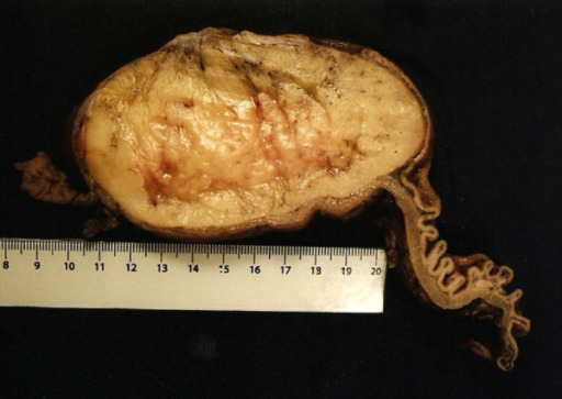
  • Soft, rubbery
  • Mobile (not fixed to surrounding tissues)
  • Round, oval, or multi-lobulated
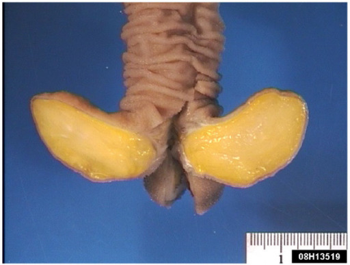
  • Bright yellow homogeneous Homogeneous Imaging of the Spleen fat with a fine fibrous capsule Fibrous capsule Hip Joint: Anatomy (superficial lesions) and trabeculae
  • Greasy cut surface

Histology:

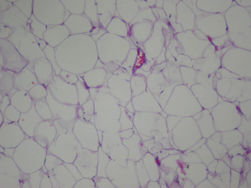
  • Mature adipose tissue Adipose tissue Adipose tissue is a specialized type of connective tissue that has both structural and highly complex metabolic functions, including energy storage, glucose homeostasis, and a multitude of endocrine capabilities. There are three types of adipose tissue, white adipose tissue, brown adipose tissue, and beige or “brite” adipose tissue, which is a transitional form. Adipose Tissue: Histology, with adipocytes Adipocytes Cells in the body that store fats, usually in the form of triglycerides. White adipocytes are the predominant type and found mostly in the abdominal cavity and subcutaneous tissue. Brown adipocytes are thermogenic cells that can be found in newborns of some species and hibernating mammals. Adipose Tissue: Histology showing no atypia Atypia Fibrocystic Change, but 2–5x variation in cell size
  • Lobular architecture with thin septae
  • No mitotic figures
  • Scant vascularity
  • Thin fibrous capsule Fibrous capsule Hip Joint: Anatomy
  • Fat necrosis Necrosis The death of cells in an organ or tissue due to disease, injury or failure of the blood supply. Ischemic Cell Damage:
    • More likely in larger lipomas
    • Focal, with histiocytes Histiocytes Macrophages found in the tissues, as opposed to those found in the blood (monocytes) or serous cavities (serous membrane). Chronic Granulomatous Disease +/- calcification
  • Some histologic variants:
    • Angiolipoma Angiolipoma A benign neoplasm composed of a mixture of adipose tissue and blood vessels. Cowden Syndrome:
      • Small, well-circumscribed, subcutaneous tumor Tumor Inflammation
      • Mature adipose cells Adipose Cells Fat Necrosis of the Breast, capillary vessels with fibrin Fibrin A protein derived from fibrinogen in the presence of thrombin, which forms part of the blood clot. Rapidly Progressive Glomerulonephritis thrombi
      • Angiolipoma Angiolipoma A benign neoplasm composed of a mixture of adipose tissue and blood vessels. Cowden Syndrome is 1 of 5 of the most common painful skin Skin The skin, also referred to as the integumentary system, is the largest organ of the body. The skin is primarily composed of the epidermis (outer layer) and dermis (deep layer). The epidermis is primarily composed of keratinocytes that undergo rapid turnover, while the dermis contains dense layers of connective tissue. Skin: Structure and Functions tumors. (The other 4 are: neuroma (traumatic), glomus tumor Tumor Inflammation, eccrine spiradenoma, and leiomyoma Leiomyoma A benign tumor derived from smooth muscle tissue, also known as a fibroid tumor. They rarely occur outside of the uterus and the gastrointestinal tract but can occur in the skin and subcutaneous tissue, probably arising from the smooth muscle of small blood vessels in these tissues. Infertility (vascular), creating the mnemonic “ANGEL.”)
    • Fibrolipoma
    • Chondroid lipoma
    • Ossifying lipoma     

Clinical Presentation

  • Subcutaneous lipomas (most common):
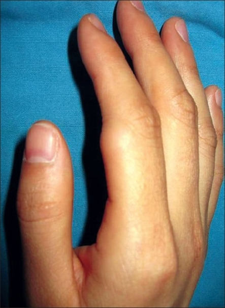
    • 2–3 cm (can be > 10 cm) soft, mobile mass Mass Three-dimensional lesion that occupies a space within the breast Imaging of the Breast, usually on the trunk or upper extremities
    • Usually solitary 
    • Painless
    • No overlying skin Skin The skin, also referred to as the integumentary system, is the largest organ of the body. The skin is primarily composed of the epidermis (outer layer) and dermis (deep layer). The epidermis is primarily composed of keratinocytes that undergo rapid turnover, while the dermis contains dense layers of connective tissue. Skin: Structure and Functions changes
    • Can occur anywhere on the body
  • Gastrointestinal lipomas:
    • Intestinal obstruction Intestinal obstruction Any impairment, arrest, or reversal of the normal flow of intestinal contents toward the anal canal. Ascaris/Ascariasis or intussusception Intussusception Intussusception occurs when a part of the intestine (intussusceptum) telescopes into another part (intussuscipiens) of the intestine. The condition can cause obstruction and, if untreated, progress to bowel ischemia. Intussusception is most common in the pediatric population, but is occasionally encountered in adults. Intussusception ( nausea Nausea An unpleasant sensation in the stomach usually accompanied by the urge to vomit. Common causes are early pregnancy, sea and motion sickness, emotional stress, intense pain, food poisoning, and various enteroviruses. Antiemetics/ vomiting Vomiting The forcible expulsion of the contents of the stomach through the mouth. Hypokalemia, abdominal pain Abdominal Pain Acute Abdomen)
    • Gastrointestinal hemorrhage Gastrointestinal hemorrhage Bleeding in any segment of the gastrointestinal tract from esophagus to rectum. Peptic Ulcer Disease (from mucosal ulceration Ulceration Corneal Abrasions, Erosion, and Ulcers)
  • Other sites, e.g. retroperitoneum, body cavities: compression Compression Blunt Chest Trauma effects on adjacent organs

Diagnosis and Management

Diagnosis

History:

  • A slowly growing lump (usually painless) for subcutaneous lipomas
  • For other types of lipomas reported, symptoms depend on the site.
  • Family history Family History Adult Health Maintenance/genetic disorders

Physical exam:

Imaging:

Endoscopy Endoscopy Procedures of applying endoscopes for disease diagnosis and treatment. Endoscopy involves passing an optical instrument through a small incision in the skin i.e., percutaneous; or through a natural orifice and along natural body pathways such as the digestive tract; and/or through an incision in the wall of a tubular structure or organ, i.e. Transluminal, to examine or perform surgery on the interior parts of the body. Gastroesophageal Reflux Disease (GERD) and endoscopic ultrasound (EUS):

  • From the esophagus Esophagus The esophagus is a muscular tube-shaped organ of around 25 centimeters in length that connects the pharynx to the stomach. The organ extends from approximately the 6th cervical vertebra to the 11th thoracic vertebra and can be divided grossly into 3 parts: the cervical part, the thoracic part, and the abdominal part. Esophagus: Anatomy to the colon Colon The large intestines constitute the last portion of the digestive system. The large intestine consists of the cecum, appendix, colon (with ascending, transverse, descending, and sigmoid segments), rectum, and anal canal. The primary function of the colon is to remove water and compact the stool prior to expulsion from the body via the rectum and anal canal. Colon, Cecum, and Appendix: Anatomy
  • Appear as smooth, round submucosal nodules
  • Possible mucosal ulceration Ulceration Corneal Abrasions, Erosion, and Ulcers if large
  • EUS can help confirm the diagnosis of a lipoma.

Biopsy Biopsy Removal and pathologic examination of specimens from the living body. Ewing Sarcoma:

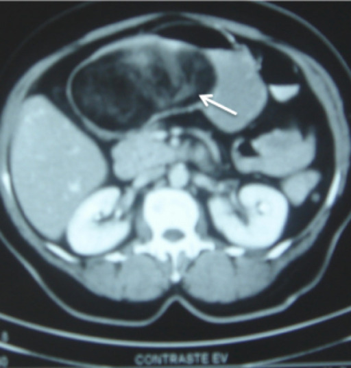
Large submucosal gastric lipoma

A large (12 x 8 x 6 cm) submucosal gastric lipoma (arrow)
Some contrast medium has leaked into the lipoma through focal ulcerated areas in the overlying mucosa, and can be seen tracking into the center from the surface.

Image: “Gastric lipoma presenting as a giant bulging mass in an oligosymptomatic patient: a case report” by Neto FA, Ferreira MC, Bertoncello LC, Neto AA, de Aveiro WC, Bento CA, Cecchino GN, Rocha MA. License: CC BY 2.0.
Management

Expectant management: appropriate for small (< 5 cm) subcutaneous asymptomatic lipomas

Surgical excision:

  • Subcutaneous lipomas:
    • Indications:
      • Pain Pain An unpleasant sensation induced by noxious stimuli which are detected by nerve endings of nociceptive neurons. Pain: Types and Pathways 
      • Cosmesis
      • Diagnosis in doubt
      • Increase in size
    • Excision needs to involve the fibrous capsule Fibrous capsule Hip Joint: Anatomy to reduce recurrence. 
  • Gastrointestinal lipomas:
    • Surgical resection if symptomatic/obstructive
    • Endoscopic removal may be feasible if small. 
  • Other sites (retroperitoneum, intraglandular):
    • Resection usually needed if malignancy Malignancy Hemothorax is in question
    • Always needed if symptomatic
    • Biopsy Biopsy Removal and pathologic examination of specimens from the living body. Ewing Sarcoma prior to resection, if feasible.

Complications of surgery:

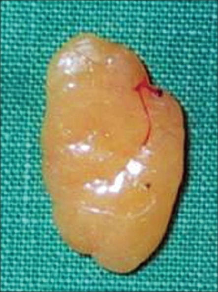
Submucosal gastric lipoma

Submucosal gastric lipoma (surgical specimen)
Note the thickened wall of the proximal gastric wall on the right side, and the attenuated focally ulcerated overlying gastric mucosa.

Image: “Gastric lipoma presenting as a giant bulging mass in an oligosymptomatic patient: a case report” by Neto FA, Ferreira MC, Bertoncello LC, Neto AA, de Aveiro WC, Bento CA, Cecchino GN, Rocha MA. License: CC BY 2.0.

Differential Diagnosis

Subcutaneous lesions

  • Keratin-filled cysts Cysts Any fluid-filled closed cavity or sac that is lined by an epithelium. Cysts can be of normal, abnormal, non-neoplastic, or neoplastic tissues. Fibrocystic Change: benign Benign Fibroadenoma cysts Cysts Any fluid-filled closed cavity or sac that is lined by an epithelium. Cysts can be of normal, abnormal, non-neoplastic, or neoplastic tissues. Fibrocystic Change lined by epidermal cells and filled with keratin Keratin A class of fibrous proteins or scleroproteins that represents the principal constituent of epidermis; hair; nails; horny tissues, and the organic matrix of tooth enamel. Two major conformational groups have been characterized, alpha-keratin, whose peptide backbone forms a coiled-coil alpha helical structure consisting of type I keratin and a type II keratin, and beta-keratin, whose backbone forms a zigzag or pleated sheet structure. Alpha-keratins have been classified into at least 20 subtypes. In addition multiple isoforms of subtypes have been found which may be due to gene duplication. Seborrheic Keratosis. Also called by the misnomer “sebaceous cysts Cysts Any fluid-filled closed cavity or sac that is lined by an epithelium. Cysts can be of normal, abnormal, non-neoplastic, or neoplastic tissues. Fibrocystic Change.” Two types:
    • Epidermal inclusion cyst: formed by the invagination and cystic Cystic Fibrocystic Change expansion of the epidermis Epidermis The external, nonvascular layer of the skin. It is made up, from within outward, of five layers of epithelium: (1) basal layer (stratum basale epidermidis); (2) spinous layer (stratum spinosum epidermidis); (3) granular layer (stratum granulosum epidermidis); (4) clear layer (stratum lucidum epidermidis); and (5) horny layer (stratum corneum epidermidis). Skin: Structure and Functions. Commonly found on the head, face, or neck Neck The part of a human or animal body connecting the head to the rest of the body. Peritonsillar Abscess. Firm and has a central punctum; prone to rupture.
    • Trichilemmal cyst (pilar cyst): originates from the outer hair root sheath. Ninety percent occur on the scalp or scrotum Scrotum A cutaneous pouch of skin containing the testicles and spermatic cords. Testicles: Anatomy. Often have an autosomal dominant Autosomal dominant Autosomal inheritance, both dominant and recessive, refers to the transmission of genes from the 22 autosomal chromosomes. Autosomal dominant diseases are expressed when only 1 copy of the dominant allele is inherited. Autosomal Recessive and Autosomal Dominant Inheritance pattern of inheritance. Usually firm with a thick cyst wall.
  • Abscess Abscess Accumulation of purulent material in tissues, organs, or circumscribed spaces, usually associated with signs of infection. Chronic Granulomatous Disease: pus-filled cyst with fibrotic wall; indurated, fixed, and erythematous; can be infectious (bacterial or fungal) or sterile Sterile Basic Procedures (if an irritant drug or substance is injected into the skin Skin The skin, also referred to as the integumentary system, is the largest organ of the body. The skin is primarily composed of the epidermis (outer layer) and dermis (deep layer). The epidermis is primarily composed of keratinocytes that undergo rapid turnover, while the dermis contains dense layers of connective tissue. Skin: Structure and Functions, resulting in aseptic inflammation Inflammation Inflammation is a complex set of responses to infection and injury involving leukocytes as the principal cellular mediators in the body’s defense against pathogenic organisms. Inflammation is also seen as a response to tissue injury in the process of wound healing. The 5 cardinal signs of inflammation are pain, heat, redness, swelling, and loss of function. Inflammation and abscess Abscess Accumulation of purulent material in tissues, organs, or circumscribed spaces, usually associated with signs of infection. Chronic Granulomatous Disease formation).
  • Well-differentiated liposarcoma (“atypical lipomatous tumor Tumor Inflammation”): mostly in the deep thigh Thigh The thigh is the region of the lower limb found between the hip and the knee joint. There is a single bone in the thigh called the femur, which is surrounded by large muscles grouped into 3 fascial compartments. Thigh: Anatomy or retroperitoneum; multi-lobulated, with focal firm areas. Atypical spindle cells Spindle Cells Acoustic Neuroma present; overexpresses MDM2 (which blocks p53 tumor Tumor Inflammation suppressor function). Well-differentiated liposarcoma tend to recur if not completely resected. Do not metastasize if dedifferentiation into higher-grade elements does not occur. 
  • Pseudolipoma: tension on subcutaneous fat Subcutaneous fat Fatty tissue under the skin throughout the body. Erythema Nodosum tissue by adjacent fibrous Fibrous Fibrocystic Change bands, which may be post-traumatic or related to scirrhous breast cancer Breast cancer Breast cancer is a disease characterized by malignant transformation of the epithelial cells of the breast. Breast cancer is the most common form of cancer and 2nd most common cause of cancer-related death among women. Breast Cancer

Deep lesions

  • Liposarcoma: a malignant neoplasm of adipocytes Adipocytes Cells in the body that store fats, usually in the form of triglycerides. White adipocytes are the predominant type and found mostly in the abdominal cavity and subcutaneous tissue. Brown adipocytes are thermogenic cells that can be found in newborns of some species and hibernating mammals. Adipose Tissue: Histology; usually arises de novo (not from a lipoma). Often intramuscular or retroperitoneal Retroperitoneal Peritoneum: Anatomy. Appears heterogeneous on imaging. Biopsy Biopsy Removal and pathologic examination of specimens from the living body. Ewing Sarcoma is needed for confirmation. 
  • Hibernoma: a benign Benign Fibroadenoma tumor Tumor Inflammation arising from brown fat; most commonly in inter-scapular area but also in the neck Neck The part of a human or animal body connecting the head to the rest of the body. Peritonsillar Abscess, axillae, thigh Thigh The thigh is the region of the lower limb found between the hip and the knee joint. There is a single bone in the thigh called the femur, which is surrounded by large muscles grouped into 3 fascial compartments. Thigh: Anatomy, or intrathoracic areas. Hibernomas have higher vascularity and density compared with lipomas.
  • Mammary hamartoma Hamartoma A focal malformation resembling a neoplasm, composed of an overgrowth of mature cells and tissues that normally occur in the affected area. Colorectal Cancer: a benign Benign Fibroadenoma proliferation of fibrous Fibrous Fibrocystic Change, glandular, and fatty tissue surrounded by a thin, fibrous capsule Fibrous capsule Hip Joint: Anatomy.
  • Gastrointestinal submucosal tumors: include leiomyomas, granular cell tumors, gastrointestinal stromal tumors Gastrointestinal stromal tumors All tumors in the gastrointestinal tract arising from mesenchymal cells (mesoderm) except those of smooth muscle cells (leiomyoma) or schwann cells (schwannoma). Gastric Cancer (GISTs), metastatic lesions, and others. Diagnosed by endoscopy Endoscopy Procedures of applying endoscopes for disease diagnosis and treatment. Endoscopy involves passing an optical instrument through a small incision in the skin i.e., percutaneous; or through a natural orifice and along natural body pathways such as the digestive tract; and/or through an incision in the wall of a tubular structure or organ, i.e. Transluminal, to examine or perform surgery on the interior parts of the body. Gastroesophageal Reflux Disease (GERD), EUS, and biopsy Biopsy Removal and pathologic examination of specimens from the living body. Ewing Sarcoma.
  • Spermatic cord Spermatic Cord Either of a pair of tubular structures formed by ductus deferens; arteries; veins; lymphatic vessels; and nerves. The spermatic cord extends from the deep inguinal ring through the inguinal canal to the testis in the scrotum. Testicles: Anatomy “lipoma”: not a true lipoma (i.e., a benign Benign Fibroadenoma neoplasm of adipose tissue Adipose tissue Adipose tissue is a specialized type of connective tissue that has both structural and highly complex metabolic functions, including energy storage, glucose homeostasis, and a multitude of endocrine capabilities. There are three types of adipose tissue, white adipose tissue, brown adipose tissue, and beige or “brite” adipose tissue, which is a transitional form. Adipose Tissue: Histology), but is only preperitoneal, mature, benign Benign Fibroadenoma, fatty tissue that has gained access to the spermatic cord Spermatic Cord Either of a pair of tubular structures formed by ductus deferens; arteries; veins; lymphatic vessels; and nerves. The spermatic cord extends from the deep inguinal ring through the inguinal canal to the testis in the scrotum. Testicles: Anatomy.

Related videos

References

  1. Nickloes, T.A. (2020). Lipomas: Background, pathophysiology, etiology. https://emedicine.medscape.com/article/191233-overview#a4
  2. Kim, K.S., & Yang, H.S. (2014). Unusual locations of lipoma: Differential diagnosis of head and neck mass. Australian Family Physician, 43(12), 867–870.  https://www.racgp.org.au/afp/2014/december/unusual-locations-of-lipoma-differential-diagnosis-of-head-and-neck-mass/#5
  3. Lipoma (2020). In Kumar, V., Abbas, A. K., Aster, J.C., (Eds.). Robbins & Cotran Pathologic Basis of Disease. (10th ed., p. 1209). Elsevier, Inc.
  4. Rouse, R.V. (2017). Lipoma—Surgical pathology criteria—Stanford University School of Medicine. http://surgpathcriteria.stanford.edu/softfat/lipoma/
  5. Fairweather, M., & Raut, C.P. (2019). To Biopsy, or Not to Biopsy [a suspected retroperitoneal sarcoma]: Is There Really a Question?. Ann Surg Oncol 26, 4182–4184. https://doi.org/10.1245/s10434-019-07723-y
  6. Rotunda, A. M., Ablon, G., & Kolodney, M. S. (2005). Lipomas treated with subcutaneous deoxycholate injections. Journal of the American Academy of Dermatology, 53(6), 973–978. https://doi.org/10.1016/j.jaad.2005.07.068

Create your free account or log in to continue reading!

Sign up now and get free access to Lecturio with concept pages, medical videos, and questions for your medical education.

User Reviews

Details