Advertisement

Advertisement

Advertisement

Advertisement

Lincosamides

The lincosamides, lincomycin and clindamycin, are inhibitors of bacterial protein synthesis Synthesis Polymerase Chain Reaction (PCR). Drugs in this class share the same binding site as that of macrolides Macrolides Macrolides and ketolides are antibiotics that inhibit bacterial protein synthesis by binding to the 50S ribosomal subunit and blocking transpeptidation. These antibiotics have a broad spectrum of antimicrobial activity but are best known for their coverage of atypical microorganisms. Macrolides and Ketolides and amphenicols; however, they differ in chemical structure. Lincosamides target the 50S ribosomal subunit and interfere with transpeptidation. The antimicrobial coverage of lincosamides encompasses gram-positive Gram-Positive Penicillins cocci Cocci Bacteriology (including MRSA MRSA A strain of Staphylococcus aureus that is non-susceptible to the action of methicillin. The mechanism of resistance usually involves modification of normal or the presence of acquired penicillin binding proteins. Staphylococcus) and anaerobes. Clindamycin can also be used to treat toxic shock syndrome Toxic Shock Syndrome Toxic shock syndrome (TSS) is an acute, multi-systemic disease caused by the toxin-producing bacteria, Staphylococcus aureus and Streptococcus pyogenes. Staphylococcal TSS is more common and associated with tampons and nasal packing. Toxic Shock Syndrome and necrotizing fasciitis Necrotizing fasciitis Necrotizing fasciitis is a life-threatening infection that causes rapid destruction and necrosis of the fascia and subcutaneous tissues. Patients may present with significant pain out of proportion to the presenting symptoms and rapidly progressive erythema of the affected area. Necrotizing Fasciitis owing to its antitoxin effect. Diarrhea Diarrhea Diarrhea is defined as ≥ 3 watery or loose stools in a 24-hour period. There are a multitude of etiologies, which can be classified based on the underlying mechanism of disease. The duration of symptoms (acute or chronic) and characteristics of the stools (e.g., watery, bloody, steatorrheic, mucoid) can help guide further diagnostic evaluation. Diarrhea is a common adverse effect, and clindamycin is often associated with a higher risk of Clostridioides difficile colitis Colitis Inflammation of the colon section of the large intestine, usually with symptoms such as diarrhea (often with blood and mucus), abdominal pain, and fever. Pseudomembranous Colitis.

Last updated: Oct 6, 2022

Editorial responsibility: Stanley Oiseth, Lindsay Jones, Evelin Maza

Advertisement

Advertisement

Advertisement

Advertisement

Advertisement

Advertisement

Chemistry and Pharmacodynamics

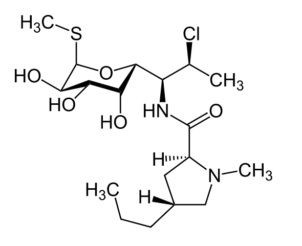
Chemical structure

  • The chemical structure consists of amino acid Amino acid Amino acids (AAs) are composed of a central carbon atom attached to a carboxyl group, an amino group, a hydrogen atom, and a side chain (R group). Basics of Amino Acids and sugar moieties.
  • Lincomycin is the prototype drug.
  • Clindamycin is a chlorinated derivative.
Chemical structure of clindamycin lincosamides

Chemical structure of clindamycin

Image: “Clindamycin” by Jü. License: Public Domain
Mechanism of action

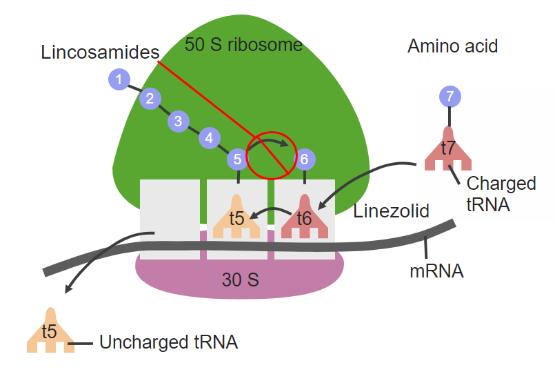
  • Inhibits bacterial protein synthesis Synthesis Polymerase Chain Reaction (PCR) by binding reversibly to the 50S ribosomal subunit: 
    • Binds near the peptidyltransferase center → prevents peptidyltransferase from adding amino acids Amino acids Organic compounds that generally contain an amino (-NH2) and a carboxyl (-COOH) group. Twenty alpha-amino acids are the subunits which are polymerized to form proteins. Basics of Amino Acids to the growing peptide (prevents transpeptidation)
    • Similar binding site as that of macrolides Macrolides Macrolides and ketolides are antibiotics that inhibit bacterial protein synthesis by binding to the 50S ribosomal subunit and blocking transpeptidation. These antibiotics have a broad spectrum of antimicrobial activity but are best known for their coverage of atypical microorganisms. Macrolides and Ketolides and chloramphenicol Chloramphenicol Chloramphenicol, the only clinically relevant drug in the amphenicol class, is a potent inhibitor of bacterial protein synthesis by binding to the 50S ribosomal subunit and preventing peptide bond formation. Chloramphenicol is a broad-spectrum antibiotic with wide distribution; however, due to its toxicity, its use is limited to severe infections. Chloramphenicol
  • Limits bacterial growth → bacteriostatic Bacteriostatic Sulfonamides and Trimethoprim
  • Can exert a time-dependent killing effect for some bacteria Bacteria Bacteria are prokaryotic single-celled microorganisms that are metabolically active and divide by binary fission. Some of these organisms play a significant role in the pathogenesis of diseases. Bacteriology
  • Additional effect: inhibits the production of staphylococcal and streptococcal toxins
Site of action clindamycin lincosamides

Site of action of clindamycin at the 50S ribosomal subunit

Image by Lecturio. License: CC BY-NC-SA 4.0

Pharmacokinetics

Since lincomycin is rarely used, the pharmacokinetics Pharmacokinetics Pharmacokinetics is the science that analyzes how the human body interacts with a drug. Pharmacokinetics examines how the drug is absorbed, distributed, metabolized, and excreted by the body. Pharmacokinetics and Pharmacodynamics of clindamycin are have been listed:

Absorption Absorption Absorption involves the uptake of nutrient molecules and their transfer from the lumen of the GI tract across the enterocytes and into the interstitial space, where they can be taken up in the venous or lymphatic circulation. Digestion and Absorption and distribution

  • Rapid absorption Absorption Absorption involves the uptake of nutrient molecules and their transfer from the lumen of the GI tract across the enterocytes and into the interstitial space, where they can be taken up in the venous or lymphatic circulation. Digestion and Absorption
    • Approximately 90% absorption Absorption Absorption involves the uptake of nutrient molecules and their transfer from the lumen of the GI tract across the enterocytes and into the interstitial space, where they can be taken up in the venous or lymphatic circulation. Digestion and Absorption of oral doses
    • Plasma Plasma The residual portion of blood that is left after removal of blood cells by centrifugation without prior blood coagulation. Transfusion Products concentration peaks 2–4 hours after oral dose and 1–2 hours after IM dose.
  • Protein bound
  • Taken up by phagocytic cells
  • Widely distributed in body fluids, bone Bone Bone is a compact type of hardened connective tissue composed of bone cells, membranes, an extracellular mineralized matrix, and central bone marrow. The 2 primary types of bone are compact and spongy. Bones: Structure and Types, and tissues
    • Crosses placenta Placenta A highly vascularized mammalian fetal-maternal organ and major site of transport of oxygen, nutrients, and fetal waste products. It includes a fetal portion (chorionic villi) derived from trophoblasts and a maternal portion (decidua) derived from the uterine endometrium. The placenta produces an array of steroid, protein and peptide hormones (placental hormones). Placenta, Umbilical Cord, and Amniotic Cavity
    • Does not cross the blood-brain barrier Blood-brain barrier Specialized non-fenestrated tightly-joined endothelial cells with tight junctions that form a transport barrier for certain substances between the cerebral capillaries and the brain tissue. Systemic and Special Circulations

Metabolism and excretion

Indications

Antimicrobial coverage

  • Gram-positive Gram-Positive Penicillins microbes:
    • Streptococcus Streptococcus Streptococcus is one of the two medically important genera of gram-positive cocci, the other being Staphylococcus. Streptococci are identified as different species on blood agar on the basis of their hemolytic pattern and sensitivity to optochin and bacitracin. There are many pathogenic species of streptococci, including S. pyogenes, S. agalactiae, S. pneumoniae, and the viridans streptococci. Streptococcus
    • Staphylococcus Staphylococcus Staphylococcus is a medically important genera of Gram-positive, aerobic cocci. These bacteria form clusters resembling grapes on culture plates. Staphylococci are ubiquitous for humans, and many strains compose the normal skin flora. Staphylococcus (including MRSA MRSA A strain of Staphylococcus aureus that is non-susceptible to the action of methicillin. The mechanism of resistance usually involves modification of normal or the presence of acquired penicillin binding proteins. Staphylococcus)
  • Anaerobes:
    • Bacteroides Bacteroides Bacteroides is a genus of opportunistic, anaerobic, gram-negative bacilli. Bacteroides fragilis is the most common species involved in human disease and is part of the normal flora of the large intestine. Bacteroides
    • Clostridium perfringens Clostridium perfringens The most common etiologic agent of gas gangrene. It is differentiable into several distinct types based on the distribution of twelve different toxins. Gas Gangrene
    • Fusobacterium Fusobacterium A genus of gram-negative, anaerobic, rod-shaped bacteria found in cavities of humans and other animals. No endospores are formed. Some species are pathogenic and occur in various purulent or gangrenous infections. Dog and Cat Bites
    • Prevotella Prevotella A genus of gram-negative, anaerobic, nonsporeforming, nonmotile rods. Organisms of this genus had originally been classified as members of the bacteroides genus but overwhelming biochemical and chemical findings in 1990 indicated the need to separate them from other bacteroides species, and hence, this new genus was established. Dog and Cat Bites melaninogenicus
    • Peptostreptococcus Peptostreptococcus A genus of gram-positive, anaerobic, coccoid bacteria that is part of the normal flora of humans. Its organisms are opportunistic pathogens causing bacteremias and soft tissue infections. Perianal and Perirectal Abscess

Type of infections Infections Invasion of the host organism by microorganisms or their toxins or by parasites that can cause pathological conditions or diseases. Chronic Granulomatous Disease

  • Skin Skin The skin, also referred to as the integumentary system, is the largest organ of the body. The skin is primarily composed of the epidermis (outer layer) and dermis (deep layer). The epidermis is primarily composed of keratinocytes that undergo rapid turnover, while the dermis contains dense layers of connective tissue. Skin: Structure and Functions and soft tissue Soft Tissue Soft Tissue Abscess infections Infections Invasion of the host organism by microorganisms or their toxins or by parasites that can cause pathological conditions or diseases. Chronic Granulomatous Disease:
    • Acne
    • Cellulitis Cellulitis Cellulitis is a common infection caused by bacteria that affects the dermis and subcutaneous tissue of the skin. It is frequently caused by Staphylococcus aureus and Streptococcus pyogenes. The skin infection presents as an erythematous and edematous area with warmth and tenderness. Cellulitis
  • Respiratory infections Infections Invasion of the host organism by microorganisms or their toxins or by parasites that can cause pathological conditions or diseases. Chronic Granulomatous Disease:
    • Oral infections Infections Invasion of the host organism by microorganisms or their toxins or by parasites that can cause pathological conditions or diseases. Chronic Granulomatous Disease
    • Pneumonia Pneumonia Pneumonia or pulmonary inflammation is an acute or chronic inflammation of lung tissue. Causes include infection with bacteria, viruses, or fungi. In more rare cases, pneumonia can also be caused through toxic triggers through inhalation of toxic substances, immunological processes, or in the course of radiotherapy. Pneumonia 
    • Lung abscess Abscess Accumulation of purulent material in tissues, organs, or circumscribed spaces, usually associated with signs of infection. Chronic Granulomatous Disease
  • Bone Bone Bone is a compact type of hardened connective tissue composed of bone cells, membranes, an extracellular mineralized matrix, and central bone marrow. The 2 primary types of bone are compact and spongy. Bones: Structure and Types and joint infections Infections Invasion of the host organism by microorganisms or their toxins or by parasites that can cause pathological conditions or diseases. Chronic Granulomatous Disease:
    • Osteomyelitis Osteomyelitis Osteomyelitis is an infection of the bone that results from the spread of microorganisms from the blood (hematogenous), nearby infected tissue, or open wounds (non-hematogenous). Infections are most commonly caused by Staphylococcus aureus. Osteomyelitis
    • Prosthetic joint infection
    • Septic arthritis Arthritis Acute or chronic inflammation of joints. Osteoarthritis
  • Intra-abdominal infections Infections Invasion of the host organism by microorganisms or their toxins or by parasites that can cause pathological conditions or diseases. Chronic Granulomatous Disease
  • Gynecologic infections Infections Invasion of the host organism by microorganisms or their toxins or by parasites that can cause pathological conditions or diseases. Chronic Granulomatous Disease:
    • Bacterial vaginosis Bacterial vaginosis Polymicrobial, nonspecific vaginitis associated with positive cultures of gardnerella vaginalis and other anaerobic organisms and a decrease in lactobacilli. It remains unclear whether the initial pathogenic event is caused by the growth of anaerobes or a primary decrease in lactobacilli. Vulvovaginitis
    • Pelvic inflammatory disease Pelvic inflammatory disease Pelvic inflammatory disease (PID) is defined as a polymicrobial infection of the upper female reproductive system. The disease can affect the uterus, fallopian tubes, ovaries, and adjacent structures. Pelvic inflammatory disease is closely linked with sexually transmitted diseases, most commonly caused by Chlamydia trachomatis, Neisseria gonorrhoeae, and Gardnerella vaginalis. Pelvic Inflammatory Disease
    • Endometritis Endometritis Endometritis is an inflammation of the endometrium, the inner layer of the uterus. The most common subtype is postpartum endometritis, resulting from the ascension of normal vaginal flora to the previously aseptic uterus. Postpartum Endometritis
  • Used as part of combination therapy for:
    • Babesiosis Babesiosis Babesiosis is an infection caused by a protozoa belonging to the genus, Babesia. The most common Babesia seen in the United States is B. microti, which is transmitted by the Ixodes tick. The protozoa thrive and replicate within host erythrocytes. Lysis of erythrocytes and the body’s immune response result in clinical symptoms. Babesia/Babesiosis
    • Toxoplasmosis Toxoplasmosis Toxoplasmosis is an infectious disease caused by Toxoplasma gondii, an obligate intracellular protozoan parasite. Felines are the definitive host, but transmission to humans can occur through contact with cat feces or the consumption of contaminated foods. The clinical presentation and complications depend on the host’s immune status. Toxoplasma/Toxoplasmosis
    • Pneumocystis jirovecii pneumonia Pneumonia Pneumonia or pulmonary inflammation is an acute or chronic inflammation of lung tissue. Causes include infection with bacteria, viruses, or fungi. In more rare cases, pneumonia can also be caused through toxic triggers through inhalation of toxic substances, immunological processes, or in the course of radiotherapy. Pneumonia
    • Toxic shock syndrome Toxic Shock Syndrome Toxic shock syndrome (TSS) is an acute, multi-systemic disease caused by the toxin-producing bacteria, Staphylococcus aureus and Streptococcus pyogenes. Staphylococcal TSS is more common and associated with tampons and nasal packing. Toxic Shock Syndrome and necrotizing fasciitis Necrotizing fasciitis Necrotizing fasciitis is a life-threatening infection that causes rapid destruction and necrosis of the fascia and subcutaneous tissues. Patients may present with significant pain out of proportion to the presenting symptoms and rapidly progressive erythema of the affected area. Necrotizing Fasciitis (antitoxin effect)

Adverse Effects and Contraindications

Adverse effects

  • Topical:
    • Pruritis
    • Xeroderma
    • Erythema Erythema Redness of the skin produced by congestion of the capillaries. This condition may result from a variety of disease processes. Chalazion
    • Exfoliation
  • Vaginal:
    • Candidiasis Candidiasis Candida is a genus of dimorphic, opportunistic fungi. Candida albicans is part of the normal human flora and is the most common cause of candidiasis. The clinical presentation varies and can include localized mucocutaneous infections (e.g., oropharyngeal, esophageal, intertriginous, and vulvovaginal candidiasis) and invasive disease (e.g., candidemia, intraabdominal abscess, pericarditis, and meningitis). Candida/Candidiasis
    • Vulvovaginitis Vulvovaginitis The term vulvovaginitis is used to describe an acute inflammation of the vulva and vagina. Vulvovaginitis can be caused by several infectious and non-infectious etiologies, and results from disruption of the normal vaginal environment. Common signs and symptoms include pain, pruritus, erythema, edema, vaginal discharge and dyspareunia. Vulvovaginitis
  • Systemic:
    • GI upset and diarrhea Diarrhea Diarrhea is defined as ≥ 3 watery or loose stools in a 24-hour period. There are a multitude of etiologies, which can be classified based on the underlying mechanism of disease. The duration of symptoms (acute or chronic) and characteristics of the stools (e.g., watery, bloody, steatorrheic, mucoid) can help guide further diagnostic evaluation. Diarrhea
    • Pseudomembranous colitis Colitis Inflammation of the colon section of the large intestine, usually with symptoms such as diarrhea (often with blood and mucus), abdominal pain, and fever. Pseudomembranous Colitis (Clostridioides difficile)
    • Esophagitis Esophagitis Esophagitis is the inflammation or irritation of the esophagus. The major types of esophagitis are medication-induced, infectious, eosinophilic, corrosive, and acid reflux. Patients typically present with odynophagia, dysphagia, and retrosternal chest pain. Esophagitis
    • Metallic taste
    • Azotemia Azotemia A biochemical abnormality referring to an elevation of blood urea nitrogen and creatinine. Azotemia can be produced by kidney diseases or other extrarenal disorders. When azotemia becomes associated with a constellation of clinical signs, it is termed uremia. Acute Kidney Injury
    • Agranulocytosis
    • Allergic reactions Allergic Reactions Type I hypersensitivity reaction against plasma proteins in donor blood Transfusion Reactions:
      • Erythema Erythema Redness of the skin produced by congestion of the capillaries. This condition may result from a variety of disease processes. Chalazion multiforme
      • Stevens-Johnson syndrome Stevens-Johnson syndrome Stevens-Johnson syndrome (SJS) is a cutaneous, immune-mediated hypersensitivity reaction that is commonly triggered by medications, including antiepileptics and antibiotics. The condition runs on a spectrum with toxic epidermal necrolysis (TEN) based on the amount of body surface area (BSA) involved. Stevens-Johnson Syndrome
      • Anaphylaxis Anaphylaxis An acute hypersensitivity reaction due to exposure to a previously encountered antigen. The reaction may include rapidly progressing urticaria, respiratory distress, vascular collapse, systemic shock, and death. Type I Hypersensitivity Reaction

Contraindications Contraindications A condition or factor associated with a recipient that makes the use of a drug, procedure, or physical agent improper or inadvisable. Contraindications may be absolute (life threatening) or relative (higher risk of complications in which benefits may outweigh risks). Noninvasive Ventilation

Drug interactions

  • Inducers of CYP3A4 CYP3A4 Class 3 Antiarrhythmic Drugs (Potassium Channel Blockers): ↓ clindamycin concentrations
  • Neuromuscular blocking agents: ↑ neuromuscular blocking effect
  • Should not be used in combination with macrolides Macrolides Macrolides and ketolides are antibiotics that inhibit bacterial protein synthesis by binding to the 50S ribosomal subunit and blocking transpeptidation. These antibiotics have a broad spectrum of antimicrobial activity but are best known for their coverage of atypical microorganisms. Macrolides and Ketolides or chloramphenicol Chloramphenicol Chloramphenicol, the only clinically relevant drug in the amphenicol class, is a potent inhibitor of bacterial protein synthesis by binding to the 50S ribosomal subunit and preventing peptide bond formation. Chloramphenicol is a broad-spectrum antibiotic with wide distribution; however, due to its toxicity, its use is limited to severe infections. Chloramphenicol (due to the same ribosomal target site)

Mechanisms of Resistance

There are 3 main routes of resistance Resistance Physiologically, the opposition to flow of air caused by the forces of friction. As a part of pulmonary function testing, it is the ratio of driving pressure to the rate of air flow. Ventilation: Mechanics of Breathing:

  • Target site modification: 
    • Ribosomal methylation Ribosomal Methylation Macrolides and Ketolides or mutation Mutation Genetic mutations are errors in DNA that can cause protein misfolding and dysfunction. There are various types of mutations, including chromosomal, point, frameshift, and expansion mutations. Types of Mutations prevents the binding of the antibiotic to its ribosomal target. 
    • Most prevalent mechanism of resistance Resistance Physiologically, the opposition to flow of air caused by the forces of friction. As a part of pulmonary function testing, it is the ratio of driving pressure to the rate of air flow. Ventilation: Mechanics of Breathing to lincosamides
  • Efflux pumps
  • Enzymatic inactivation (uncommon, mostly seen in Staphylococcus Staphylococcus Staphylococcus is a medically important genera of Gram-positive, aerobic cocci. These bacteria form clusters resembling grapes on culture plates. Staphylococci are ubiquitous for humans, and many strains compose the normal skin flora. Staphylococcus)

Comparison of Antibiotics

The following table compares several classes of bacterial protein synthesis Synthesis Polymerase Chain Reaction (PCR) inhibitor antibiotics:

Table: Comparison of several classes of bacterial protein synthesis Synthesis Polymerase Chain Reaction (PCR) inhibitor antibiotics
Drug class Mechanism of action Coverage Adverse effects
Amphenicols
  • Gram positives
  • Gram negatives
  • Atypicals
  • GI upset
  • Optic neuritis Optic neuritis Inflammation of the optic nerve. Commonly associated conditions include autoimmune disorders such as multiple sclerosis, infections, and granulomatous diseases. Clinical features include retro-orbital pain that is aggravated by eye movement, loss of color vision, and contrast sensitivity that may progress to severe visual loss, an afferent pupillary defect (Marcus-Gunn pupil), and in some instances optic disc hyperemia and swelling. Inflammation may occur in the portion of the nerve within the globe (neuropapillitis or anterior optic neuritis) or the portion behind the globe (retrobulbar neuritis or posterior optic neuritis). Cranial Nerve Palsies
  • Aplastic anemia Aplastic Anemia Aplastic anemia (AA) is a rare, life-threatening condition characterized by pancytopenia and hypocellularity of the bone marrow (in the absence of any abnormal cells) reflecting damage to hematopoietic stem cells. Aplastic anemia can be acquired or inherited, however, most cases of AA are acquired and caused by autoimmune damage to hematopoietic stem cells. Aplastic Anemia
  • Gray baby syndrome Gray Baby Syndrome Chloramphenicol
Lincosamides
  • Gram-positive Gram-Positive Penicillins cocci Cocci Bacteriology:
    • MSSA
    • MRSA MRSA A strain of Staphylococcus aureus that is non-susceptible to the action of methicillin. The mechanism of resistance usually involves modification of normal or the presence of acquired penicillin binding proteins. Staphylococcus
    • Streptococcus Streptococcus Streptococcus is one of the two medically important genera of gram-positive cocci, the other being Staphylococcus. Streptococci are identified as different species on blood agar on the basis of their hemolytic pattern and sensitivity to optochin and bacitracin. There are many pathogenic species of streptococci, including S. pyogenes, S. agalactiae, S. pneumoniae, and the viridans streptococci. Streptococcus
  • Anaerobes
  • GI upset
  • Allergic reactions Allergic Reactions Type I hypersensitivity reaction against plasma proteins in donor blood Transfusion Reactions
  • Pseudomembranous colitis Colitis Inflammation of the colon section of the large intestine, usually with symptoms such as diarrhea (often with blood and mucus), abdominal pain, and fever. Pseudomembranous Colitis
Macrolides Macrolides Macrolides and ketolides are antibiotics that inhibit bacterial protein synthesis by binding to the 50S ribosomal subunit and blocking transpeptidation. These antibiotics have a broad spectrum of antimicrobial activity but are best known for their coverage of atypical microorganisms. Macrolides and Ketolides
  • Gram positives
  • Gram negatives
  • Atypicals
  • Mycobacterium avium Mycobacterium avium A bacterium causing tuberculosis in domestic fowl and other birds. In pigs, it may cause localized and sometimes disseminated disease. The organism occurs occasionally in sheep and cattle. It should be distinguished from the m. avium complex, which infects primarily humans. Mycobacterium complex
  • GI upset
  • QT prolongation
  • Hepatotoxicity Hepatotoxicity Acetaminophen
  • Myasthenia gravis Myasthenia Gravis Myasthenia gravis (MG) is an autoimmune neuromuscular disorder characterized by weakness and fatigability of skeletal muscles caused by dysfunction/destruction of acetylcholine receptors at the neuromuscular junction. MG presents with fatigue, ptosis, diplopia, dysphagia, respiratory difficulties, and progressive weakness in the limbs, leading to difficulty in movement. Myasthenia Gravis exacerbation
Oxazolidinones Oxazolidinones The oxazolidinones (linezolid and tedizolid) are bacterial protein synthesis inhibitors. Their unique binding site on the 23S ribosomal RNA of the 50S ribosome gives them zero cross-resistance with other antibiotics. Oxazolidinones
  • Bind BIND Hyperbilirubinemia of the Newborn to the 23S rRNA rRNA The most abundant form of RNA. Together with proteins, it forms the ribosomes, playing a structural role and also a role in ribosomal binding of mRNA and tRNAs. Individual chains are conventionally designated by their sedimentation coefficients. In eukaryotes, four large chains exist, synthesized in the nucleolus and constituting about 50% of the ribosome. RNA Types and Structure of the 50S subunit
  • Prevent initiation complex formation Initiation Complex Formation Oxazolidinones
Gram-positive Gram-Positive Penicillins cocci Cocci Bacteriology:
  • MSSA
  • MRSA MRSA A strain of Staphylococcus aureus that is non-susceptible to the action of methicillin. The mechanism of resistance usually involves modification of normal or the presence of acquired penicillin binding proteins. Staphylococcus
  • VRE
  • Streptococcus Streptococcus Streptococcus is one of the two medically important genera of gram-positive cocci, the other being Staphylococcus. Streptococci are identified as different species on blood agar on the basis of their hemolytic pattern and sensitivity to optochin and bacitracin. There are many pathogenic species of streptococci, including S. pyogenes, S. agalactiae, S. pneumoniae, and the viridans streptococci. Streptococcus
rRNA: ribosomal RNA
VRE: vancomycin-resistant Enterococcus
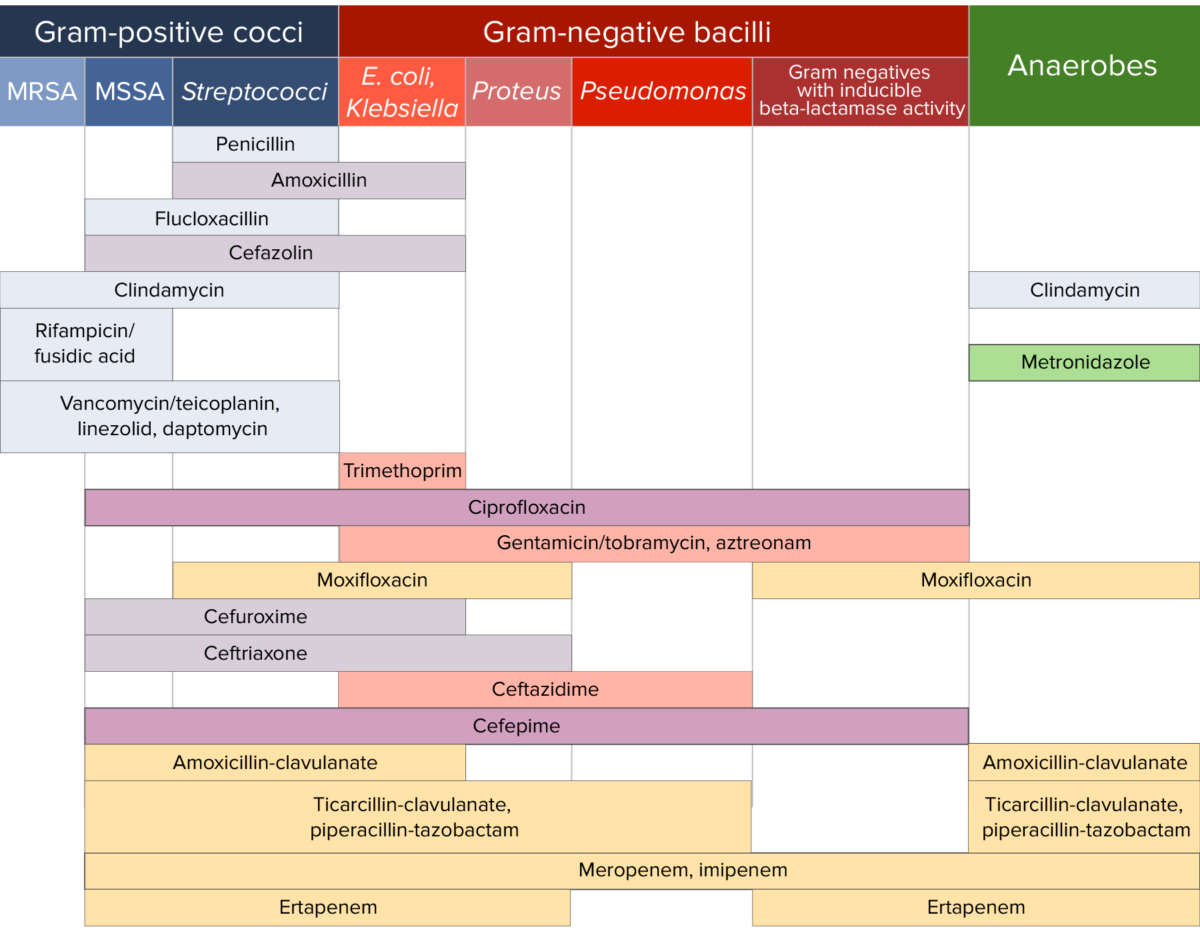
Antibiotic sensitivity chart

Antibiotic sensitivity:
Chart comparing the microbial coverage of different antibiotics for gram-positive cocci, gram-negative bacilli, and anaerobes.

Image by Lecturio. License: CC BY-NC-SA 4.0

References

  1. Deck, D.H., Winston, L.G. (2012). Tetracyclines, macrolides, clindamycin, chloramphenicol, streptogramins, & oxazolidinones. In Katzung, B.G., Masters, S.B., Trevor, A.J. (Eds.), Basic & Clinical Pharmacology (12th edition, pp. 809-819). https://pharmacomedicale.org/images/cnpm/CNPM_2016/katzung-pharmacology.pdf
  2. Leclercq, R. (2002). Mechanisms of resistance to macrolides and lincosamides: Nature of the resistance elements and their clinical implications. Clinical Infectious Diseases, 34(4), 482-492. https://academic.oup.com/cid/article/34/4/482/412492
  3. Johnson, M. (2020). Clindamycin: An overview. In Bond, S. (Ed.), UpToDate. Retrieved June 30, 2021, from https://www.uptodate.com/contents/clindamycin-an-overview
  4. Murphy, P.B., Bistas, K.G., Le, J.K. (2020). Clindamycin. StatPearls. Retrieved June 30, 2021, from https://www.ncbi.nlm.nih.gov/books/NBK519574/
  5. Werth, B.J. (2020). Clindamycin. MSD Manual Professional Version. Retrieved June 30, 2021, from https://www.msdmanuals.com/professional/infectious-diseases/bacteria-and-antibacterial-drugs/clindamycin

Create your free account or log in to continue reading!

Sign up now and get free access to Lecturio with concept pages, medical videos, and questions for your medical education.

User Reviews

Details