Advertisement

Advertisement

Advertisement

Advertisement

Larynx: Anatomy

The larynx, also commonly called the voice box, is a cylindrical space located in the neck Neck The part of a human or animal body connecting the head to the rest of the body. Peritonsillar Abscess at the level of the C3–C6 vertebrae. The larynx is continuous superiorly with the pharynx Pharynx The pharynx is a component of the digestive system that lies posterior to the nasal cavity, oral cavity, and larynx. The pharynx can be divided into the oropharynx, nasopharynx, and laryngopharynx. Pharyngeal muscles play an integral role in vital processes such as breathing, swallowing, and speaking. Pharynx: Anatomy and inferiorly with the trachea Trachea The trachea is a tubular structure that forms part of the lower respiratory tract. The trachea is continuous superiorly with the larynx and inferiorly becomes the bronchial tree within the lungs. The trachea consists of a support frame of semicircular, or C-shaped, rings made out of hyaline cartilage and reinforced by collagenous connective tissue. Trachea: Anatomy. This structure is made up of 9 cartilages that are connected by membranes, ligaments, and muscles and that house the vocal cords. The major structures forming the framework of the larynx are the thyroid Thyroid The thyroid gland is one of the largest endocrine glands in the human body. The thyroid gland is a highly vascular, brownish-red gland located in the visceral compartment of the anterior region of the neck. Thyroid Gland: Anatomy cartilage Cartilage Cartilage is a type of connective tissue derived from embryonic mesenchyme that is responsible for structural support, resilience, and the smoothness of physical actions. Perichondrium (connective tissue membrane surrounding cartilage) compensates for the absence of vasculature in cartilage by providing nutrition and support. Cartilage: Histology, cricoid cartilage Cartilage Cartilage is a type of connective tissue derived from embryonic mesenchyme that is responsible for structural support, resilience, and the smoothness of physical actions. Perichondrium (connective tissue membrane surrounding cartilage) compensates for the absence of vasculature in cartilage by providing nutrition and support. Cartilage: Histology, and epiglottis. The larynx serves to produce sound (phonation), conducts air to the trachea Trachea The trachea is a tubular structure that forms part of the lower respiratory tract. The trachea is continuous superiorly with the larynx and inferiorly becomes the bronchial tree within the lungs. The trachea consists of a support frame of semicircular, or C-shaped, rings made out of hyaline cartilage and reinforced by collagenous connective tissue. Trachea: Anatomy, and prevents large molecules from reaching the lungs Lungs Lungs are the main organs of the respiratory system. Lungs are paired viscera located in the thoracic cavity and are composed of spongy tissue. The primary function of the lungs is to oxygenate blood and eliminate CO2. Lungs: Anatomy.

Last updated: Nov 19, 2024

Editorial responsibility: Stanley Oiseth, Lindsay Jones, Evelin Maza

Advertisement

Advertisement

Advertisement

Advertisement

Advertisement

Advertisement

Development and Gross Anatomy

Development

  • The laryngeal cartilages develop from the 4th and 6th pairs of the pharyngeal arches Pharyngeal arches The branchial arches, also known as pharyngeal or visceral arches, are embryonic structures seen in the development of vertebrates that serve as precursors for many structures of the face, neck, and head. These arches are composed of a central core of mesoderm, which is covered externally by ectoderm and internally by endoderm. Branchial Apparatus and Aortic Arches.
  • The cricothyroid muscle originates from the 4th pharyngeal arch.
  • The rest of the intrinsic muscles of the larynx originate from the 6th pharyngeal arch.

Gross anatomy

The larynx is a framework composed mainly of cartilages held together by muscles and ligaments, both of which are divided into intrinsic and extrinsic forms.

Table: Gross anatomy
Cartilage Cartilage Cartilage is a type of connective tissue derived from embryonic mesenchyme that is responsible for structural support, resilience, and the smoothness of physical actions. Perichondrium (connective tissue membrane surrounding cartilage) compensates for the absence of vasculature in cartilage by providing nutrition and support. Cartilage: Histology Unpaired
  • Epiglottis
  • Cricoid
  • Thyroid Thyroid The thyroid gland is one of the largest endocrine glands in the human body. The thyroid gland is a highly vascular, brownish-red gland located in the visceral compartment of the anterior region of the neck. Thyroid Gland: Anatomy
Paired
  • Arytenoid
  • Corniculate
  • Cuneiform
Ligaments Extrinsic
  • Thyrohyoid membrane
  • Hyoepiglottic ligament
Intrinsic
  • Cricothyroid ligament (also called cricovocal or cricothyroid membrane)
  • Vocal ligaments
  • Quadrangular membrane
  • Vestibular ligaments
  • Aryepiglottic membrane
Muscles Extrinsic
  • Suprahyoid group: stylohyoid, digastric, mylohyoid, and geniohyoid
  • Infrahyoid group: sternohyoid, omohyoid, sternothyroid, and thyrohyoid
Intrinsic
  • Cricothyroid
  • Thyroarytenoid
  • Posterior cricoarytenoid
  • Lateral cricoarytenoid
  • Transverse arytenoid
  • Oblique arytenoid
  • Vocalis
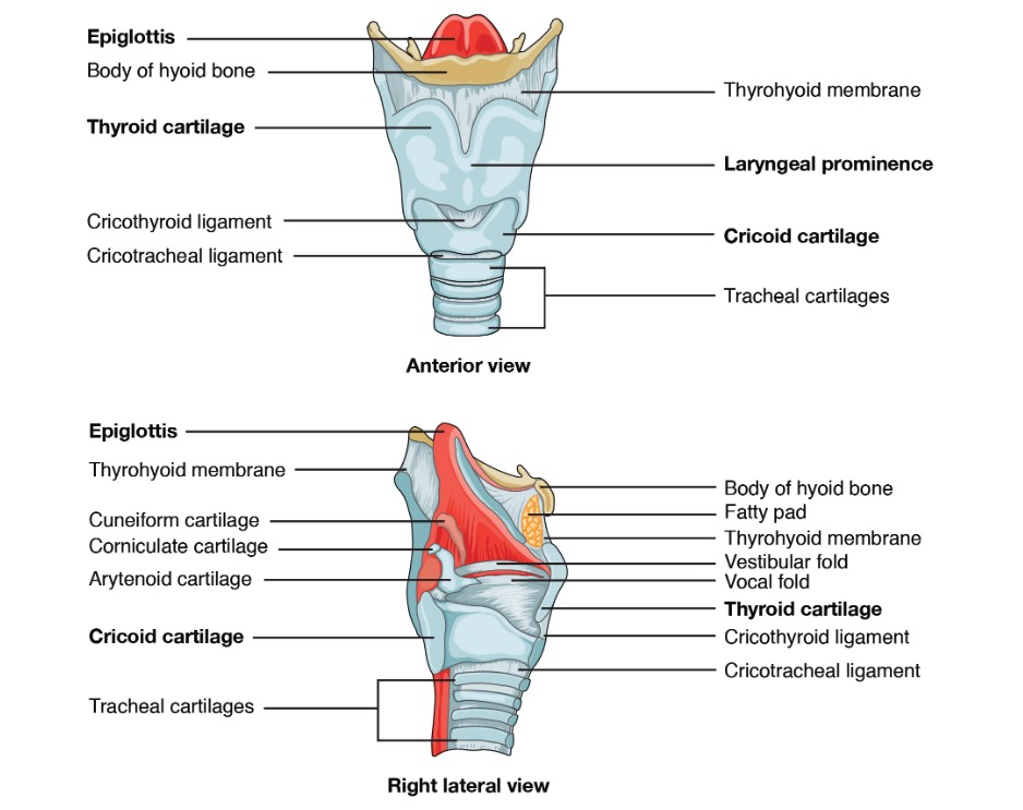
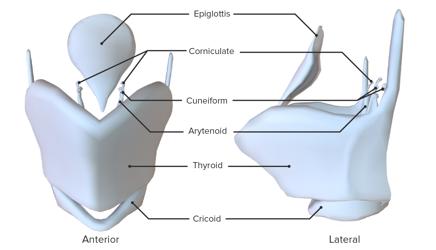
Anterior and right lateral views of the larynx

Anterior (top) and right lateral (bottom) views of the larynx, displaying its anatomical landmarks

Image: “Larynx” by OpenStax. License: CC BY 4.0

Joints of the larynx: 

  • Cricothyroid: allows forward and down tilting movement of the thyroid Thyroid The thyroid gland is one of the largest endocrine glands in the human body. The thyroid gland is a highly vascular, brownish-red gland located in the visceral compartment of the anterior region of the neck. Thyroid Gland: Anatomy cartilage Cartilage Cartilage is a type of connective tissue derived from embryonic mesenchyme that is responsible for structural support, resilience, and the smoothness of physical actions. Perichondrium (connective tissue membrane surrounding cartilage) compensates for the absence of vasculature in cartilage by providing nutrition and support. Cartilage: Histology between the inferior horns of the thyroid Thyroid The thyroid gland is one of the largest endocrine glands in the human body. The thyroid gland is a highly vascular, brownish-red gland located in the visceral compartment of the anterior region of the neck. Thyroid Gland: Anatomy cartilage Cartilage Cartilage is a type of connective tissue derived from embryonic mesenchyme that is responsible for structural support, resilience, and the smoothness of physical actions. Perichondrium (connective tissue membrane surrounding cartilage) compensates for the absence of vasculature in cartilage by providing nutrition and support. Cartilage: Histology and the cricoid cartilage Cartilage Cartilage is a type of connective tissue derived from embryonic mesenchyme that is responsible for structural support, resilience, and the smoothness of physical actions. Perichondrium (connective tissue membrane surrounding cartilage) compensates for the absence of vasculature in cartilage by providing nutrition and support. Cartilage: Histology 
  • Cricoarytenoid: allows abduction Abduction Examination of the Upper Limbs and adduction Adduction Examination of the Upper Limbs of the vocal ligaments by means of the cricoid and arytenoid cartilages

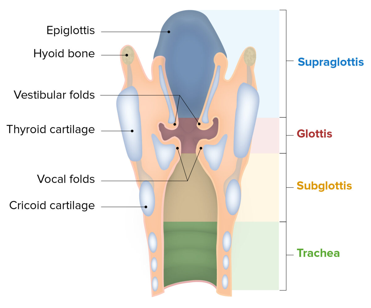
Regions of the larynx:

  • Supraglottis: also called the vestibule Vestibule An oval, bony chamber of the inner ear, part of the bony labyrinth. It is continuous with bony cochlea anteriorly, and semicircular canals posteriorly. The vestibule contains two communicating sacs (utricle and saccule) of the balancing apparatus. The oval window on its lateral wall is occupied by the base of the stapes of the middle ear. Ear: Anatomy, extends from the entrance of the larynx to the vestibular folds
  • Glottis: extends from the vestibular folds to the vocal folds
  • Subglottis: extends from the vocal folds to the lower margin of the cricoid cartilage Cartilage Cartilage is a type of connective tissue derived from embryonic mesenchyme that is responsible for structural support, resilience, and the smoothness of physical actions. Perichondrium (connective tissue membrane surrounding cartilage) compensates for the absence of vasculature in cartilage by providing nutrition and support. Cartilage: Histology (beginning of the trachea Trachea The trachea is a tubular structure that forms part of the lower respiratory tract. The trachea is continuous superiorly with the larynx and inferiorly becomes the bronchial tree within the lungs. The trachea consists of a support frame of semicircular, or C-shaped, rings made out of hyaline cartilage and reinforced by collagenous connective tissue. Trachea: Anatomy)
Components and regions of the larynx

Components and regions of the larynx

Image by Lecturio.
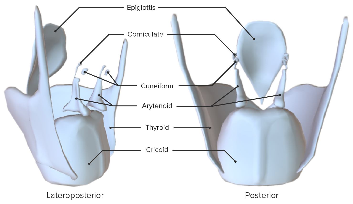
Cartilages

Unpaired cartilages

  • Epiglottis:
    • Elastic Elastic Connective Tissue: Histology cartilage Cartilage Cartilage is a type of connective tissue derived from embryonic mesenchyme that is responsible for structural support, resilience, and the smoothness of physical actions. Perichondrium (connective tissue membrane surrounding cartilage) compensates for the absence of vasculature in cartilage by providing nutrition and support. Cartilage: Histology
    • Leaf-shaped body
    • Entirely covered by mucosa
    • Projects superiorly and posteriorly into the pharynx Pharynx The pharynx is a component of the digestive system that lies posterior to the nasal cavity, oral cavity, and larynx. The pharynx can be divided into the oropharynx, nasopharynx, and laryngopharynx. Pharyngeal muscles play an integral role in vital processes such as breathing, swallowing, and speaking. Pharynx: Anatomy and functions to close the larynx during swallowing Swallowing The act of taking solids and liquids into the gastrointestinal tract through the mouth and throat. Gastrointestinal Motility to prevent food from entering the airway Airway ABCDE Assessment
    • Attaches to the hyoid bone Bone Bone is a compact type of hardened connective tissue composed of bone cells, membranes, an extracellular mineralized matrix, and central bone marrow. The 2 primary types of bone are compact and spongy. Bones: Structure and Types superiorly via the hyoepiglottic ligament 
    • Attaches to the thyroid Thyroid The thyroid gland is one of the largest endocrine glands in the human body. The thyroid gland is a highly vascular, brownish-red gland located in the visceral compartment of the anterior region of the neck. Thyroid Gland: Anatomy cartilage Cartilage Cartilage is a type of connective tissue derived from embryonic mesenchyme that is responsible for structural support, resilience, and the smoothness of physical actions. Perichondrium (connective tissue membrane surrounding cartilage) compensates for the absence of vasculature in cartilage by providing nutrition and support. Cartilage: Histology via the midline thyroepiglottic ligament 
  • Cricoid:
    • Hyaline cartilage Hyaline cartilage A type of cartilage characterized by a homogeneous amorphous matrix containing predominantly type II collagen and ground substance. Hyaline cartilage is found in articular cartilage; costal cartilage; laryngeal cartilages; and the nasal septum. Cartilage: Histology
    • Ring-shaped body, making it the only complete ring of cartilage Cartilage Cartilage is a type of connective tissue derived from embryonic mesenchyme that is responsible for structural support, resilience, and the smoothness of physical actions. Perichondrium (connective tissue membrane surrounding cartilage) compensates for the absence of vasculature in cartilage by providing nutrition and support. Cartilage: Histology in the larynx
    • Attaches to the thyroid Thyroid The thyroid gland is one of the largest endocrine glands in the human body. The thyroid gland is a highly vascular, brownish-red gland located in the visceral compartment of the anterior region of the neck. Thyroid Gland: Anatomy cartilage Cartilage Cartilage is a type of connective tissue derived from embryonic mesenchyme that is responsible for structural support, resilience, and the smoothness of physical actions. Perichondrium (connective tissue membrane surrounding cartilage) compensates for the absence of vasculature in cartilage by providing nutrition and support. Cartilage: Histology via the cricothyroid ligament
    • Attaches to the 1st tracheal ring via the cricotracheal ligament
  • Thyroid Thyroid The thyroid gland is one of the largest endocrine glands in the human body. The thyroid gland is a highly vascular, brownish-red gland located in the visceral compartment of the anterior region of the neck. Thyroid Gland: Anatomy:
    • The largest of the laryngeal cartilages
    • Hyaline cartilage Hyaline cartilage A type of cartilage characterized by a homogeneous amorphous matrix containing predominantly type II collagen and ground substance. Hyaline cartilage is found in articular cartilage; costal cartilage; laryngeal cartilages; and the nasal septum. Cartilage: Histology
    • V-shaped body, with an anterior connection point and no posterior components
    • The anterior connection forms the laryngeal prominence, also known as the Adam’s apple.
    • The posterior aspects of the thyroid Thyroid The thyroid gland is one of the largest endocrine glands in the human body. The thyroid gland is a highly vascular, brownish-red gland located in the visceral compartment of the anterior region of the neck. Thyroid Gland: Anatomy’s 2 lateral laminae project upward and downward, creating the superior (bigger) and inferior horns.
    • Attaches to the hyoid bone Bone Bone is a compact type of hardened connective tissue composed of bone cells, membranes, an extracellular mineralized matrix, and central bone marrow. The 2 primary types of bone are compact and spongy. Bones: Structure and Types superiorly via the thyrohyoid membrane and median and lateral thyrohyoid ligaments
    • Attaches to the cricoid cartilage Cartilage Cartilage is a type of connective tissue derived from embryonic mesenchyme that is responsible for structural support, resilience, and the smoothness of physical actions. Perichondrium (connective tissue membrane surrounding cartilage) compensates for the absence of vasculature in cartilage by providing nutrition and support. Cartilage: Histology inferiorly via the cricothyroid ligament

Paired cartilages

  • Arytenoid:
    • Hyaline cartilage Hyaline cartilage A type of cartilage characterized by a homogeneous amorphous matrix containing predominantly type II collagen and ground substance. Hyaline cartilage is found in articular cartilage; costal cartilage; laryngeal cartilages; and the nasal septum. Cartilage: Histology
    • Pyramid-shaped bodies:
      • Superior apex
      • Anterior vocal process (site of attachment of the vocalis muscle)
      • Lateral muscular process
    • Located superiorly to the posterior portion of the cricoid cartilage Cartilage Cartilage is a type of connective tissue derived from embryonic mesenchyme that is responsible for structural support, resilience, and the smoothness of physical actions. Perichondrium (connective tissue membrane surrounding cartilage) compensates for the absence of vasculature in cartilage by providing nutrition and support. Cartilage: Histology, between the lateral laminae of the thyroid Thyroid The thyroid gland is one of the largest endocrine glands in the human body. The thyroid gland is a highly vascular, brownish-red gland located in the visceral compartment of the anterior region of the neck. Thyroid Gland: Anatomy cartilage Cartilage Cartilage is a type of connective tissue derived from embryonic mesenchyme that is responsible for structural support, resilience, and the smoothness of physical actions. Perichondrium (connective tissue membrane surrounding cartilage) compensates for the absence of vasculature in cartilage by providing nutrition and support. Cartilage: Histology
    • Attaches to the corniculate cartilages at their superior apexes
  • Corniculate:
    • Elastic Elastic Connective Tissue: Histology cartilage Cartilage Cartilage is a type of connective tissue derived from embryonic mesenchyme that is responsible for structural support, resilience, and the smoothness of physical actions. Perichondrium (connective tissue membrane surrounding cartilage) compensates for the absence of vasculature in cartilage by providing nutrition and support. Cartilage: Histology
    • Cone-shaped bodies
    • Located superiorly, posteriorly, and medially to the arytenoids and serves to prolong them
    • Enclosed within the aryepiglottic folds of mucosa
  • Cuneiform:
    • Elastic Elastic Connective Tissue: Histology cartilage Cartilage Cartilage is a type of connective tissue derived from embryonic mesenchyme that is responsible for structural support, resilience, and the smoothness of physical actions. Perichondrium (connective tissue membrane surrounding cartilage) compensates for the absence of vasculature in cartilage by providing nutrition and support. Cartilage: Histology
    • Club-shaped bodies
    • Located superiorly and anteriorly to the corniculate cartilages
    • Enclosed within the aryepiglottic folds of mucosa

Ligaments and Membranes

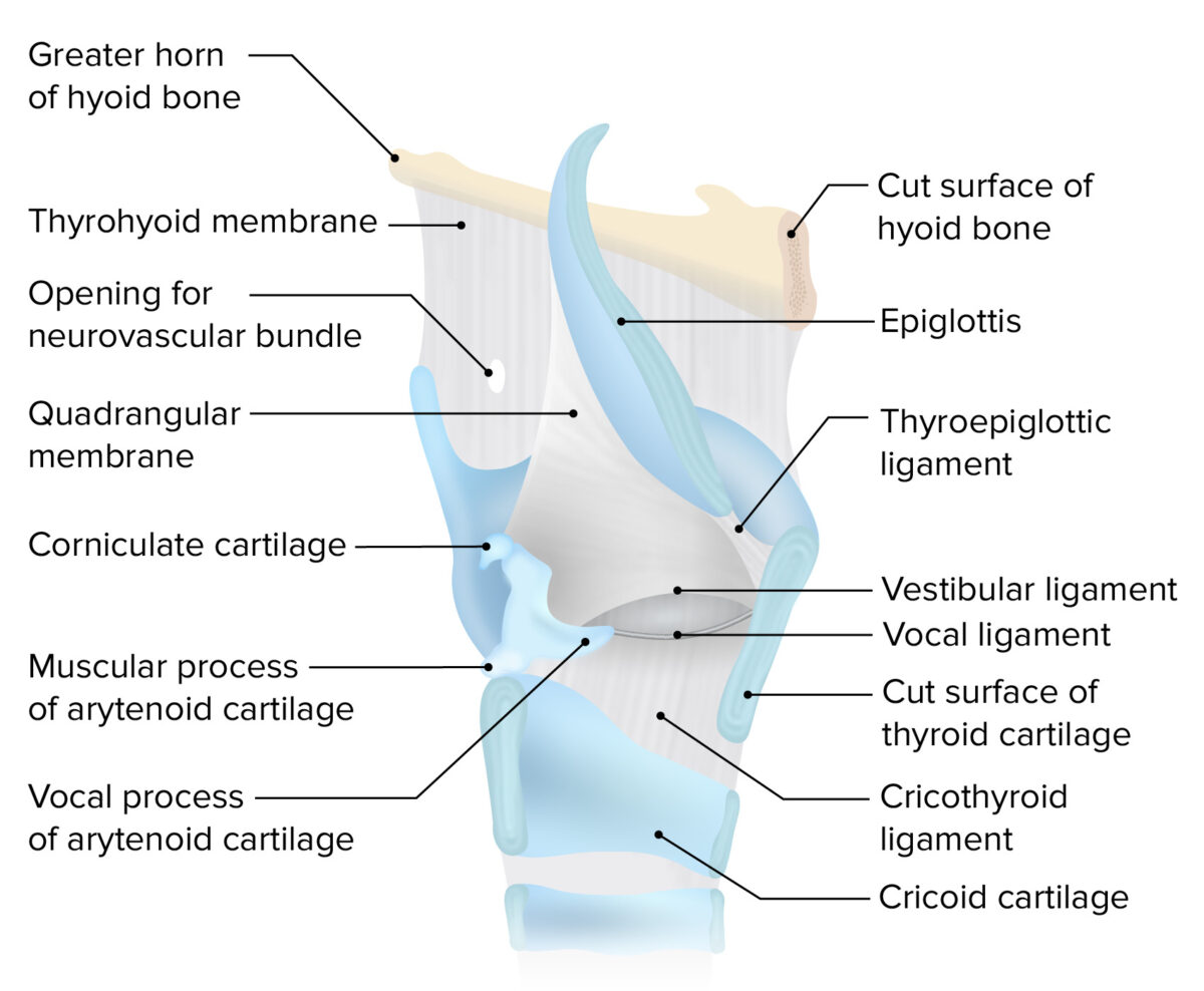
The ligaments and membranes of the larynx are responsible for connecting the cartilages and forming 1 single fibrocartilaginous structure. The membranes also fold over and enclose certain cartilages and membranes to comprise the moving and functional parts of the larynx (e.g., vocal cords).

Extrinsic

Thyrohyoid membrane:

  • Wide, fibrous Fibrous Fibrocystic Change sheet of tissue
  • Connects the hyoid bone Bone Bone is a compact type of hardened connective tissue composed of bone cells, membranes, an extracellular mineralized matrix, and central bone marrow. The 2 primary types of bone are compact and spongy. Bones: Structure and Types to the thyroid Thyroid The thyroid gland is one of the largest endocrine glands in the human body. The thyroid gland is a highly vascular, brownish-red gland located in the visceral compartment of the anterior region of the neck. Thyroid Gland: Anatomy cartilage Cartilage Cartilage is a type of connective tissue derived from embryonic mesenchyme that is responsible for structural support, resilience, and the smoothness of physical actions. Perichondrium (connective tissue membrane surrounding cartilage) compensates for the absence of vasculature in cartilage by providing nutrition and support. Cartilage: Histology
  • Pierced by the internal laryngeal artery and the internal laryngeal nerve
  • Thickened areas comprise the single median thyrohyoid ligament and the two thinner and smaller thyrohyoid ligaments.

Hyoepiglottic ligament:

  • Thin, elastic Elastic Connective Tissue: Histology tissue
  • Connects the anterior surface of the epiglottis to the upper border of the hyoid bone Bone Bone is a compact type of hardened connective tissue composed of bone cells, membranes, an extracellular mineralized matrix, and central bone marrow. The 2 primary types of bone are compact and spongy. Bones: Structure and Types
  • Protects the supraglottic larynx
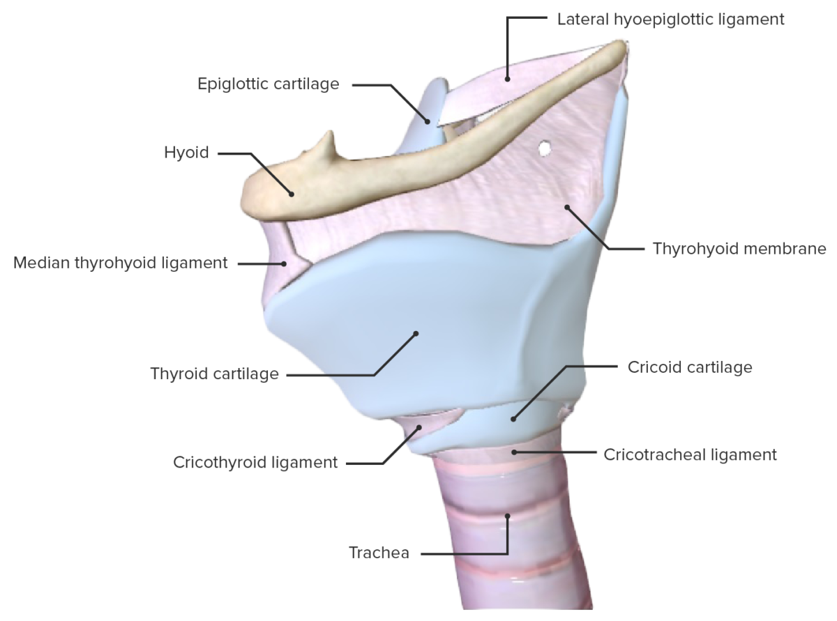
Lateral view of the larynx, featuring the membranes and cartilages

Lateral view of the larynx, featuring the membranes and cartilages

Image by Lecturio.

Intrinsic

Cricothyroid ligament:

  • Also called the cricovocal or cricothyroid membrane
  • Connects the lower border of the thyroid Thyroid The thyroid gland is one of the largest endocrine glands in the human body. The thyroid gland is a highly vascular, brownish-red gland located in the visceral compartment of the anterior region of the neck. Thyroid Gland: Anatomy cartilage Cartilage Cartilage is a type of connective tissue derived from embryonic mesenchyme that is responsible for structural support, resilience, and the smoothness of physical actions. Perichondrium (connective tissue membrane surrounding cartilage) compensates for the absence of vasculature in cartilage by providing nutrition and support. Cartilage: Histology to the upper border of the cricoid cartilage Cartilage Cartilage is a type of connective tissue derived from embryonic mesenchyme that is responsible for structural support, resilience, and the smoothness of physical actions. Perichondrium (connective tissue membrane surrounding cartilage) compensates for the absence of vasculature in cartilage by providing nutrition and support. Cartilage: Histology
  • Composed of the median and lateral cricothyroid ligaments:
    • Median: a thickened area at the anterior midline between the cartilages
    • Lateral: also known as the conus elasticus ( elastic Elastic Connective Tissue: Histology cone); thinner than the median ligament and extend from the superior border of the cricoid to the inferior border of the vocal ligaments, with which they are continuous
  • An incision is made through this structure during a cricothyrotomy Cricothyrotomy ABCDE Assessment to establish an emergency patent airway Airway ABCDE Assessment.

Vocal ligaments:

  • The free borders of the conus elasticus (lateral cricothyroid ligaments)
  • Extend from the vocal processes of the arytenoid cartilages to the angle of the thyroid Thyroid The thyroid gland is one of the largest endocrine glands in the human body. The thyroid gland is a highly vascular, brownish-red gland located in the visceral compartment of the anterior region of the neck. Thyroid Gland: Anatomy cartilage Cartilage Cartilage is a type of connective tissue derived from embryonic mesenchyme that is responsible for structural support, resilience, and the smoothness of physical actions. Perichondrium (connective tissue membrane surrounding cartilage) compensates for the absence of vasculature in cartilage by providing nutrition and support. Cartilage: Histology

Quadrangular membrane:

  • Thin, extends between the lateral borders of the epiglottis and the anterolateral margins of the arytenoid cartilages
  • Vestibular ligament:
    • The thick free lower edge of the quadrangular membrane
    • Enclosed in a fold of mucosa to form the vestibular folds (false vocal cords) that extend from the thyroid Thyroid The thyroid gland is one of the largest endocrine glands in the human body. The thyroid gland is a highly vascular, brownish-red gland located in the visceral compartment of the anterior region of the neck. Thyroid Gland: Anatomy cartilage Cartilage Cartilage is a type of connective tissue derived from embryonic mesenchyme that is responsible for structural support, resilience, and the smoothness of physical actions. Perichondrium (connective tissue membrane surrounding cartilage) compensates for the absence of vasculature in cartilage by providing nutrition and support. Cartilage: Histology to the arytenoid cartilages
  • Aryepiglottic membrane:
    • The free upper edge of the quadrangular membrane
    • Enclosed in a fold of mucosa to form the aryepiglottic folds
    • Reinforced in its posterior portion by the corniculate and cuneiform cartilages, seen as small nodules within the fold
The ligaments and membranes of the larynx

The ligaments and membranes of the larynx

Image by Lecturio.

Muscles

Extrinsic laryngeal muscles

Suprahyoid group:

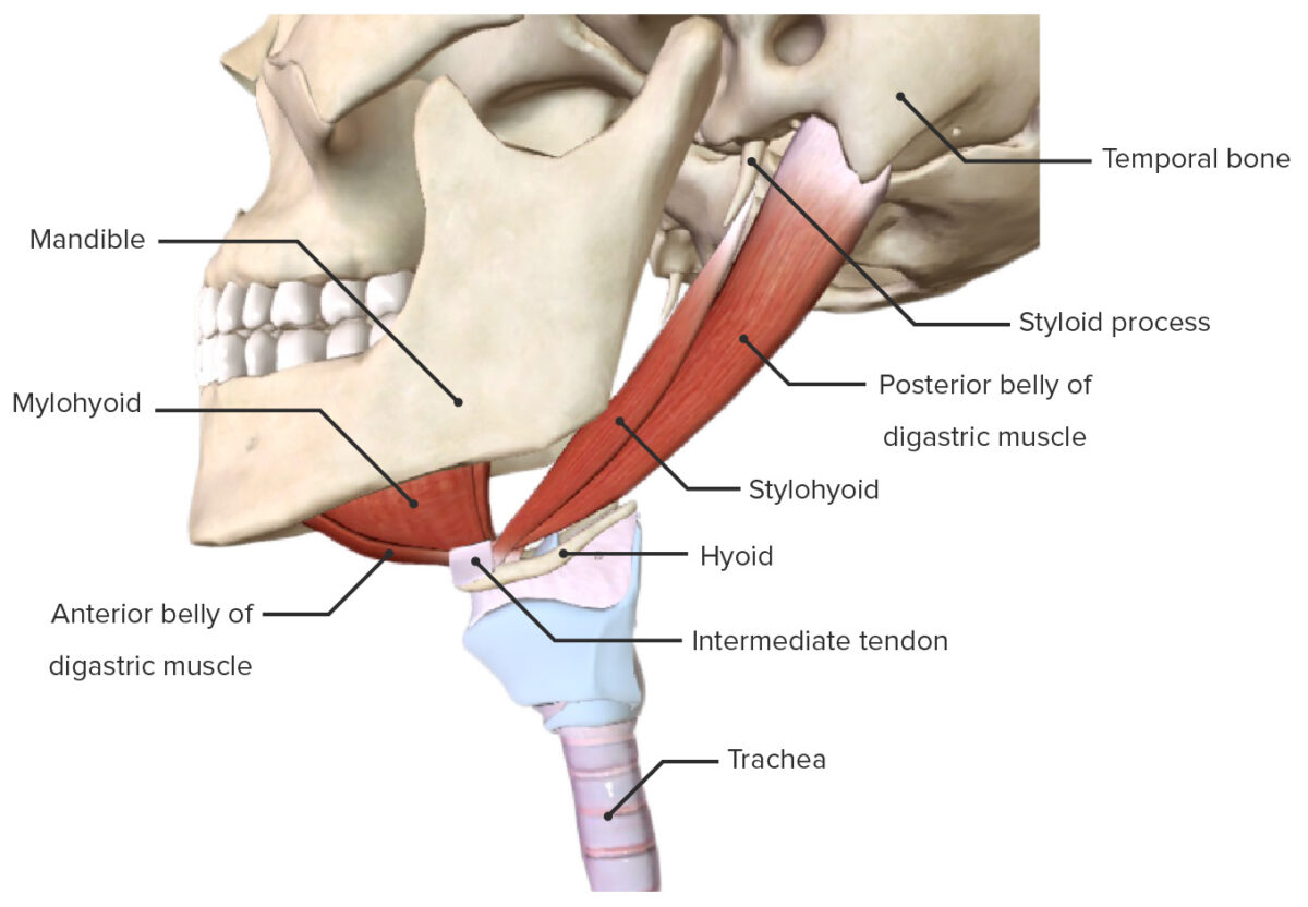
The suprahyoid laryngeal muscles are characterized by their location above the hyoid bone Bone Bone is a compact type of hardened connective tissue composed of bone cells, membranes, an extracellular mineralized matrix, and central bone marrow. The 2 primary types of bone are compact and spongy. Bones: Structure and Types and function of elevating the hyoid bone Bone Bone is a compact type of hardened connective tissue composed of bone cells, membranes, an extracellular mineralized matrix, and central bone marrow. The 2 primary types of bone are compact and spongy. Bones: Structure and Types and larynx during swallowing Swallowing The act of taking solids and liquids into the gastrointestinal tract through the mouth and throat. Gastrointestinal Motility and phonation.

Table: Suprahyoid group
Muscle Origin Insertion Innervation
Stylohyoid Styloid process of the temporal bone Temporal bone Either of a pair of compound bones forming the lateral (left and right) surfaces and base of the skull which contains the organs of hearing. It is a large bone formed by the fusion of parts: the squamous (the flattened anterior-superior part), the tympanic (the curved anterior-inferior part), the mastoid (the irregular posterior portion), and the petrous (the part at the base of the skull). Jaw and Temporomandibular Joint: Anatomy Body of the hyoid bone Bone Bone is a compact type of hardened connective tissue composed of bone cells, membranes, an extracellular mineralized matrix, and central bone marrow. The 2 primary types of bone are compact and spongy. Bones: Structure and Types Facial nerve Facial nerve The 7th cranial nerve. The facial nerve has two parts, the larger motor root which may be called the facial nerve proper, and the smaller intermediate or sensory root. Together they provide efferent innervation to the muscles of facial expression and to the lacrimal and salivary glands, and convey afferent information for taste from the anterior two-thirds of the tongue and for touch from the external ear. The 12 Cranial Nerves: Overview and Functions
Digastric Anterior belly: digastric fossa of the mandible Mandible The largest and strongest bone of the face constituting the lower jaw. It supports the lower teeth. Jaw and Temporomandibular Joint: Anatomy Anterior and posterior bellies: intermediate tendon Anterior belly: mylohyoid nerve, branch of the mandibular nerve Mandibular nerve A branch of the trigeminal (5th cranial) nerve. The mandibular nerve carries motor fibers to the muscles of mastication and sensory fibers to the teeth and gingivae, the face in the region of the mandible, and parts of the dura. Jaw and Temporomandibular Joint: Anatomy
Posterior belly: mastoid notch of the temporal bone Temporal bone Either of a pair of compound bones forming the lateral (left and right) surfaces and base of the skull which contains the organs of hearing. It is a large bone formed by the fusion of parts: the squamous (the flattened anterior-superior part), the tympanic (the curved anterior-inferior part), the mastoid (the irregular posterior portion), and the petrous (the part at the base of the skull). Jaw and Temporomandibular Joint: Anatomy Posterior belly: facial nerve Facial nerve The 7th cranial nerve. The facial nerve has two parts, the larger motor root which may be called the facial nerve proper, and the smaller intermediate or sensory root. Together they provide efferent innervation to the muscles of facial expression and to the lacrimal and salivary glands, and convey afferent information for taste from the anterior two-thirds of the tongue and for touch from the external ear. The 12 Cranial Nerves: Overview and Functions
Mylohyoid Mylohyoid line of mandible Mandible The largest and strongest bone of the face constituting the lower jaw. It supports the lower teeth. Jaw and Temporomandibular Joint: Anatomy Body of the hyoid bone Bone Bone is a compact type of hardened connective tissue composed of bone cells, membranes, an extracellular mineralized matrix, and central bone marrow. The 2 primary types of bone are compact and spongy. Bones: Structure and Types and median raphe Raphe Testicles: Anatomy Mylohyoid nerve, branch of the mandibular nerve Mandibular nerve A branch of the trigeminal (5th cranial) nerve. The mandibular nerve carries motor fibers to the muscles of mastication and sensory fibers to the teeth and gingivae, the face in the region of the mandible, and parts of the dura. Jaw and Temporomandibular Joint: Anatomy
Geniohyoid Mental spine Spine The human spine, or vertebral column, is the most important anatomical and functional axis of the human body. It consists of 7 cervical vertebrae, 12 thoracic vertebrae, and 5 lumbar vertebrae and is limited cranially by the skull and caudally by the sacrum. Vertebral Column: Anatomy of the inner surface of the mandible Mandible The largest and strongest bone of the face constituting the lower jaw. It supports the lower teeth. Jaw and Temporomandibular Joint: Anatomy Body of the hyoid bone Bone Bone is a compact type of hardened connective tissue composed of bone cells, membranes, an extracellular mineralized matrix, and central bone marrow. The 2 primary types of bone are compact and spongy. Bones: Structure and Types C1–C2 of the cervical plexus Cervical Plexus A network of nerve fibers originating in the upper four cervical spinal cord segments. The cervical plexus distributes cutaneous nerves to parts of the neck, shoulders, and back of the head. It also distributes motor fibers to muscles of the cervical spinal column, infrahyoid muscles, and the diaphragm. Peripheral Nerve Injuries in the Cervicothoracic Region and the hypoglossal nerve Hypoglossal nerve The 12th cranial nerve. The hypoglossal nerve originates in the hypoglossal nucleus of the medulla and supplies motor innervation to all of the muscles of the tongue except the palatoglossus (which is supplied by the vagus). This nerve also contains proprioceptive afferents from the tongue muscles. Lips and Tongue: Anatomy
The suprahyoid group of the extrinsic laryngeal muscles

The suprahyoid group of the extrinsic laryngeal muscles:
The geniohyoid muscle is not shown.

Image by BioDigital, edited by Lecturio

Infrahyoid group:

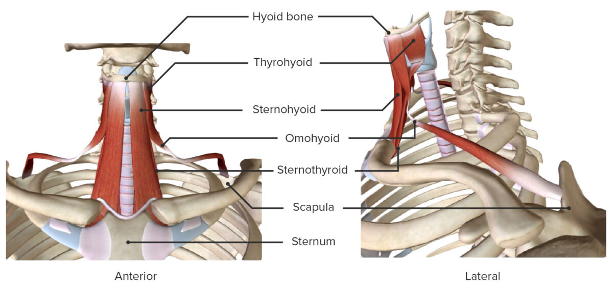
The infrahyoid laryngeal muscles are characterized by their location below the hyoid bone Bone Bone is a compact type of hardened connective tissue composed of bone cells, membranes, an extracellular mineralized matrix, and central bone marrow. The 2 primary types of bone are compact and spongy. Bones: Structure and Types and by their function of depressing the hyoid bone Bone Bone is a compact type of hardened connective tissue composed of bone cells, membranes, an extracellular mineralized matrix, and central bone marrow. The 2 primary types of bone are compact and spongy. Bones: Structure and Types and larynx during swallowing Swallowing The act of taking solids and liquids into the gastrointestinal tract through the mouth and throat. Gastrointestinal Motility and phonation.

Table: Infrahyoid group
Muscle Origin Insertion Innervation
Sternohyoid Dorsal surface of the manubrium Manubrium The upper or most anterior segment of the sternum which articulates with the clavicle and first two pairs of ribs. Chest Wall: Anatomy and the sternoclavicular joint Sternoclavicular Joint Examination of the Upper Limbs Body of the hyoid bone Bone Bone is a compact type of hardened connective tissue composed of bone cells, membranes, an extracellular mineralized matrix, and central bone marrow. The 2 primary types of bone are compact and spongy. Bones: Structure and Types C1–C3 of the cervical plexus Cervical Plexus A network of nerve fibers originating in the upper four cervical spinal cord segments. The cervical plexus distributes cutaneous nerves to parts of the neck, shoulders, and back of the head. It also distributes motor fibers to muscles of the cervical spinal column, infrahyoid muscles, and the diaphragm. Peripheral Nerve Injuries in the Cervicothoracic Region
Omohyoid
  • Superior border of the scapula
  • Central tendon Central tendon Diaphragm: Anatomy: deep cervical fascia Fascia Layers of connective tissue of variable thickness. The superficial fascia is found immediately below the skin; the deep fascia invests muscles, nerves, and other organs. Cellulitis ( clavicle Clavicle A bone on the ventral side of the shoulder girdle, which in humans is commonly called the collar bone. Clavicle Fracture, 1st rib)
Body of the hyoid bone Bone Bone is a compact type of hardened connective tissue composed of bone cells, membranes, an extracellular mineralized matrix, and central bone marrow. The 2 primary types of bone are compact and spongy. Bones: Structure and Types (connected to the carotid sheath)
Sternothyroid Dorsal surface of the manubrium Manubrium The upper or most anterior segment of the sternum which articulates with the clavicle and first two pairs of ribs. Chest Wall: Anatomy Oblique line of the thyroid Thyroid The thyroid gland is one of the largest endocrine glands in the human body. The thyroid gland is a highly vascular, brownish-red gland located in the visceral compartment of the anterior region of the neck. Thyroid Gland: Anatomy cartilage Cartilage Cartilage is a type of connective tissue derived from embryonic mesenchyme that is responsible for structural support, resilience, and the smoothness of physical actions. Perichondrium (connective tissue membrane surrounding cartilage) compensates for the absence of vasculature in cartilage by providing nutrition and support. Cartilage: Histology
Thyrohyoid (continuation of sternothyroid) Oblique line of the thyroid Thyroid The thyroid gland is one of the largest endocrine glands in the human body. The thyroid gland is a highly vascular, brownish-red gland located in the visceral compartment of the anterior region of the neck. Thyroid Gland: Anatomy cartilage Cartilage Cartilage is a type of connective tissue derived from embryonic mesenchyme that is responsible for structural support, resilience, and the smoothness of physical actions. Perichondrium (connective tissue membrane surrounding cartilage) compensates for the absence of vasculature in cartilage by providing nutrition and support. Cartilage: Histology Body and greater horns of the hyoid bone Bone Bone is a compact type of hardened connective tissue composed of bone cells, membranes, an extracellular mineralized matrix, and central bone marrow. The 2 primary types of bone are compact and spongy. Bones: Structure and Types Hypoglossal nerve Hypoglossal nerve The 12th cranial nerve. The hypoglossal nerve originates in the hypoglossal nucleus of the medulla and supplies motor innervation to all of the muscles of the tongue except the palatoglossus (which is supplied by the vagus). This nerve also contains proprioceptive afferents from the tongue muscles. Lips and Tongue: Anatomy (cranial nerve XII), via the anterior rami of C1
Anterior and lateral views of the infrahyoid group of the extrinsic laryngeal muscles

Anterior and lateral views of the infrahyoid group of the extrinsic laryngeal muscles

Image by BioDigital, edited by Lecturio

Intrinsic laryngeal muscles

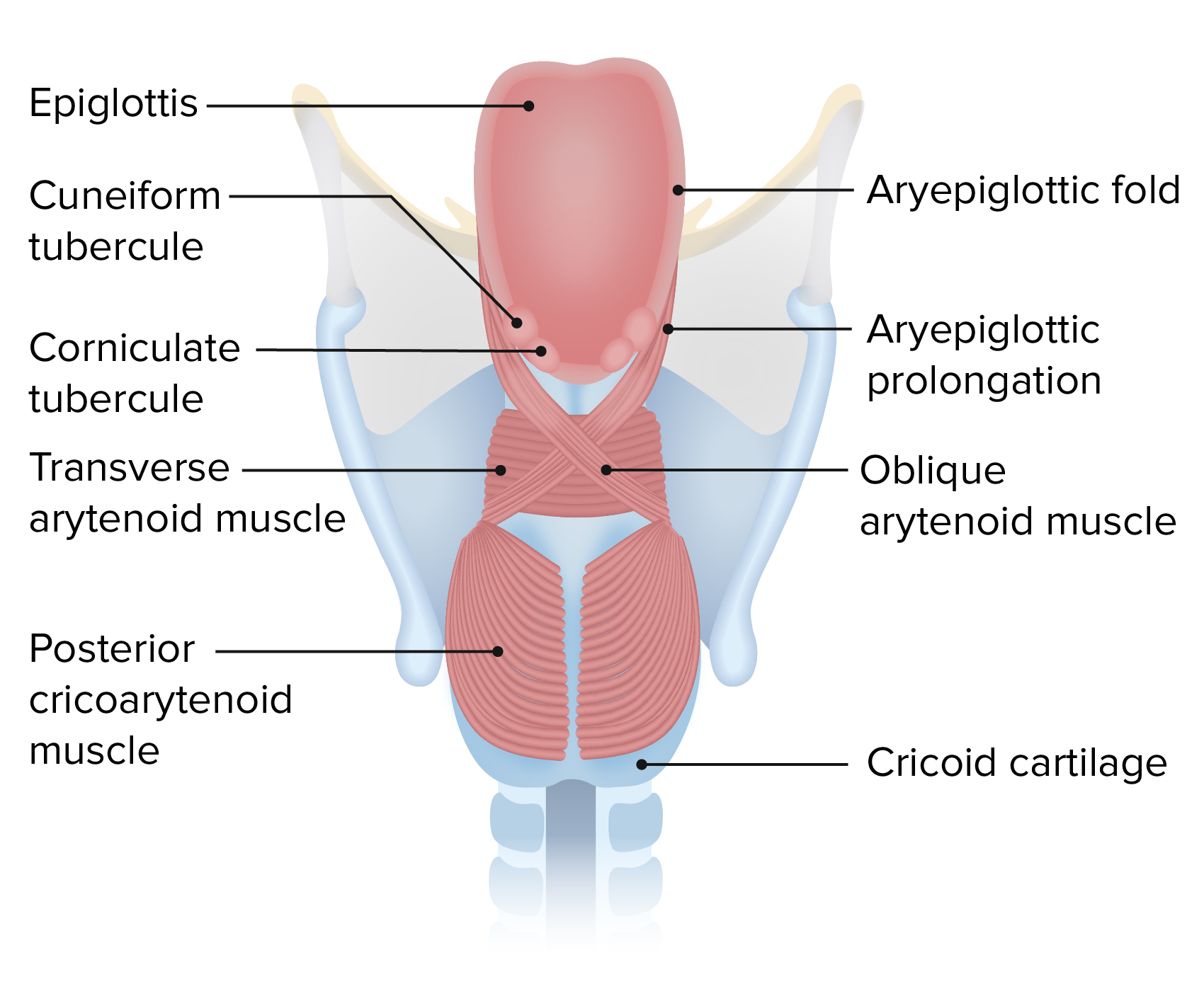
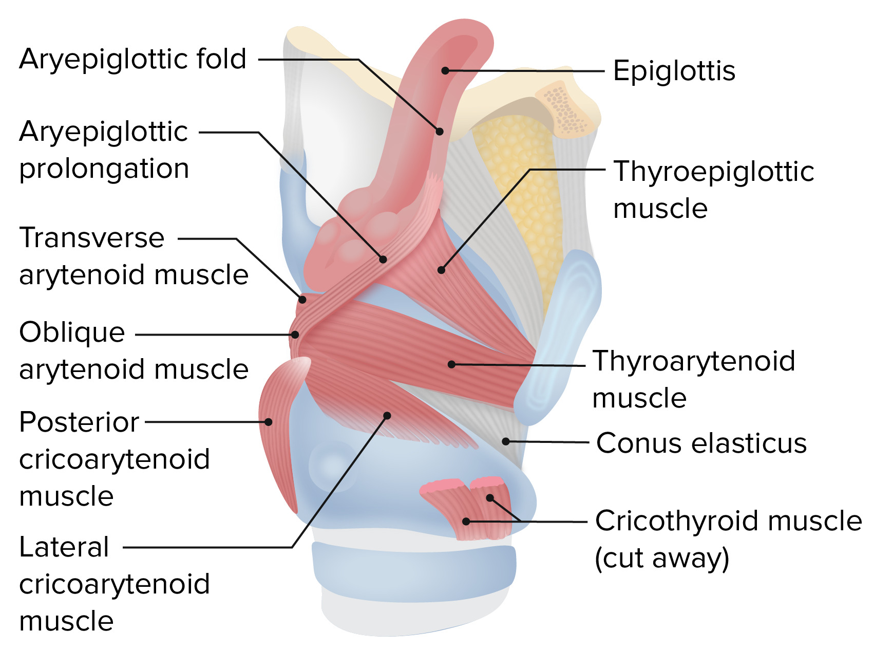
The intrinsic laryngeal muscles serve to produce phonation by modifying the length and tension of the vocal cords and the size of the rima glottidis (the opening between the vocal cords).

Table: Intrinsic laryngeal muscles
Muscle Origin Insertion Innervation
Cricothyroid Anterolateral portion of the cricoid
  • Oblique portion: inferior horn of the thyroid Thyroid The thyroid gland is one of the largest endocrine glands in the human body. The thyroid gland is a highly vascular, brownish-red gland located in the visceral compartment of the anterior region of the neck. Thyroid Gland: Anatomy cartilage Cartilage Cartilage is a type of connective tissue derived from embryonic mesenchyme that is responsible for structural support, resilience, and the smoothness of physical actions. Perichondrium (connective tissue membrane surrounding cartilage) compensates for the absence of vasculature in cartilage by providing nutrition and support. Cartilage: Histology
  • Straight portion: inferior margin of the thyroid Thyroid The thyroid gland is one of the largest endocrine glands in the human body. The thyroid gland is a highly vascular, brownish-red gland located in the visceral compartment of the anterior region of the neck. Thyroid Gland: Anatomy cartilage Cartilage Cartilage is a type of connective tissue derived from embryonic mesenchyme that is responsible for structural support, resilience, and the smoothness of physical actions. Perichondrium (connective tissue membrane surrounding cartilage) compensates for the absence of vasculature in cartilage by providing nutrition and support. Cartilage: Histology
External laryngeal nerve, branch of the superior laryngeal nerve
Thyroarytenoid Angle of the thyroid Thyroid The thyroid gland is one of the largest endocrine glands in the human body. The thyroid gland is a highly vascular, brownish-red gland located in the visceral compartment of the anterior region of the neck. Thyroid Gland: Anatomy cartilage Cartilage Cartilage is a type of connective tissue derived from embryonic mesenchyme that is responsible for structural support, resilience, and the smoothness of physical actions. Perichondrium (connective tissue membrane surrounding cartilage) compensates for the absence of vasculature in cartilage by providing nutrition and support. Cartilage: Histology and cricothyroid ligament Anterolateral surface of the arytenoids Inferior laryngeal nerve, branch of the recurrent laryngeal nerve
Posterior cricoarytenoid Posterior surface of the cricoid Muscular process of the arytenoids
Lateral cricoarytenoid Arch of the cricoid
Transverse arytenoid Lateral border and muscular process of the arytenoids Lateral border and muscular process of the opposite arytenoid
Oblique arytenoid Muscular process of the arytenoids Apex of the opposite arytenoid (with a prolongation to the aryepiglottic folds) Recurrent laryngeal nerve
Vocalis Lateral portions of the vocal processes of the arytenoids Anterior portion of the ipsilateral vocal ligament
  • Recurrent laryngeal nerve
  • External laryngeal nerve

Neurovasculature

Blood supply

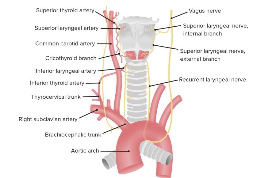
  • Superior laryngeal artery:
    • Branch of the superior thyroid artery Superior thyroid artery Thyroid Gland: Anatomy
    • Originates near the upper margin of the thyroid Thyroid The thyroid gland is one of the largest endocrine glands in the human body. The thyroid gland is a highly vascular, brownish-red gland located in the visceral compartment of the anterior region of the neck. Thyroid Gland: Anatomy cartilage Cartilage Cartilage is a type of connective tissue derived from embryonic mesenchyme that is responsible for structural support, resilience, and the smoothness of physical actions. Perichondrium (connective tissue membrane surrounding cartilage) compensates for the absence of vasculature in cartilage by providing nutrition and support. Cartilage: Histology
    • Accompanies the internal branch of the superior laryngeal nerve
    • Supplies the internal surface of the larynx
  • Cricothyroid artery:
  • Inferior laryngeal artery:

Venous drainage

  • Superior laryngeal vein:
    • Joins the superior thyroid Thyroid The thyroid gland is one of the largest endocrine glands in the human body. The thyroid gland is a highly vascular, brownish-red gland located in the visceral compartment of the anterior region of the neck. Thyroid Gland: Anatomy vein
    • Drains into the superior vena cava Superior vena cava The venous trunk which returns blood from the head, neck, upper extremities and chest. Mediastinum and Great Vessels: Anatomy
  • Inferior laryngeal vein:
    • Joins the inferior thyroid Thyroid The thyroid gland is one of the largest endocrine glands in the human body. The thyroid gland is a highly vascular, brownish-red gland located in the visceral compartment of the anterior region of the neck. Thyroid Gland: Anatomy vein
    • Drains into the brachiocephalic vein
Larynx blood supply and innervation

Larynx blood supply and innervation

Image by Lecturio.

Innervation

The innervation of the larynx is supplied by 2 branches of the vagus nerve Vagus nerve The 10th cranial nerve. The vagus is a mixed nerve which contains somatic afferents (from skin in back of the ear and the external auditory meatus), visceral afferents (from the pharynx, larynx, thorax, and abdomen), parasympathetic efferents (to the thorax and abdomen), and efferents to striated muscle (of the larynx and pharynx). Pharynx: Anatomy: the superior and inferior laryngeal nerves.

Superior laryngeal nerve:

  • Originates from the inferior vagal ganglion
  • Divides into 2 branches:
    • Internal laryngeal nerve:
      • Sensory Sensory Neurons which conduct nerve impulses to the central nervous system. Nervous System: Histology and autonomic
      • Larger of the 2 branches
      • Supplies sensory Sensory Neurons which conduct nerve impulses to the central nervous system. Nervous System: Histology fibers to the mucous membranes in the laryngeal vestibule Vestibule An oval, bony chamber of the inner ear, part of the bony labyrinth. It is continuous with bony cochlea anteriorly, and semicircular canals posteriorly. The vestibule contains two communicating sacs (utricle and saccule) of the balancing apparatus. The oval window on its lateral wall is occupied by the base of the stapes of the middle ear. Ear: Anatomy and middle laryngeal cavity and to the superior surface of vocal folds
    • External laryngeal nerve:
      • Motor Motor Neurons which send impulses peripherally to activate muscles or secretory cells. Nervous System: Histology
      • Smaller of the 2 branches
      • Innervates the pharyngeal constrictor and cricothyroid muscle

Inferior laryngeal nerve:

  • Constitutes the main motor Motor Neurons which send impulses peripherally to activate muscles or secretory cells. Nervous System: Histology nerve of the larynx
  • Continuation of the recurrent laryngeal nerve (branch of the vagus)
    • The left recurrent laryngeal nerve originates in the thorax.
    • The right recurrent laryngeal nerve originates in the root of neck Neck The part of a human or animal body connecting the head to the rest of the body. Peritonsillar Abscess.
  • Accompanies the inferior laryngeal artery
  • Divides into anterior and posterior branches
    • Anterior branch supplies the following muscles:
      • Lateral cricoarytenoid 
      • Thyroarytenoid
      • Vocalis
    • Posterior branch supplies the following muscles:
      • Posterior cricoarytenoid
      • Transverse and oblique arytenoids

Mnemonics:

  • “The larynx is supplied by the Xth (10th) cranial nerve.”
  • SCAR Scar Dermatologic Examination:
    • Superior laryngeal nerve innervates the Cricothyroid muscle.
    • All other muscles are innervated by the Recurrent laryngeal nerve.

Microscopic Anatomy

  • Larynx: mucous membrane Mucous membrane An epithelium with mucus-secreting cells, such as goblet cells. It forms the lining of many body cavities, such as the digestive tract, the respiratory tract, and the reproductive tract. Mucosa, rich in blood and lymph vessels, comprises an inner epithelium, a middle layer (lamina propria) of loose connective tissue, and an outer layer (muscularis mucosae) of smooth muscle cells that separates the mucosa from submucosa. Barrett Esophagus containing ciliated columnar epithelium Epithelium The epithelium is a complex of specialized cellular organizations arranged into sheets and lining cavities and covering the surfaces of the body. The cells exhibit polarity, having an apical and a basal pole. Structures important for the epithelial integrity and function involve the basement membrane, the semipermeable sheet on which the cells rest, and interdigitations, as well as cellular junctions. Surface Epithelium: Histology (respiratory epithelium Epithelium The epithelium is a complex of specialized cellular organizations arranged into sheets and lining cavities and covering the surfaces of the body. The cells exhibit polarity, having an apical and a basal pole. Structures important for the epithelial integrity and function involve the basement membrane, the semipermeable sheet on which the cells rest, and interdigitations, as well as cellular junctions. Surface Epithelium: Histology)
  • Folds:
    • Vestibular cords ( plicae Plicae Small Intestine: Anatomy vestibulares):
      • Stratified nonkeratinizing squamous epithelium Epithelium The epithelium is a complex of specialized cellular organizations arranged into sheets and lining cavities and covering the surfaces of the body. The cells exhibit polarity, having an apical and a basal pole. Structures important for the epithelial integrity and function involve the basement membrane, the semipermeable sheet on which the cells rest, and interdigitations, as well as cellular junctions. Surface Epithelium: Histology
      • Seromucous glands
    • Vocal cords ( plicae Plicae Small Intestine: Anatomy vocales):
      • Stratified nonkeratinizing squamous 
      • No glands
      • Vocal ligament
      • Vocal muscle

Functions

Table: Functions of the larynx
Muscle Function
Cricothyroid
  • Forward and downward rotation Rotation Motion of an object in which either one or more points on a line are fixed. It is also the motion of a particle about a fixed point. X-rays of the thyroid Thyroid The thyroid gland is one of the largest endocrine glands in the human body. The thyroid gland is a highly vascular, brownish-red gland located in the visceral compartment of the anterior region of the neck. Thyroid Gland: Anatomy cartilage Cartilage Cartilage is a type of connective tissue derived from embryonic mesenchyme that is responsible for structural support, resilience, and the smoothness of physical actions. Perichondrium (connective tissue membrane surrounding cartilage) compensates for the absence of vasculature in cartilage by providing nutrition and support. Cartilage: Histology
  • Tautens and thins the vocal cords → production of higher-pitched sounds
Posterior cricoarytenoid
Lateral cricoarytenoid
Transverse arytenoid
Oblique arytenoid
Thyroarytenoid Sphincter of vestibule Vestibule An oval, bony chamber of the inner ear, part of the bony labyrinth. It is continuous with bony cochlea anteriorly, and semicircular canals posteriorly. The vestibule contains two communicating sacs (utricle and saccule) of the balancing apparatus. The oval window on its lateral wall is occupied by the base of the stapes of the middle ear. Ear: Anatomy and laryngeal inlet
Vocal muscle Adjusts tension of the vocal folds

Movements of the larynx: 

Depending on the action (e.g., respiration Respiration The act of breathing with the lungs, consisting of inhalation, or the taking into the lungs of the ambient air, and of exhalation, or the expelling of the modified air which contains more carbon dioxide than the air taken in. Nose Anatomy (External & Internal), phonation, etc ETC The electron transport chain (ETC) sends electrons through a series of proteins, which generate an electrochemical proton gradient that produces energy in the form of adenosine triphosphate (ATP). Electron Transport Chain (ETC).), the laryngeal muscles regulate the movements of the vocal cords and determine the size of the rima glottidis, the opening between the vocal cords.

Table: Movements of the larynx
Movements
Respiration Respiration The act of breathing with the lungs, consisting of inhalation, or the taking into the lungs of the ambient air, and of exhalation, or the expelling of the modified air which contains more carbon dioxide than the air taken in. Nose Anatomy (External & Internal) In quiet respiration Respiration The act of breathing with the lungs, consisting of inhalation, or the taking into the lungs of the ambient air, and of exhalation, or the expelling of the modified air which contains more carbon dioxide than the air taken in. Nose Anatomy (External & Internal):
  • Arytenoid cartilages are abducted.
  • Laryngeal inlet is open.
  • Rima glottidis is open and triangle-shaped.
In forced inspiration Inspiration Ventilation: Mechanics of Breathing:
  • Arytenoid cartilages are rotated laterally.
  • Vocal folds are abducted.
  • Rima glottidis is open wider.
Phonation
  • Arytenoid cartilages and vocal folds are adducted.
  • Air is forced through rima glottidis.
Effort closure Rima glottidis is completely closed.
Swallowing Swallowing The act of taking solids and liquids into the gastrointestinal tract through the mouth and throat. Gastrointestinal Motility
  • Laryngeal inlet is narrowed.
  • Rima glottidis is closed.
  • Larynx moves up and forward.
  • Epiglottis swings downward.
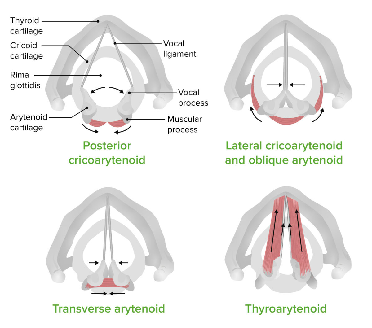
The functions of the intrinsic laryngeal muscles

The functions of the intrinsic laryngeal muscles:
Note the effects on the vocal cords and the rima glottidis.
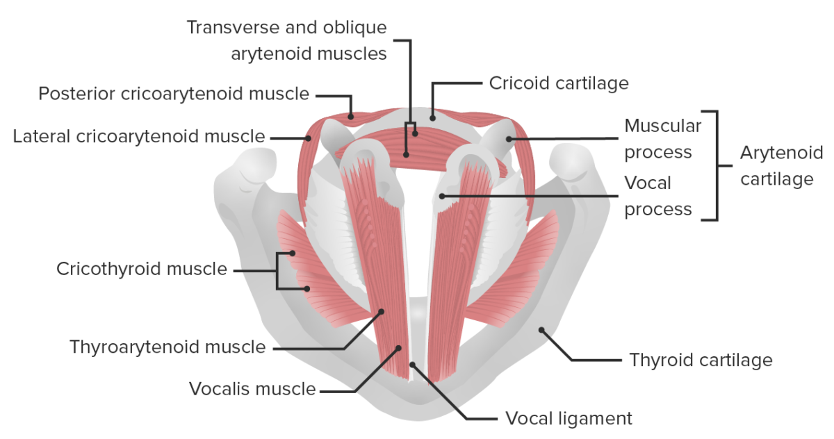
Image by Lecturio.
Head and neck larynx and epiglottis

Intrinsic muscles of the larynx

Image by Lecturio.

Clinical Relevance

Infectious disorders of the larynx

  • Laryngitis Laryngitis Laryngitis is an inflammation of the larynx most commonly due to infection or trauma that can be either acute or chronic. In this condition, the 2 folds of mucous membranes that make up the vocal cords become inflamed and irritated. The inflammation results in a distortion of the voice produced, resulting in a hoarse sound or aphonia. Laryngitis: acute inflammation Acute Inflammation Inflammation of the larynx; can be infectious or noninfectious: Laryngitis Laryngitis Laryngitis is an inflammation of the larynx most commonly due to infection or trauma that can be either acute or chronic. In this condition, the 2 folds of mucous membranes that make up the vocal cords become inflamed and irritated. The inflammation results in a distortion of the voice produced, resulting in a hoarse sound or aphonia. Laryngitis results in fever Fever Fever is defined as a measured body temperature of at least 38°C (100.4°F). Fever is caused by circulating endogenous and/or exogenous pyrogens that increase levels of prostaglandin E2 in the hypothalamus. Fever is commonly associated with chills, rigors, sweating, and flushing of the skin. Fever, hoarseness Hoarseness An unnaturally deep or rough quality of voice. Parapharyngeal Abscess, pain Pain An unpleasant sensation induced by noxious stimuli which are detected by nerve endings of nociceptive neurons. Pain: Types and Pathways, or an irritating cough that worsens at night, along with a dry throat Throat The pharynx is a component of the digestive system that lies posterior to the nasal cavity, oral cavity, and larynx. The pharynx can be divided into the oropharynx, nasopharynx, and laryngopharynx. Pharyngeal muscles play an integral role in vital processes such as breathing, swallowing, and speaking. Pharynx: Anatomy and malaise Malaise Tick-borne Encephalitis Virus. Runny nose Nose The nose is the human body’s primary organ of smell and functions as part of the upper respiratory system. The nose may be best known for inhaling oxygen and exhaling carbon dioxide, but it also contributes to other important functions, such as tasting. The anatomy of the nose can be divided into the external nose and the nasal cavity. Nose Anatomy (External & Internal) and headache Headache The symptom of pain in the cranial region. It may be an isolated benign occurrence or manifestation of a wide variety of headache disorders. Brain Abscess may be associated symptoms.
  • Epiglottitis Epiglottitis Epiglottitis (or “supraglottitis”) is an inflammation of the epiglottis and adjacent supraglottic structures. The majority of cases are caused by bacterial infection. Symptoms are rapid in onset and severe. Epiglottitis: acute inflammation Acute Inflammation Inflammation of the epiglottis and surrounding structures in the supraglottic space: Epiglottitis Epiglottitis Epiglottitis (or “supraglottitis”) is an inflammation of the epiglottis and adjacent supraglottic structures. The majority of cases are caused by bacterial infection. Symptoms are rapid in onset and severe. Epiglottitis is more life-threatening and sudden in onset than laryngitis Laryngitis Laryngitis is an inflammation of the larynx most commonly due to infection or trauma that can be either acute or chronic. In this condition, the 2 folds of mucous membranes that make up the vocal cords become inflamed and irritated. The inflammation results in a distortion of the voice produced, resulting in a hoarse sound or aphonia. Laryngitis. Haemophilus influenzae Haemophilus Influenzae A species of Haemophilus found on the mucous membranes of humans and a variety of animals. The species is further divided into biotypes I through viii. Haemophilus is the culprit in children. Patients Patients Individuals participating in the health care system for the purpose of receiving therapeutic, diagnostic, or preventive procedures. Clinician–Patient Relationship present with dysphagia Dysphagia Dysphagia is the subjective sensation of difficulty swallowing. Symptoms can range from a complete inability to swallow, to the sensation of solids or liquids becoming “stuck.” Dysphagia is classified as either oropharyngeal or esophageal, with esophageal dysphagia having 2 sub-types: functional and mechanical. Dysphagia and sore throat Sore throat Pharyngitis is an inflammation of the back of the throat (pharynx). Pharyngitis is usually caused by an upper respiratory tract infection, which is viral in most cases. It typically results in a sore throat and fever. Other symptoms may include a runny nose, cough, headache, and hoarseness. Pharyngitis, along with dyspnea Dyspnea Dyspnea is the subjective sensation of breathing discomfort. Dyspnea is a normal manifestation of heavy physical or psychological exertion, but also may be caused by underlying conditions (both pulmonary and extrapulmonary). Dyspnea with or without stridor Stridor Laryngomalacia and Tracheomalacia.
  • Diphtheria Diphtheria Diphtheria is an infectious disease caused by Corynebacterium diphtheriae that most often results in respiratory disease with membranous inflammation of the pharynx, sore throat, fever, swollen glands, and weakness. The hallmark sign is a sheet of thick, gray material covering the back of the throat. Diphtheria: serious bacterial infection caused by Corynebacterium diphtheriae Corynebacterium diphtheriae Diphtheria is an infectious disease caused by corynebacterium diphtheriae that most often results in respiratory disease with membranous inflammation of the pharynx, sore throat, fever, swollen glands, and weakness. The hallmark sign is a sheet of thick, gray material covering the back of the throat. Diphtheria: Diphtheria Diphtheria Diphtheria is an infectious disease caused by Corynebacterium diphtheriae that most often results in respiratory disease with membranous inflammation of the pharynx, sore throat, fever, swollen glands, and weakness. The hallmark sign is a sheet of thick, gray material covering the back of the throat. Diphtheria affects the mucous membranes in the nose Nose The nose is the human body’s primary organ of smell and functions as part of the upper respiratory system. The nose may be best known for inhaling oxygen and exhaling carbon dioxide, but it also contributes to other important functions, such as tasting. The anatomy of the nose can be divided into the external nose and the nasal cavity. Nose Anatomy (External & Internal) and throat Throat The pharynx is a component of the digestive system that lies posterior to the nasal cavity, oral cavity, and larynx. The pharynx can be divided into the oropharynx, nasopharynx, and laryngopharynx. Pharyngeal muscles play an integral role in vital processes such as breathing, swallowing, and speaking. Pharynx: Anatomy, resulting in sore throat Sore throat Pharyngitis is an inflammation of the back of the throat (pharynx). Pharyngitis is usually caused by an upper respiratory tract infection, which is viral in most cases. It typically results in a sore throat and fever. Other symptoms may include a runny nose, cough, headache, and hoarseness. Pharyngitis, fever Fever Fever is defined as a measured body temperature of at least 38°C (100.4°F). Fever is caused by circulating endogenous and/or exogenous pyrogens that increase levels of prostaglandin E2 in the hypothalamus. Fever is commonly associated with chills, rigors, sweating, and flushing of the skin. Fever, swollen glands, and weakness. 
  • Croup Croup Croup, also known as laryngotracheobronchitis, is a disease most commonly caused by a viral infection that leads to severe inflammation of the upper airway. It usually presents in children < 5 years of age. Patients develop a hoarse, "seal-like" barking cough and inspiratory stridor. Croup: Croup Croup Croup, also known as laryngotracheobronchitis, is a disease most commonly caused by a viral infection that leads to severe inflammation of the upper airway. It usually presents in children < 5 years of age. Patients develop a hoarse, "seal-like" barking cough and inspiratory stridor. Croup is a disease caused most commonly by a viral infection or rarely by a bacterial infection that results in swelling Swelling Inflammation inside the trachea Trachea The trachea is a tubular structure that forms part of the lower respiratory tract. The trachea is continuous superiorly with the larynx and inferiorly becomes the bronchial tree within the lungs. The trachea consists of a support frame of semicircular, or C-shaped, rings made out of hyaline cartilage and reinforced by collagenous connective tissue. Trachea: Anatomy and interferes with normal breathing.

Congenital disorders of the larynx

Laryngomalacia Laryngomalacia A congenital or acquired condition of underdeveloped or degeneration of cartilage in the larynx. This results in a floppy laryngeal wall making patency difficult to maintain. Laryngomalacia and Tracheomalacia: excessive flaccidity of the supraglottic larynx leads it to be sucked out of position during inspiration Inspiration Ventilation: Mechanics of Breathing, which can produce stridor Stridor Laryngomalacia and Tracheomalacia: This condition manifests at birth and disappears after 2 years of age.

Miscellaneous disorders of the larynx

Foreign body Foreign Body Foreign Body Aspiration aspiration: potentially life-threatening emergency that most commonly occurs in children ages 1–3 years. Presents as sudden onset of coughing, choking, stridor Stridor Laryngomalacia and Tracheomalacia, and dyspnea Dyspnea Dyspnea is the subjective sensation of breathing discomfort. Dyspnea is a normal manifestation of heavy physical or psychological exertion, but also may be caused by underlying conditions (both pulmonary and extrapulmonary). Dyspnea.

References

  1. Drake, R., et al. Gray’s Anatomy for Students E-Book. Elsevier Health Sciences, 2014.
  2. Standring, S. Gray’s Anatomy: The Anatomical Basis of Clinical Practice, 41st ed. Edinburgh: Churchill Livingstone/Elsevier, 2016.

Create your free account or log in to continue reading!

Sign up now and get free access to Lecturio with concept pages, medical videos, and questions for your medical education.

User Reviews

Details