Krabbe Disease

Krabbe disease, also known as globoid cell leukodystrophy or galactosylceramide lipidosis, is a rare autosomal recessive Autosomal recessive Autosomal inheritance, both dominant and recessive, refers to the transmission of genes from the 22 autosomal chromosomes. Autosomal recessive diseases are only expressed when 2 copies of the recessive allele are inherited. Autosomal Recessive and Autosomal Dominant Inheritance lysosomal storage disorder caused by a deficiency of the enzyme galactocerebrosidase. Accumulation of galactocerebroside results in destruction of myelin-producing cells throughout the peripheral and central nervous systems, leading to demyelination Demyelination Multiple Sclerosis and clinical symptoms. The disease is classified into infantile-onset (classic) and late-onset subtypes. Clinical manifestations for classic Krabbe disease include irritability, hypertonia Hypertonia Abnormal increase in skeletal or smooth muscle tone. Skeletal muscle hypertonicity may be associated with pyramidal tract lesions or basal ganglia diseases. Neurological Examination, difficulty feeding, failure to thrive Failure to Thrive Failure to thrive (FTT), or faltering growth, describes suboptimal weight gain and growth in children. The majority of cases are due to inadequate caloric intake; however, genetic, infectious, and oncological etiologies are also common. Failure to Thrive, and rapid neurodegeneration. Death occurs from infection or respiratory failure Respiratory failure Respiratory failure is a syndrome that develops when the respiratory system is unable to maintain oxygenation and/or ventilation. Respiratory failure may be acute or chronic and is classified as hypoxemic, hypercapnic, or a combination of the two. Respiratory Failure. Diagnosis is made by measuring enzyme activity. There is no cure for Krabbe disease. Management is supportive. Stem cell transplantation is an option for some infants.

Last updated: Sep 12, 2022

Editorial responsibility: Stanley Oiseth, Lindsay Jones, Evelin Maza

Overview

Epidemiology

  • Incidence Incidence The number of new cases of a given disease during a given period in a specified population. It also is used for the rate at which new events occur in a defined population. It is differentiated from prevalence, which refers to all cases in the population at a given time. Measures of Disease Frequency
    • 1 case per 250,000 births in the United States
    • 1 case per 100,000 births in Europe
  • Both sexes are affected equally.
  • Panethnic, but higher incidence Incidence The number of new cases of a given disease during a given period in a specified population. It also is used for the rate at which new events occur in a defined population. It is differentiated from prevalence, which refers to all cases in the population at a given time. Measures of Disease Frequency noted in:
    • European ancestry
    • Druze community in Israel

Etiology

  • A lysosomal storage disorder
  • Autosomal recessive inheritance Autosomal recessive inheritance Autosomal Recessive and Autosomal Dominant Inheritance
  • Mutation Mutation Genetic mutations are errors in DNA that can cause protein misfolding and dysfunction. There are various types of mutations, including chromosomal, point, frameshift, and expansion mutations. Types of Mutations of the galactocerebrosidase gene Gene A category of nucleic acid sequences that function as units of heredity and which code for the basic instructions for the development, reproduction, and maintenance of organisms. Basic Terms of Genetics (GALC) on chromosome Chromosome In a prokaryotic cell or in the nucleus of a eukaryotic cell, a structure consisting of or containing DNA which carries the genetic information essential to the cell. Basic Terms of Genetics 14q31
  • Results in a deficiency of the enzyme GALC

Pathophysiology

  • Many aspects of the pathophysiology remain unknown.
  • Accumulation of galactocerebroside is toxic to: 
    • Oligodendrocytes in the CNS
    • Schwann cells in the peripheral nervous system Peripheral nervous system The nervous system outside of the brain and spinal cord. The peripheral nervous system has autonomic and somatic divisions. The autonomic nervous system includes the enteric, parasympathetic, and sympathetic subdivisions. The somatic nervous system includes the cranial and spinal nerves and their ganglia and the peripheral sensory receptors. Nervous System: Anatomy, Structure, and Classification (PNS)
  • Results in demyelination Demyelination Multiple Sclerosis and neurologic manifestations
  • Pathologic findings include: 
    • Globoid cells (hallmark): abnormal, multinucleated macrophages Macrophages The relatively long-lived phagocytic cell of mammalian tissues that are derived from blood monocytes. Main types are peritoneal macrophages; alveolar macrophages; histiocytes; kupffer cells of the liver; and osteoclasts. They may further differentiate within chronic inflammatory lesions to epithelioid cells or may fuse to form foreign body giant cells or langhans giant cells. Innate Immunity: Phagocytes and Antigen Presentation
    • Generalized loss of myelin
    • Absence of oligodendroglia
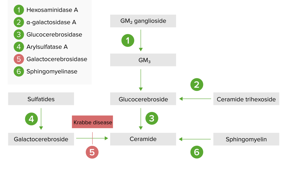
Lysosomal storage pathway diagram

The lysosomal storage pathway:
Krabbe disease results from galactocerebrosidase enzyme deficiency (step 5) and the subsequent buildup of galactocerebroside.

Image by Lecturio.

Clinical Presentation

Time course

  • Infantile (classic):
    • Majority of cases
    • Onset within 6 months
    • Death by approximately 13 months
  • Late-onset subtypes:
    • Onset at ≥ 13 months of age and subdivided into:
      • Late infantile 
      • Juvenile
      • Adult
    • Disease progression varies (usually faster in younger-onset subtypes).

Signs and symptoms

The classic, infantile form progresses through the following stages:
  • Stage 1 Stage 1 Trypanosoma brucei/African trypanosomiasis:
    • Irritability (most common initial symptom)
    • Feeding difficulties
    • Failure to thrive Failure to Thrive Failure to thrive (FTT), or faltering growth, describes suboptimal weight gain and growth in children. The majority of cases are due to inadequate caloric intake; however, genetic, infectious, and oncological etiologies are also common. Failure to Thrive
    • Vomiting Vomiting The forcible expulsion of the contents of the stomach through the mouth. Hypokalemia
    • Gastroesophageal reflux
    • Hypertonia Hypertonia Abnormal increase in skeletal or smooth muscle tone. Skeletal muscle hypertonicity may be associated with pyramidal tract lesions or basal ganglia diseases. Neurological Examination, rigidity Rigidity Continuous involuntary sustained muscle contraction which is often a manifestation of basal ganglia diseases. When an affected muscle is passively stretched, the degree of resistance remains constant regardless of the rate at which the muscle is stretched. This feature helps to distinguish rigidity from muscle spasticity. Megacolon
    • Hyperesthesia—auditory, tactile and visual
    • Peripheral neuropathy Neuropathy Leprosy
    • Hyperpyrexia
  • Stage 2:
    • Hyporeflexia Hyporeflexia Duchenne Muscular Dystrophy or hyperreflexia
    • Optic atrophy Atrophy Decrease in the size of a cell, tissue, organ, or multiple organs, associated with a variety of pathological conditions such as abnormal cellular changes, ischemia, malnutrition, or hormonal changes. Cellular Adaptation and blindness Blindness The inability to see or the loss or absence of perception of visual stimuli. This condition may be the result of eye diseases; optic nerve diseases; optic chiasm diseases; or brain diseases affecting the visual pathways or occipital lobe. Retinopathy of Prematurity
    • Opisthotonus Opisthotonus Tetanus (muscle spasm resulting in a backward arching of the head, neck Neck The part of a human or animal body connecting the head to the rest of the body. Peritonsillar Abscess, and spine Spine The human spine, or vertebral column, is the most important anatomical and functional axis of the human body. It consists of 7 cervical vertebrae, 12 thoracic vertebrae, and 5 lumbar vertebrae and is limited cranially by the skull and caudally by the sacrum. Vertebral Column: Anatomy)
    • Seizures Seizures A seizure is abnormal electrical activity of the neurons in the cerebral cortex that can manifest in numerous ways depending on the region of the brain affected. Seizures consist of a sudden imbalance that occurs between the excitatory and inhibitory signals in cortical neurons, creating a net excitation. The 2 major classes of seizures are focal and generalized. Seizures (do not respond to anticonvulsants)
    • Psychomotor deterioration (rapid and severe)
  • Stage 3:
    • Deafness
    • Decerebrate posturing Decerebrate posturing A condition characterized by abnormal posturing of the limbs that is associated with injury to the brainstem. This may occur as a clinical manifestation or induced experimentally in animals. The extensor reflexes are exaggerated leading to rigid extension of the limbs accompanied by hyperreflexia and opisthotonus. This condition is usually caused by lesions which occur in the region of the brainstem that lies between the red nuclei and the vestibular nuclei. In contrast, decorticate rigidity is characterized by flexion of the elbows and wrists with extension of the legs and feet. The causative lesion for this condition is located above the red nuclei and usually consists of diffuse cerebral damage. Increased Intracranial Pressure (ICP)
    • Severe spasticity Spasticity Spinal Disk Herniation
    • Death is often due to infection or respiratory failure Respiratory failure Respiratory failure is a syndrome that develops when the respiratory system is unable to maintain oxygenation and/or ventilation. Respiratory failure may be acute or chronic and is classified as hypoxemic, hypercapnic, or a combination of the two. Respiratory Failure.

Diagnosis and Management

Diagnosis

  • Prenatal screening Screening Preoperative Care is standard in certain locations.
  • Diagnostic testing:
    • Measurement of GALC activity: 
      • Often tested on blood leukocytes Leukocytes White blood cells. These include granular leukocytes (basophils; eosinophils; and neutrophils) as well as non-granular leukocytes (lymphocytes and monocytes). White Myeloid Cells: Histology or skin Skin The skin, also referred to as the integumentary system, is the largest organ of the body. The skin is primarily composed of the epidermis (outer layer) and dermis (deep layer). The epidermis is primarily composed of keratinocytes that undergo rapid turnover, while the dermis contains dense layers of connective tissue. Skin: Structure and Functions fibroblasts Fibroblasts Connective tissue cells which secrete an extracellular matrix rich in collagen and other macromolecules. Sarcoidosis
      • Low or absent GALC confirms the diagnosis.
    • Molecular genetic testing Genetic Testing Detection of a mutation; genotype; karyotype; or specific alleles associated with genetic traits, heritable diseases, or predisposition to a disease, or that may lead to the disease in descendants. It includes prenatal genetic testing. Myotonic Dystrophies for the genetic mutation Mutation Genetic mutations are errors in DNA that can cause protein misfolding and dysfunction. There are various types of mutations, including chromosomal, point, frameshift, and expansion mutations. Types of Mutations is also available.
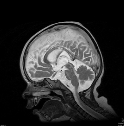
  • Additional testing may show:
Krabbe disease on imaging

A 13-month-old boy with diffuse demyelination (hyperintensity) of the corpus callosum on MRI, characteristic of Krabbe disease

Image: “Krabbe disease on imaging” by Department of Diagnostic Imaging, Institute of Mother and Child, ul. Kasprzaka, 17a, 01-211 Warsaw, Poland. License: CC BY 3.0

Management

  • There is no cure for Krabbe disease.
  • Management is supportive, focused on controlling symptoms.
  • Stem cell transplantation is an option for infants who screen positive and have not yet developed symptoms.

Differential Diagnosis

  • Gaucher disease Gaucher disease Gaucher Disease (GD) is an autosomal recessive lysosomal storage disorder caused by a deficiency of glucocerebrosidase enzyme activity, resulting in accumulation of glucocerebroside in cells and certain organs. The disease is categorized into 3 types with variable clinical presentation. Gaucher Disease ( GD GD Gaucher disease (GD) is an autosomal recessive lysosomal storage disorder caused by a deficiency of glucocerebrosidase enzyme activity, resulting in accumulation of glucocerebroside in cells and certain organs. The disease is categorized into 3 types with variable clinical presentation. Gaucher Disease): lysosomal storage disorder caused by a deficiency of glucocerebrosidase enzyme activity, resulting in accumulation of glucocerebroside. Infantile GD GD Gaucher disease (GD) is an autosomal recessive lysosomal storage disorder caused by a deficiency of glucocerebrosidase enzyme activity, resulting in accumulation of glucocerebroside in cells and certain organs. The disease is categorized into 3 types with variable clinical presentation. Gaucher Disease presents within 6 months of life with progressive neurodegeneration, loss of motor Motor Neurons which send impulses peripherally to activate muscles or secretory cells. Nervous System: Histology skills, hypotonia Hypotonia Duchenne Muscular Dystrophy, hepatosplenomegaly Hepatosplenomegaly Cytomegalovirus, feeding difficulties, and death before age 3 years. Diagnosis is made with measurement of acid beta-glucosidase Beta-glucosidase An exocellulase with specificity for a variety of beta-d-glucoside substrates. It catalyzes the hydrolysis of terminal non-reducing residues in beta-d-glucosides with release of glucose. Gaucher Disease activity and confirmed with genetic analysis. Management is supportive.
  • Fabry disease Fabry disease Fabry disease (FD), also known as Anderson-Fabry disease, is an X-linked recessive lysosomal storage disorder and the 2nd most common of the lysosomal storage disorders. Fabry disease is caused by a deficiency in the alpha-galactosidase enzyme (alpha-Gal A), resulting in the accumulation of the glycosphingolipid globotriaosylceramide (Gb3) in lysosomes. Fabry Disease: lysosomal storage disorder caused by alpha-galactosidase Alpha-galactosidase An enzyme that catalyzes the hydrolysis of terminal, non-reducing alpha-d-galactose residues in alpha-galactosides including galactose oligosaccharides, galactomannans, and galactolipids. Fabry Disease A (GLA) deficiency, resulting in glycophospholipid deposition in the vascular endothelium Endothelium A layer of epithelium that lines the heart, blood vessels (vascular endothelium), lymph vessels (lymphatic endothelium), and the serous cavities of the body. Arteries: Histology and smooth muscle cells. Clinical manifestations include acroparesthesias, purplish skin Skin The skin, also referred to as the integumentary system, is the largest organ of the body. The skin is primarily composed of the epidermis (outer layer) and dermis (deep layer). The epidermis is primarily composed of keratinocytes that undergo rapid turnover, while the dermis contains dense layers of connective tissue. Skin: Structure and Functions lesions, decreased sweating, cerebrovascular and cardiovascular complications, and renal disease. Diagnosis is made with GLA enzyme activity. Management is supportive and can include enzyme replacement Enzyme replacement Therapeutic replacement or supplementation of defective or missing enzymes to alleviate the effects of enzyme deficiency (e.g., glucosylceramidase replacement for gaucher disease). Severe Combined Immunodeficiency (SCID) and chaperone therapies.
  • Niemann-Pick disease Niemann-Pick disease Niemann-Pick disease (NPD) is a rare, inherited, lysosomal storage disorder. The disease is classified on the basis of the genetic mutation. Type A and type B result from mutations in the SMPD-1 gene, resulting in acid sphingomyelinase enzyme deficiency. Type C results from NPC1 or NPC2 gene mutations, which are needed for intracellular transport of lipids. Niemann-Pick Disease type A (NPD-A): lysosomal storage disorder caused by acid sphingomyelinase Acid sphingomyelinase Niemann-Pick Disease enzyme deficiency. The disease is characterized by progressive neurodegeneration starting within a few months of life and resulting in death by age 3. Clinical manifestations include a macular cherry-red spot, difficulty feeding, loss of motor Motor Neurons which send impulses peripherally to activate muscles or secretory cells. Nervous System: Histology skills, hypotonia Hypotonia Duchenne Muscular Dystrophy, and organomegaly. Diagnosis includes measurement of sphingomyelinase enzyme activity and genetic testing Genetic Testing Detection of a mutation; genotype; karyotype; or specific alleles associated with genetic traits, heritable diseases, or predisposition to a disease, or that may lead to the disease in descendants. It includes prenatal genetic testing. Myotonic Dystrophies. Management is supportive. 
  • Tay-Sachs disease Tay-Sachs disease Tay-Sachs disease is an autosomal recessive lysosomal storage disorder caused by genetic mutations in the hexosaminidase A (HEXA) gene, leading to progressive neurodegeneration. Classic symptoms in infants include rapid degeneration of cognitive and neuromuscular abilities, progressive blindness, and a macular cherry-red spot on physical examination. Tay-Sachs Disease: autosomal recessive Autosomal recessive Autosomal inheritance, both dominant and recessive, refers to the transmission of genes from the 22 autosomal chromosomes. Autosomal recessive diseases are only expressed when 2 copies of the recessive allele are inherited. Autosomal Recessive and Autosomal Dominant Inheritance lysosomal storage disorder caused by mutations in the hexosaminidase A Hexosaminidase A A mammalian beta-hexosaminidase isoform that is a heteromeric protein comprised of both hexosaminidase alpha and hexosaminidase beta subunits. Deficiency of hexosaminidase A due to mutations in the gene encoding the hexosaminidase alpha subunit is a case of Tay-sachs disease. Deficiency of hexosaminidase A and hexosaminidase B due to mutations in the gene encoding the hexosaminidase beta subunit is a case of Sandhoff disease. Tay-Sachs Disease (HEXA) gene Gene A category of nucleic acid sequences that function as units of heredity and which code for the basic instructions for the development, reproduction, and maintenance of organisms. Basic Terms of Genetics, leading to progressive neurodegeneration. Clinical manifestations include rapid degeneration of cognitive and neuromuscular abilities, progressive blindness Blindness The inability to see or the loss or absence of perception of visual stimuli. This condition may be the result of eye diseases; optic nerve diseases; optic chiasm diseases; or brain diseases affecting the visual pathways or occipital lobe. Retinopathy of Prematurity, and a macular cherry-red spot on physical examination. Diagnosis is made with enzyme activity testing and molecular genetic analysis. Management is supportive.
  • Sandhoff disease ( SD SD The standard deviation (SD) is a measure of how far each observed value is from the mean in a data set. Measures of Central Tendency and Dispersion): lysosomal storage disorder caused by a deficiency in both HEXA and HEXB. Juvenile SD SD The standard deviation (SD) is a measure of how far each observed value is from the mean in a data set. Measures of Central Tendency and Dispersion is characterized by progressive neurodegeneration starting at 6 months of age. Clinical manifestations include organomegaly, skeletal abnormalities, hyperacusis Hyperacusis An abnormally disproportionate increase in the sensation of loudness in response to auditory stimuli of normal volume. Cochlear disease, vestibulocochlear nerve diseases, facial nerve diseases, stapes surgery, and other disorders may be associated with this condition. Cranial Nerve Palsies, macular cherry-red spot, blindness Blindness The inability to see or the loss or absence of perception of visual stimuli. This condition may be the result of eye diseases; optic nerve diseases; optic chiasm diseases; or brain diseases affecting the visual pathways or occipital lobe. Retinopathy of Prematurity, and seizures Seizures A seizure is abnormal electrical activity of the neurons in the cerebral cortex that can manifest in numerous ways depending on the region of the brain affected. Seizures consist of a sudden imbalance that occurs between the excitatory and inhibitory signals in cortical neurons, creating a net excitation. The 2 major classes of seizures are focal and generalized. Seizures. Diagnosis is made by measurement of HEXA and HEXB, as well as genetic analysis. Management is supportive. 
  • Pompe disease ( glycogen storage disease Glycogen storage disease A group of inherited metabolic disorders involving the enzymes responsible for the synthesis and degradation of glycogen. In some patients, prominent liver involvement is presented. In others, more generalized storage of glycogen occurs, sometimes with prominent cardiac involvement. Benign Liver Tumors II): lysosomal and glycogen storage disorder caused by acid alpha glucosidase (GAA) deficiency. There are 3 types of Pompe disease, with variable Variable Variables represent information about something that can change. The design of the measurement scales, or of the methods for obtaining information, will determine the data gathered and the characteristics of that data. As a result, a variable can be qualitative or quantitative, and may be further classified into subgroups. Types of Variables onset and presentations. Clinical manifestations include failure to thrive Failure to Thrive Failure to thrive (FTT), or faltering growth, describes suboptimal weight gain and growth in children. The majority of cases are due to inadequate caloric intake; however, genetic, infectious, and oncological etiologies are also common. Failure to Thrive, feeding difficulty, hypotonia Hypotonia Duchenne Muscular Dystrophy, progressive muscle weakness, hypertrophic cardiomyopathy Hypertrophic Cardiomyopathy Hypertrophic cardiomyopathy (HCM) is the most commonly inherited cardiomyopathy, which is characterized by an asymmetric increase in thickness (hypertrophy) of the left ventricular wall, diastolic dysfunction, and often left ventricular outflow tract obstruction. Hypertrophic Cardiomyopathy, and respiratory failure Respiratory failure Respiratory failure is a syndrome that develops when the respiratory system is unable to maintain oxygenation and/or ventilation. Respiratory failure may be acute or chronic and is classified as hypoxemic, hypercapnic, or a combination of the two. Respiratory Failure. The diagnosis is made by measuring enzyme activity and molecular genetic analysis. Management includes supportive measures and enzyme replacement Enzyme replacement Therapeutic replacement or supplementation of defective or missing enzymes to alleviate the effects of enzyme deficiency (e.g., glucosylceramidase replacement for gaucher disease). Severe Combined Immunodeficiency (SCID).

References

  1. Escolar, M. (n.d.). Krabbe disease. UptoDate. Retrieved August 2, 2021, from https://www.uptodate.com/contents/krabbe-disease
  2. Brenda, A.V. (2019). Medscape. Retrieved August 7, 2021, from https://emedicine.medscape.com/article/951722-overview
  3. NIH Genetic and Rare Diseases Information Center. (n.d.). Krabbe disease. Retrieved August 8, 2021, from https://rarediseases.info.nih.gov/diseases/6844/krabbe-disease
  4. National Organization for Rare Disorders. (n.d.). Krabbe disease. Retrieved August 7, 2021, from https://rarediseases.org/rare-diseases/leukodystrophy-krabbes/
  5. Jain, M., De Jesus, O. (2021). Krabbe disease. StatPearls. Retrieved August 9, 2021, from https://www.ncbi.nlm.nih.gov/books/NBK562315/

Create your free account or log in to continue reading!

Sign up now and get free access to Lecturio with concept pages, medical videos, and questions for your medical education.

User Reviews

Details