Kidney Function Tests

The kidneys Kidneys The kidneys are a pair of bean-shaped organs located retroperitoneally against the posterior wall of the abdomen on either side of the spine. As part of the urinary tract, the kidneys are responsible for blood filtration and excretion of water-soluble waste in the urine. Kidneys: Anatomy, solid organs located in the lower back, are extremely important in regulating homeostasis Homeostasis The processes whereby the internal environment of an organism tends to remain balanced and stable. Cell Injury and Death through their role in maintaining blood volume, electrolyte balance, acid–base equilibrium Equilibrium Occurs when tumor cells survive the initial elimination attempt These cells are not able to progress, being maintained in a state of dormancy by the adaptive immune system. In this phase, tumor immunogenicity is edited, where T cells keep selectively attacking highly immunogenic tumor cells.This attack leaves other cells with less immunogenicity to potentially develop resistance to the immune response. Cancer Immunotherapy, blood-pressure regulation, and removal of metabolic waste from the blood. Evaluation of renal function and early detection of kidney dysfunction is of primary importance. Tests of renal function are useful in identifying the presence of renal disease, monitoring the response of kidneys Kidneys The kidneys are a pair of bean-shaped organs located retroperitoneally against the posterior wall of the abdomen on either side of the spine. As part of the urinary tract, the kidneys are responsible for blood filtration and excretion of water-soluble waste in the urine. Kidneys: Anatomy to treatment, and determining the progression of renal disease.

Last updated: May 17, 2024

Editorial responsibility: Stanley Oiseth, Lindsay Jones, Evelin Maza

Overview

Anatomy of the nephron Nephron The functional units of the kidney, consisting of the glomerulus and the attached tubule. Kidneys: Anatomy

Nephron Nephron The functional units of the kidney, consisting of the glomerulus and the attached tubule. Kidneys: Anatomy:
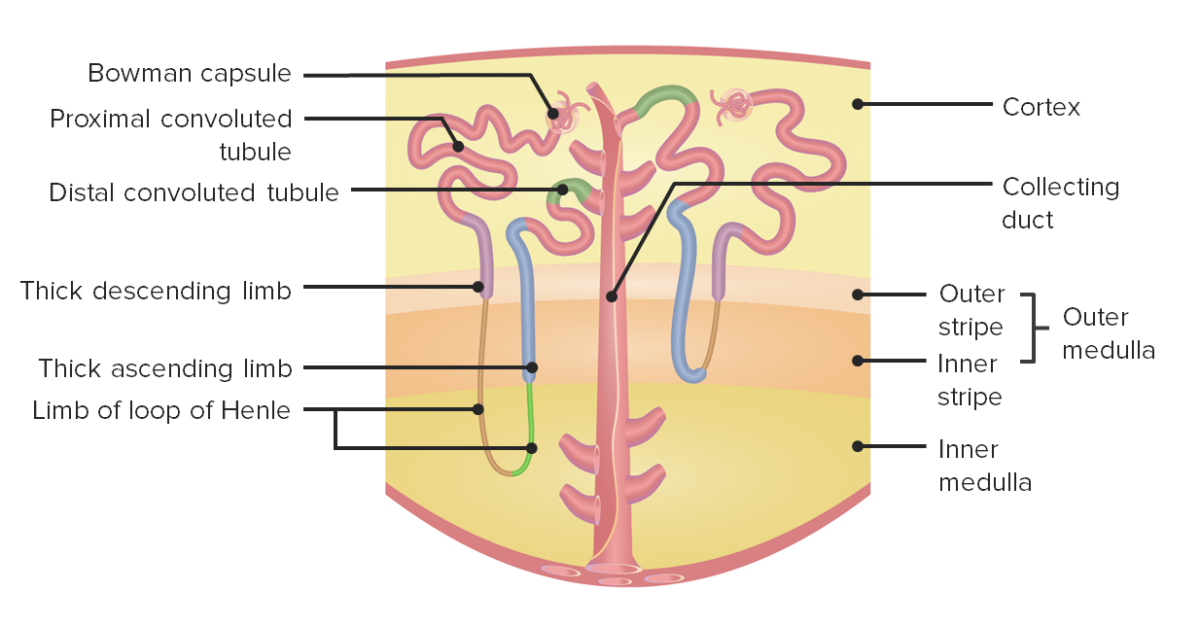
  • The basic structural and functional unit of the kidney
  • Filters and cleanses the blood and produces urine
  • Each kidney contains > 1 million nephrons.

Parts of nephron Nephron The functional units of the kidney, consisting of the glomerulus and the attached tubule. Kidneys: Anatomy:

  • Afferent arteriole Afferent arteriole Glomerular Filtration: renal artery Renal artery A branch of the abdominal aorta which supplies the kidneys, adrenal glands and ureters. Glomerular Filtration arteriole that enters the glomerulus 
  • Efferent arteriole Efferent arteriole Glomerular Filtration: renal artery Renal artery A branch of the abdominal aorta which supplies the kidneys, adrenal glands and ureters. Glomerular Filtration arteriole that exits the glomerulus
  • Glomerulus: capillary tuft that receives its blood supply from an afferent arteriole Afferent arteriole Glomerular Filtration of the renal circulation Circulation The movement of the blood as it is pumped through the cardiovascular system. ABCDE Assessment
  • Bowman capsule Bowman Capsule A double-walled epithelial capsule that is the bulbous closed proximal end of the kidney tubular system. It surrounds the cluster of convoluted capillaries of kidney glomerulus and is continuous with the convoluted proximal kidney tubule. Kidneys: Anatomy: surrounds the glomerulus
  • Renal tubule: coiled tube that converts blood filtrate into urine.
    • Proximal convoluted tubule Proximal convoluted tubule The renal tubule portion that extends from the bowman capsule in the kidney cortex into the kidney medulla. The proximal tubule consists of a convoluted proximal segment in the cortex, and a distal straight segment descending into the medulla where it forms the u-shaped loop of henle. Osmotic Diuretics (PCT): portion close to the glomerulus in the renal cortex
    • Loop of Henle Loop of Henle The U-shaped portion of the renal tubule in the kidney medulla, consisting of a descending limb and an ascending limb. It is situated between the proximal kidney tubule and the distal kidney tubule. Tubular System: forms a loop (with descending and ascending limbs) that goes through the renal medulla
    • Distal convoluted tubule Distal convoluted tubule The portion of renal tubule that begins from the enlarged segment of the ascending limb of the loop of henle. It reenters the kidney cortex and forms the convoluted segments of the distal tubule. Gitelman Syndrome (DCT): portion of the tubule restricted to the renal cortex
Nephron anatomy

Nephron anatomy:
The left side shows a juxtamedullary nephron, while the right side shows a cortical nephron.

Image by Lecturio.

Physiology of urine production

Urine is a waste by-product composed of excess water and metabolic waste filtered out of the bloodstream by nephrons. Urine formation is a stepwise process.  
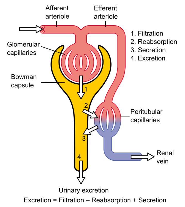
  • Glomerular filtration Glomerular filtration The kidneys are primarily in charge of the maintenance of water and solute homeostasis through the processes of filtration, reabsorption, secretion, and excretion. Glomerular filtration is the process of converting the systemic blood supply into a filtrate, which will ultimately become the urine. Glomerular Filtration:
    • The kidneys Kidneys The kidneys are a pair of bean-shaped organs located retroperitoneally against the posterior wall of the abdomen on either side of the spine. As part of the urinary tract, the kidneys are responsible for blood filtration and excretion of water-soluble waste in the urine. Kidneys: Anatomy filter about 180 L of plasma Plasma The residual portion of blood that is left after removal of blood cells by centrifugation without prior blood coagulation. Transfusion Products per day (125 mL/min).
    • Takes place inside glomerulus
    • Water and nitrogenous waste are filtered out of the blood and form glomerular filtrate.
    • Nonfilterable components exit the glomerulus via the efferent arteriole Efferent arteriole Glomerular Filtration.
  • Tubular reabsorption:
    • Reabsorption of nutrients shed by glomerulus
    • Occurs in the PCT 
    • Can be passive (along gradients) or active (using ATP-generated energy)
    • Water and electrolyte reabsorption hormonally regulated
  • Tubular secretion Secretion Coagulation Studies:
    • Undesirable products like metabolic wastes, urea Urea A compound formed in the liver from ammonia produced by the deamination of amino acids. It is the principal end product of protein catabolism and constitutes about one half of the total urinary solids. Urea Cycle, uric acid Uric acid An oxidation product, via xanthine oxidase, of oxypurines such as xanthine and hypoxanthine. It is the final oxidation product of purine catabolism in humans and primates, whereas in most other mammals urate oxidase further oxidizes it to allantoin. Nephrolithiasis, and certain drugs
    • Most of the tubular secretion Secretion Coagulation Studies happens in the DCT.
The basic anatomy of the nephron and the steps involved in urine formation

The basic anatomy of the nephron and the steps involved in urine formation

Image: “Physiology of Nephron” by Madhero88. License: CC BY 3.0, edited by Lecturio.

Serologic Parameters of Renal Function

Creatinine and estimated glomerular filtration Glomerular filtration The kidneys are primarily in charge of the maintenance of water and solute homeostasis through the processes of filtration, reabsorption, secretion, and excretion. Glomerular filtration is the process of converting the systemic blood supply into a filtrate, which will ultimately become the urine. Glomerular Filtration rate (eGFR)

The most commonly used marker to assess kidney function is the measurement of excreted creatinine, based on which the eGFR can be calculated.

  • Creatinine: by-product of creatine Creatine An amino acid that occurs in vertebrate tissues and in urine. In muscle tissue, creatine generally occurs as phosphocreatine. Creatine is excreted as creatinine in the urine. Acute Kidney Injury phosphate Phosphate Inorganic salts of phosphoric acid. Electrolytes metabolism in muscle:
    • Produced at a constant rate by the body
    • Amount produced per day depends on muscle bulk and meat consumption
    • Cleared from the blood entirely by the kidney
    • Commonly used endogenous marker for assessment of glomerular function: calculated clearance of creatinine → eGFR
  • Mean serum values:
    • Dependent on sex Sex The totality of characteristics of reproductive structure, functions, phenotype, and genotype, differentiating the male from the female organism. Gender Dysphoria and ethnicity 
    • 1.13 mg/dL for men (higher muscle mass Mass Three-dimensional lesion that occupies a space within the breast Imaging of the Breast and creatinine excretion)
    • 0.93 mg/dL for women (lower muscle mass Mass Three-dimensional lesion that occupies a space within the breast Imaging of the Breast and creatinine excretion)
    • Mean values have been shown to be higher in non-Hispanic Black Americans → considered when calculating the eGFR  
  • Creatinine clearance should be corrected for body surface area.
  • Creatinine clearance overestimates GFR by around 10%–20% because of tubular secretion Secretion Coagulation Studies.

Glomerular filtration Glomerular filtration The kidneys are primarily in charge of the maintenance of water and solute homeostasis through the processes of filtration, reabsorption, secretion, and excretion. Glomerular filtration is the process of converting the systemic blood supply into a filtrate, which will ultimately become the urine. Glomerular Filtration rate (GFR)

  • The most exact overall indicator Indicator Methods for assessing flow through a system by injection of a known quantity of an indicator, such as a dye, radionuclide, or chilled liquid, into the system and monitoring its concentration over time at a specific point in the system. Body Fluid Compartments of glomerular function 
  • Rate (in milliliters per minute) at which a substance in plasma Plasma The residual portion of blood that is left after removal of blood cells by centrifugation without prior blood coagulation. Transfusion Products is filtered through the glomeruli
  • Commonly measured by administering inulin Inulin A starch found in the tubers and roots of many plants. Since it is hydrolyzable to fructose, it is classified as a fructosan. It has been used in physiologic investigation for determination of the rate of glomerular function. Glomerular Filtration:
    • Polysaccharide that cannot be broken down and is completely filtered
    • Used as a marker by measuring rate at which it appears in the urine
  • Time-consuming and expensive to measure → performed only in specialized centers

Blood urea Urea A compound formed in the liver from ammonia produced by the deamination of amino acids. It is the principal end product of protein catabolism and constitutes about one half of the total urinary solids. Urea Cycle nitrogen Nitrogen An element with the atomic symbol n, atomic number 7, and atomic weight [14. 00643; 14. 00728]. Nitrogen exists as a diatomic gas and makes up about 78% of the earth’s atmosphere by volume. It is a constituent of proteins and nucleic acids and found in all living cells. Urea Cycle (BUN)

  • Normal metabolic processes (including the urea Urea A compound formed in the liver from ammonia produced by the deamination of amino acids. It is the principal end product of protein catabolism and constitutes about one half of the total urinary solids. Urea Cycle cycle) result in the production of nitrogen-rich ammonia Ammonia A colorless alkaline gas. It is formed in the body during decomposition of organic materials during a large number of metabolically important reactions. Note that the aqueous form of ammonia is referred to as ammonium hydroxide. Acid-Base Balance in the liver Liver The liver is the largest gland in the human body. The liver is found in the superior right quadrant of the abdomen and weighs approximately 1.5 kilograms. Its main functions are detoxification, metabolism, nutrient storage (e.g., iron and vitamins), synthesis of coagulation factors, formation of bile, filtration, and storage of blood. Liver: Anatomy.
  • Ammonia Ammonia A colorless alkaline gas. It is formed in the body during decomposition of organic materials during a large number of metabolically important reactions. Note that the aqueous form of ammonia is referred to as ammonium hydroxide. Acid-Base Balance is further refined to urea Urea A compound formed in the liver from ammonia produced by the deamination of amino acids. It is the principal end product of protein catabolism and constitutes about one half of the total urinary solids. Urea Cycle and excreted primarily (85%) by the kidneys Kidneys The kidneys are a pair of bean-shaped organs located retroperitoneally against the posterior wall of the abdomen on either side of the spine. As part of the urinary tract, the kidneys are responsible for blood filtration and excretion of water-soluble waste in the urine. Kidneys: Anatomy.
  • Measurement of urea Urea A compound formed in the liver from ammonia produced by the deamination of amino acids. It is the principal end product of protein catabolism and constitutes about one half of the total urinary solids. Urea Cycle excretion as a marker of kidney function is less accurate than creatinine but increases earlier in renal disease.
  • Other pathological states may lead to elevated urea Urea A compound formed in the liver from ammonia produced by the deamination of amino acids. It is the principal end product of protein catabolism and constitutes about one half of the total urinary solids. Urea Cycle excretion:

Ratio of BUN:creatinine

Provides more useful information than either tests alone:

  • Distinguish prerenal from renal causes of kidney failure 
  • In prerenal disease, ratio is close to 20:1
  • In intrinsic renal disease, ratio is closer to 10:1 

Cystatin C

  • Low-molecular-weight protein, functions as a protease Protease Enzyme of the human immunodeficiency virus that is required for post-translational cleavage of gag and gag-pol precursor polyproteins into functional products needed for viral assembly. HIV protease is an aspartic protease encoded by the amino terminus of the pol gene. HIV Infection and AIDS inhibitor 
  • Cystatin C is measured in serum and urine: normally not found in urine.
  • Serum levels of cystatin C are inversely correlated with the glomerular filtration Glomerular filtration The kidneys are primarily in charge of the maintenance of water and solute homeostasis through the processes of filtration, reabsorption, secretion, and excretion. Glomerular filtration is the process of converting the systemic blood supply into a filtrate, which will ultimately become the urine. Glomerular Filtration rate (GFR).
  • Formed at a constant rate→filtered by the kidneys Kidneys The kidneys are a pair of bean-shaped organs located retroperitoneally against the posterior wall of the abdomen on either side of the spine. As part of the urinary tract, the kidneys are responsible for blood filtration and excretion of water-soluble waste in the urine. Kidneys: Anatomy→ reabsorbed and metabolized by proximal renal tubules
  • Less dependent on age, gender Gender Gender Dysphoria, ethnicity, diet, and muscle mass Mass Three-dimensional lesion that occupies a space within the breast Imaging of the Breast compared to creatinine
  • May be more specific than creatinine for estimation of GFR

Urinalysis

There are 3 basic components to urinalysis Urinalysis Examination of urine by chemical, physical, or microscopic means. Routine urinalysis usually includes performing chemical screening tests, determining specific gravity, observing any unusual color or odor, screening for bacteriuria, and examining the sediment microscopically. Urinary Tract Infections (UTIs) in Children

  • Gross examination
  • Chemical evaluation (usually done using urine dipstick)
  • Microscopic examination

Gross examination

  • Turbidity: a visual evaluation of how cloudy the urine appears
  • Color:
    • Yellow-amber: normal
    • Yellow-cloudy: infectious causes ( pyuria Pyuria The presence of white blood cells (leukocytes) in the urine. It is often associated with bacterial infections of the urinary tract. Pyuria without bacteriuria can be caused by tuberculosis, stones, or cancer. Urinary Tract Infections (UTIs))
    • Orange: dehydration Dehydration The condition that results from excessive loss of water from a living organism. Volume Depletion and Dehydration
    • Red: hemolysis
    • Dark brown: myoglobinuria Myoglobinuria The presence of myoglobin in urine usually as a result of rhabdomyolysis. Rhabdomyolysis (due to rhabdomyolysis Rhabdomyolysis Rhabdomyolysis is characterized by muscle necrosis and the release of toxic intracellular contents, especially myoglobin, into the circulation. Rhabdomyolysis)
    • Brownish black to black: alkaptonuria
    • Green, or dark with a greenish hue: jaundice Jaundice Jaundice is the abnormal yellowing of the skin and/or sclera caused by the accumulation of bilirubin. Hyperbilirubinemia is caused by either an increase in bilirubin production or a decrease in the hepatic uptake, conjugation, or excretion of bilirubin. Jaundice (bilirubinuria)

Chemical evaluation

  • Nitrite: indicates the presence of coliform bacteria Bacteria Bacteria are prokaryotic single-celled microorganisms that are metabolically active and divide by binary fission. Some of these organisms play a significant role in the pathogenesis of diseases. Bacteriology
  • Leukocyte esterase: indicates the presence of WBCs
  • Proteins Proteins Linear polypeptides that are synthesized on ribosomes and may be further modified, crosslinked, cleaved, or assembled into complex proteins with several subunits. The specific sequence of amino acids determines the shape the polypeptide will take, during protein folding, and the function of the protein. Energy Homeostasis:
    • Albuminuria: increased permeability of glomerulus, usually secondary to chronic diseases such as diabetes Diabetes Diabetes mellitus (DM) is a metabolic disease characterized by hyperglycemia and dysfunction of the regulation of glucose metabolism by insulin. Type 1 DM is diagnosed mostly in children and young adults as the result of autoimmune destruction of β cells in the pancreas and the resulting lack of insulin. Type 2 DM has a significant association with obesity and is characterized by insulin resistance. Diabetes Mellitus mellitus 
    • hCG: appears during pregnancy Pregnancy The status during which female mammals carry their developing young (embryos or fetuses) in utero before birth, beginning from fertilization to birth. Pregnancy: Diagnosis, Physiology, and Care in females and testicular cancer Testicular cancer Testicular cancer is the most common solid malignancy affecting men 15-35 years of age. Most of the testicular cancers are of the germ cell tumor type, and they can be classified as seminomas and nonseminomas. The most common presentation of testicular cancer is a painless testicular mass. Testicular Cancer in males
  • pH pH The quantitative measurement of the acidity or basicity of a solution. Acid-Base Balance: Normal range is 4.5–8; elevated or decreased urine pH pH The quantitative measurement of the acidity or basicity of a solution. Acid-Base Balance levels are associated with kidney stones Kidney stones Nephrolithiasis is the formation of a stone, or calculus, anywhere along the urinary tract caused by precipitations of solutes in the urine. The most common type of kidney stone is the calcium oxalate stone, but other types include calcium phosphate, struvite (ammonium magnesium phosphate), uric acid, and cystine stones. Nephrolithiasis or infections Infections Invasion of the host organism by microorganisms or their toxins or by parasites that can cause pathological conditions or diseases. Chronic Granulomatous Disease.
  • Specific gravity:
    • Detects the ion concentration of urine
    • Value < 1.010 indicates diluted urine.
    • Value > 1.020 indicates concentrated urine.
  • Glucose Glucose A primary source of energy for living organisms. It is naturally occurring and is found in fruits and other parts of plants in its free state. It is used therapeutically in fluid and nutrient replacement. Lactose Intolerance ( glucosuria Glucosuria Diabetes Mellitus), seen in diabetes Diabetes Diabetes mellitus (DM) is a metabolic disease characterized by hyperglycemia and dysfunction of the regulation of glucose metabolism by insulin. Type 1 DM is diagnosed mostly in children and young adults as the result of autoimmune destruction of β cells in the pancreas and the resulting lack of insulin. Type 2 DM has a significant association with obesity and is characterized by insulin resistance. Diabetes Mellitus mellitus 
  • Ketone bodies Ketone bodies The metabolic substances acetone; 3-hydroxybutyric acid; and acetoacetic acid (acetoacetates). They are produced in the liver and kidney during fatty acids oxidation and used as a source of energy by the heart, muscle and brain. Ketone Body Metabolism, seen in diabetic ketoacidosis Ketoacidosis A life-threatening complication of diabetes mellitus, primarily of type 1 diabetes mellitus with severe insulin deficiency and extreme hyperglycemia. It is characterized by ketosis; dehydration; and depressed consciousness leading to coma. Metabolic Acidosis
  • Bilirubin Bilirubin A bile pigment that is a degradation product of heme. Heme Metabolism: increased due to RBC hemolysis and liver Liver The liver is the largest gland in the human body. The liver is found in the superior right quadrant of the abdomen and weighs approximately 1.5 kilograms. Its main functions are detoxification, metabolism, nutrient storage (e.g., iron and vitamins), synthesis of coagulation factors, formation of bile, filtration, and storage of blood. Liver: Anatomy damage
  • Urobilinogen Urobilinogen A colorless compound formed in the intestines by the reduction of bilirubin. Some is excreted in the feces where it is oxidized to urobilin. Some is reabsorbed and re-excreted in the bile as bilirubin. At times, it is re-excreted in the urine, where it may be later oxidized to urobilin. Heme Metabolism: Increased levels are seen in liver Liver The liver is the largest gland in the human body. The liver is found in the superior right quadrant of the abdomen and weighs approximately 1.5 kilograms. Its main functions are detoxification, metabolism, nutrient storage (e.g., iron and vitamins), synthesis of coagulation factors, formation of bile, filtration, and storage of blood. Liver: Anatomy damage.

Microscopic examination

  • Cells:
    • RBCs RBCs Erythrocytes, or red blood cells (RBCs), are the most abundant cells in the blood. While erythrocytes in the fetus are initially produced in the yolk sac then the liver, the bone marrow eventually becomes the main site of production. Erythrocytes: Histology ( hematuria Hematuria Presence of blood in the urine. Renal Cell Carcinoma): various renal, bladder Bladder A musculomembranous sac along the urinary tract. Urine flows from the kidneys into the bladder via the ureters, and is held there until urination. Pyelonephritis and Perinephric Abscess, or urethral etiologies
    • Acanthocytes: dysmorphic RBCs RBCs Erythrocytes, or red blood cells (RBCs), are the most abundant cells in the blood. While erythrocytes in the fetus are initially produced in the yolk sac then the liver, the bone marrow eventually becomes the main site of production. Erythrocytes: Histology seen in glomerular disease
    • RBC casts: seen in glomerulonephritis or hypertensive emergency Hypertensive emergency A condition of markedly elevated blood pressure with diastolic pressure usually greater than 120 mm hg. Malignant hypertension is characterized by widespread vascular damage, papilledema, retinopathy, hypertensive encephalopathy, and renal dysfunction. Uncontrolled Hypertension 
    • WBCs: infectious causes ( pyuria Pyuria The presence of white blood cells (leukocytes) in the urine. It is often associated with bacterial infections of the urinary tract. Pyuria without bacteriuria can be caused by tuberculosis, stones, or cancer. Urinary Tract Infections (UTIs))
  • Urinary casts:
    • Microscopic cylindrical structures formed in the DCT and collecting ducts of the kidneys Kidneys The kidneys are a pair of bean-shaped organs located retroperitoneally against the posterior wall of the abdomen on either side of the spine. As part of the urinary tract, the kidneys are responsible for blood filtration and excretion of water-soluble waste in the urine. Kidneys: Anatomy
    • Present in the urine in certain disease states
Table: Urinary casts and their clinical significance
Cast Clinical significance/diseases
RBC casts
  • Glomerulonephritis
  • Hypertensive emergency Hypertensive emergency A condition of markedly elevated blood pressure with diastolic pressure usually greater than 120 mm hg. Malignant hypertension is characterized by widespread vascular damage, papilledema, retinopathy, hypertensive encephalopathy, and renal dysfunction. Uncontrolled Hypertension
WBC casts
  • Tubulointerstitial inflammation Inflammation Inflammation is a complex set of responses to infection and injury involving leukocytes as the principal cellular mediators in the body’s defense against pathogenic organisms. Inflammation is also seen as a response to tissue injury in the process of wound healing. The 5 cardinal signs of inflammation are pain, heat, redness, swelling, and loss of function. Inflammation
  • Acute pyelonephritis Acute pyelonephritis Inflammation of the kidney involving the renal parenchyma (the nephrons); kidney pelvis; and kidney calices. It is characterized by abdominal pain; fever; nausea; vomiting; and occasionally diarrhea. Imaging of the Urinary System
  • Transplant rejection
Epithelial cell casts
  • Glomerulonephritis
  • Interstitial nephritis
Fatty casts Fatty Casts Minimal Change Disease Nephrotic syndrome Nephrotic syndrome Nephrotic syndrome is characterized by severe proteinuria, hypoalbuminemia, and peripheral edema. In contrast, the nephritic syndromes present with hematuria, variable loss of renal function, and hypertension, although there is sometimes overlap of > 1 glomerular disease in the same individual. Nephrotic Syndrome
Granular, muddy brown casts Acute tubular necrosis Necrosis The death of cells in an organ or tissue due to disease, injury or failure of the blood supply. Ischemic Cell Damage (ATN)
Waxy casts
  • End-stage renal disease (ESRD)
  • Chronic renal failure Renal failure Conditions in which the kidneys perform below the normal level in the ability to remove wastes, concentrate urine, and maintain electrolyte balance; blood pressure; and calcium metabolism. Renal insufficiency can be classified by the degree of kidney damage (as measured by the level of proteinuria) and reduction in glomerular filtration rate. Crush Syndrome
Hyaline casts
  • Precipitation of Tamm–Horsfall mucoprotein (secreted by renal tubule cells)
  • Nonspecific, can be a normal finding

Clinical Relevance

The following conditions are associated with abnormal kidney function tests:

  • Acute renal failure Renal failure Conditions in which the kidneys perform below the normal level in the ability to remove wastes, concentrate urine, and maintain electrolyte balance; blood pressure; and calcium metabolism. Renal insufficiency can be classified by the degree of kidney damage (as measured by the level of proteinuria) and reduction in glomerular filtration rate. Crush Syndrome: classified into prerenal, intrinsic, and postrenal categories, depending on the etiology. Acute renal failure Renal failure Conditions in which the kidneys perform below the normal level in the ability to remove wastes, concentrate urine, and maintain electrolyte balance; blood pressure; and calcium metabolism. Renal insufficiency can be classified by the degree of kidney damage (as measured by the level of proteinuria) and reduction in glomerular filtration rate. Crush Syndrome is marked by decreased urine output, metabolic acidosis Acidosis A pathologic condition of acid accumulation or depletion of base in the body. The two main types are respiratory acidosis and metabolic acidosis, due to metabolic acid build up. Respiratory Acidosis, fatigue Fatigue The state of weariness following a period of exertion, mental or physical, characterized by a decreased capacity for work and reduced efficiency to respond to stimuli. Fibromyalgia, confusion, and nausea Nausea An unpleasant sensation in the stomach usually accompanied by the urge to vomit. Common causes are early pregnancy, sea and motion sickness, emotional stress, intense pain, food poisoning, and various enteroviruses. Antiemetics. Laboratory test shows elevated creatinine levels. 
  • Chronic kidney disease Chronic Kidney Disease Chronic kidney disease (CKD) is kidney impairment that lasts for ≥ 3 months, implying that it is irreversible. Hypertension and diabetes are the most common causes; however, there are a multitude of other etiologies. In the early to moderate stages, CKD is usually asymptomatic and is primarily diagnosed by laboratory abnormalities. Chronic Kidney Disease: characterized by progressive deterioration of renal function. Associated symptoms and findings of CKD CKD Chronic kidney disease (CKD) is kidney impairment that lasts for ≥ 3 months, implying that it is irreversible. Hypertension and diabetes are the most common causes; however, there are a multitude of other etiologies. In the early to moderate stages, CKD is usually asymptomatic and is primarily diagnosed by laboratory abnormalities. Chronic Kidney Disease include fatigue Fatigue The state of weariness following a period of exertion, mental or physical, characterized by a decreased capacity for work and reduced efficiency to respond to stimuli. Fibromyalgia, water retention, pruritus Pruritus An intense itching sensation that produces the urge to rub or scratch the skin to obtain relief. Atopic Dermatitis (Eczema), muscle weakness, confusion, and peripheral neuropathy Neuropathy Leprosy. Treatment includes hemodialysis Hemodialysis Procedures which temporarily or permanently remedy insufficient cleansing of body fluids by the kidneys. Crush Syndrome or peritoneal dialysis Peritoneal dialysis Dialysis fluid being introduced into and removed from the peritoneal cavity as either a continuous or an intermittent procedure. Peritoneal Dialysis and Hemodialysis and renal transplantation in combination with supportive measures.
  • Tumor lysis Tumor Lysis Tumor Lysis Syndrome syndrome: complication of cancer treatment when large amounts of cells are suddenly lysed as a result of chemotherapy Chemotherapy Osteosarcoma. Tumor lysis Tumor Lysis Tumor Lysis Syndrome syndrome usually occurs with acute leukemias and non-Hodgkin’s lymphomas but can also occur with other hematologic malignancies or solid tumors. The syndrome can lead to AKI AKI Acute kidney injury refers to sudden and often reversible loss of renal function, which develops over days or weeks. Azotemia refers to elevated levels of nitrogen-containing substances in the blood that accompany AKI, which include BUN and creatinine. Acute Kidney Injury in association with a number of metabolic abnormalities ( hypocalcemia Hypocalcemia Hypocalcemia, a serum calcium < 8.5 mg/dL, can result from various conditions. The causes may include hypoparathyroidism, drugs, disorders leading to vitamin D deficiency, and more. Calcium levels are regulated and affected by different elements such as dietary intake, parathyroid hormone (PTH), vitamin D, pH, and albumin. Presentation can range from an asymptomatic (mild deficiency) to a life-threatening condition (acute, significant deficiency). Hypocalcemia, hyperphosphatemia Hyperphosphatemia A condition of abnormally high level of phosphates in the blood, usually significantly above the normal range of 0. 84-1. 58 mmol per liter of serum. Hypocalcemia, hyperkalemia Hyperkalemia Hyperkalemia is defined as a serum potassium (K+) concentration >5.2 mEq/L. Homeostatic mechanisms maintain the serum K+ concentration between 3.5 and 5.2 mEq/L, despite marked variation in dietary intake. Hyperkalemia can be due to a variety of causes, which include transcellular shifts, tissue breakdown, inadequate renal excretion, and drugs. Hyperkalemia, and hyperuricemia Hyperuricemia Excessive uric acid or urate in blood as defined by its solubility in plasma at 37 degrees c; greater than 0. 42 mmol per liter (7. 0 mg/dl) in men or 0. 36 mmol per liter (6. 0 mg/dl) in women. Gout). Uric acid Uric acid An oxidation product, via xanthine oxidase, of oxypurines such as xanthine and hypoxanthine. It is the final oxidation product of purine catabolism in humans and primates, whereas in most other mammals urate oxidase further oxidizes it to allantoin. Nephrolithiasis–lowering medications can prevent the syndrome and should be initiated prior to chemotherapy Chemotherapy Osteosarcoma.

The following conditions are associated with abnormal urinalysis Urinalysis Examination of urine by chemical, physical, or microscopic means. Routine urinalysis usually includes performing chemical screening tests, determining specific gravity, observing any unusual color or odor, screening for bacteriuria, and examining the sediment microscopically. Urinary Tract Infections (UTIs) in Children:

  • Glomerulonephritis: Acute glomerulonephritis Acute Glomerulonephritis Nephritic Syndrome in Children is defined as glomerular injury that is accompanied by inflammation Inflammation Inflammation is a complex set of responses to infection and injury involving leukocytes as the principal cellular mediators in the body’s defense against pathogenic organisms. Inflammation is also seen as a response to tissue injury in the process of wound healing. The 5 cardinal signs of inflammation are pain, heat, redness, swelling, and loss of function. Inflammation of the glomeruli. Acute glomerulonephritis Acute Glomerulonephritis Nephritic Syndrome in Children is a clinical constellation of sudden onset of hematuria Hematuria Presence of blood in the urine. Renal Cell Carcinoma and proteinuria Proteinuria The presence of proteins in the urine, an indicator of kidney diseases. Nephrotic Syndrome in Children, edema Edema Edema is a condition in which excess serous fluid accumulates in the body cavity or interstitial space of connective tissues. Edema is a symptom observed in several medical conditions. It can be categorized into 2 types, namely, peripheral (in the extremities) and internal (in an organ or body cavity). Edema, and hypertension Hypertension Hypertension, or high blood pressure, is a common disease that manifests as elevated systemic arterial pressures. Hypertension is most often asymptomatic and is found incidentally as part of a routine physical examination or during triage for an unrelated medical encounter. Hypertension with or without RBC casts and can be due to a number of underlying pathologies, such as infection, connective tissue Connective tissue Connective tissues originate from embryonic mesenchyme and are present throughout the body except inside the brain and spinal cord. The main function of connective tissues is to provide structural support to organs. Connective tissues consist of cells and an extracellular matrix. Connective Tissue: Histology disorders, drug toxicity Toxicity Dosage Calculation, hematologic dyscrasias, glomerular basement membrane Basement membrane A darkly stained mat-like extracellular matrix (ecm) that separates cell layers, such as epithelium from endothelium or a layer of connective tissue. The ecm layer that supports an overlying epithelium or endothelium is called basal lamina. Basement membrane (bm) can be formed by the fusion of either two adjacent basal laminae or a basal lamina with an adjacent reticular lamina of connective tissue. Bm, composed mainly of type IV collagen; glycoprotein laminin; and proteoglycan, provides barriers as well as channels between interacting cell layers. Thin Basement Membrane Nephropathy (TBMN) diseases, and hereditary disorders.
  • Rhabdomyolysis Rhabdomyolysis Rhabdomyolysis is characterized by muscle necrosis and the release of toxic intracellular contents, especially myoglobin, into the circulation. Rhabdomyolysis: clinical syndrome characterized by the breakdown of skeletal muscle. The acute  release of muscle protein ( myoglobin Myoglobin A conjugated protein which is the oxygen-transporting pigment of muscle. It is made up of one globin polypeptide chain and one heme group. Rhabdomyolysis) into the circulation Circulation The movement of the blood as it is pumped through the cardiovascular system. ABCDE Assessment is followed by myoglobinuria Myoglobinuria The presence of myoglobin in urine usually as a result of rhabdomyolysis. Rhabdomyolysis, which can lead to AKI AKI Acute kidney injury refers to sudden and often reversible loss of renal function, which develops over days or weeks. Azotemia refers to elevated levels of nitrogen-containing substances in the blood that accompany AKI, which include BUN and creatinine. Acute Kidney Injury. Rhabdomyolysis Rhabdomyolysis Rhabdomyolysis is characterized by muscle necrosis and the release of toxic intracellular contents, especially myoglobin, into the circulation. Rhabdomyolysis can follow trauma, sepsis Sepsis Systemic inflammatory response syndrome with a proven or suspected infectious etiology. When sepsis is associated with organ dysfunction distant from the site of infection, it is called severe sepsis. When sepsis is accompanied by hypotension despite adequate fluid infusion, it is called septic shock. Sepsis and Septic Shock, or exposure to drugs. The diagnosis is established by history and the finding of elevated CK in the serum. 
  • Diabetic ketoacidosis Ketoacidosis A life-threatening complication of diabetes mellitus, primarily of type 1 diabetes mellitus with severe insulin deficiency and extreme hyperglycemia. It is characterized by ketosis; dehydration; and depressed consciousness leading to coma. Metabolic Acidosis: acute metabolic complication of diabetes Diabetes Diabetes mellitus (DM) is a metabolic disease characterized by hyperglycemia and dysfunction of the regulation of glucose metabolism by insulin. Type 1 DM is diagnosed mostly in children and young adults as the result of autoimmune destruction of β cells in the pancreas and the resulting lack of insulin. Type 2 DM has a significant association with obesity and is characterized by insulin resistance. Diabetes Mellitus characterized by hyperglycemia Hyperglycemia Abnormally high blood glucose level. Diabetes Mellitus (> 11 mmol/L), ketonemia (> 31 mg/mL)/ ketonuria Ketonuria Hyperglycemic Crises (3+), and metabolic acidosis Acidosis A pathologic condition of acid accumulation or depletion of base in the body. The two main types are respiratory acidosis and metabolic acidosis, due to metabolic acid build up. Respiratory Acidosis ( pH pH The quantitative measurement of the acidity or basicity of a solution. Acid-Base Balance < 7.3). Diabetic ketoacidosis Ketoacidosis A life-threatening complication of diabetes mellitus, primarily of type 1 diabetes mellitus with severe insulin deficiency and extreme hyperglycemia. It is characterized by ketosis; dehydration; and depressed consciousness leading to coma. Metabolic Acidosis occurs mostly with type 1 Type 1 Spinal Muscular Atrophy diabetes Diabetes Diabetes mellitus (DM) is a metabolic disease characterized by hyperglycemia and dysfunction of the regulation of glucose metabolism by insulin. Type 1 DM is diagnosed mostly in children and young adults as the result of autoimmune destruction of β cells in the pancreas and the resulting lack of insulin. Type 2 DM has a significant association with obesity and is characterized by insulin resistance. Diabetes Mellitus mellitus. Treatment involves insulin Insulin Insulin is a peptide hormone that is produced by the beta cells of the pancreas. Insulin plays a role in metabolic functions such as glucose uptake, glycolysis, glycogenesis, lipogenesis, and protein synthesis. Exogenous insulin may be needed for individuals with diabetes mellitus, in whom there is a deficiency in endogenous insulin or increased insulin resistance. Insulin, IV fluids IV fluids Intravenous fluids are one of the most common interventions administered in medicine to approximate physiologic bodily fluids. Intravenous fluids are divided into 2 categories: crystalloid and colloid solutions. Intravenous fluids have a wide variety of indications, including intravascular volume expansion, electrolyte manipulation, and maintenance fluids. Intravenous Fluids, and prevention of hypokalemia Hypokalemia Hypokalemia is defined as plasma potassium (K+) concentration < 3.5 mEq/L. Homeostatic mechanisms maintain plasma concentration between 3.5-5.2 mEq/L despite marked variation in dietary intake. Hypokalemia can be due to renal losses, GI losses, transcellular shifts, or poor dietary intake. Hypokalemia.

References

  1. Teo SH, Endre ZH. (2017). Biomarkers in acute kidney injury (AKI). Best Pract Res Clin Anaesthesiol 31:331–344. https://pubmed.ncbi.nlm.nih.gov/29248140/
  2. Gounden V, Bhatt H, Jialal I. (2020). Renal function tests. StatPearls. https://pubmed.ncbi.nlm.nih.gov/29939598/
  3. Ferguson TW, Komenda P, Tangri N. (2015). Cystatin C as a biomarker for estimating glomerular filtration rate. Curr Opin Nephrol Hypertens. https://pubmed.ncbi.nlm.nih.gov/26066476/
  4. Le T, Bhusan V, Sochat M, et al. (Eds.) (2020). First Aid for the USMLE Step 1, 30th ed., pp. 582, 590. McGraw-Hill Education.
  5. Soveri I, Berg UB, Björk J, Elinder CG, Grubb A, Mejare I, Sterner G, Bäck SE. (2014). SBU GFR Review Group. Measuring GFR: a systematic review. Am J Kidney Dis. https://pubmed.ncbi.nlm.nih.gov/24840668/

Create your free account or log in to continue reading!

Sign up now and get free access to Lecturio with concept pages, medical videos, and questions for your medical education.

User Reviews

Details