Impetigo

Impetigo is a highly contagious superficial bacterial infection typically caused by Staphylococcus aureus Staphylococcus aureus Potentially pathogenic bacteria found in nasal membranes, skin, hair follicles, and perineum of warm-blooded animals. They may cause a wide range of infections and intoxications. Brain Abscess (most common) and Streptococcus Streptococcus Streptococcus is one of the two medically important genera of gram-positive cocci, the other being Staphylococcus. Streptococci are identified as different species on blood agar on the basis of their hemolytic pattern and sensitivity to optochin and bacitracin. There are many pathogenic species of streptococci, including S. pyogenes, S. agalactiae, S. pneumoniae, and the viridans streptococci. Streptococcus pyogenes. Impetigo most commonly presents in children aged 2 to 5 years with lesions that evolve from papules to vesicles Vesicles Female Genitourinary Examination to pustules, which eventually break down to form characteristic “honey-colored” crusts. Infection can either be primary (bacterial infection of healthy, intact skin Skin The skin, also referred to as the integumentary system, is the largest organ of the body. The skin is primarily composed of the epidermis (outer layer) and dermis (deep layer). The epidermis is primarily composed of keratinocytes that undergo rapid turnover, while the dermis contains dense layers of connective tissue. Skin: Structure and Functions) or secondary (infection of pre-existing abrasions Abrasions Corneal Abrasions, Erosion, and Ulcers). The diagnosis is clinical, and management includes topical or systemic antibiotic therapy. Complications of impetigo include post-streptococcal glomerulonephritis, cellulitis Cellulitis Cellulitis is a common infection caused by bacteria that affects the dermis and subcutaneous tissue of the skin. It is frequently caused by Staphylococcus aureus and Streptococcus pyogenes. The skin infection presents as an erythematous and edematous area with warmth and tenderness. Cellulitis, and scarlet fever Scarlet fever Infection with group a Streptococci that is characterized by tonsillitis and pharyngitis. An erythematous rash is commonly present. Scarlet Fever.

Last updated: May 17, 2024

Editorial responsibility: Stanley Oiseth, Lindsay Jones, Evelin Maza

Epidemiology and Etiology

Epidemiology

  • Any age can be affected
  • Most commonly affects children aged 25 years
  • Known to cause epidemics Epidemics Sudden outbreaks of a disease in a country or region not previously recognized in that area, or a rapid increase in the number of new cases of a previous existing endemic disease. Epidemics can also refer to outbreaks of disease in animal or plant populations. Influenza Viruses/Influenza or outbreaks Outbreaks Sudden increase in the incidence of a disease. The concept includes epidemics and pandemics. Influenza Viruses/Influenza in preschools and schools (easily spread among individuals with close contact)

Etiology

  • Impetigo is a bacterial skin Skin The skin, also referred to as the integumentary system, is the largest organ of the body. The skin is primarily composed of the epidermis (outer layer) and dermis (deep layer). The epidermis is primarily composed of keratinocytes that undergo rapid turnover, while the dermis contains dense layers of connective tissue. Skin: Structure and Functions infection caused by:
    • Staphylococcus aureus Staphylococcus aureus Potentially pathogenic bacteria found in nasal membranes, skin, hair follicles, and perineum of warm-blooded animals. They may cause a wide range of infections and intoxications. Brain Abscess: causes 80% of cases, both bullous and non-bullous forms
    • Streptococcus Streptococcus Streptococcus is one of the two medically important genera of gram-positive cocci, the other being Staphylococcus. Streptococci are identified as different species on blood agar on the basis of their hemolytic pattern and sensitivity to optochin and bacitracin. There are many pathogenic species of streptococci, including S. pyogenes, S. agalactiae, S. pneumoniae, and the viridans streptococci. Streptococcus pyogenes (group A beta-hemolytic streptococcus Streptococcus Streptococcus is one of the two medically important genera of gram-positive cocci, the other being Staphylococcus. Streptococci are identified as different species on blood agar on the basis of their hemolytic pattern and sensitivity to optochin and bacitracin. There are many pathogenic species of streptococci, including S. pyogenes, S. agalactiae, S. pneumoniae, and the viridans streptococci. Streptococcus): causes approximately 10% of cases, non-bullous forms only
    • S. aureus S. aureus Potentially pathogenic bacteria found in nasal membranes, skin, hair follicles, and perineum of warm-blooded animals. They may cause a wide range of infections and intoxications. Staphylococcus and S. pyogenes co-infection: occurs in approximately 10% of cases
  • 2 forms:
    1. Primary impetigo: bacterial infection of intact, healthy skin Skin The skin, also referred to as the integumentary system, is the largest organ of the body. The skin is primarily composed of the epidermis (outer layer) and dermis (deep layer). The epidermis is primarily composed of keratinocytes that undergo rapid turnover, while the dermis contains dense layers of connective tissue. Skin: Structure and Functions
    2. Secondary impetigo (impetiginization): more common, secondary infection of pre-existing skin Skin The skin, also referred to as the integumentary system, is the largest organ of the body. The skin is primarily composed of the epidermis (outer layer) and dermis (deep layer). The epidermis is primarily composed of keratinocytes that undergo rapid turnover, while the dermis contains dense layers of connective tissue. Skin: Structure and Functions lesions, such as scabies Scabies Scabies is an infestation of the skin by the Sarcoptes scabiei mite, which presents most commonly with intense pruritus, characteristic linear burrows, and erythematous papules, particularly in the interdigital folds and the flexor aspects of the wrists. Scabies, insect bites, small cuts, eczema Eczema Atopic dermatitis, also known as eczema, is a chronic, relapsing, pruritic, inflammatory skin disease that occurs more frequently in children, although adults can also be affected. The condition is often associated with elevated serum levels of IgE and a personal or family history of atopy. Skin dryness, erythema, oozing, crusting, and lichenification are present. Atopic Dermatitis (Eczema), etc ETC The electron transport chain (ETC) sends electrons through a series of proteins, which generate an electrochemical proton gradient that produces energy in the form of adenosine triphosphate (ATP). Electron Transport Chain (ETC).
  • Transmission: 
    • Highly contagious
    • Spread by direct contact with lesions or with nasal carrier Carrier Vaccination
  • Risk factors
    • Children aged 25 years
    • Skin Skin The skin, also referred to as the integumentary system, is the largest organ of the body. The skin is primarily composed of the epidermis (outer layer) and dermis (deep layer). The epidermis is primarily composed of keratinocytes that undergo rapid turnover, while the dermis contains dense layers of connective tissue. Skin: Structure and Functions trauma (e.g., abrasion Abrasion Soft Tissue Abscess, lacerations, animal bite, or sting)
    • Poor hygiene
    • Crowded conditions
    • Warm, humid weather (increased incidence Incidence The number of new cases of a given disease during a given period in a specified population. It also is used for the rate at which new events occur in a defined population. It is differentiated from prevalence, which refers to all cases in the population at a given time. Measures of Disease Frequency in the summer)
    • Participation in sports with skin-to-skin contact
    • Diabetes Diabetes Diabetes mellitus (DM) is a metabolic disease characterized by hyperglycemia and dysfunction of the regulation of glucose metabolism by insulin. Type 1 DM is diagnosed mostly in children and young adults as the result of autoimmune destruction of β cells in the pancreas and the resulting lack of insulin. Type 2 DM has a significant association with obesity and is characterized by insulin resistance. Diabetes Mellitus mellitus
    • Malnutrition Malnutrition Malnutrition is a clinical state caused by an imbalance or deficiency of calories and/or micronutrients and macronutrients. The 2 main manifestations of acute severe malnutrition are marasmus (total caloric insufficiency) and kwashiorkor (protein malnutrition with characteristic edema). Malnutrition in children in resource-limited countries

Clinical Presentation

There are 3 variants of impetigo:

Non-bullous impetigo

  • Most common: approximately 70% of cases
  • Previously known as “contagious impetigo”
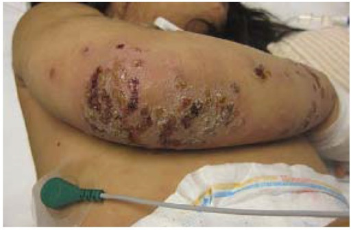
  • Begins as a rash Rash Rocky Mountain Spotted Fever with papules vesicles Vesicles Female Genitourinary Examination surrounded by erythema Erythema Redness of the skin produced by congestion of the capillaries. This condition may result from a variety of disease processes. Chalazion → pustules, which rupture and ooze exudate Exudate Exudates are fluids, cells, or other cellular substances that are slowly discharged from blood vessels usually from inflamed tissues. Pleural Effusion (pus and serous fluid) that dries → pruritic honey-colored crusts that heal with no scarring Scarring Inflammation
  • Lesions are non-tender and pruritic
  • Lesions usually occur around the mouth and nose Nose The nose is the human body’s primary organ of smell and functions as part of the upper respiratory system. The nose may be best known for inhaling oxygen and exhaling carbon dioxide, but it also contributes to other important functions, such as tasting. The anatomy of the nose can be divided into the external nose and the nasal cavity. Nose Anatomy (External & Internal) and/or on the hands

Bullous impetigo

  • 30% of cases
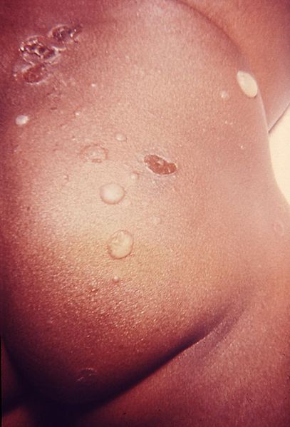
  • Begins as a rash Rash Rocky Mountain Spotted Fever with papules → vesicles Vesicles Female Genitourinary Examination → large, flaccid bullae Bullae Erythema Multiforme, which are pruritic and rupture, oozing cloudy or yellow fluid (pus) → dries into brown crusts → may lead to scarring Scarring Inflammation in severe infections Infections Invasion of the host organism by microorganisms or their toxins or by parasites that can cause pathological conditions or diseases. Chronic Granulomatous Disease 
  • Positive Nikolsky sign Nikolsky Sign Dermatologic Examination: sloughing off of the outermost layer of skin Skin The skin, also referred to as the integumentary system, is the largest organ of the body. The skin is primarily composed of the epidermis (outer layer) and dermis (deep layer). The epidermis is primarily composed of keratinocytes that undergo rapid turnover, while the dermis contains dense layers of connective tissue. Skin: Structure and Functions upon slight rubbing or lateral traction
  • Lesions usually occur on the trunk
  • May also present with systemic symptoms ( fatigue Fatigue The state of weariness following a period of exertion, mental or physical, characterized by a decreased capacity for work and reduced efficiency to respond to stimuli. Fibromyalgia, fever Fever Fever is defined as a measured body temperature of at least 38°C (100.4°F). Fever is caused by circulating endogenous and/or exogenous pyrogens that increase levels of prostaglandin E2 in the hypothalamus. Fever is commonly associated with chills, rigors, sweating, and flushing of the skin. Fever, weakness, general malaise Malaise Tick-borne Encephalitis Virus)

Ecthyma

  • Rare; exact incidence Incidence The number of new cases of a given disease during a given period in a specified population. It also is used for the rate at which new events occur in a defined population. It is differentiated from prevalence, which refers to all cases in the population at a given time. Measures of Disease Frequency unknown
  • Ulcerative form that affects deeper layers of the dermis Dermis A layer of vascularized connective tissue underneath the epidermis. The surface of the dermis contains innervated papillae. Embedded in or beneath the dermis are sweat glands; hair follicles; and sebaceous glands. Skin: Structure and Functions (also known as “deep impetigo”)
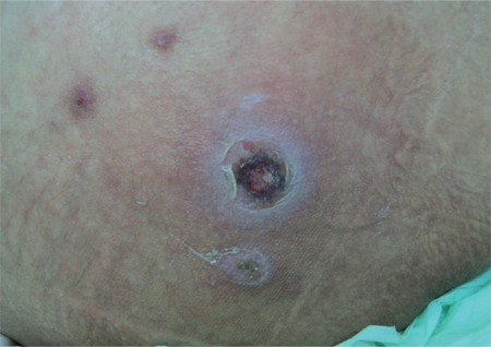
  • Begins as a rash Rash Rocky Mountain Spotted Fever with papules → vesicles Vesicles Female Genitourinary Examination → sores that are painful, erythematous, and fluid- or pus-filled → coin-sized ulcers with a “punched-out” appearance covered with thick gray-yellow scabs → usually lead to scarring Scarring Inflammation 
  • Lesions usually occur on the extremities
  • Ecthyma gangrenosum Ecthyma gangrenosum Pseudomonas is most commonly associated with Pseudomonas Pseudomonas Pseudomonas is a non-lactose-fermenting, gram-negative bacillus that produces pyocyanin, which gives it a characteristic blue-green color. Pseudomonas is found ubiquitously in the environment, as well as in moist reservoirs, such as hospital sinks and respiratory equipment. Pseudomonas bacteremia Bacteremia The presence of viable bacteria circulating in the blood. Fever, chills, tachycardia, and tachypnea are common acute manifestations of bacteremia. The majority of cases are seen in already hospitalized patients, most of whom have underlying diseases or procedures which render their bloodstreams susceptible to invasion. Glycopeptides, seen in patients Patients Individuals participating in the health care system for the purpose of receiving therapeutic, diagnostic, or preventive procedures. Clinician–Patient Relationship who are critically ill and immunocompromised immunocompromised A human or animal whose immunologic mechanism is deficient because of an immunodeficiency disorder or other disease or as the result of the administration of immunosuppressive drugs or radiation. Gastroenteritis; hemorrhagic pustules evolve into necrotic ulcers.

Diagnosis

Diagnosis is usually clinical, based on the natural sequence of the lesions and the presence of honey-colored crusts on pediatric patients Patients Individuals participating in the health care system for the purpose of receiving therapeutic, diagnostic, or preventive procedures. Clinician–Patient Relationship aged 2–5 years.

  • Gram stain Gram stain Klebsiella and culture of exudate Exudate Exudates are fluids, cells, or other cellular substances that are slowly discharged from blood vessels usually from inflamed tissues. Pleural Effusion are not usually required; only needed in the case of:
    • Resistance Resistance Physiologically, the opposition to flow of air caused by the forces of friction. As a part of pulmonary function testing, it is the ratio of driving pressure to the rate of air flow. Ventilation: Mechanics of Breathing to therapy
    • Recurrence
    • Inconclusive clinical diagnosis
  • Imaging: X-ray X-ray Penetrating electromagnetic radiation emitted when the inner orbital electrons of an atom are excited and release radiant energy. X-ray wavelengths range from 1 pm to 10 nm. Hard x-rays are the higher energy, shorter wavelength x-rays. Soft x-rays or grenz rays are less energetic and longer in wavelength. The short wavelength end of the x-ray spectrum overlaps the gamma rays wavelength range. The distinction between gamma rays and x-rays is based on their radiation source. Pulmonary Function Tests, magnetic resonance imaging (MRI), and/or bone Bone Bone is a compact type of hardened connective tissue composed of bone cells, membranes, an extracellular mineralized matrix, and central bone marrow. The 2 primary types of bone are compact and spongy. Bones: Structure and Types scintigraphy Scintigraphy Sjögren’s Syndrome may be performed in the case of severe joint pain Pain An unpleasant sensation induced by noxious stimuli which are detected by nerve endings of nociceptive neurons. Pain: Types and Pathways to confirm or rule out septic arthritis Arthritis Acute or chronic inflammation of joints. Osteoarthritis or osteomyelitis Osteomyelitis Osteomyelitis is an infection of the bone that results from the spread of microorganisms from the blood (hematogenous), nearby infected tissue, or open wounds (non-hematogenous). Infections are most commonly caused by Staphylococcus aureus. Osteomyelitis

Management and Complications

Management

Management depends on the type and severity of the infection.

  • Mild to moderate cases of non-bullous impetigo:
    • Topical antibiotic creams or ointments for 7 days (e.g., mupirocin, retapamulin)
  • Bullous impetigo, ecthyma, and severe cases of non-bullous impetigo:
    • First-line: oral antibiotics including cephalexin and dicloxacillin Dicloxacillin One of the penicillins which is resistant to penicillinase. Penicillins
    • If β-hemolytic streptococci are detected, oral penicillin Penicillin Rheumatic Fever is preferred.
  • Cases of methicillin-resistant S. aureus S. aureus Potentially pathogenic bacteria found in nasal membranes, skin, hair follicles, and perineum of warm-blooded animals. They may cause a wide range of infections and intoxications. Staphylococcus ( MRSA MRSA A strain of Staphylococcus aureus that is non-susceptible to the action of methicillin. The mechanism of resistance usually involves modification of normal or the presence of acquired penicillin binding proteins. Staphylococcus) impetigo:
    • Clindamycin Clindamycin An antibacterial agent that is a semisynthetic analog of lincomycin. Lincosamides, trimethoprim-sulfamethoxazole, or doxycycline
    • Note: Retrospective data indicate that at standard doses, children < 8 years can receive up to 5 courses of doxycycline without detectable evidence of tooth staining. Drug-induced tooth discoloration in children had been reported with older classes of tetracyclines Tetracyclines Tetracyclines are a class of broad-spectrum antibiotics indicated for a wide variety of bacterial infections. These medications bind the 30S ribosomal subunit to inhibit protein synthesis of bacteria. Tetracyclines cover gram-positive and gram-negative organisms, as well as atypical bacteria such as chlamydia, mycoplasma, spirochetes, and even protozoa. Tetracyclines.

Prevention of spread

  • Regular Regular Insulin handwashing
  • Covering of open skin Skin The skin, also referred to as the integumentary system, is the largest organ of the body. The skin is primarily composed of the epidermis (outer layer) and dermis (deep layer). The epidermis is primarily composed of keratinocytes that undergo rapid turnover, while the dermis contains dense layers of connective tissue. Skin: Structure and Functions lesions
  • Children may return to school 24 hours after beginning antibiotic treatment

Complications

  • Scarring Scarring Inflammation or disfigurement
  • Poststreptococcal glomerulonephritis Poststreptococcal Glomerulonephritis Post-streptococcal glomerulonephritis (PSGN) is a type of nephritis that is caused by a prior infection with group A beta-hemolytic Streptococcus (GAS). The clinical presentation of PSGN can range from asymptomatic, microscopic hematuria to full-blown acute nephritic syndrome, which is characterized by red-to-brown urine, proteinuria, edema, and acute kidney injury. Postinfectious Glomerulonephritis
  • Scarlet fever Scarlet fever Infection with group a Streptococci that is characterized by tonsillitis and pharyngitis. An erythematous rash is commonly present. Scarlet Fever
  • Cellulitis Cellulitis Cellulitis is a common infection caused by bacteria that affects the dermis and subcutaneous tissue of the skin. It is frequently caused by Staphylococcus aureus and Streptococcus pyogenes. The skin infection presents as an erythematous and edematous area with warmth and tenderness. Cellulitis
  • Septic arthritis Arthritis Acute or chronic inflammation of joints. Osteoarthritis 
  • Staphylococcal scalded skin syndrome Staphylococcal Scalded Skin Syndrome Staphylococcal scalded skin syndrome (SSSS), also known as Ritter disease and staphylococcal epidermal necrolysis, is a toxin-mediated condition caused by Staphylococcus aureus. The exfoliative toxin produced disseminates and cleaves desmoglein 1 in the epidermis, causing separation and detachment of the skin. Staphylococcal Scalded Skin Syndrome (SSSS)
  • Septicemia or sepsis Sepsis Systemic inflammatory response syndrome with a proven or suspected infectious etiology. When sepsis is associated with organ dysfunction distant from the site of infection, it is called severe sepsis. When sepsis is accompanied by hypotension despite adequate fluid infusion, it is called septic shock. Sepsis and Septic Shock
  • Superimposed bacterial infection of primary impetigo lesions

Differential Diagnosis

The following conditions also present with bullae Bullae Erythema Multiforme and serve as differential diagnoses for bullous impetigo:

  • Pemphigus vulgaris Pemphigus vulgaris Bullous pemphigoid and pemphigus vulgaris are two different blistering autoimmune diseases. In pemphigus vulgaris, autoantibodies attack the desmosomal proteins, which connect the keratinocytes to one another. This attack results in a more severe, potentially fatal condition with fragile, flaccid blisters, usually with significant mucosal involvement. Bullous Pemphigoid and Pemphigus Vulgaris and bullous pemphigoid Bullous pemphigoid Bullous pemphigoid and pemphigus vulgaris are two different blistering autoimmune diseases. In bullous pemphigoid, autoantibodies attack the hemidesmosomes, which connect epidermal keratinocytes to the basement membrane. This attack results in large, tense subepidermal blisters. Bullous Pemphigoid and Pemphigus Vulgaris: autoimmune blistering disorders that present with tense blisters and bullae Bullae Erythema Multiforme in the case of bullous pemphigoid Bullous pemphigoid Bullous pemphigoid and pemphigus vulgaris are two different blistering autoimmune diseases. In bullous pemphigoid, autoantibodies attack the hemidesmosomes, which connect epidermal keratinocytes to the basement membrane. This attack results in large, tense subepidermal blisters. Bullous Pemphigoid and Pemphigus Vulgaris and flaccid bullae Bullae Erythema Multiforme and mucosal erosions Erosions Corneal Abrasions, Erosion, and Ulcers in the case of pemphigus vulgaris Pemphigus vulgaris Bullous pemphigoid and pemphigus vulgaris are two different blistering autoimmune diseases. In pemphigus vulgaris, autoantibodies attack the desmosomal proteins, which connect the keratinocytes to one another. This attack results in a more severe, potentially fatal condition with fragile, flaccid blisters, usually with significant mucosal involvement. Bullous Pemphigoid and Pemphigus Vulgaris
  • Second-degree burns Burns A burn is a type of injury to the skin and deeper tissues caused by exposure to heat, electricity, chemicals, friction, or radiation. Burns are classified according to their depth as superficial (1st-degree), partial-thickness (2nd-degree), full-thickness (3rd-degree), and 4th-degree burns. Burns: tissue injury caused by local heat Heat Inflammation exposure and heating of the respective tissue. Second-degree burns Burns A burn is a type of injury to the skin and deeper tissues caused by exposure to heat, electricity, chemicals, friction, or radiation. Burns are classified according to their depth as superficial (1st-degree), partial-thickness (2nd-degree), full-thickness (3rd-degree), and 4th-degree burns. Burns are superficial, limited to the epidermis Epidermis The external, nonvascular layer of the skin. It is made up, from within outward, of five layers of epithelium: (1) basal layer (stratum basale epidermidis); (2) spinous layer (stratum spinosum epidermidis); (3) granular layer (stratum granulosum epidermidis); (4) clear layer (stratum lucidum epidermidis); and (5) horny layer (stratum corneum epidermidis). Skin: Structure and Functions, and present as hyperemia, wet wound bed, intact sensibility, and blistering. 
  • Varicella: primary infection Primary infection Herpes Simplex Virus 1 and 2 with varicella zoster virus Virus Viruses are infectious, obligate intracellular parasites composed of a nucleic acid core surrounded by a protein capsid. Viruses can be either naked (non-enveloped) or enveloped. The classification of viruses is complex and based on many factors, including type and structure of the nucleoid and capsid, the presence of an envelope, the replication cycle, and the host range. Virology. Occurs most commonly in children and presents as a pruritic rash Rash Rocky Mountain Spotted Fever, with macules that rapidly develop into papules and then vesicles Vesicles Female Genitourinary Examination

The following conditions are other types of superficial skin Skin The skin, also referred to as the integumentary system, is the largest organ of the body. The skin is primarily composed of the epidermis (outer layer) and dermis (deep layer). The epidermis is primarily composed of keratinocytes that undergo rapid turnover, while the dermis contains dense layers of connective tissue. Skin: Structure and Functions infections Infections Invasion of the host organism by microorganisms or their toxins or by parasites that can cause pathological conditions or diseases. Chronic Granulomatous Disease that are included in the differential diagnoses of impetigo:

  • Erysipelas Erysipelas Erysipelas is a bacterial infection of the superficial layer of the skin extending to the skin’s superficial lymphatic vessels. This infection presents as a raised, well-defined, tender, and bright red rash. Typically on the legs or face, but erysipelas can occur anywhere on the skin. Erysipelas: acute superficial infection of the upper dermis Dermis A layer of vascularized connective tissue underneath the epidermis. The surface of the dermis contains innervated papillae. Embedded in or beneath the dermis are sweat glands; hair follicles; and sebaceous glands. Skin: Structure and Functions and involving lymphatics. Usually caused by bacterial agents such as group A β-hemolytic streptococcus Streptococcus Streptococcus is one of the two medically important genera of gram-positive cocci, the other being Staphylococcus. Streptococci are identified as different species on blood agar on the basis of their hemolytic pattern and sensitivity to optochin and bacitracin. There are many pathogenic species of streptococci, including S. pyogenes, S. agalactiae, S. pneumoniae, and the viridans streptococci. Streptococcus, it presents as a sharply demarcated skin Skin The skin, also referred to as the integumentary system, is the largest organ of the body. The skin is primarily composed of the epidermis (outer layer) and dermis (deep layer). The epidermis is primarily composed of keratinocytes that undergo rapid turnover, while the dermis contains dense layers of connective tissue. Skin: Structure and Functions lesion with erythema Erythema Redness of the skin produced by congestion of the capillaries. This condition may result from a variety of disease processes. Chalazion, edema Edema Edema is a condition in which excess serous fluid accumulates in the body cavity or interstitial space of connective tissues. Edema is a symptom observed in several medical conditions. It can be categorized into 2 types, namely, peripheral (in the extremities) and internal (in an organ or body cavity). Edema, and warmth. 
  • Dermatitis Dermatitis Any inflammation of the skin. Atopic Dermatitis (Eczema): a general term for an edematous skin Skin The skin, also referred to as the integumentary system, is the largest organ of the body. The skin is primarily composed of the epidermis (outer layer) and dermis (deep layer). The epidermis is primarily composed of keratinocytes that undergo rapid turnover, while the dermis contains dense layers of connective tissue. Skin: Structure and Functions rash Rash Rocky Mountain Spotted Fever caused by an allergic reaction or a non-bacterial infection 
  • Cutaneous candidiasis Candidiasis Candida is a genus of dimorphic, opportunistic fungi. Candida albicans is part of the normal human flora and is the most common cause of candidiasis. The clinical presentation varies and can include localized mucocutaneous infections (e.g., oropharyngeal, esophageal, intertriginous, and vulvovaginal candidiasis) and invasive disease (e.g., candidemia, intraabdominal abscess, pericarditis, and meningitis). Candida/Candidiasis: a superficial infection caused by Candida Candida Candida is a genus of dimorphic, opportunistic fungi. Candida albicans is part of the normal human flora and is the most common cause of candidiasis. The clinical presentation varies and can include localized mucocutaneous infections (e.g., oropharyngeal, esophageal, intertriginous, and vulvovaginal candidiasis) and invasive disease (e.g., candidemia, intraabdominal abscess, pericarditis, and meningitis). Candida/Candidiasis spp. Usually associated with immunodeficiency Immunodeficiency Chédiak-Higashi Syndrome
  • Tinea: an infection of the skin Skin The skin, also referred to as the integumentary system, is the largest organ of the body. The skin is primarily composed of the epidermis (outer layer) and dermis (deep layer). The epidermis is primarily composed of keratinocytes that undergo rapid turnover, while the dermis contains dense layers of connective tissue. Skin: Structure and Functions caused by dermatophytes Dermatophytes Tinea infections are a group of diseases caused by fungi infecting keratinized tissue (hair, nails, and skin). These infections are termed dermatomycoses and are caused by the dermatophyte fungi. There are approximately 40 dermatophyte fungi that are part of 3 genera, including Trichophyton, Epidermophyton, and Microsporum. These infections can affect any part of the body but occur most often in warm, moist regions like the groin and the feet. Dermatophytes/Tinea Infections. There are 3 types of dermatophytes Dermatophytes Tinea infections are a group of diseases caused by fungi infecting keratinized tissue (hair, nails, and skin). These infections are termed dermatomycoses and are caused by the dermatophyte fungi. There are approximately 40 dermatophyte fungi that are part of 3 genera, including Trichophyton, Epidermophyton, and Microsporum. These infections can affect any part of the body but occur most often in warm, moist regions like the groin and the feet. Dermatophytes/Tinea Infections: Trichophyton Trichophyton A mitosporic fungal genus and an anamorphic form of arthroderma. Various species attack the skin, nails, and hair. Dermatophytes/Tinea Infections, Epidermophyton Epidermophyton A fungal genus which grows in the epidermis and is the cause of tinea. Dermatophytes/Tinea Infections, and Microsporum Microsporum A mitosporic oxygenales fungal genus causing various diseases of the skin and hair. The species microsporum canis produces tinea capitis and tinea corporis, which usually are acquired from domestic cats and dogs. Teleomorphs includes arthroderma (nannizzia). Dermatophytes/Tinea Infections
  • Herpes simplex Herpes Simplex A group of acute infections caused by herpes simplex virus type 1 or type 2 that is characterized by the development of one or more small fluid-filled vesicles with a raised erythematous base on the skin or mucous membrane. It occurs as a primary infection or recurs due to a reactivation of a latent infection. Congenital TORCH Infections viral infection: a viral infection that presents as recurring, erythematous vesicles Vesicles Female Genitourinary Examination that turn into painful ulcerations, usually around in and around the mouth ( HSV HSV Herpes simplex virus (HSV) is a double-stranded DNA virus belonging to the family Herpesviridae. Herpes simplex virus commonly causes recurrent infections involving the skin and mucosal surfaces, including the mouth, lips, eyes, and genitals. Herpes Simplex Virus 1 and 2 type 1 Type 1 Spinal Muscular Atrophy is more common at this site)
  • Folliculitis: a localized inflammation Inflammation Inflammation is a complex set of responses to infection and injury involving leukocytes as the principal cellular mediators in the body’s defense against pathogenic organisms. Inflammation is also seen as a response to tissue injury in the process of wound healing. The 5 cardinal signs of inflammation are pain, heat, redness, swelling, and loss of function. Inflammation of the hair follicle Hair follicle A tube-like invagination of the epidermis from which the hair shaft develops and into which sebaceous glands open. The hair follicle is lined by a cellular inner and outer root sheath of epidermal origin and is invested with a fibrous sheath derived from the dermis. Follicles of very long hairs extend into the subcutaneous layer of tissue under the skin. Cowden Syndrome or sebaceous glands primarily caused by S. aureus S. aureus Potentially pathogenic bacteria found in nasal membranes, skin, hair follicles, and perineum of warm-blooded animals. They may cause a wide range of infections and intoxications. Staphylococcus. It is characterized by erythema Erythema Redness of the skin produced by congestion of the capillaries. This condition may result from a variety of disease processes. Chalazion, tender papules, and/or pustules at the site of hair follicles.

References

  1. Baddour LM. Impetigo. (Literature review current through: July 2020). UpToDate Evidence-Based Medicine. https://www.uptodate.com/contents/impetigo?search=impetigo&source=search_result&selectedTitle=1~122&usage_type=default&display_rank=1
  2. Emanuel P. Impetigo. (2013). DermNet New Zealand (Supported by and contributed to by New Zealand Dermatologists on behalf of the New Zealand Dermatological Society Incorporated). https://dermnetnz.org/topics/impetigo-pathology/

Create your free account or log in to continue reading!

Sign up now and get free access to Lecturio with concept pages, medical videos, and questions for your medical education.

User Reviews

Details