Advertisement

Advertisement

Advertisement

Advertisement

Hypertrophic Pyloric Stenosis

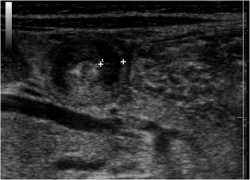
Hypertrophic pyloric stenosis Stenosis Hypoplastic Left Heart Syndrome (HLHS) is hypertrophy Hypertrophy General increase in bulk of a part or organ due to cell enlargement and accumulation of fluids and secretions, not due to tumor formation, nor to an increase in the number of cells (hyperplasia). Cellular Adaptation and hyperplasia Hyperplasia An increase in the number of cells in a tissue or organ without tumor formation. It differs from hypertrophy, which is an increase in bulk without an increase in the number of cells. Cellular Adaptation of the pyloric sphincter muscle. The condition is the most common cause of gastrointestinal obstruction in infants. Affected newborns typically present after the third to fifth week of life with progressive non-bilious vomiting Vomiting The forcible expulsion of the contents of the stomach through the mouth. Hypokalemia and a firm, olive-like mass Mass Three-dimensional lesion that occupies a space within the breast Imaging of the Breast in the epigastrium Epigastrium Surgical Anatomy of the Abdomen. Ultrasound confirms the diagnosis based on the thickness and length of the pyloric muscle and channel diameter. Initial treatment consists of fluid resuscitation Resuscitation The restoration to life or consciousness of one apparently dead. . Neonatal Respiratory Distress Syndrome with correction of electrolyte imbalances, followed by either open or laparoscopic pyloromyotomy.

Last updated: Sep 29, 2022

Editorial responsibility: Stanley Oiseth, Lindsay Jones, Evelin Maza

Advertisement

Advertisement

Advertisement

Advertisement

Advertisement

Advertisement

Advertisement

Advertisement

Overview

Definition

Hypertrophic pyloric stenosis Stenosis Hypoplastic Left Heart Syndrome (HLHS) is a functional obstruction of the gastric outlet caused by hypertrophy Hypertrophy General increase in bulk of a part or organ due to cell enlargement and accumulation of fluids and secretions, not due to tumor formation, nor to an increase in the number of cells (hyperplasia). Cellular Adaptation and hyperplasia Hyperplasia An increase in the number of cells in a tissue or organ without tumor formation. It differs from hypertrophy, which is an increase in bulk without an increase in the number of cells. Cellular Adaptation of both the circular and longitudinal layers of the pylorus Pylorus The region between the sharp indentation at the lower third of the stomach (incisura angularis) and the junction of the pylorus with the duodenum. Pyloric antral glands contain mucus-secreting cells and gastrin-secreting endocrine cells (g cells). Stomach: Anatomy in infants.
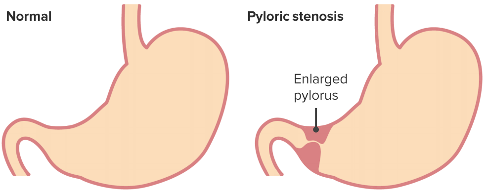
Hypertrophic pyloric stenosis

Hypertrophic pyloric stenosis

Image by Lecturio.
Epidemiology

  • The most common cause of intestinal obstruction Intestinal obstruction Any impairment, arrest, or reversal of the normal flow of intestinal contents toward the anal canal. Ascaris/Ascariasis in infants
  • Occurs in 13 in 1,000 infants in the United States
  • More common in whites, males, firstborns, blood groups B or O, and those with a positive history of pyloric stenosis Stenosis Hypoplastic Left Heart Syndrome (HLHS) in mother or father

Etiology

  • Associations include eosinophilic gastroenteritis Gastroenteritis Gastroenteritis is inflammation of the stomach and intestines, commonly caused by infections from bacteria, viruses, or parasites. Transmission may be foodborne, fecal-oral, or through animal contact. Common clinical features include abdominal pain, diarrhea, vomiting, fever, and dehydration. Gastroenteritis, Apert syndrome Apert Syndrome Craniosynostosis, Zellweger syndrome Zellweger syndrome Zellweger syndrome (ZWS), also called cerebrohepatorenal syndrome, is a rare congenital peroxisome biosynthesis disorder and is considered an inborn error of metabolism. Zellweger syndrome is the most severe form of a spectrum of conditions called Zellweger spectrum disorder (ZSD), and is characterized by the reduction or absence of functional peroxisomes. Zellweger Syndrome, trisomy Trisomy The possession of a third chromosome of any one type in an otherwise diploid cell. Types of Mutations 18, use of erythromycin Erythromycin A bacteriostatic antibiotic macrolide produced by streptomyces erythreus. Erythromycin a is considered its major active component. In sensitive organisms, it inhibits protein synthesis by binding to 50s ribosomal subunits. This binding process inhibits peptidyl transferase activity and interferes with translocation of amino acids during translation and assembly of proteins. Macrolides and Ketolides in first 2 weeks of pregnancy Pregnancy The status during which female mammals carry their developing young (embryos or fetuses) in utero before birth, beginning from fertilization to birth. Pregnancy: Diagnosis, Physiology, and Care, and maternal macrolide use during pregnancy Pregnancy The status during which female mammals carry their developing young (embryos or fetuses) in utero before birth, beginning from fertilization to birth. Pregnancy: Diagnosis, Physiology, and Care or breastfeeding Breastfeeding Breastfeeding is often the primary source of nutrition for the newborn. During pregnancy, hormonal stimulation causes the number and size of mammary glands in the breast to significantly increase. After delivery, prolactin stimulates milk production, while oxytocin stimulates milk expulsion through the lactiferous ducts, where it is sucked out through the nipple by the infant. Breastfeeding.
  • Reduced levels of neuronal nitric oxide Nitric Oxide A free radical gas produced endogenously by a variety of mammalian cells, synthesized from arginine by nitric oxide synthase. Nitric oxide is one of the endothelium-dependent relaxing factors released by the vascular endothelium and mediates vasodilation. It also inhibits platelet aggregation, induces disaggregation of aggregated platelets, and inhibits platelet adhesion to the vascular endothelium. Nitric oxide activates cytosolic guanylate cyclase and thus elevates intracellular levels of cyclic gmp. Pulmonary Hypertension Drugs synthase have been shown to be involved in the pathogenesis.

Clinical Manifestations

Symptoms

  • Initially, regurgitation Regurgitation Gastroesophageal Reflux Disease (GERD) after feeding
  • Later, non-bilious, projectile vomiting Vomiting The forcible expulsion of the contents of the stomach through the mouth. Hypokalemia immediately after feeding
  • Usually starts after 3 weeks of age (range: 1 week to 5 months)
  • Poor weight gain
  • Infant shows signs of hunger (irritability, increased sucking reflex Sucking reflex A primitive instinct present in all newborn mammals. Babies will suckle anything that comes into contact with the roof of their mouth. Breastfeeding).

Signs

Diagnosis

Laboratory studies

  • Hypochloremic metabolic alkalosis Alkalosis A pathological condition that removes acid or adds base to the body fluids. Respiratory Alkalosis
  • Hypokalemia Hypokalemia Hypokalemia is defined as plasma potassium (K+) concentration < 3.5 mEq/L. Homeostatic mechanisms maintain plasma concentration between 3.5-5.2 mEq/L despite marked variation in dietary intake. Hypokalemia can be due to renal losses, GI losses, transcellular shifts, or poor dietary intake. Hypokalemia
  • Elevated blood urea Urea A compound formed in the liver from ammonia produced by the deamination of amino acids. It is the principal end product of protein catabolism and constitutes about one half of the total urinary solids. Urea Cycle nitrogen Nitrogen An element with the atomic symbol n, atomic number 7, and atomic weight [14. 00643; 14. 00728]. Nitrogen exists as a diatomic gas and makes up about 78% of the earth’s atmosphere by volume. It is a constituent of proteins and nucleic acids and found in all living cells. Urea Cycle (BUN) and creatinine with severe dehydration Severe Dehydration Fluid Replacement Therapy in Children
  • Unconjugated (and occasionally conjugated) hyperbilirubinemia Hyperbilirubinemia A condition characterized by an abnormal increase of bilirubin in the blood, which may result in jaundice. Bilirubin, a breakdown product of heme, is normally excreted in the bile or further catabolized before excretion in the urine. Jaundice is the most common clinical association.

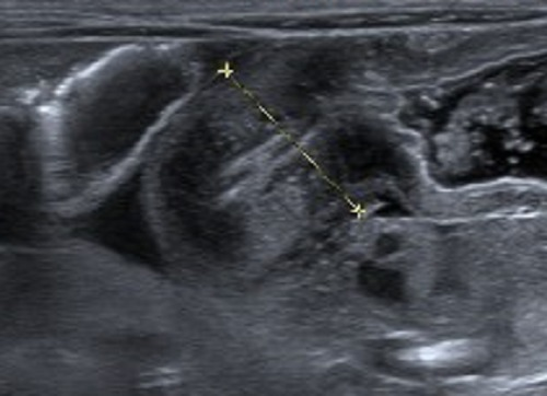
Ultrasound

  • Diagnosis based on:
    • Pyloric muscle thickness: > 3 mm MM Multiple myeloma (MM) is a malignant condition of plasma cells (activated B lymphocytes) primarily seen in the elderly. Monoclonal proliferation of plasma cells results in cytokine-driven osteoclastic activity and excessive secretion of IgG antibodies. Multiple Myeloma (the most discriminating and accurate criterion) 
    • Pyloric muscle length: > 15 mm MM Multiple myeloma (MM) is a malignant condition of plasma cells (activated B lymphocytes) primarily seen in the elderly. Monoclonal proliferation of plasma cells results in cytokine-driven osteoclastic activity and excessive secretion of IgG antibodies. Multiple Myeloma
    • Pyloric diameter: > 15 mm MM Multiple myeloma (MM) is a malignant condition of plasma cells (activated B lymphocytes) primarily seen in the elderly. Monoclonal proliferation of plasma cells results in cytokine-driven osteoclastic activity and excessive secretion of IgG antibodies. Multiple Myeloma

Contrast studies

  • If diagnosis not confirmed by ultrasound, but still suspected
  • String sign: elongated pyloric channel
  • Shoulder sign: pyloric bulging into channel
  • Double-track sign: parallel streaks of contrast in narrowed channel

Treatment

Preoperative care Preoperative Care Thorough preoperative care is important for patients scheduled to undergo surgery so that they can have the best possible outcomes after their surgical procedure. The preoperative process begins once the decision has been made to proceed with a surgical procedure. Preoperative Care

  • Fluid replacement + decompression
  • Correct electrolyte abnormalities
  • Restoration of acid-base balance

Surgery

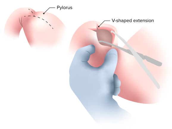
  • Ramstedt pyloromyotomy: short transverse skin Skin The skin, also referred to as the integumentary system, is the largest organ of the body. The skin is primarily composed of the epidermis (outer layer) and dermis (deep layer). The epidermis is primarily composed of keratinocytes that undergo rapid turnover, while the dermis contains dense layers of connective tissue. Skin: Structure and Functions incision plus longitudinal incision of pyloric muscle up to submucosa
  • Laparoscopy Laparoscopy Laparoscopy is surgical exploration and interventions performed through small incisions with a camera and long instruments. Laparotomy and Laparoscopy: equally effective with shorter hospital stay
Hypertrophic pyloric stenosis surgery

This is the classic definitive surgical treatment of hypertrophic pyloric stenosis called a “Ramstedt pyloromyotomy”, in which a longitudinal incision of the anterior surface of the pylorus is made down through the muscle layer only to the level of the submucosa, leaving the submucosa and mucosa in a prolapsed state.

Image by Lecturio.

Postoperative care Postoperative care After any procedure performed in the operating room, all patients must undergo close observation at least in the recovery room. After larger procedures and for patients who require hospitalization, observation must continue on the surgical ward. The primary intent of this practice is the early detection of postoperative complications. Postoperative Care

  • Oral feeding can be initiated within 1224 hours.
  • Persistent vomiting Vomiting The forcible expulsion of the contents of the stomach through the mouth. Hypokalemia suggests incomplete pyloromyotomy or an alternative diagnosis.

Conservative approach

  • If surgery not tolerated or unavailable
  • Oral and intravenous atropine Atropine An alkaloid, originally from atropa belladonna, but found in other plants, mainly solanaceae. Hyoscyamine is the 3(s)-endo isomer of atropine. Anticholinergic Drugs and cardiac monitoring → gradual introduction of feeding

Differential Diagnosis

  • Gastroesophageal reflux disease Gastroesophageal Reflux Disease Gastroesophageal reflux disease (GERD) occurs when the stomach acid frequently flows back into the esophagus. This backwash (acid reflux) can irritate the lining of the esophagus, causing symptoms such as retrosternal burning pain (heartburn). Gastroesophageal Reflux Disease (GERD) ( GERD GERD Gastroesophageal reflux disease (GERD) occurs when the stomach acid frequently flows back into the esophagus. This backwash (acid reflux) can irritate the lining of the esophagus, causing symptoms such as retrosternal burning pain (heartburn). Gastroesophageal Reflux Disease (GERD)): a chronic disorder in which gastric acid Gastric acid Hydrochloric acid present in gastric juice. Gastroesophageal Reflux Disease (GERD), food, or fluids back up from the stomach Stomach The stomach is a muscular sac in the upper left portion of the abdomen that plays a critical role in digestion. The stomach develops from the foregut and connects the esophagus with the duodenum. Structurally, the stomach is C-shaped and forms a greater and lesser curvature and is divided grossly into regions: the cardia, fundus, body, and pylorus. Stomach: Anatomy into the esophagus Esophagus The esophagus is a muscular tube-shaped organ of around 25 centimeters in length that connects the pharynx to the stomach. The organ extends from approximately the 6th cervical vertebra to the 11th thoracic vertebra and can be divided grossly into 3 parts: the cervical part, the thoracic part, and the abdominal part. Esophagus: Anatomy. Patients Patients Individuals participating in the health care system for the purpose of receiving therapeutic, diagnostic, or preventive procedures. Clinician–Patient Relationship often present with heartburn Heartburn Substernal pain or burning sensation, usually associated with regurgitation of gastric juice into the esophagus. Gastroesophageal Reflux Disease (GERD), bad breath Bad breath An offensive, foul breath odor resulting from a variety of causes such as poor oral hygiene, dental or oral infections, or the ingestion of certain foods. Oral Cancer, chest pain Pain An unpleasant sensation induced by noxious stimuli which are detected by nerve endings of nociceptive neurons. Pain: Types and Pathways, and regurgitation Regurgitation Gastroesophageal Reflux Disease (GERD). Gastroesophageal reflux disease Gastroesophageal Reflux Disease Gastroesophageal reflux disease (GERD) occurs when the stomach acid frequently flows back into the esophagus. This backwash (acid reflux) can irritate the lining of the esophagus, causing symptoms such as retrosternal burning pain (heartburn). Gastroesophageal Reflux Disease (GERD) can lead to damage to the teeth Teeth Normally, an adult has 32 teeth: 16 maxillary and 16 mandibular. These teeth are divided into 4 quadrants with 8 teeth each. Each quadrant consists of 2 incisors (dentes incisivi), 1 canine (dens caninus), 2 premolars (dentes premolares), and 3 molars (dentes molares). Teeth are composed of enamel, dentin, and dental cement. Teeth: Anatomy, esophagitis Esophagitis Esophagitis is the inflammation or irritation of the esophagus. The major types of esophagitis are medication-induced, infectious, eosinophilic, corrosive, and acid reflux. Patients typically present with odynophagia, dysphagia, and retrosternal chest pain. Esophagitis, and Barrett’s esophagus Esophagus The esophagus is a muscular tube-shaped organ of around 25 centimeters in length that connects the pharynx to the stomach. The organ extends from approximately the 6th cervical vertebra to the 11th thoracic vertebra and can be divided grossly into 3 parts: the cervical part, the thoracic part, and the abdominal part. Esophagus: Anatomy. Treatment includes lifestyle changes and medical therapy with antacids, proton pump Pump ACES and RUSH: Resuscitation Ultrasound Protocols inhibitors (PPIs), and prokinetics. 
  • Cyclical vomiting Vomiting The forcible expulsion of the contents of the stomach through the mouth. Hypokalemia syndrome (CVS): a condition of unknown pathogenesis characterized by recurrent episodes of nausea Nausea An unpleasant sensation in the stomach usually accompanied by the urge to vomit. Common causes are early pregnancy, sea and motion sickness, emotional stress, intense pain, food poisoning, and various enteroviruses. Antiemetics, vomiting Vomiting The forcible expulsion of the contents of the stomach through the mouth. Hypokalemia, and lethargy Lethargy A general state of sluggishness, listless, or uninterested, with being tired, and having difficulty concentrating and doing simple tasks. It may be related to depression or drug addiction. Hyponatremia. Symptoms can last hours or days. Cyclical vomiting Vomiting The forcible expulsion of the contents of the stomach through the mouth. Hypokalemia syndrome most commonly occurs in children but can affect individuals of any age. 
  • Gastroenteritis Gastroenteritis Gastroenteritis is inflammation of the stomach and intestines, commonly caused by infections from bacteria, viruses, or parasites. Transmission may be foodborne, fecal-oral, or through animal contact. Common clinical features include abdominal pain, diarrhea, vomiting, fever, and dehydration. Gastroenteritis: inflammation Inflammation Inflammation is a complex set of responses to infection and injury involving leukocytes as the principal cellular mediators in the body’s defense against pathogenic organisms. Inflammation is also seen as a response to tissue injury in the process of wound healing. The 5 cardinal signs of inflammation are pain, heat, redness, swelling, and loss of function. Inflammation of the gastrointestinal tract most often caused by viruses Viruses Minute infectious agents whose genomes are composed of DNA or RNA, but not both. They are characterized by a lack of independent metabolism and the inability to replicate outside living host cells. Virology, but also bacteria Bacteria Bacteria are prokaryotic single-celled microorganisms that are metabolically active and divide by binary fission. Some of these organisms play a significant role in the pathogenesis of diseases. Bacteriology or parasites. Symptoms usually last less than 14 days and include increased stool frequency or diarrhea Diarrhea Diarrhea is defined as ≥ 3 watery or loose stools in a 24-hour period. There are a multitude of etiologies, which can be classified based on the underlying mechanism of disease. The duration of symptoms (acute or chronic) and characteristics of the stools (e.g., watery, bloody, steatorrheic, mucoid) can help guide further diagnostic evaluation. Diarrhea, fever Fever Fever is defined as a measured body temperature of at least 38°C (100.4°F). Fever is caused by circulating endogenous and/or exogenous pyrogens that increase levels of prostaglandin E2 in the hypothalamus. Fever is commonly associated with chills, rigors, sweating, and flushing of the skin. Fever, vomiting Vomiting The forcible expulsion of the contents of the stomach through the mouth. Hypokalemia, or abdominal pain Abdominal Pain Acute Abdomen
  • Congenital Congenital Chorioretinitis adrenal hyperplasia Hyperplasia An increase in the number of cells in a tissue or organ without tumor formation. It differs from hypertrophy, which is an increase in bulk without an increase in the number of cells. Cellular Adaptation ( CAH CAH Congenital adrenal hyperplasia (CAH) consists of a group of autosomal recessive disorders that cause a deficiency of an enzyme needed in cortisol, aldosterone, and androgen synthesis. The most common subform of cah is 21-hydroxylase deficiency, followed by 11β-hydroxylase deficiency. Congenital Adrenal Hyperplasia): a group of rare autosomal recessive Autosomal recessive Autosomal inheritance, both dominant and recessive, refers to the transmission of genes from the 22 autosomal chromosomes. Autosomal recessive diseases are only expressed when 2 copies of the recessive allele are inherited. Autosomal Recessive and Autosomal Dominant Inheritance diseases characterized by defects in enzymes Enzymes Enzymes are complex protein biocatalysts that accelerate chemical reactions without being consumed by them. Due to the body’s constant metabolic needs, the absence of enzymes would make life unsustainable, as reactions would occur too slowly without these molecules. Basics of Enzymes of the adrenal glands Adrenal Glands The adrenal glands are a pair of retroperitoneal endocrine glands located above the kidneys. The outer parenchyma is called the adrenal cortex and has 3 distinct zones, each with its own secretory products. Beneath the cortex lies the adrenal medulla, which secretes catecholamines involved in the fight-or-flight response. Adrenal Glands: Anatomy involved in cortisol Cortisol Glucocorticoids, androgen, and aldosterone Aldosterone A hormone secreted by the adrenal cortex that regulates electrolyte and water balance by increasing the renal retention of sodium and the excretion of potassium. Hyperkalemia synthesis Synthesis Polymerase Chain Reaction (PCR). Patients Patients Individuals participating in the health care system for the purpose of receiving therapeutic, diagnostic, or preventive procedures. Clinician–Patient Relationship may present with a wide range of symptoms, including virilization in females, salt-wasting leading to hypotension Hypotension Hypotension is defined as low blood pressure, specifically < 90/60 mm Hg, and is most commonly a physiologic response. Hypotension may be mild, serious, or life threatening, depending on the cause. Hypotension and other electrolyte abnormalities, and premature Premature Childbirth before 37 weeks of pregnancy (259 days from the first day of the mother’s last menstrual period, or 245 days after fertilization). Necrotizing Enterocolitis completion of growth resulting in short stature.
  • Increased intracranial pressure Intracranial Pressure Idiopathic Intracranial Hypertension: a medical emergency defined by an intracranial pressure Intracranial Pressure Idiopathic Intracranial Hypertension exceeding 20 mm MM Multiple myeloma (MM) is a malignant condition of plasma cells (activated B lymphocytes) primarily seen in the elderly. Monoclonal proliferation of plasma cells results in cytokine-driven osteoclastic activity and excessive secretion of IgG antibodies. Multiple Myeloma Hg that can lead to a decrease in cerebral perfusion Cerebral Perfusion Syncope as well as herniation Herniation Omphalocele of the brain Brain The part of central nervous system that is contained within the skull (cranium). Arising from the neural tube, the embryonic brain is comprised of three major parts including prosencephalon (the forebrain); mesencephalon (the midbrain); and rhombencephalon (the hindbrain). The developed brain consists of cerebrum; cerebellum; and other structures in the brain stem. Nervous System: Anatomy, Structure, and Classification. Common symptoms include headache Headache The symptom of pain in the cranial region. It may be an isolated benign occurrence or manifestation of a wide variety of headache disorders. Brain Abscess, vomiting Vomiting The forcible expulsion of the contents of the stomach through the mouth. Hypokalemia, altered mentation, papilledema Papilledema Swelling of the optic disk, usually in association with increased intracranial pressure, characterized by hyperemia, blurring of the disk margins, microhemorrhages, blind spot enlargement, and engorgement of retinal veins. Chronic papilledema may cause optic atrophy and visual loss. Idiopathic Intracranial Hypertension, and bradycardia Bradycardia Bradyarrhythmia is a rhythm in which the heart rate is less than 60/min. Bradyarrhythmia can be physiologic, without symptoms or hemodynamic change. Pathologic bradyarrhythmia results in reduced cardiac output and hemodynamic instability causing syncope, dizziness, or dyspnea. Bradyarrhythmias. Causes include increased production or partially obstructed outflow of cerebrospinal fluid Cerebrospinal Fluid A watery fluid that is continuously produced in the choroid plexus and circulates around the surface of the brain; spinal cord; and in the cerebral ventricles. Ventricular System: Anatomy, bleedings, tumors, and infections Infections Invasion of the host organism by microorganisms or their toxins or by parasites that can cause pathological conditions or diseases. Chronic Granulomatous Disease.
  • Volvulus Volvulus A volvulus is the twisting or axial rotation of a portion of the bowel around its mesentery. The most common site of volvulus in adults is the colon; most frequently the sigmoid volvulus. Patients typically present with symptoms of bowel obstruction such as abdominal pain, distension, vomiting, and constipation/obstipation. Volvulus: a condition in which a loop of intestine twists around itself and the surrounding mesentery Mesentery A layer of the peritoneum which attaches the abdominal viscera to the abdominal wall and conveys their blood vessels and nerves. Peritoneum: Anatomy, causing bowel obstruction Bowel obstruction Any impairment, arrest, or reversal of the normal flow of intestinal contents toward the anal canal. Ascaris/Ascariasis. Volvulus Volvulus A volvulus is the twisting or axial rotation of a portion of the bowel around its mesentery. The most common site of volvulus in adults is the colon; most frequently the sigmoid volvulus. Patients typically present with symptoms of bowel obstruction such as abdominal pain, distension, vomiting, and constipation/obstipation. Volvulus is often caused by a birth defect and presents early in life, but can occur at any age. Common symptoms include vomiting Vomiting The forcible expulsion of the contents of the stomach through the mouth. Hypokalemia, abdominal pain Abdominal Pain Acute Abdomen and bloating Bloating Constipation, constipation Constipation Constipation is common and may be due to a variety of causes. Constipation is generally defined as bowel movement frequency < 3 times per week. Patients who are constipated often strain to pass hard stools. The condition is classified as primary (also known as idiopathic or functional constipation) or secondary, and as acute or chronic. Constipation, and bloody stool that may be gradual or sudden in onset. Emergency surgery is necessary to prevent necrosis Necrosis The death of cells in an organ or tissue due to disease, injury or failure of the blood supply. Ischemic Cell Damage of the intestinal structures.

Create your free account or log in to continue reading!

Sign up now and get free access to Lecturio with concept pages, medical videos, and questions for your medical education.

User Reviews

It's Deal Week! 💸 Save 30% on all plans!

Details