Advertisement

Advertisement

Advertisement

Advertisement

Hypertrophic Cardiomyopathy

Hypertrophic cardiomyopathy Cardiomyopathy Cardiomyopathy refers to a group of myocardial diseases associated with structural changes of the heart muscles (myocardium) and impaired systolic and/or diastolic function in the absence of other heart disorders (coronary artery disease, hypertension, valvular disease, and congenital heart disease). Cardiomyopathy: Overview and Types (HCM) is the most commonly inherited cardiomyopathy Cardiomyopathy Cardiomyopathy refers to a group of myocardial diseases associated with structural changes of the heart muscles (myocardium) and impaired systolic and/or diastolic function in the absence of other heart disorders (coronary artery disease, hypertension, valvular disease, and congenital heart disease). Cardiomyopathy: Overview and Types, which is characterized by an asymmetric increase in thickness ( hypertrophy Hypertrophy General increase in bulk of a part or organ due to cell enlargement and accumulation of fluids and secretions, not due to tumor formation, nor to an increase in the number of cells (hyperplasia). Cellular Adaptation) of the left ventricular wall, diastolic dysfunction Diastolic dysfunction Restrictive Cardiomyopathy, and often left ventricular outflow tract obstruction. Hypertrophic cardiomyopathy Cardiomyopathy Cardiomyopathy refers to a group of myocardial diseases associated with structural changes of the heart muscles (myocardium) and impaired systolic and/or diastolic function in the absence of other heart disorders (coronary artery disease, hypertension, valvular disease, and congenital heart disease). Cardiomyopathy: Overview and Types is caused by various gene Gene A category of nucleic acid sequences that function as units of heredity and which code for the basic instructions for the development, reproduction, and maintenance of organisms. Basic Terms of Genetics mutations affecting the contractile components of the heart, known as sarcomeres. Inheritance of HCM is typically autosomal dominant Autosomal dominant Autosomal inheritance, both dominant and recessive, refers to the transmission of genes from the 22 autosomal chromosomes. Autosomal dominant diseases are expressed when only 1 copy of the dominant allele is inherited. Autosomal Recessive and Autosomal Dominant Inheritance, although sporadic Sporadic Selective IgA Deficiency mutations also occur. Patients Patients Individuals participating in the health care system for the purpose of receiving therapeutic, diagnostic, or preventive procedures. Clinician–Patient Relationship may be asymptomatic, present with dyspnea Dyspnea Dyspnea is the subjective sensation of breathing discomfort. Dyspnea is a normal manifestation of heavy physical or psychological exertion, but also may be caused by underlying conditions (both pulmonary and extrapulmonary). Dyspnea and chest pain Pain An unpleasant sensation induced by noxious stimuli which are detected by nerve endings of nociceptive neurons. Pain: Types and Pathways or suffer sudden cardiac death Sudden cardiac death Cardiac arrest is the sudden, complete cessation of cardiac output with hemodynamic collapse. Patients present as pulseless, unresponsive, and apneic. Rhythms associated with cardiac arrest are ventricular fibrillation/tachycardia, asystole, or pulseless electrical activity. Cardiac Arrest without prior symptoms. Diagnosis is made based on ECG ECG An electrocardiogram (ECG) is a graphic representation of the electrical activity of the heart plotted against time. Adhesive electrodes are affixed to the skin surface allowing measurement of cardiac impulses from many angles. The ECG provides 3-dimensional information about the conduction system of the heart, the myocardium, and other cardiac structures. Electrocardiogram (ECG), echocardiography Echocardiography Ultrasonic recording of the size, motion, and composition of the heart and surrounding tissues. The standard approach is transthoracic. Tricuspid Valve Atresia (TVA), stress test, and cardiac MRI Cardiac MRI Imaging of the Heart and Great Vessels. Symptomatic HCM is typically treated with beta-blockers Beta-blockers Drugs that bind to but do not activate beta-adrenergic receptors thereby blocking the actions of beta-adrenergic agonists. Adrenergic beta-antagonists are used for treatment of hypertension, cardiac arrhythmias, angina pectoris, glaucoma, migraine headaches, and anxiety. Class 2 Antiarrhythmic Drugs (Beta Blockers) as the 1st-line therapy. Additional management depends on the presence of left ventricular outflow tract obstruction.

Last updated: May 16, 2024

Editorial responsibility: Stanley Oiseth, Lindsay Jones, Evelin Maza

Advertisement

Advertisement

Advertisement

Advertisement

Advertisement

Advertisement

Overview

Epidemiology

  • Most commonly inherited cardiomyopathy Cardiomyopathy Cardiomyopathy refers to a group of myocardial diseases associated with structural changes of the heart muscles (myocardium) and impaired systolic and/or diastolic function in the absence of other heart disorders (coronary artery disease, hypertension, valvular disease, and congenital heart disease). Cardiomyopathy: Overview and Types 
  • Prevalence Prevalence The total number of cases of a given disease in a specified population at a designated time. It is differentiated from incidence, which refers to the number of new cases in the population at a given time. Measures of Disease Frequency: 1 in 200–500 adults
  • Higher prevalence Prevalence The total number of cases of a given disease in a specified population at a designated time. It is differentiated from incidence, which refers to the number of new cases in the population at a given time. Measures of Disease Frequency in men than in women
  • Patients Patients Individuals participating in the health care system for the purpose of receiving therapeutic, diagnostic, or preventive procedures. Clinician–Patient Relationship present most commonly in their 30s.
  • Well-known cause of sudden cardiac death Sudden cardiac death Cardiac arrest is the sudden, complete cessation of cardiac output with hemodynamic collapse. Patients present as pulseless, unresponsive, and apneic. Rhythms associated with cardiac arrest are ventricular fibrillation/tachycardia, asystole, or pulseless electrical activity. Cardiac Arrest in athletes (approximately 50% of deaths occurring during or immediately after physical activity)
  • Left ventricular outflow tract obstructions seen in 70%–75% cases

Etiology

Approximately 60%–70% of cases are caused by mutations affecting the thick or thin myofilament proteins Proteins Linear polypeptides that are synthesized on ribosomes and may be further modified, crosslinked, cleaved, or assembled into complex proteins with several subunits. The specific sequence of amino acids determines the shape the polypeptide will take, during protein folding, and the function of the protein. Energy Homeostasis of the sarcomeres (contractile components of the heart).

  • Mutations leading to hypertrophic cardiomyopathy Cardiomyopathy Cardiomyopathy refers to a group of myocardial diseases associated with structural changes of the heart muscles (myocardium) and impaired systolic and/or diastolic function in the absence of other heart disorders (coronary artery disease, hypertension, valvular disease, and congenital heart disease). Cardiomyopathy: Overview and Types (HCM) found in 6 different genes Genes A category of nucleic acid sequences that function as units of heredity and which code for the basic instructions for the development, reproduction, and maintenance of organisms. DNA Types and Structure over 4 different chromosomes Chromosomes In a prokaryotic cell or in the nucleus of a eukaryotic cell, a structure consisting of or containing DNA which carries the genetic information essential to the cell. DNA Types and Structure 
  • 50+ associated gene Gene A category of nucleic acid sequences that function as units of heredity and which code for the basic instructions for the development, reproduction, and maintenance of organisms. Basic Terms of Genetics mutations identified
  • The majority are missense mutations (single amino acid Amino acid Amino acids (AAs) are composed of a central carbon atom attached to a carboxyl group, an amino group, a hydrogen atom, and a side chain (R group). Basics of Amino Acids is replaced).
  • Inheritance pattern most commonly autosomal dominant Autosomal dominant Autosomal inheritance, both dominant and recessive, refers to the transmission of genes from the 22 autosomal chromosomes. Autosomal dominant diseases are expressed when only 1 copy of the dominant allele is inherited. Autosomal Recessive and Autosomal Dominant Inheritance
  • De novo ( sporadic Sporadic Selective IgA Deficiency) mutations without a familial pattern may also occur.
  • Penetrance Penetrance The percent frequency with which a dominant or homozygous recessive gene or gene combination manifests itself in the phenotype of the carriers. Familial Juvenile Polyposis and phenotypic expression vary due to a vast number of mutations and sarcomeric genes Genes A category of nucleic acid sequences that function as units of heredity and which code for the basic instructions for the development, reproduction, and maintenance of organisms. DNA Types and Structure that cause HCM.

Pathophysiology

  • Left ventricle (LV) becomes thickened and hypertrophied (asymmetrically with septal predominance) → left ventricular cavity becomes small/noncompliant → harder for the heart to pump Pump ACES and RUSH: Resuscitation Ultrasound Protocols blood and relax to refill during diastole Diastole Post-systolic relaxation of the heart, especially the heart ventricles. Cardiac Cycle
  • Depending on the location and severity of wall thickening within the ventricle:
    • Left ventricular outflow tract obstruction impairs blood flow Blood flow Blood flow refers to the movement of a certain volume of blood through the vasculature over a given unit of time (e.g., mL per minute). Vascular Resistance, Flow, and Mean Arterial Pressure from the heart to rest of the body, especially with exertional activity:
      • Dynamic and affected by ↑ LV contractility, ↓ preload Preload Cardiac Mechanics, and ↓ afterload Afterload Afterload is the resistance in the aorta that prevents blood from leaving the heart. Afterload represents the pressure the LV needs to overcome to eject blood into the aorta. Cardiac Mechanics
      • ↑ Left ventricular outflow tract obstruction → ↓ cardiac output Cardiac output The volume of blood passing through the heart per unit of time. It is usually expressed as liters (volume) per minute so as not to be confused with stroke volume (volume per beat). Cardiac Mechanics
    • Diastolic dysfunction Diastolic dysfunction Restrictive Cardiomyopathy
    • Mitral regurgitation Regurgitation Gastroesophageal Reflux Disease (GERD)
    • Myocardial ischemia Myocardial ischemia A disorder of cardiac function caused by insufficient blood flow to the muscle tissue of the heart. The decreased blood flow may be due to narrowing of the coronary arteries (coronary artery disease), to obstruction by a thrombus (coronary thrombosis), or less commonly, to diffuse narrowing of arterioles and other small vessels within the heart. Coronary Heart Disease
    • Sudden cardiac death Sudden cardiac death Cardiac arrest is the sudden, complete cessation of cardiac output with hemodynamic collapse. Patients present as pulseless, unresponsive, and apneic. Rhythms associated with cardiac arrest are ventricular fibrillation/tachycardia, asystole, or pulseless electrical activity. Cardiac Arrest
  • Mechanisms of left ventricular outflow tract obstruction:
    • Anterior displacement Displacement The process by which an emotional or behavioral response that is appropriate for one situation appears in another situation for which it is inappropriate. Defense Mechanisms of the mitral valve Mitral valve The valve between the left atrium and left ventricle of the heart. Heart: Anatomy during systole Systole Period of contraction of the heart, especially of the heart ventricles. Cardiac Cycle, also known as systolic anterior motion
    • Venturi effect: High-velocity blood flow Blood flow Blood flow refers to the movement of a certain volume of blood through the vasculature over a given unit of time (e.g., mL per minute). Vascular Resistance, Flow, and Mean Arterial Pressure through the left ventricular outflow tract during systole Systole Period of contraction of the heart, especially of the heart ventricles. Cardiac Cycle creates negative pressure, pulling the mitral valve Mitral valve The valve between the left atrium and left ventricle of the heart. Heart: Anatomy leaflets toward the ventricular septum and pressing the leaflets against it.
    • Abnormally elongated mitral valve Mitral valve The valve between the left atrium and left ventricle of the heart. Heart: Anatomy leaflets in patients Patients Individuals participating in the health care system for the purpose of receiving therapeutic, diagnostic, or preventive procedures. Clinician–Patient Relationship with HCM 
    • Muscular obstruction due to a hypertrophic septum narrowing cavity space 
  • Hypertrophy Hypertrophy General increase in bulk of a part or organ due to cell enlargement and accumulation of fluids and secretions, not due to tumor formation, nor to an increase in the number of cells (hyperplasia). Cellular Adaptation or anomalous insertion of papillary muscle may also occur, increasing left ventricular apical pressure and the risk of apical ventricular aneurysms.

Clinical Presentation

Many individuals affected by HCM are asymptomatic throughout their lives. For some, typically during adolescence, sudden cardiac death Sudden cardiac death Cardiac arrest is the sudden, complete cessation of cardiac output with hemodynamic collapse. Patients present as pulseless, unresponsive, and apneic. Rhythms associated with cardiac arrest are ventricular fibrillation/tachycardia, asystole, or pulseless electrical activity. Cardiac Arrest is the 1st symptom.

History

Symptoms:

  • Shortness of breath Shortness of breath Dyspnea is the subjective sensation of breathing discomfort. Dyspnea is a normal manifestation of heavy physical or psychological exertion, but also may be caused by underlying conditions (both pulmonary and extrapulmonary). Dyspnea, particularly with physical activity (most common symptom)
  • Chest pain Pain An unpleasant sensation induced by noxious stimuli which are detected by nerve endings of nociceptive neurons. Pain: Types and Pathways, especially upon exertion
  • Fatigue Fatigue The state of weariness following a period of exertion, mental or physical, characterized by a decreased capacity for work and reduced efficiency to respond to stimuli. Fibromyalgia
  • Palpitations Palpitations Ebstein’s Anomaly
  • Dizziness Dizziness An imprecise term which may refer to a sense of spatial disorientation, motion of the environment, or lightheadedness. Lateral Medullary Syndrome (Wallenberg Syndrome)/light-headedness
  • Syncope Syncope Syncope is a short-term loss of consciousness and loss of postural stability followed by spontaneous return of consciousness to the previous neurologic baseline without the need for resuscitation. The condition is caused by transient interruption of cerebral blood flow that may be benign or related to a underlying life-threatening condition. Syncope, particularly during exercise:
    • 15%–25% of patients Patients Individuals participating in the health care system for the purpose of receiving therapeutic, diagnostic, or preventive procedures. Clinician–Patient Relationship report at least 1 episode of syncope Syncope Syncope is a short-term loss of consciousness and loss of postural stability followed by spontaneous return of consciousness to the previous neurologic baseline without the need for resuscitation. The condition is caused by transient interruption of cerebral blood flow that may be benign or related to a underlying life-threatening condition. Syncope.
    • More common in individuals < 30 years of age
    • Unexplained syncope Syncope Syncope is a short-term loss of consciousness and loss of postural stability followed by spontaneous return of consciousness to the previous neurologic baseline without the need for resuscitation. The condition is caused by transient interruption of cerebral blood flow that may be benign or related to a underlying life-threatening condition. Syncope = ↑ risk sudden cardiac death Sudden cardiac death Cardiac arrest is the sudden, complete cessation of cardiac output with hemodynamic collapse. Patients present as pulseless, unresponsive, and apneic. Rhythms associated with cardiac arrest are ventricular fibrillation/tachycardia, asystole, or pulseless electrical activity. Cardiac Arrest

Family history Family History Adult Health Maintenance:
  • Important to determine the history of HCM to help in the diagnosis
  • History of sudden cardiac death Sudden cardiac death Cardiac arrest is the sudden, complete cessation of cardiac output with hemodynamic collapse. Patients present as pulseless, unresponsive, and apneic. Rhythms associated with cardiac arrest are ventricular fibrillation/tachycardia, asystole, or pulseless electrical activity. Cardiac Arrest in a family member worsens prognosis Prognosis A prediction of the probable outcome of a disease based on a individual’s condition and the usual course of the disease as seen in similar situations. Non-Hodgkin Lymphomas:
    • Sometimes, sudden cardiac death Sudden cardiac death Cardiac arrest is the sudden, complete cessation of cardiac output with hemodynamic collapse. Patients present as pulseless, unresponsive, and apneic. Rhythms associated with cardiac arrest are ventricular fibrillation/tachycardia, asystole, or pulseless electrical activity. Cardiac Arrest is not known as the cause of a family member’s death.
    • Important to ask about unexplained deaths ( drowning Drowning Drowning occurs due to respiratory impairment from submersion or immersion in a liquid medium. Aspiration of water leads to hypoxemia, which affects all organ systems, resulting in respiratory insufficiency and acute respiratory distress syndrome (ARDS), cardiac arrhythmias, and neuronal damage. Drowning, car accidents, etc ETC The electron transport chain (ETC) sends electrons through a series of proteins, which generate an electrochemical proton gradient that produces energy in the form of adenosine triphosphate (ATP). Electron Transport Chain (ETC))

Physical exam

  • Patients Patients Individuals participating in the health care system for the purpose of receiving therapeutic, diagnostic, or preventive procedures. Clinician–Patient Relationship with minimal or no left ventricular outflow tract obstruction tend to have a normal exam.
  • Cardiac auscultation must be performed in at least 2 different positions (sitting and standing).
  • Patients Patients Individuals participating in the health care system for the purpose of receiving therapeutic, diagnostic, or preventive procedures. Clinician–Patient Relationship with left ventricular outflow tract obstruction may present with:
    • Crescendo-decrescendo, harsh systolic murmur best heard at the apex and lower left sternal border:
      • May radiate into axilla Axilla The axilla is a pyramid-shaped space located between the upper thorax and the arm. The axilla has a base, an apex, and 4 walls (anterior, medial, lateral, posterior). The base of the pyramid is made up of the axillary skin. The apex is the axillary inlet, located between the 1st rib, superior border of the scapula, and clavicle. Axilla and Brachial Plexus: Anatomy, but not into neck Neck The part of a human or animal body connecting the head to the rest of the body. Peritonsillar Abscess
        • Intensity ↑ with standing from sitting or lying position
        • Intensity ↑ with Valsalva maneuver Valsalva maneuver Forced expiratory effort against a closed glottis. Rectal Prolapse
        • Intensity ↓ with squatting
      • Pansystolic murmur Pansystolic Murmur Transposition of the Great Vessels of mitral regurgitation Regurgitation Gastroesophageal Reflux Disease (GERD) is sometimes heard
      • Systolic thrill Systolic Thrill Ventricular Septal Defect (VSD) (palpable vibratory sensation) at the apex or lower left sternal border
      • Palpable left ventricular heave (upward push/motion on hand Hand The hand constitutes the distal part of the upper limb and provides the fine, precise movements needed in activities of daily living. It consists of 5 metacarpal bones and 14 phalanges, as well as numerous muscles innervated by the median and ulnar nerves. Hand: Anatomy)
      • 2nd heart sound may split.
      • S3 S3 Heart Sounds or S4 gallop S4 gallop Heart Sounds may be heard in younger patients Patients Individuals participating in the health care system for the purpose of receiving therapeutic, diagnostic, or preventive procedures. Clinician–Patient Relationship.
      • Prominent, forceful impulse at apex
      • Double-beat (bifid) carotid or arterial pulse may be present.
      • High BP
      • Tachycardia Tachycardia Abnormally rapid heartbeat, usually with a heart rate above 100 beats per minute for adults. Tachycardia accompanied by disturbance in the cardiac depolarization (cardiac arrhythmia) is called tachyarrhythmia. Sepsis in Children
      • Hypotension Hypotension Hypotension is defined as low blood pressure, specifically < 90/60 mm Hg, and is most commonly a physiologic response. Hypotension may be mild, serious, or life threatening, depending on the cause. Hypotension in setting of acute hemodynamic collapse
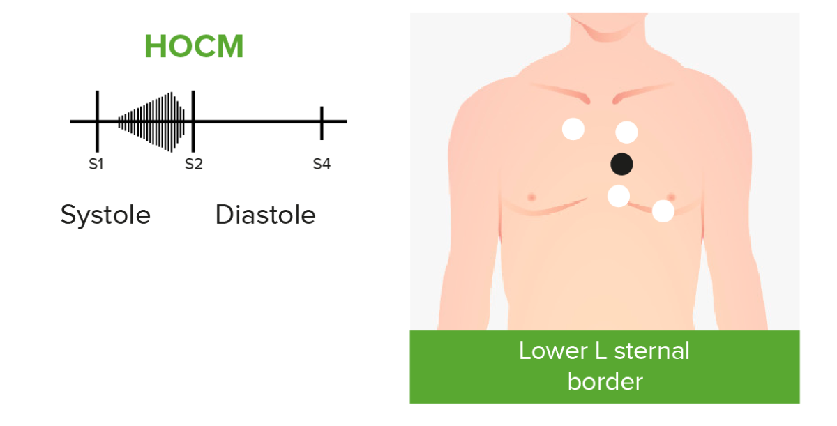
Cardiac murmurs in hocm

Phonocardiograms of abnormal heart sounds caused by hypertrophic obstructive cardiomyopathy (HOCM):
Patients with HOCM and left ventricular outflow tract obstruction may present with an audible crescendo-decrescendo systolic murmur best heard at the apex and lower left sternal border.
Image by Lecturio.

Diagnosis

  • Initial diagnosis based on history and physical examination:
    • Given the prevalence Prevalence The total number of cases of a given disease in a specified population at a designated time. It is differentiated from incidence, which refers to the number of new cases in the population at a given time. Measures of Disease Frequency of a lack of symptoms, physical examination and screening Screening Preoperative Care of all athletes annually during pre-participation is essential.
    • May require further investigation:
      • Unexplained chest pain Pain An unpleasant sensation induced by noxious stimuli which are detected by nerve endings of nociceptive neurons. Pain: Types and Pathways, shortness of breath Shortness of breath Dyspnea is the subjective sensation of breathing discomfort. Dyspnea is a normal manifestation of heavy physical or psychological exertion, but also may be caused by underlying conditions (both pulmonary and extrapulmonary). Dyspnea on exertion, syncope Syncope Syncope is a short-term loss of consciousness and loss of postural stability followed by spontaneous return of consciousness to the previous neurologic baseline without the need for resuscitation. The condition is caused by transient interruption of cerebral blood flow that may be benign or related to a underlying life-threatening condition. Syncope, palpitations Palpitations Ebstein’s Anomaly
      • Family history Family History Adult Health Maintenance of HCM or sudden cardiac death Sudden cardiac death Cardiac arrest is the sudden, complete cessation of cardiac output with hemodynamic collapse. Patients present as pulseless, unresponsive, and apneic. Rhythms associated with cardiac arrest are ventricular fibrillation/tachycardia, asystole, or pulseless electrical activity. Cardiac Arrest at an early age
      • Systolic heart murmur
  • ECG ECG An electrocardiogram (ECG) is a graphic representation of the electrical activity of the heart plotted against time. Adhesive electrodes are affixed to the skin surface allowing measurement of cardiac impulses from many angles. The ECG provides 3-dimensional information about the conduction system of the heart, the myocardium, and other cardiac structures. Electrocardiogram (ECG):
    • Abnormal in > 90% of cases
    • Left-axis deviation
    • P wave P wave Electrocardiogram (ECG) abnormalities (diphasic) consistent with left-atrial or biatrial enlargement
    • Pathological Q wave abnormalities in inferior (II, III, and aVF) and lateral leads (I, aVL, V5, V6)
    • Tall R waves in lateral leads with deep S waves in V1, V2:
    • May or may not show QRS widening 
    • ST-T wave abnormalities in lateral leads, including horizontal or down-sloping ST-segment with T wave T wave Electrocardiogram (ECG) inversions
  • Echocardiography Echocardiography Ultrasonic recording of the size, motion, and composition of the heart and surrounding tissues. The standard approach is transthoracic. Tricuspid Valve Atresia (TVA) with Doppler Doppler Ultrasonography applying the doppler effect, with frequency-shifted ultrasound reflections produced by moving targets (usually red blood cells) in the bloodstream along the ultrasound axis in direct proportion to the velocity of movement of the targets, to determine both direction and velocity of blood flow. Ultrasound (Sonography):
    • Assessment for:
    • Findings suggestive of HCM
      • Asymmetric thickening of the left ventricular wall
      • Variable Variable Variables represent information about something that can change. The design of the measurement scales, or of the methods for obtaining information, will determine the data gathered and the characteristics of that data. As a result, a variable can be qualitative or quantitative, and may be further classified into subgroups. Types of Variables wall thickening, most commonly seen in interventricular septum Interventricular Septum Ventricular Septal Defect (VSD)
    • Diagnostic criteria:
      • LVH with a maximum wall thickness > 15 mm in adults or a z-score Z-score Standard deviation difference between patient’s bone mass density and that of age-matched population. Osteoporosis > 3 in children
      • In case of positive family history Family History Adult Health Maintenance of HCM or pathogenic sarcomere Sarcomere The repeating contractile units of the myofibril, delimited by z bands along its length. Muscle Tissue: Histology variant, LVH with a maximum wall thickness of > 13 mm supports diagnosis 
    • The majority of patients Patients Individuals participating in the health care system for the purpose of receiving therapeutic, diagnostic, or preventive procedures. Clinician–Patient Relationship have normal left ventricular ejection fraction Ejection fraction Cardiac Cycle (LVEF).
  • Exercise stress testing:
    • Treadmill stress test preferred over pharmacological intervention → gives better idea of functional capacity and cardiovascular changes with physical activity
    • Should be performed in conjunction with echocardiography Echocardiography Ultrasonic recording of the size, motion, and composition of the heart and surrounding tissues. The standard approach is transthoracic. Tricuspid Valve Atresia (TVA) 
  • Ambulatory monitoring (Holter monitor):
    • Should be performed for 24–48 hours
    • Used to evaluate for atrial and ventricular arrhythmias
  • Cardiac MRI Cardiac MRI Imaging of the Heart and Great Vessels with gadolinium Gadolinium An element of the rare earth family of metals. It has the atomic symbol gd, atomic number 64, and atomic weight 157. 25. Its oxide is used in the control rods of some nuclear reactors. Magnetic Resonance Imaging (MRI): provides more advanced and reliable assessment of cardiac structure, anatomy, and function than echocardiography Echocardiography Ultrasonic recording of the size, motion, and composition of the heart and surrounding tissues. The standard approach is transthoracic. Tricuspid Valve Atresia (TVA)
  • Coronary angiography Angiography Radiography of blood vessels after injection of a contrast medium. Cardiac Surgery and/or cardiac catheterization Cardiac Catheterization Procedures in which placement of cardiac catheters is performed for therapeutic or diagnostic procedures. Cardiac Surgery is reserved for patients Patients Individuals participating in the health care system for the purpose of receiving therapeutic, diagnostic, or preventive procedures. Clinician–Patient Relationship in whom coronary artery Coronary Artery Truncus Arteriosus disease needs to be ruled out.
  • Genetic testing Genetic Testing Detection of a mutation; genotype; karyotype; or specific alleles associated with genetic traits, heritable diseases, or predisposition to a disease, or that may lead to the disease in descendants. It includes prenatal genetic testing. Myotonic Dystrophies: only if familial HCM suspected

Management

Treatment

No pharmacological treatment is known to improve prognosis Prognosis A prediction of the probable outcome of a disease based on a individual’s condition and the usual course of the disease as seen in similar situations. Non-Hodgkin Lymphomas.

Goals of treatment:

  • Reduce symptoms.
  • Reduce left ventricular outflow tract obstruction.
  • Control HR to ↑ filling time (with caution to avoid bradycardia Bradycardia Bradyarrhythmia is a rhythm in which the heart rate is less than 60/min. Bradyarrhythmia can be physiologic, without symptoms or hemodynamic change. Pathologic bradyarrhythmia results in reduced cardiac output and hemodynamic instability causing syncope, dizziness, or dyspnea. Bradyarrhythmias)
  • Maintain atrial contractions.
  • Manage/correct any conduction disturbances.
  • Avoid volume depletion Volume depletion Volume status is a balance between water and solutes, the majority of which is Na. Volume depletion refers to a loss of both water and Na, whereas dehydration refers only to a loss of water. Volume depletion can be caused by GI losses, renal losses, bleeding, poor oral Na intake, or third spacing of fluids. Volume Depletion and Dehydration.

Asymptomatic cases:

  • Observed with routine follow up and surveillance Surveillance Developmental Milestones and Normal Growth (including repeat ECG ECG An electrocardiogram (ECG) is a graphic representation of the electrical activity of the heart plotted against time. Adhesive electrodes are affixed to the skin surface allowing measurement of cardiac impulses from many angles. The ECG provides 3-dimensional information about the conduction system of the heart, the myocardium, and other cardiac structures. Electrocardiogram (ECG) and echocardiography Echocardiography Ultrasonic recording of the size, motion, and composition of the heart and surrounding tissues. The standard approach is transthoracic. Tricuspid Valve Atresia (TVA)
  • Avoid strenuous exercise Strenuous exercise Physical activity which is usually regular and done with the intention of improving or maintaining physical fitness or health. Contrast with physical exertion which is concerned largely with the physiologic and metabolic response to energy expenditure. Cardiovascular Response to Exercise.

In setting of heart failure Heart Failure A heterogeneous condition in which the heart is unable to pump out sufficient blood to meet the metabolic need of the body. Heart failure can be caused by structural defects, functional abnormalities (ventricular dysfunction), or a sudden overload beyond its capacity. Chronic heart failure is more common than acute heart failure which results from sudden insult to cardiac function, such as myocardial infarction. Total Anomalous Pulmonary Venous Return (TAPVR) symptoms without left ventricular outflow tract obstruction:

  • Beta-blockers Beta-blockers Drugs that bind to but do not activate beta-adrenergic receptors thereby blocking the actions of beta-adrenergic agonists. Adrenergic beta-antagonists are used for treatment of hypertension, cardiac arrhythmias, angina pectoris, glaucoma, migraine headaches, and anxiety. Class 2 Antiarrhythmic Drugs (Beta Blockers) (treatment of choice) or nondihydropyridine calcium-channel blockers Calcium-Channel Blockers A class of drugs that act by selective inhibition of calcium influx through cellular membranes. Heart Failure and Angina Medication ( CCBs CCBs Calcium channel blockers (CCBS) are a class of medications that inhibit voltage-dependent L-type calcium channels of cardiac and vascular smooth muscle cells. The inhibition of these channels produces vasodilation and myocardial depression. There are 2 major classes of CCBS: dihydropyridines and non-dihydropyridines. Class 4 Antiarrhythmic Drugs (Calcium Channel Blockers))
  • Diuretics Diuretics Agents that promote the excretion of urine through their effects on kidney function. Heart Failure and Angina Medication if signs or symptoms of fluid overload
  • If severe symptoms persist and in case of reconfirmation of the absence of left ventricular outflow tract obstruction, a heart transplant Heart transplant The transference of a heart from one human or animal to another. Dilated Cardiomyopathy may be needed.

In setting of heart failure Heart Failure A heterogeneous condition in which the heart is unable to pump out sufficient blood to meet the metabolic need of the body. Heart failure can be caused by structural defects, functional abnormalities (ventricular dysfunction), or a sudden overload beyond its capacity. Chronic heart failure is more common than acute heart failure which results from sudden insult to cardiac function, such as myocardial infarction. Total Anomalous Pulmonary Venous Return (TAPVR) symptoms and left ventricular outflow tract obstruction:

  • Monotherapy with beta-blockers Beta-blockers Drugs that bind to but do not activate beta-adrenergic receptors thereby blocking the actions of beta-adrenergic agonists. Adrenergic beta-antagonists are used for treatment of hypertension, cardiac arrhythmias, angina pectoris, glaucoma, migraine headaches, and anxiety. Class 2 Antiarrhythmic Drugs (Beta Blockers): 1st-line treatment
  • Monotherapy with nondihydropyridine CCBs CCBs Calcium channel blockers (CCBS) are a class of medications that inhibit voltage-dependent L-type calcium channels of cardiac and vascular smooth muscle cells. The inhibition of these channels produces vasodilation and myocardial depression. There are 2 major classes of CCBS: dihydropyridines and non-dihydropyridines. Class 4 Antiarrhythmic Drugs (Calcium Channel Blockers) (i.e., verapamil Verapamil A calcium channel blocker that is a class IV anti-arrhythmia agent. Pulmonary Hypertension Drugs) may be used if:
    • Beta-blockers Beta-blockers Drugs that bind to but do not activate beta-adrenergic receptors thereby blocking the actions of beta-adrenergic agonists. Adrenergic beta-antagonists are used for treatment of hypertension, cardiac arrhythmias, angina pectoris, glaucoma, migraine headaches, and anxiety. Class 2 Antiarrhythmic Drugs (Beta Blockers) are contraindicated or not tolerated
    • No signs or symptoms of hypotension Hypotension Hypotension is defined as low blood pressure, specifically < 90/60 mm Hg, and is most commonly a physiologic response. Hypotension may be mild, serious, or life threatening, depending on the cause. Hypotension, volume overload, or severe dyspnea Dyspnea Dyspnea is the subjective sensation of breathing discomfort. Dyspnea is a normal manifestation of heavy physical or psychological exertion, but also may be caused by underlying conditions (both pulmonary and extrapulmonary). Dyspnea 
  • If monotherapy is not sufficient:
    • Addition of disopyramide Disopyramide A class I antiarrhythmic agent (one that interferes directly with the depolarization of the cardiac membrane and thus serves as a membrane-stabilizing agent) with a depressant action on the heart similar to that of guanidine. It also possesses some anticholinergic and local anesthetic properties. Class 1 Antiarrhythmic Drugs (Sodium Channel Blockers) (should not be used as monotherapy or in a setting of prolonged QT interval QT interval Electrocardiogram (ECG) on ECG ECG An electrocardiogram (ECG) is a graphic representation of the electrical activity of the heart plotted against time. Adhesive electrodes are affixed to the skin surface allowing measurement of cardiac impulses from many angles. The ECG provides 3-dimensional information about the conduction system of the heart, the myocardium, and other cardiac structures. Electrocardiogram (ECG))
    • Combination therapy beta-blocker + nondihydropyridine CCBs CCBs Calcium channel blockers (CCBS) are a class of medications that inhibit voltage-dependent L-type calcium channels of cardiac and vascular smooth muscle cells. The inhibition of these channels produces vasodilation and myocardial depression. There are 2 major classes of CCBS: dihydropyridines and non-dihydropyridines. Class 4 Antiarrhythmic Drugs (Calcium Channel Blockers)
  • Avoid
  • If limiting symptoms persist:
    • Surgical septal myectomy (partial resection of the thickened septal wall)
    • Septal ablation with alcohol ( ethanol Ethanol A clear, colorless liquid rapidly absorbed from the gastrointestinal tract and distributed throughout the body. It has bactericidal activity and is used often as a topical disinfectant. It is widely used as a solvent and preservative in pharmaceutical preparations as well as serving as the primary ingredient in alcoholic beverages. Ethanol Metabolism) via cardiac catheterization Cardiac Catheterization Procedures in which placement of cardiac catheters is performed for therapeutic or diagnostic procedures. Cardiac Surgery (creates localized myocardial infarct Infarct Area of necrotic cells in an organ, arising mainly from hypoxia and ischemia Ischemic Cell Damage resulting in cardiac remodeling)

Complications

  • Concurrent atrial fibrillation Atrial fibrillation Atrial fibrillation (AF or Afib) is a supraventricular tachyarrhythmia and the most common kind of arrhythmia. It is caused by rapid, uncontrolled atrial contractions and uncoordinated ventricular responses. Atrial Fibrillation:
    • Beta-blockers Beta-blockers Drugs that bind to but do not activate beta-adrenergic receptors thereby blocking the actions of beta-adrenergic agonists. Adrenergic beta-antagonists are used for treatment of hypertension, cardiac arrhythmias, angina pectoris, glaucoma, migraine headaches, and anxiety. Class 2 Antiarrhythmic Drugs (Beta Blockers), CCBs CCBs Calcium channel blockers (CCBS) are a class of medications that inhibit voltage-dependent L-type calcium channels of cardiac and vascular smooth muscle cells. The inhibition of these channels produces vasodilation and myocardial depression. There are 2 major classes of CCBS: dihydropyridines and non-dihydropyridines. Class 4 Antiarrhythmic Drugs (Calcium Channel Blockers), or antiarrhythmic drug therapy 
    • Cardioversion Cardioversion Atrial Fibrillation as a last resort
    • Atrial fibrillation Atrial fibrillation Atrial fibrillation (AF or Afib) is a supraventricular tachyarrhythmia and the most common kind of arrhythmia. It is caused by rapid, uncontrolled atrial contractions and uncoordinated ventricular responses. Atrial Fibrillation: thromboembolism Thromboembolism Obstruction of a blood vessel (embolism) by a blood clot (thrombus) in the blood stream. Systemic Lupus Erythematosus prophylaxis Prophylaxis Cephalosporins with a blood thinner such as warfarin Warfarin An anticoagulant that acts by inhibiting the synthesis of vitamin K-dependent coagulation factors. Warfarin is indicated for the prophylaxis and/or treatment of venous thrombosis and its extension, pulmonary embolism, and atrial fibrillation with embolization. It is also used as an adjunct in the prophylaxis of systemic embolism after myocardial infarction. Warfarin is also used as a rodenticide. Anticoagulants
    • Ventricular arrhythmias: Implantable cardioverter-defibrillator (ICD) should be considered.
  • LV apical aneurysm Aneurysm An aneurysm is a bulging, weakened area of a blood vessel that causes an abnormal widening of its diameter > 1.5 times the size of the native vessel. Aneurysms occur more often in arteries than in veins and are at risk of dissection and rupture, which can be life-threatening. Thoracic Aortic Aneurysms: thromboembolism Thromboembolism Obstruction of a blood vessel (embolism) by a blood clot (thrombus) in the blood stream. Systemic Lupus Erythematosus prophylaxis Prophylaxis Cephalosporins with a blood thinner such as warfarin Warfarin An anticoagulant that acts by inhibiting the synthesis of vitamin K-dependent coagulation factors. Warfarin is indicated for the prophylaxis and/or treatment of venous thrombosis and its extension, pulmonary embolism, and atrial fibrillation with embolization. It is also used as an adjunct in the prophylaxis of systemic embolism after myocardial infarction. Warfarin is also used as a rodenticide. Anticoagulants

Prognosis Prognosis A prediction of the probable outcome of a disease based on a individual’s condition and the usual course of the disease as seen in similar situations. Non-Hodgkin Lymphomas

Differential Diagnosis

  • Hypertensive heart disease: long-standing, untreated, and uncontrolled high BP may cause left ventricular cardiac hypertrophy Hypertrophy General increase in bulk of a part or organ due to cell enlargement and accumulation of fluids and secretions, not due to tumor formation, nor to an increase in the number of cells (hyperplasia). Cellular Adaptation. Hypertensive heart disease is typically seen in adults who have had high BP for > 10 years and have signs of other end-organ damage. Diagnosis is made based on ECG ECG An electrocardiogram (ECG) is a graphic representation of the electrical activity of the heart plotted against time. Adhesive electrodes are affixed to the skin surface allowing measurement of cardiac impulses from many angles. The ECG provides 3-dimensional information about the conduction system of the heart, the myocardium, and other cardiac structures. Electrocardiogram (ECG) and echocardiography Echocardiography Ultrasonic recording of the size, motion, and composition of the heart and surrounding tissues. The standard approach is transthoracic. Tricuspid Valve Atresia (TVA). Left ventricular wall thickness rarely exceeds 15 mm in hypertensive disease. Treatment is with anti-hypertensive medications, weight loss Weight loss Decrease in existing body weight. Bariatric Surgery, and sodium Sodium A member of the alkali group of metals. It has the atomic symbol na, atomic number 11, and atomic weight 23. Hyponatremia restriction. Gradual regression Regression Corneal Abrasions, Erosion, and Ulcers of LVH may occur over time with adequate treatment. 
  • Aortic valve Aortic valve The valve between the left ventricle and the ascending aorta which prevents backflow into the left ventricle. Heart: Anatomy stenosis Stenosis Hypoplastic Left Heart Syndrome (HLHS): may occur due to congenital Congenital Chorioretinitis valve abnormalities in younger patients Patients Individuals participating in the health care system for the purpose of receiving therapeutic, diagnostic, or preventive procedures. Clinician–Patient Relationship or secondary to atherosclerotic narrowing of the valve later in life. Aortic valve Aortic valve The valve between the left ventricle and the ascending aorta which prevents backflow into the left ventricle. Heart: Anatomy stenosis Stenosis Hypoplastic Left Heart Syndrome (HLHS) is the most common cause of left ventricular outflow tract obstruction causing left ventricular wall hypertrophy Hypertrophy General increase in bulk of a part or organ due to cell enlargement and accumulation of fluids and secretions, not due to tumor formation, nor to an increase in the number of cells (hyperplasia). Cellular Adaptation. Signs and symptoms are similar to those in HCM. Echocardiography Echocardiography Ultrasonic recording of the size, motion, and composition of the heart and surrounding tissues. The standard approach is transthoracic. Tricuspid Valve Atresia (TVA) confirms the diagnosis, showing thickened and calcified aortic leaflets with a narrowed aortic orifice. Treatment is with medications commonly used in heart failure Heart Failure A heterogeneous condition in which the heart is unable to pump out sufficient blood to meet the metabolic need of the body. Heart failure can be caused by structural defects, functional abnormalities (ventricular dysfunction), or a sudden overload beyond its capacity. Chronic heart failure is more common than acute heart failure which results from sudden insult to cardiac function, such as myocardial infarction. Total Anomalous Pulmonary Venous Return (TAPVR). In severe cases, valve-replacement surgery is recommended. 
  • Fabry disease Fabry disease Fabry disease (FD), also known as Anderson-Fabry disease, is an X-linked recessive lysosomal storage disorder and the 2nd most common of the lysosomal storage disorders. Fabry disease is caused by a deficiency in the alpha-galactosidase enzyme (alpha-Gal A), resulting in the accumulation of the glycosphingolipid globotriaosylceramide (Gb3) in lysosomes. Fabry Disease: an X-linked recessive X-Linked Recessive Duchenne Muscular Dystrophy glycolipid storage disease caused by a deficiency of the lysosomal enzyme, alpha-galactosidase Alpha-galactosidase An enzyme that catalyzes the hydrolysis of terminal, non-reducing alpha-d-galactose residues in alpha-galactosides including galactose oligosaccharides, galactomannans, and galactolipids. Fabry Disease A. Men are affected more than women. Fabry disease Fabry disease Fabry disease (FD), also known as Anderson-Fabry disease, is an X-linked recessive lysosomal storage disorder and the 2nd most common of the lysosomal storage disorders. Fabry disease is caused by a deficiency in the alpha-galactosidase enzyme (alpha-Gal A), resulting in the accumulation of the glycosphingolipid globotriaosylceramide (Gb3) in lysosomes. Fabry Disease causes concentric (most common), eccentric, and asymmetric left ventricular hypertrophy Ventricular Hypertrophy Tetralogy of Fallot, but left ventricular outflow tract obstruction is rare. Although ECG ECG An electrocardiogram (ECG) is a graphic representation of the electrical activity of the heart plotted against time. Adhesive electrodes are affixed to the skin surface allowing measurement of cardiac impulses from many angles. The ECG provides 3-dimensional information about the conduction system of the heart, the myocardium, and other cardiac structures. Electrocardiogram (ECG), echocardiography Echocardiography Ultrasonic recording of the size, motion, and composition of the heart and surrounding tissues. The standard approach is transthoracic. Tricuspid Valve Atresia (TVA), and cardiac MRI Cardiac MRI Imaging of the Heart and Great Vessels are useful in evaluation, determination of leukocyte alpha-galactosidase Alpha-galactosidase An enzyme that catalyzes the hydrolysis of terminal, non-reducing alpha-d-galactose residues in alpha-galactosides including galactose oligosaccharides, galactomannans, and galactolipids. Fabry Disease A activity and genetic testing Genetic Testing Detection of a mutation; genotype; karyotype; or specific alleles associated with genetic traits, heritable diseases, or predisposition to a disease, or that may lead to the disease in descendants. It includes prenatal genetic testing. Myotonic Dystrophies are needed for diagnosis. Treatment involves enzyme-replacement therapy in addition to pharmacological therapy for heart failure Heart Failure A heterogeneous condition in which the heart is unable to pump out sufficient blood to meet the metabolic need of the body. Heart failure can be caused by structural defects, functional abnormalities (ventricular dysfunction), or a sudden overload beyond its capacity. Chronic heart failure is more common than acute heart failure which results from sudden insult to cardiac function, such as myocardial infarction. Total Anomalous Pulmonary Venous Return (TAPVR)
  • Athlete’s heart: non-pathological cardiac hypertrophy Hypertrophy General increase in bulk of a part or organ due to cell enlargement and accumulation of fluids and secretions, not due to tumor formation, nor to an increase in the number of cells (hyperplasia). Cellular Adaptation with left ventricular wall thickness up to 13–15 mm may also be seen in highly trained athletes. Intense strength training and endurance training can induce increases in left ventricular mass Mass Three-dimensional lesion that occupies a space within the breast Imaging of the Breast, symmetrical Symmetrical Dermatologic Examination wall thickness, and cavity size. A thorough history including family history Family History Adult Health Maintenance, and differentiating the presenting features using ECG ECG An electrocardiogram (ECG) is a graphic representation of the electrical activity of the heart plotted against time. Adhesive electrodes are affixed to the skin surface allowing measurement of cardiac impulses from many angles. The ECG provides 3-dimensional information about the conduction system of the heart, the myocardium, and other cardiac structures. Electrocardiogram (ECG), echocardiography Echocardiography Ultrasonic recording of the size, motion, and composition of the heart and surrounding tissues. The standard approach is transthoracic. Tricuspid Valve Atresia (TVA), cardiac MRI Cardiac MRI Imaging of the Heart and Great Vessels, and stress testing help distinguish athlete’s heart from HCM. No treatment is required for athlete’s heart as it is a benign Benign Fibroadenoma condition. 
  • Restrictive cardiomyopathy Restrictive Cardiomyopathy Restrictive cardiomyopathy (RCM) is a fairly uncommon condition characterized by progressive stiffening of the cardiac muscle, which causes impaired relaxation and refilling of the heart during diastole, resulting in diastolic dysfunction and eventual heart failure. Restrictive Cardiomyopathy: a condition that occurs secondary to scarring Scarring Inflammation and/or infiltration of the heart muscle. Restrictive cardiomyopathy Restrictive Cardiomyopathy Restrictive cardiomyopathy (RCM) is a fairly uncommon condition characterized by progressive stiffening of the cardiac muscle, which causes impaired relaxation and refilling of the heart during diastole, resulting in diastolic dysfunction and eventual heart failure. Restrictive Cardiomyopathy is characterized by a stiffening of the ventricles causing impaired relaxation and refilling of the heart, resulting in diastolic dysfunction Diastolic dysfunction Restrictive Cardiomyopathy and eventual heart failure Heart Failure A heterogeneous condition in which the heart is unable to pump out sufficient blood to meet the metabolic need of the body. Heart failure can be caused by structural defects, functional abnormalities (ventricular dysfunction), or a sudden overload beyond its capacity. Chronic heart failure is more common than acute heart failure which results from sudden insult to cardiac function, such as myocardial infarction. Total Anomalous Pulmonary Venous Return (TAPVR). Patients Patients Individuals participating in the health care system for the purpose of receiving therapeutic, diagnostic, or preventive procedures. Clinician–Patient Relationship typically present with signs and symptoms of heart failure Heart Failure A heterogeneous condition in which the heart is unable to pump out sufficient blood to meet the metabolic need of the body. Heart failure can be caused by structural defects, functional abnormalities (ventricular dysfunction), or a sudden overload beyond its capacity. Chronic heart failure is more common than acute heart failure which results from sudden insult to cardiac function, such as myocardial infarction. Total Anomalous Pulmonary Venous Return (TAPVR). Diagnostic studies similar to those used for HCM help make the diagnosis. Treatment includes drug therapy and the use of implantable devices. A heart transplant Heart transplant The transference of a heart from one human or animal to another. Dilated Cardiomyopathy may be needed in refractory cases. 
  • Dilated cardiomyopathy Dilated Cardiomyopathy Dilated cardiomyopathy (DCM) is the most common type of non-ischemic cardiomyopathy and a common cause of heart failure (HF). The cause may be idiopathic, familial, or secondary to a variety of underlying conditions. The disease is characterized by the enlargement of 1 or both ventricles and reduced systolic function. Dilated Cardiomyopathy ( DCM DCM Dilated cardiomyopathy (DCM) is the most common type of non-ischemic cardiomyopathy and a common cause of heart failure (HF). The cause may be idiopathic, familial, or secondary to a variety of underlying conditions. The disease is characterized by the enlargement of 1 or both ventricles and reduced systolic function. Dilated Cardiomyopathy): the most common type of non-ischemic cardiomyopathy Cardiomyopathy Cardiomyopathy refers to a group of myocardial diseases associated with structural changes of the heart muscles (myocardium) and impaired systolic and/or diastolic function in the absence of other heart disorders (coronary artery disease, hypertension, valvular disease, and congenital heart disease). Cardiomyopathy: Overview and Types. Dilated cardiomyopathy Dilated Cardiomyopathy Dilated cardiomyopathy (DCM) is the most common type of non-ischemic cardiomyopathy and a common cause of heart failure (HF). The cause may be idiopathic, familial, or secondary to a variety of underlying conditions. The disease is characterized by the enlargement of 1 or both ventricles and reduced systolic function. Dilated Cardiomyopathy causes systolic heart failure Heart Failure A heterogeneous condition in which the heart is unable to pump out sufficient blood to meet the metabolic need of the body. Heart failure can be caused by structural defects, functional abnormalities (ventricular dysfunction), or a sudden overload beyond its capacity. Chronic heart failure is more common than acute heart failure which results from sudden insult to cardiac function, such as myocardial infarction. Total Anomalous Pulmonary Venous Return (TAPVR). The etiology may be idiopathic Idiopathic Dermatomyositis, familial, or secondary to several underlying conditions. Dilated cardiomyopathy Dilated Cardiomyopathy Dilated cardiomyopathy (DCM) is the most common type of non-ischemic cardiomyopathy and a common cause of heart failure (HF). The cause may be idiopathic, familial, or secondary to a variety of underlying conditions. The disease is characterized by the enlargement of 1 or both ventricles and reduced systolic function. Dilated Cardiomyopathy is characterized by the enlargement of 1 or both ventricles and reduced systolic function. Patients Patients Individuals participating in the health care system for the purpose of receiving therapeutic, diagnostic, or preventive procedures. Clinician–Patient Relationship typically present with signs and symptoms of heart failure Heart Failure A heterogeneous condition in which the heart is unable to pump out sufficient blood to meet the metabolic need of the body. Heart failure can be caused by structural defects, functional abnormalities (ventricular dysfunction), or a sudden overload beyond its capacity. Chronic heart failure is more common than acute heart failure which results from sudden insult to cardiac function, such as myocardial infarction. Total Anomalous Pulmonary Venous Return (TAPVR). Diagnostic studies similar to those used for HCM help make the diagnosis. Treatment includes drug therapy and the use of implantable devices. Dilated cardiomyopathy Dilated Cardiomyopathy Dilated cardiomyopathy (DCM) is the most common type of non-ischemic cardiomyopathy and a common cause of heart failure (HF). The cause may be idiopathic, familial, or secondary to a variety of underlying conditions. The disease is characterized by the enlargement of 1 or both ventricles and reduced systolic function. Dilated Cardiomyopathy is the most common cause of cardiac transplantation.

References

  1. Iantorno, M. (2020). Hypertrophic cardiomyopathy. MedlinePlus. Retrieved February 6, 2021, from https://medlineplus.gov/ency/article/000192.htm
  2. Morita, H., et al. (2006). Single-gene mutations and increased left ventricular wall thickness in the community: The Framingham Heart Study. Circulation. https://pubmed.ncbi.nlm.nih.gov/16754800/ 
  3. Veselka, J., Anavekar, N.S., Charron, P. (2017). Hypertrophic obstructive cardiomyopathy. Lancet. https://pubmed.ncbi.nlm.nih.gov/27912983/ 
  4. Wigle, E.D., Rakowski, H., Kimball, B.P., Williams, W.G. (1995). Hypertrophic cardiomyopathy. Clinical spectrum and treatment. Circulation. https://pubmed.ncbi.nlm.nih.gov/7671349/ 
  5. Elliott, P.M., Kaski, J.C., Prasad, K., Seo, H., Slade, A.K., Goldman, J.H., McKenna, W.J. (1996). Chest pain during daily life in patients with hypertrophic cardiomyopathy: An ambulatory electrocardiographic study. Eur Heart J. https://pubmed.ncbi.nlm.nih.gov/8809524/ 
  6. Gersh, B.J. et al. (2011). American College of Cardiology Foundation/American Heart Association Task Force on Practice Guidelines; American Association for Thoracic Surgery; American Society of Echocardiography; American Society of Nuclear Cardiology; Heart Failure Society of America; Heart Rhythm Society; Society for Cardiovascular Angiography and Interventions; Society of Thoracic Surgeons. 2011 ACCF/AHA guideline for the diagnosis and treatment of hypertrophic cardiomyopathy: Executive summary: A report of the American College of Cardiology Foundation/American Heart Association Task Force on Practice Guidelines. Circulation. https://pubmed.ncbi.nlm.nih.gov/22068435/ 
  7. Veselka, J., Anavekar, N.S., Charron, P. Hypertrophic obstructive cardiomyopathy. Lancet. 2017 Mar 25;389(10075):1253-1267. https://pubmed.ncbi.nlm.nih.gov/27912983/ 
  8. Cirino, A.L., Ho, C. (2008, updated 2019) Hypertrophic Cardiomyopathy Overview. GeneReviews® [Internet]. Retrieved February 6, 2021, from https://www.ncbi.nlm.nih.gov/books/NBK1768/
  9. Shore, S. (2020). 2020 AHA/ACC Guideline for Hypertrophic Cardiomyopathy: Key Perspectives. American College of Cardiology. Retrieved February 8, 2021, from https://www.acc.org/latest-in-cardiology/ten-points-to-remember/2020/11/18/18/47/2020-aha-acc-guideline-for-hcm-gl-hcm

Create your free account or log in to continue reading!

Sign up now and get free access to Lecturio with concept pages, medical videos, and questions for your medical education.

User Reviews

Details