Hydrocephalus in Children

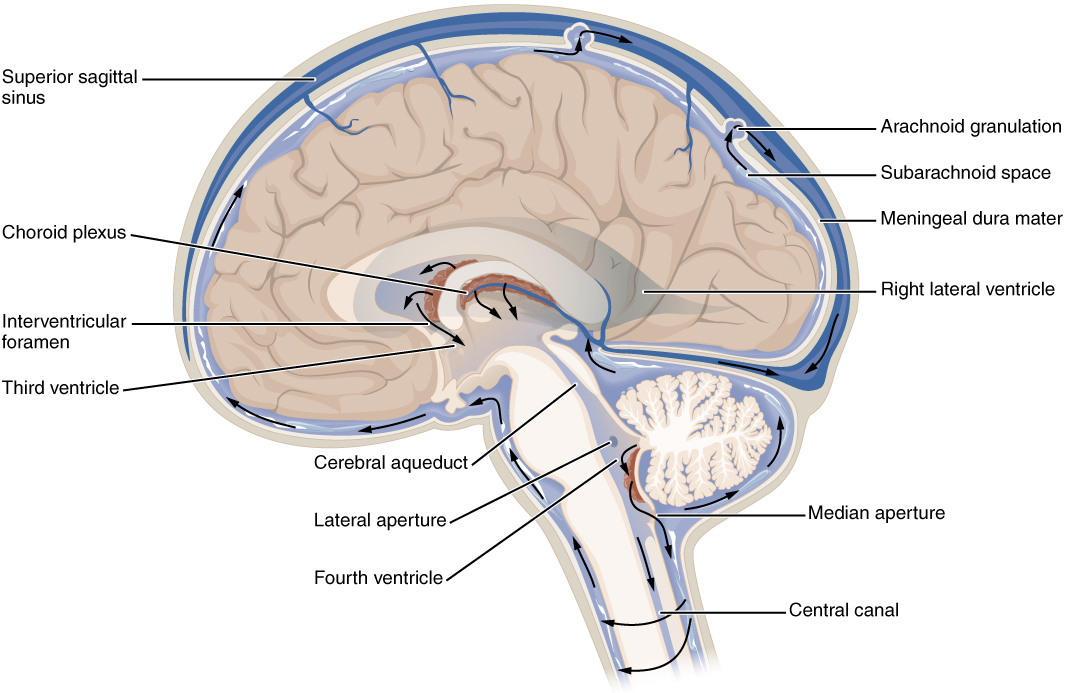
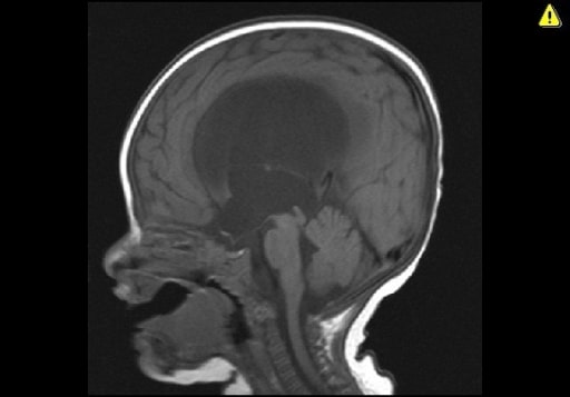
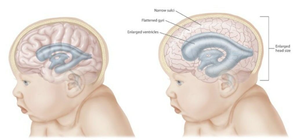
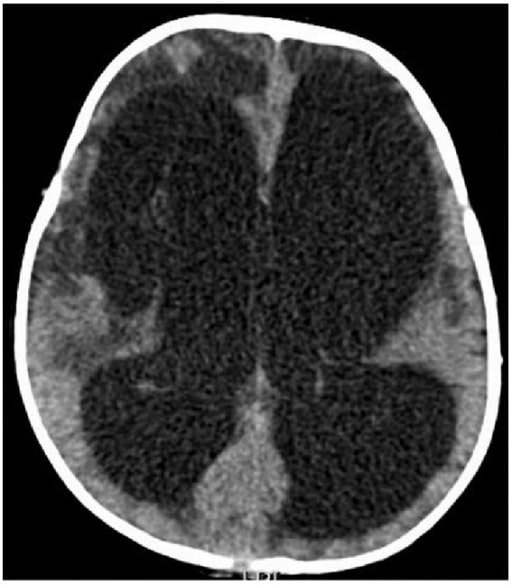
Hydrocephalus Hydrocephalus Excessive accumulation of cerebrospinal fluid within the cranium which may be associated with dilation of cerebral ventricles, intracranial. Subarachnoid Hemorrhage is a potentially life-threatening condition caused by the excess accumulation of cerebrospinal fluid Cerebrospinal Fluid A watery fluid that is continuously produced in the choroid plexus and circulates around the surface of the brain; spinal cord; and in the cerebral ventricles. Ventricular System: Anatomy (CSF) within the ventricular system Ventricular System The ventricular system is an extension of the subarachnoid space into the brain consisting of a series of interconnecting spaces and channels. Four chambers are filled with cerebrospinal fluid (CSF): the paired lateral ventricles, the unpaired 3rd ventricle, and the unpaired 4th ventricle. Ventricular System: Anatomy. Hydrocephalus Hydrocephalus Excessive accumulation of cerebrospinal fluid within the cranium which may be associated with dilation of cerebral ventricles, intracranial. Subarachnoid Hemorrhage can be classified as communicating, which is caused by either impaired CSF absorption Absorption Absorption involves the uptake of nutrient molecules and their transfer from the lumen of the GI tract across the enterocytes and into the interstitial space, where they can be taken up in the venous or lymphatic circulation. Digestion and Absorption or excess CSF production; or non-communicating, which is caused by a structural blockage in CSF flow Flow Blood flows through the heart, arteries, capillaries, and veins in a closed, continuous circuit. Flow is the movement of volume per unit of time. Flow is affected by the pressure gradient and the resistance fluid encounters between 2 points. Vascular resistance is the opposition to flow, which is caused primarily by blood friction against vessel walls. Vascular Resistance, Flow, and Mean Arterial Pressure. The causes of hydrocephalus Hydrocephalus Excessive accumulation of cerebrospinal fluid within the cranium which may be associated with dilation of cerebral ventricles, intracranial. Subarachnoid Hemorrhage can be congenital Congenital Chorioretinitis or acquired. The clinical presentation is often nonspecific with symptoms including headache Headache The symptom of pain in the cranial region. It may be an isolated benign occurrence or manifestation of a wide variety of headache disorders. Brain Abscess, behavioral changes, developmental delays, or nausea Nausea An unpleasant sensation in the stomach usually accompanied by the urge to vomit. Common causes are early pregnancy, sea and motion sickness, emotional stress, intense pain, food poisoning, and various enteroviruses. Antiemetics and vomiting Vomiting The forcible expulsion of the contents of the stomach through the mouth. Hypokalemia. If the hydrocephalus Hydrocephalus Excessive accumulation of cerebrospinal fluid within the cranium which may be associated with dilation of cerebral ventricles, intracranial. Subarachnoid Hemorrhage is congenital Congenital Chorioretinitis or acquired at a young age, infants often present with macrocephaly Macrocephaly Cowden Syndrome. Diagnosis is confirmed with neuroimaging Neuroimaging Non-invasive methods of visualizing the central nervous system, especially the brain, by various imaging modalities. Febrile Infant (ultrasound, computed tomography of the head, or magnetic resonance imaging) showing ventriculomegaly. Treatment is most commonly surgical with the placement of a CSF shunt.

Last updated: Feb 14, 2023

Editorial responsibility: Stanley Oiseth, Lindsay Jones, Evelin Maza

Epidemiology

Incidence Incidence The number of new cases of a given disease during a given period in a specified population. It also is used for the rate at which new events occur in a defined population. It is differentiated from prevalence, which refers to all cases in the population at a given time. Measures of Disease Frequency

  • Highest in Latin America (316 per 100,000 births) and Africa (145 per 100,000 births)
  • Lowest in United States and Canada (68 per 100,000 births)
  • Higher incidence Incidence The number of new cases of a given disease during a given period in a specified population. It also is used for the rate at which new events occur in a defined population. It is differentiated from prevalence, which refers to all cases in the population at a given time. Measures of Disease Frequency in low- and middle-income countries due to:
    • High crude birth rates
    • High prevalence Prevalence The total number of cases of a given disease in a specified population at a designated time. It is differentiated from incidence, which refers to the number of new cases in the population at a given time. Measures of Disease Frequency of neglected tropical diseases ( NTDs NTDs Neural tube defects (NTDs) are the 2nd-most common type of congenital birth defects. Neural tube defects can range from asymptomatic (closed ntd) to very severe malformations of the spine or brain (open ntd). Neural tube defects are caused by the failure of the neural tube to close properly during the 3rd and 4th week of embryological development. Neural Tube Defects)

Risk factors

Classification

There are different ways to characterize hydrocephalus Hydrocephalus Excessive accumulation of cerebrospinal fluid within the cranium which may be associated with dilation of cerebral ventricles, intracranial. Subarachnoid Hemorrhage, each of which can help physicians Physicians Individuals licensed to practice medicine. Clinician–Patient Relationship better understand the underlying cause and determine the appropriate treatment.
Communicating versus non-communicating

  • Communicating (non-obstructive): caused by impaired absorption Absorption Absorption involves the uptake of nutrient molecules and their transfer from the lumen of the GI tract across the enterocytes and into the interstitial space, where they can be taken up in the venous or lymphatic circulation. Digestion and Absorption (most common) or excess production of cerebrospinal fluid Cerebrospinal Fluid A watery fluid that is continuously produced in the choroid plexus and circulates around the surface of the brain; spinal cord; and in the cerebral ventricles. Ventricular System: Anatomy (CSF) 
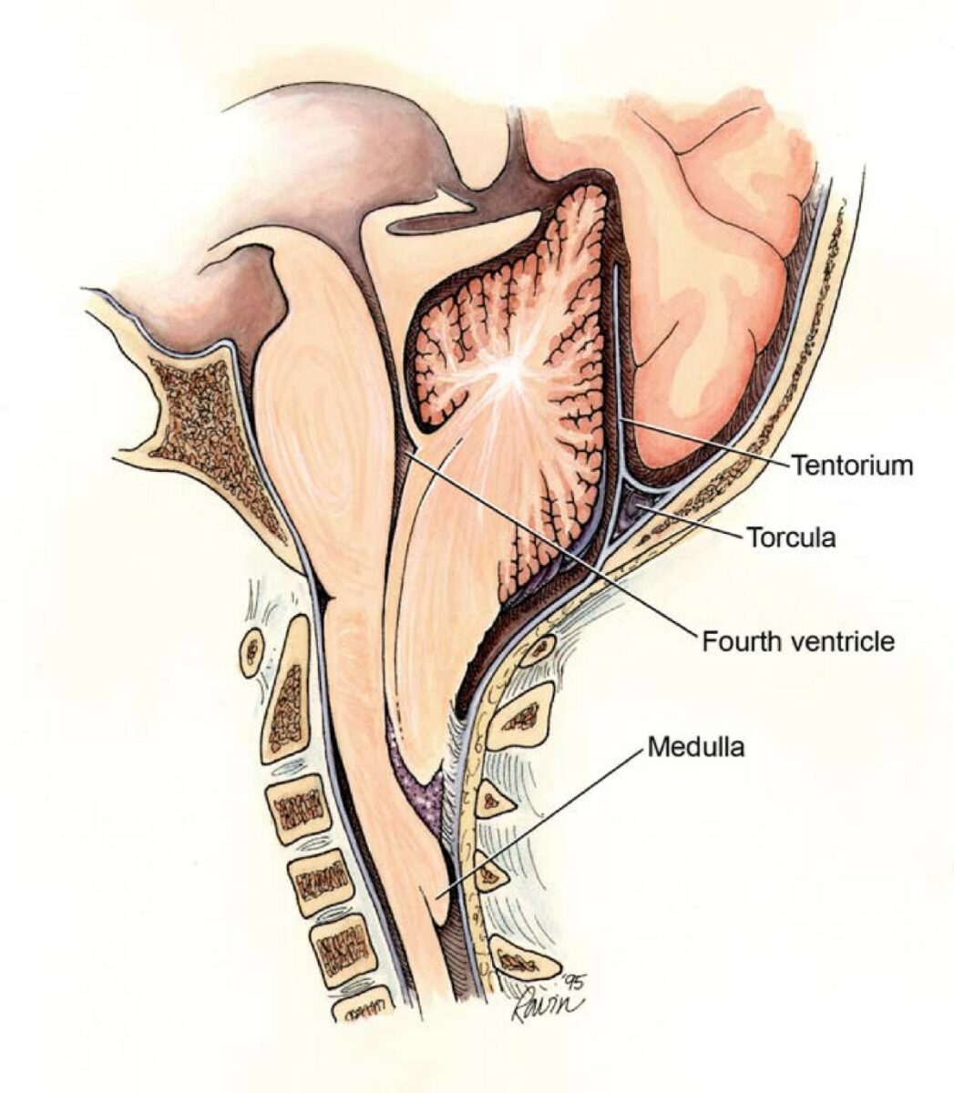
  • Non-communicating (obstructive): caused by a structural blockage of CSF flow Flow Blood flows through the heart, arteries, capillaries, and veins in a closed, continuous circuit. Flow is the movement of volume per unit of time. Flow is affected by the pressure gradient and the resistance fluid encounters between 2 points. Vascular resistance is the opposition to flow, which is caused primarily by blood friction against vessel walls. Vascular Resistance, Flow, and Mean Arterial Pressure in the ventricular system Ventricular System The ventricular system is an extension of the subarachnoid space into the brain consisting of a series of interconnecting spaces and channels. Four chambers are filled with cerebrospinal fluid (CSF): the paired lateral ventricles, the unpaired 3rd ventricle, and the unpaired 4th ventricle. Ventricular System: Anatomy. Obstruction of CSF can be:
    • Proximal
      • 3rd ventricle
      • Aqueduct
    • Distal
      • 4th ventricle 
      • Foramen magnum

Congenital Congenital Chorioretinitis versus acquired

  • Congenital Congenital Chorioretinitis
    • CNS malformations
      • Myelomeningocele
      • Chiari II malformation
      • Dandy-Walker malformation
      • Encephalocele Encephalocele Brain tissue herniation through a congenital or acquired defect in the skull. The majority of congenital encephaloceles occur in the occipital or frontal regions. Clinical features include a protuberant mass that may be pulsatile. The quantity and location of protruding neural tissue determines the type and degree of neurologic deficit. Visual defects, psychomotor developmental delay, and persistent motor deficits frequently occur. Neural Tube Defects
      • Aqueductal stenosis Stenosis Hypoplastic Left Heart Syndrome (HLHS)
    • Infections Infections Invasion of the host organism by microorganisms or their toxins or by parasites that can cause pathological conditions or diseases. Chronic Granulomatous Disease
      • Intrauterine infections Infections Invasion of the host organism by microorganisms or their toxins or by parasites that can cause pathological conditions or diseases. Chronic Granulomatous Disease
      • Rubella Rubella An acute infectious disease caused by the rubella virus. The virus enters the respiratory tract via airborne droplet and spreads to the lymphatic system. Rubella Virus, cytomegalovirus Cytomegalovirus CMV is a ubiquitous double-stranded DNA virus belonging to the Herpesviridae family. CMV infections can be transmitted in bodily fluids, such as blood, saliva, urine, semen, and breast milk. The initial infection is usually asymptomatic in the immunocompetent host, or it can present with symptoms of mononucleosis. Cytomegalovirus (CMV), toxoplasmosis Toxoplasmosis Toxoplasmosis is an infectious disease caused by Toxoplasma gondii, an obligate intracellular protozoan parasite. Felines are the definitive host, but transmission to humans can occur through contact with cat feces or the consumption of contaminated foods. The clinical presentation and complications depend on the host’s immune status. Toxoplasma/Toxoplasmosis, Zika virus Zika virus An arbovirus in the flavivirus genus of the family flaviviridae. Originally isolated in the zika forest of uganda it has been introduced to Asia and the americas. Zika Virus Infection, enterovirus Enterovirus A genus of the family picornaviridae whose members preferentially inhabit the intestinal tract of a variety of hosts. The genus contains many species. Newly described members of human enteroviruses are assigned continuous numbers with the species designated ‘human enterovirus’. Coxsackievirus
    • Intraventricular hemorrhage Intraventricular hemorrhage Bleeding within the cerebral ventricles. It is associated with intraventricular trauma, aneurysm, vascular malformations, hypertension and in very low birth weight infants. Intracerebral Hemorrhage: associated with prematurity Prematurity Neonatal Respiratory Distress Syndrome
    • Medication during pregnancy Pregnancy The status during which female mammals carry their developing young (embryos or fetuses) in utero before birth, beginning from fertilization to birth. Pregnancy: Diagnosis, Physiology, and Care: isotretinoin, misoprostol Misoprostol A synthetic analog of natural prostaglandin e1. It produces a dose-related inhibition of gastric acid and pepsin secretion, and enhances mucosal resistance to injury. It is an effective anti-ulcer agent and also has oxytocic properties. Eicosanoids, metronidazole Metronidazole A nitroimidazole used to treat amebiasis; vaginitis; trichomonas infections; giardiasis; anaerobic bacteria; and treponemal infections. Pyogenic Liver Abscess, antidepressants 
    • Genetics Genetics Genetics is the study of genes and their functions and behaviors. Basic Terms of Genetics 
  • Acquired
    • Hemorrhage (intraventricular or subarachnoid)
    • Infections Infections Invasion of the host organism by microorganisms or their toxins or by parasites that can cause pathological conditions or diseases. Chronic Granulomatous Disease: meningitis Meningitis Meningitis is inflammation of the meninges, the protective membranes of the brain, and spinal cord. The causes of meningitis are varied, with the most common being bacterial or viral infection. The classic presentation of meningitis is a triad of fever, altered mental status, and nuchal rigidity. Meningitis 
    • CNS tumors
Chiari 2 malformation

Chiari II malformation showing the points of potential obstruction that yield different subtypes of hydrocephalus

Image: “Chiari2” by Rekate HL. License: CC BY 2.0
Pathophysiology

Normal CSF production

  • CSF is produced in the choroid Choroid The thin, highly vascular membrane covering most of the posterior of the eye between the retina and sclera. Eye: Anatomy plexus of the ventricles → circulates through the ventricular system Ventricular System The ventricular system is an extension of the subarachnoid space into the brain consisting of a series of interconnecting spaces and channels. Four chambers are filled with cerebrospinal fluid (CSF): the paired lateral ventricles, the unpaired 3rd ventricle, and the unpaired 4th ventricle. Ventricular System: Anatomy → 4th ventricle → subarachnoid space Subarachnoid space The space between the arachnoid membrane and pia mater, filled with cerebrospinal fluid. It contains large blood vessels that supply the brain and spinal cord. Subarachnoid Hemorrhage → absorbed by arachnoid villi (located in the subarachnoid space Subarachnoid space The space between the arachnoid membrane and pia mater, filled with cerebrospinal fluid. It contains large blood vessels that supply the brain and spinal cord. Subarachnoid Hemorrhage) → venous sinuses Venous sinuses Veins: Histology + systemic circulation Circulation The movement of the blood as it is pumped through the cardiovascular system. ABCDE Assessment 
  • Normal volume of CSF: 150 ml (reached by 5 years of age)

Acquired hydrocephalus Hydrocephalus Excessive accumulation of cerebrospinal fluid within the cranium which may be associated with dilation of cerebral ventricles, intracranial. Subarachnoid Hemorrhage

  • Most commonly hemorrhage as a result of prematurity Prematurity Neonatal Respiratory Distress Syndrome 
  • Most common defect: resorption of CSF
  • Impaired CSF absorption Absorption Absorption involves the uptake of nutrient molecules and their transfer from the lumen of the GI tract across the enterocytes and into the interstitial space, where they can be taken up in the venous or lymphatic circulation. Digestion and Absorption most often occurs due to subarachnoid villi inflammation Inflammation Inflammation is a complex set of responses to infection and injury involving leukocytes as the principal cellular mediators in the body’s defense against pathogenic organisms. Inflammation is also seen as a response to tissue injury in the process of wound healing. The 5 cardinal signs of inflammation are pain, heat, redness, swelling, and loss of function. Inflammation.
  • Subarachnoid villi inflammation Inflammation Inflammation is a complex set of responses to infection and injury involving leukocytes as the principal cellular mediators in the body’s defense against pathogenic organisms. Inflammation is also seen as a response to tissue injury in the process of wound healing. The 5 cardinal signs of inflammation are pain, heat, redness, swelling, and loss of function. Inflammation results in CSF accumulation causing:
    • Distension of the ventricular system Ventricular System The ventricular system is an extension of the subarachnoid space into the brain consisting of a series of interconnecting spaces and channels. Four chambers are filled with cerebrospinal fluid (CSF): the paired lateral ventricles, the unpaired 3rd ventricle, and the unpaired 4th ventricle. Ventricular System: Anatomy (ventricular dilation)
    • Increased intracranial pressure Intracranial Pressure Idiopathic Intracranial Hypertension ( ICP ICP Normal intracranial pressure (ICP) is defined as < 15 mm Hg, whereas pathologically increased ICP is any pressure ≥ 20 mm Hg. Increased ICP may result from several etiologies, including trauma, intracranial hemorrhage, mass lesions, cerebral edema, increased CSF production, and decreased CSF absorption. Increased Intracranial Pressure (ICP))

Clinical Presentation

Symptoms

  • Neonates and infants:
    • Irritability
    • Vomiting Vomiting The forcible expulsion of the contents of the stomach through the mouth. Hypokalemia
    • Poor feeding
    • Seizures Seizures A seizure is abnormal electrical activity of the neurons in the cerebral cortex that can manifest in numerous ways depending on the region of the brain affected. Seizures consist of a sudden imbalance that occurs between the excitatory and inhibitory signals in cortical neurons, creating a net excitation. The 2 major classes of seizures are focal and generalized. Seizures
  • Children:
    • Failure to thrive Failure to Thrive Failure to thrive (FTT), or faltering growth, describes suboptimal weight gain and growth in children. The majority of cases are due to inadequate caloric intake; however, genetic, infectious, and oncological etiologies are also common. Failure to Thrive
    • Behavioral changes → altered mental status Altered Mental Status Sepsis in Children
    • Developmental delays
    • Severe headache Headache The symptom of pain in the cranial region. It may be an isolated benign occurrence or manifestation of a wide variety of headache disorders. Brain Abscess
    • Persistent nausea Nausea An unpleasant sensation in the stomach usually accompanied by the urge to vomit. Common causes are early pregnancy, sea and motion sickness, emotional stress, intense pain, food poisoning, and various enteroviruses. Antiemetics and vomiting Vomiting The forcible expulsion of the contents of the stomach through the mouth. Hypokalemia
    • Drowsiness
    • Abnormal gait Gait Manner or style of walking. Neurological Examination

Physical examination

  • Neonates:
    • Tense or bulging fontanelles Fontanelles Physical Examination of the Newborn
    • Frontal Frontal The bone that forms the frontal aspect of the skull. Its flat part forms the forehead, articulating inferiorly with the nasal bone and the cheek bone on each side of the face. Skull: Anatomy bossing
    • Papilledema Papilledema Swelling of the optic disk, usually in association with increased intracranial pressure, characterized by hyperemia, blurring of the disk margins, microhemorrhages, blind spot enlargement, and engorgement of retinal veins. Chronic papilledema may cause optic atrophy and visual loss. Idiopathic Intracranial Hypertension
    • Irritability
    • Vomiting Vomiting The forcible expulsion of the contents of the stomach through the mouth. Hypokalemia
  • Infants:
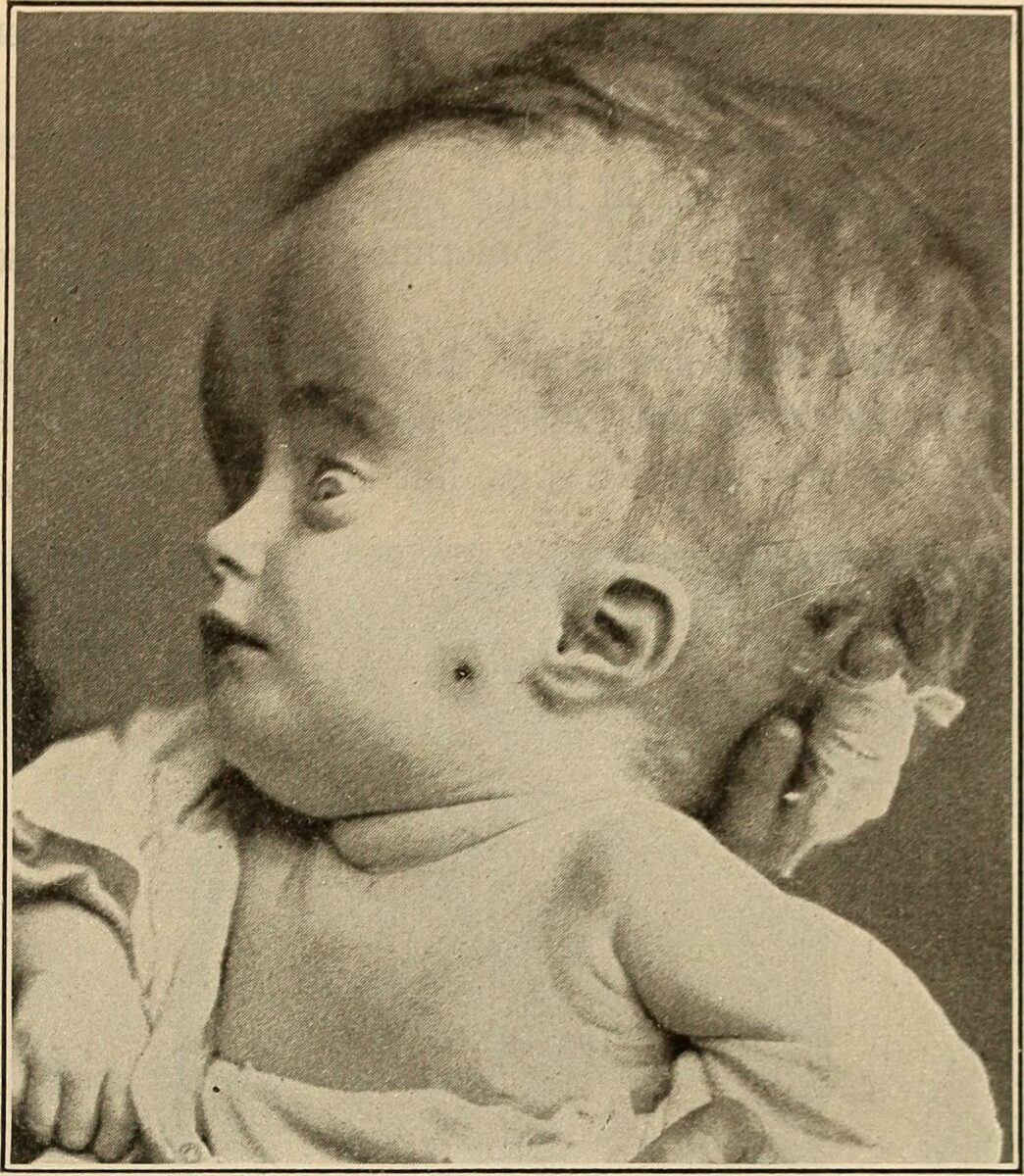
    • “Setting sun” sign: upward gaze paresis Paresis A general term referring to a mild to moderate degree of muscular weakness, occasionally used as a synonym for paralysis (severe or complete loss of motor function). In the older literature, paresis often referred specifically to paretic neurosyphilis. ‘general paresis’ and ‘general paralysis’ may still carry that connotation. Bilateral lower extremity paresis is referred to as paraparesis. Spinal Disk Herniation, eyes appear driven down (early symptom, seen in 40% of obstructive hydrocephalus Hydrocephalus Excessive accumulation of cerebrospinal fluid within the cranium which may be associated with dilation of cerebral ventricles, intracranial. Subarachnoid Hemorrhage)
  • Children:
    • “Setting sun” sign
    • Cognitive deterioration
    • Imbalance and gait Gait Manner or style of walking. Neurological Examination disturbances
    • Urinary incontinence Urinary incontinence Urinary incontinence (UI) is involuntary loss of bladder control or unintentional voiding, which represents a hygienic or social problem to the patient. Urinary incontinence is a symptom, a sign, and a disorder. The 5 types of UI include stress, urge, mixed, overflow, and functional. Urinary Incontinence
    • Papilledema Papilledema Swelling of the optic disk, usually in association with increased intracranial pressure, characterized by hyperemia, blurring of the disk margins, microhemorrhages, blind spot enlargement, and engorgement of retinal veins. Chronic papilledema may cause optic atrophy and visual loss. Idiopathic Intracranial Hypertension
Sunset sign hydrocephalus

Photo of a child with hydrocephalus and a noticeable “setting sun” sign

Image: “Sunset sign” by Louis Fischer. License: Public Domain
Diagnosis

Management

Any patient with hydrocephalus Hydrocephalus Excessive accumulation of cerebrospinal fluid within the cranium which may be associated with dilation of cerebral ventricles, intracranial. Subarachnoid Hemorrhage and symptoms of ICP ICP Normal intracranial pressure (ICP) is defined as < 15 mm Hg, whereas pathologically increased ICP is any pressure ≥ 20 mm Hg. Increased ICP may result from several etiologies, including trauma, intracranial hemorrhage, mass lesions, cerebral edema, increased CSF production, and decreased CSF absorption. Increased Intracranial Pressure (ICP) requires intervention. The method used to relieve the pressure and the speed at which it is deployed is based on the severity of the patient’s ICP ICP Normal intracranial pressure (ICP) is defined as < 15 mm Hg, whereas pathologically increased ICP is any pressure ≥ 20 mm Hg. Increased ICP may result from several etiologies, including trauma, intracranial hemorrhage, mass lesions, cerebral edema, increased CSF production, and decreased CSF absorption. Increased Intracranial Pressure (ICP).
  • Acutely symptomatic patients Patients Individuals participating in the health care system for the purpose of receiving therapeutic, diagnostic, or preventive procedures. Clinician–Patient Relationship with life-threatening hydrocephalus Hydrocephalus Excessive accumulation of cerebrospinal fluid within the cranium which may be associated with dilation of cerebral ventricles, intracranial. Subarachnoid Hemorrhage:
    • Extra ventricular drain (EVD): transcranial catheter connected to an external closed reservoir Reservoir Animate or inanimate sources which normally harbor disease-causing organisms and thus serve as potential sources of disease outbreaks. Reservoirs are distinguished from vectors (disease vectors) and carriers, which are agents of disease transmission rather than continuing sources of potential disease outbreaks. Humans may serve both as disease reservoirs and carriers. Escherichia coli
      • Can be placed rapidly and emergently
      • Collects CSF to relieve pressure 
      • Allows serial measurement of ICP ICP Normal intracranial pressure (ICP) is defined as < 15 mm Hg, whereas pathologically increased ICP is any pressure ≥ 20 mm Hg. Increased ICP may result from several etiologies, including trauma, intracranial hemorrhage, mass lesions, cerebral edema, increased CSF production, and decreased CSF absorption. Increased Intracranial Pressure (ICP) and serial CSF sampling
    • Diuretics Diuretics Agents that promote the excretion of urine through their effects on kidney function. Heart Failure and Angina Medication: may be acutely effective in select cases but do not have a long-term role
      • Acetazolamide Acetazolamide One of the carbonic anhydrase inhibitors that is sometimes effective against absence seizures. It is sometimes useful also as an adjunct in the treatment of tonic-clonic, myoclonic, and atonic seizures, particularly in women whose seizures occur or are exacerbated at specific times in the menstrual cycle. However, its usefulness is transient often because of rapid development of tolerance. Its antiepileptic effect may be due to its inhibitory effect on brain carbonic anhydrase, which leads to an increased transneuronal chloride gradient, increased chloride current, and increased inhibition. Carbonic Anhydrase Inhibitors: decreases CSF production at the level of the choroid Choroid The thin, highly vascular membrane covering most of the posterior of the eye between the retina and sclera. Eye: Anatomy plexus by inhibiting carbonic anhydrase Carbonic anhydrase A family of zinc-containing enzymes that catalyze the reversible hydration of carbon dioxide. They play an important role in the transport of carbon dioxide from the tissues to the lung. Carbonic Anhydrase Inhibitors activity
      • Furosemide Furosemide A benzoic-sulfonamide-furan. It is a diuretic with fast onset and short duration that is used for edema and chronic renal insufficiency. Loop Diuretics: similar effect as acetazolamide Acetazolamide One of the carbonic anhydrase inhibitors that is sometimes effective against absence seizures. It is sometimes useful also as an adjunct in the treatment of tonic-clonic, myoclonic, and atonic seizures, particularly in women whose seizures occur or are exacerbated at specific times in the menstrual cycle. However, its usefulness is transient often because of rapid development of tolerance. Its antiepileptic effect may be due to its inhibitory effect on brain carbonic anhydrase, which leads to an increased transneuronal chloride gradient, increased chloride current, and increased inhibition. Carbonic Anhydrase Inhibitors, but not as potent and not usually used alone
    • Lumbar puncture Lumbar Puncture Febrile Infant (LP):
      • Used for diagnostic sampling of CSF, can also be used to reduce CSF volume and thus ICP ICP Normal intracranial pressure (ICP) is defined as < 15 mm Hg, whereas pathologically increased ICP is any pressure ≥ 20 mm Hg. Increased ICP may result from several etiologies, including trauma, intracranial hemorrhage, mass lesions, cerebral edema, increased CSF production, and decreased CSF absorption. Increased Intracranial Pressure (ICP)
      • Due to the risk of infection and unclear benefit, serial LPs are no longer commonly performed.
  • Symptomatic but clinically stable or nonprogressive hydrocephalus Hydrocephalus Excessive accumulation of cerebrospinal fluid within the cranium which may be associated with dilation of cerebral ventricles, intracranial. Subarachnoid Hemorrhage patients Patients Individuals participating in the health care system for the purpose of receiving therapeutic, diagnostic, or preventive procedures. Clinician–Patient Relationship:
    • More intensive surgery is often tolerated.
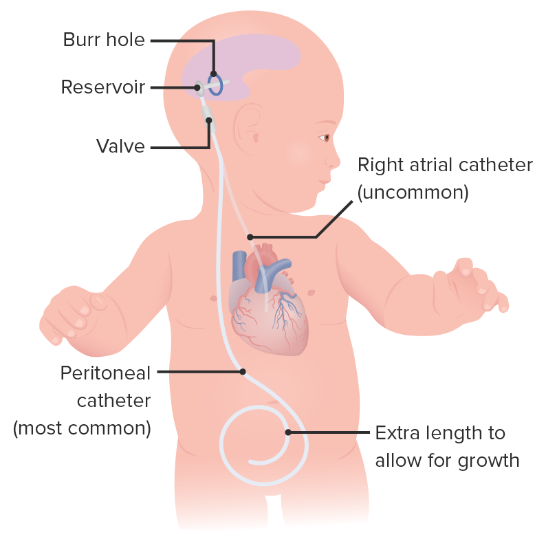
    • CSF shunting: ventriculoperitoneal or ventriculopleural shunt
      • Catheter draining CSF to spaces in the body with high surface area and high capillary concentration where it can be safely reabsorbed
      • Risks: infection and clogging of the catheters
    • Endoscopic 3rd ventriculostomy Ventriculostomy Surgical creation of an opening in a cerebral ventricle. Neurosurgery: creating a passageway (or ostomy) in the bottom of the 3rd ventricle allowing CSF to drain into the subarachnoid space Subarachnoid space The space between the arachnoid membrane and pia mater, filled with cerebrospinal fluid. It contains large blood vessels that supply the brain and spinal cord. Subarachnoid Hemorrhage
  • Asymptomatic, clinically stable patients Stable Patients Blunt Chest Trauma:
    • May be followed clinically
Illustration of a ventriculoperitoneal shunt in a baby

A diagram of a ventriculoperitoneal shunt in a baby

Image by Lecturio.

Complications

Disease progression

  • Herniation Herniation Omphalocele of the cerebral parenchyma
    • Visual problems secondary to compression Compression Blunt Chest Trauma of the optic nerve Optic nerve The 2nd cranial nerve which conveys visual information from the retina to the brain. The nerve carries the axons of the retinal ganglion cells which sort at the optic chiasm and continue via the optic tracts to the brain. The largest projection is to the lateral geniculate nuclei; other targets include the superior colliculi and the suprachiasmatic nuclei. Though known as the second cranial nerve, it is considered part of the central nervous system. The 12 Cranial Nerves: Overview and Functions
    • Gait Gait Manner or style of walking. Neurological Examination changes
    • Death
  • Cognitive dysfunction and loss of developmental milestones Developmental milestones Developmental milestones are the skills or abilities that most children are able to perform when they reach a certain age. Understanding the appropriate milestones and at what age they are reached helps clinicians identify symptoms of delayed development. Developmental milestones are divided into 5 important domains: gross motor, fine motor, language, social, and cognitive. Developmental Milestones and Normal Growth

Shunt placement

  • Shunt infection (following 5%15% of procedures)
    • May progress to meningitis Meningitis Meningitis is inflammation of the meninges, the protective membranes of the brain, and spinal cord. The causes of meningitis are varied, with the most common being bacterial or viral infection. The classic presentation of meningitis is a triad of fever, altered mental status, and nuchal rigidity. Meningitis
    • May require shunt removal for treatment
  • Shunt failure: obstruction of shunt 
  • Overdrainage
  • Seizures Seizures A seizure is abnormal electrical activity of the neurons in the cerebral cortex that can manifest in numerous ways depending on the region of the brain affected. Seizures consist of a sudden imbalance that occurs between the excitatory and inhibitory signals in cortical neurons, creating a net excitation. The 2 major classes of seizures are focal and generalized. Seizures
  • Subdural hematoma Hematoma A collection of blood outside the blood vessels. Hematoma can be localized in an organ, space, or tissue. Intussusception: traumatic injury to the vascular system during stent insertion
  • Abdominal complications: ascites Ascites Ascites is the pathologic accumulation of fluid within the peritoneal cavity that occurs due to an osmotic and/or hydrostatic pressure imbalance secondary to portal hypertension (cirrhosis, heart failure) or non-portal hypertension (hypoalbuminemia, malignancy, infection). Ascites and an increase in intra-abdominal pressure

Mnemonic

“Don’t pump Pump ACES and RUSH: Resuscitation Ultrasound Protocols the shunt unless you’re stuck.”: The shunt pump Pump ACES and RUSH: Resuscitation Ultrasound Protocols should be accessed as little as possible.

Differential Diagnosis

The differential diagnosis of hydrocephalus Hydrocephalus Excessive accumulation of cerebrospinal fluid within the cranium which may be associated with dilation of cerebral ventricles, intracranial. Subarachnoid Hemorrhage includes:

  • Reduction in brain Brain The part of central nervous system that is contained within the skull (cranium). Arising from the neural tube, the embryonic brain is comprised of three major parts including prosencephalon (the forebrain); mesencephalon (the midbrain); and rhombencephalon (the hindbrain). The developed brain consists of cerebrum; cerebellum; and other structures in the brain stem. Nervous System: Anatomy, Structure, and Classification volume: occurs due to cerebral atrophy Cerebral Atrophy Subdural Hemorrhage or other brain Brain The part of central nervous system that is contained within the skull (cranium). Arising from the neural tube, the embryonic brain is comprised of three major parts including prosencephalon (the forebrain); mesencephalon (the midbrain); and rhombencephalon (the hindbrain). The developed brain consists of cerebrum; cerebellum; and other structures in the brain stem. Nervous System: Anatomy, Structure, and Classification malformations. Unlike hydrocephalus Hydrocephalus Excessive accumulation of cerebrospinal fluid within the cranium which may be associated with dilation of cerebral ventricles, intracranial. Subarachnoid Hemorrhage, ICP ICP Normal intracranial pressure (ICP) is defined as < 15 mm Hg, whereas pathologically increased ICP is any pressure ≥ 20 mm Hg. Increased ICP may result from several etiologies, including trauma, intracranial hemorrhage, mass lesions, cerebral edema, increased CSF production, and decreased CSF absorption. Increased Intracranial Pressure (ICP) is not elevated.
  • Normal pressure hydrocephalus Hydrocephalus Excessive accumulation of cerebrospinal fluid within the cranium which may be associated with dilation of cerebral ventricles, intracranial. Subarachnoid Hemorrhage ( NPH NPH Normal pressure hydrocephalus (NPH) is a neurodegenerative disorder characterized by the triad of gait abnormalities, dementia, and urinary urgency or incontinence. Normal pressure hydrocephalus can be either idiopathic or secondary to intraventricular or subarachnoid hemorrhage. Normal Pressure Hydrocephalus): enlargement of ventricles without detectable elevations in ICP ICP Normal intracranial pressure (ICP) is defined as < 15 mm Hg, whereas pathologically increased ICP is any pressure ≥ 20 mm Hg. Increased ICP may result from several etiologies, including trauma, intracranial hemorrhage, mass lesions, cerebral edema, increased CSF production, and decreased CSF absorption. Increased Intracranial Pressure (ICP). Occurs most commonly in adults > 60 years old. Symptoms include a triad of urinary incontinence Urinary incontinence Urinary incontinence (UI) is involuntary loss of bladder control or unintentional voiding, which represents a hygienic or social problem to the patient. Urinary incontinence is a symptom, a sign, and a disorder. The 5 types of UI include stress, urge, mixed, overflow, and functional. Urinary Incontinence, ataxia Ataxia Impairment of the ability to perform smoothly coordinated voluntary movements. This condition may affect the limbs, trunk, eyes, pharynx, larynx, and other structures. Ataxia may result from impaired sensory or motor function. Sensory ataxia may result from posterior column injury or peripheral nerve diseases. Motor ataxia may be associated with cerebellar diseases; cerebral cortex diseases; thalamic diseases; basal ganglia diseases; injury to the red nucleus; and other conditions. Ataxia-telangiectasia, and cognitive dysfunction (i.e., “wet, wobbly, and wacky.”)

References

  1. Kahle, K. T., M.D., Kulkarni, A. V., M.D., Limbrick, D. D., M.D., & Warf, B. C., Dr. (2016). Hydrocephalus in children. Lancet, The, 387(10020), 788799. doi:http://dx.doi.org/10.1016/S0140-6736(15)60694-8
  2. Blázquez, M., & Zarranz, J. J. (2018). Síndrome meníngeo. edema cerebral. hipertensión intracraneal. hidrocefalias. hipotensión intracraneal. In J. J. Zarranz (Ed.), Neurología (pp. 219233). https://www.clinicalkey.es/#!/content/3-s2.0-B978849113071000012X
  3. Sato, O., Yamguchi, T., Kittaka, M., & Toyama, H. (2001). Hydrocephalus and epilepsy. Child’s nervous system: ChNS: official journal of the International Society for Pediatric Neurosurgery, 17(12), 76–86. DOI:10.1007/s003810000381
  4. Tully, H. and Dobyns, W. (2014). Infantile hydrocephalus: a review of epidemiology, classification and causes. https://www.ncbi.nlm.nih.gov/pmc/articles/PMC4334358/

Create your free account or log in to continue reading!

Sign up now and get free access to Lecturio with concept pages, medical videos, and questions for your medical education.

User Reviews

Details