Hip Fractures

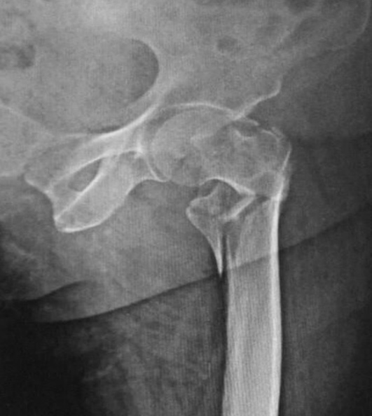
A hip fracture Fracture A fracture is a disruption of the cortex of any bone and periosteum and is commonly due to mechanical stress after an injury or accident. Open fractures due to trauma can be a medical emergency. Fractures are frequently associated with automobile accidents, workplace injuries, and trauma. Overview of Bone Fractures is a disruption in the cortex of the femur at the hip joint Hip joint The hip joint is a ball-and-socket joint formed by the head of the femur and the acetabulum of the pelvis. The hip joint is the most stable joint in the body and is supported by a very strong capsule and several ligaments, allowing the joint to sustain forces that can be multiple times the total body weight. Hip Joint: Anatomy, either between the trochanters (intertrochanteric) or at the femoral neck Neck The part of a human or animal body connecting the head to the rest of the body. Peritonsillar Abscess. Hip fracture Fracture A fracture is a disruption of the cortex of any bone and periosteum and is commonly due to mechanical stress after an injury or accident. Open fractures due to trauma can be a medical emergency. Fractures are frequently associated with automobile accidents, workplace injuries, and trauma. Overview of Bone Fractures is a serious injury and can result in life-threatening complications. The causes include high-energy impact due to trauma such as in motor vehicle accidents Motor Vehicle Accidents Spinal Cord Injuries, or low-energy trauma Low-Energy Trauma Toddler’s Fractures (falls) in patients Patients Individuals participating in the health care system for the purpose of receiving therapeutic, diagnostic, or preventive procedures. Clinician–Patient Relationship with osteoporosis Osteoporosis Osteoporosis refers to a decrease in bone mass and density leading to an increased number of fractures. There are 2 forms of osteoporosis: primary, which is commonly postmenopausal or senile; and secondary, which is a manifestation of immobilization, underlying medical disorders, or long-term use of certain medications. Osteoporosis. The clinical presentation includes groin Groin The external junctural region between the lower part of the abdomen and the thigh. Male Genitourinary Examination pain Pain An unpleasant sensation induced by noxious stimuli which are detected by nerve endings of nociceptive neurons. Pain: Types and Pathways, tenderness on palpation Palpation Application of fingers with light pressure to the surface of the body to determine consistency of parts beneath in physical diagnosis; includes palpation for determining the outlines of organs. Dermatologic Examination, immobility, and deformity Deformity Examination of the Upper Limbs of the lower limbs. Diagnosis is made clinically and by imaging studies. Definitive management is usually surgical, but depends on the type of fracture Fracture A fracture is a disruption of the cortex of any bone and periosteum and is commonly due to mechanical stress after an injury or accident. Open fractures due to trauma can be a medical emergency. Fractures are frequently associated with automobile accidents, workplace injuries, and trauma. Overview of Bone Fractures and status of the patient.

Last updated: Dec 5, 2022

Editorial responsibility: Stanley Oiseth, Lindsay Jones, Evelin Maza

Overview

Definition

A hip fracture Fracture A fracture is a disruption of the cortex of any bone and periosteum and is commonly due to mechanical stress after an injury or accident. Open fractures due to trauma can be a medical emergency. Fractures are frequently associated with automobile accidents, workplace injuries, and trauma. Overview of Bone Fractures is a disruption in the cortex of the femur at the femoroacetabular (hip) joint, either between the greater and lesser trochanters (intertrochanteric) or at the femoral neck Neck The part of a human or animal body connecting the head to the rest of the body. Peritonsillar Abscess.

Anatomy

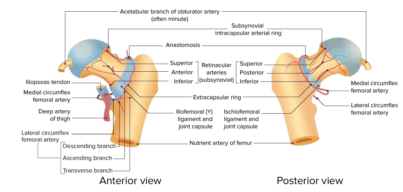
The hip is a ball-and-socket joint made up of the femoral head and acetabulum (socket). Blood supply to the femoral head is from the circumflex artery.
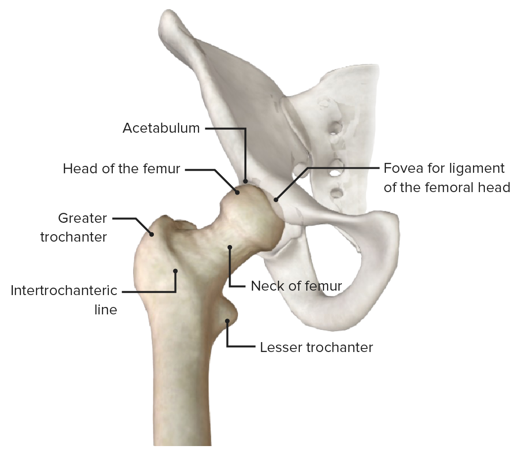
Anterior view of hip joint

Anterior view of the hip joint (pelvis faded), featuring the bony landmarks of the proximal end of the femur

Image by BioDigital, edited by Lecturio

Classification

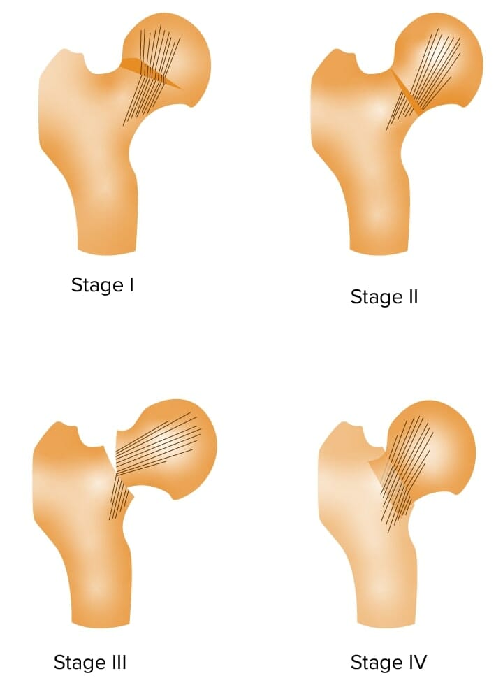
Hip fractures are classified by anatomic location and fracture Fracture A fracture is a disruption of the cortex of any bone and periosteum and is commonly due to mechanical stress after an injury or accident. Open fractures due to trauma can be a medical emergency. Fractures are frequently associated with automobile accidents, workplace injuries, and trauma. Overview of Bone Fractures type. Recognizing these fractures is important for surgical management.
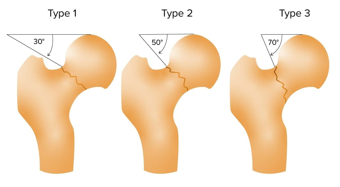
  • Intracapsular (femoral neck Neck The part of a human or animal body connecting the head to the rest of the body. Peritonsillar Abscess) hip fracture Fracture A fracture is a disruption of the cortex of any bone and periosteum and is commonly due to mechanical stress after an injury or accident. Open fractures due to trauma can be a medical emergency. Fractures are frequently associated with automobile accidents, workplace injuries, and trauma. Overview of Bone Fractures:
  • Extracapsular (intertrochanteric) hip fracture Fracture A fracture is a disruption of the cortex of any bone and periosteum and is commonly due to mechanical stress after an injury or accident. Open fractures due to trauma can be a medical emergency. Fractures are frequently associated with automobile accidents, workplace injuries, and trauma. Overview of Bone Fractures:
    • 2018 AO/OTA is the standard classification for fractures used by trauma surgeons and physicians Physicians Individuals licensed to practice medicine. Clinician–Patient Relationship dealing with musculoskeletal orthopedic trauma care:
    • Extracapsular fractures have a lower risk of complications.

Epidemiology

  • Prevalence Prevalence The total number of cases of a given disease in a specified population at a designated time. It is differentiated from incidence, which refers to the number of new cases in the population at a given time. Measures of Disease Frequency:
    • One of the most frequent fractures in patients Patients Individuals participating in the health care system for the purpose of receiving therapeutic, diagnostic, or preventive procedures. Clinician–Patient Relationship presenting to EDs EDS Ehlers-danlos syndrome (EDS) is a heterogeneous group of inherited connective tissue disorders that are characterized by hyperextensible skin, hypermobile joints, and fragility of the skin and connective tissue. Ehlers-Danlos Syndrome
    • 15% of women and 5% of men will sustain a hip fracture Fracture A fracture is a disruption of the cortex of any bone and periosteum and is commonly due to mechanical stress after an injury or accident. Open fractures due to trauma can be a medical emergency. Fractures are frequently associated with automobile accidents, workplace injuries, and trauma. Overview of Bone Fractures in their lifetime due to osteoporosis Osteoporosis Osteoporosis refers to a decrease in bone mass and density leading to an increased number of fractures. There are 2 forms of osteoporosis: primary, which is commonly postmenopausal or senile; and secondary, which is a manifestation of immobilization, underlying medical disorders, or long-term use of certain medications. Osteoporosis.
  • Incidence Incidence The number of new cases of a given disease during a given period in a specified population. It also is used for the rate at which new events occur in a defined population. It is differentiated from prevalence, which refers to all cases in the population at a given time. Measures of Disease Frequency:
    • Approximately 550/100,000 women per year in the US
    • Approximately 200/100,000 men per year in the US
  • Average age of presentation: 80 years
  • 5% of affected individuals have no history of trauma.

Etiology

  • High-energy trauma in healthy adults:
  • Low-energy trauma Low-Energy Trauma Toddler’s Fractures in the elderly due to falls and/or osteoporosis Osteoporosis Osteoporosis refers to a decrease in bone mass and density leading to an increased number of fractures. There are 2 forms of osteoporosis: primary, which is commonly postmenopausal or senile; and secondary, which is a manifestation of immobilization, underlying medical disorders, or long-term use of certain medications. Osteoporosis:
    • Risk factors for falls:
      • Prior history of falls
      • Gait Gait Manner or style of walking. Neurological Examination abnormalities
      • Use of walking aids AIDS Chronic HIV infection and depletion of CD4 cells eventually results in acquired immunodeficiency syndrome (AIDS), which can be diagnosed by the presence of certain opportunistic diseases called AIDS-defining conditions. These conditions include a wide spectrum of bacterial, viral, fungal, and parasitic infections as well as several malignancies and generalized conditions. HIV Infection and AIDS
      • Vertigo Vertigo Vertigo is defined as the perceived sensation of rotational motion while remaining still. A very common complaint in primary care and the ER, vertigo is more frequently experienced by women and its prevalence increases with age. Vertigo is classified into peripheral or central based on its etiology. Vertigo
      • Parkinson’s disease
      • Epilepsy Epilepsy Epilepsy is a chronic brain disorder marked by recurrent and unprovoked seizures. These seizures can be classified as focal or generalized and idiopathic or secondary to another condition. Clinical presentation correlates to the classification of the epileptic disorder. Epilepsy
    • Risk factors for osteoporosis Osteoporosis Osteoporosis refers to a decrease in bone mass and density leading to an increased number of fractures. There are 2 forms of osteoporosis: primary, which is commonly postmenopausal or senile; and secondary, which is a manifestation of immobilization, underlying medical disorders, or long-term use of certain medications. Osteoporosis:
      • Smoking Smoking Willful or deliberate act of inhaling and exhaling smoke from burning substances or agents held by hand. Interstitial Lung Diseases
      • Older age
      • Hormone deficiency ( menopause Menopause Menopause is a physiologic process in women characterized by the permanent cessation of menstruation that occurs after the loss of ovarian activity. Menopause can only be diagnosed retrospectively, after 12 months without menstrual bleeding. Menopause in women and hypogonadism Hypogonadism Hypogonadism is a condition characterized by reduced or no sex hormone production by the testes or ovaries. Hypogonadism can result from primary (hypergonadotropic) or secondary (hypogonadotropic) failure. Symptoms include infertility, increased risk of osteoporosis, erectile dysfunction, decreased libido, and regression (or absence) of secondary sexual characteristics. Hypogonadism in men)
      • Use of steroids Steroids A group of polycyclic compounds closely related biochemically to terpenes. They include cholesterol, numerous hormones, precursors of certain vitamins, bile acids, alcohols (sterols), and certain natural drugs and poisons. Steroids have a common nucleus, a fused, reduced 17-carbon atom ring system, cyclopentanoperhydrophenanthrene. Most steroids also have two methyl groups and an aliphatic side-chain attached to the nucleus. Benign Liver Tumors (glucocorticoid therapy)
      • Low body weight
      • Excess alcohol intake
  • Pathological fractures caused by a disease process (not related to trauma):
    • Osteoporosis Osteoporosis Osteoporosis refers to a decrease in bone mass and density leading to an increased number of fractures. There are 2 forms of osteoporosis: primary, which is commonly postmenopausal or senile; and secondary, which is a manifestation of immobilization, underlying medical disorders, or long-term use of certain medications. Osteoporosis
    • Paget’s disease of bone Bone Bone is a compact type of hardened connective tissue composed of bone cells, membranes, an extracellular mineralized matrix, and central bone marrow. The 2 primary types of bone are compact and spongy. Bones: Structure and Types
    • Bone Bone Bone is a compact type of hardened connective tissue composed of bone cells, membranes, an extracellular mineralized matrix, and central bone marrow. The 2 primary types of bone are compact and spongy. Bones: Structure and Types tumors:
      • Osteosarcoma Osteosarcoma Osteosarcoma is a primary malignant tumor of the bone characterized by the production of osteoid or immature bone by the tumor cells. The disease is most common in children and young adults and most frequently affects growth plates of the long bones, although it can involve any bone. Osteosarcoma
      • Multiple myeloma Multiple myeloma Multiple myeloma (MM) is a malignant condition of plasma cells (activated B lymphocytes) primarily seen in the elderly. Monoclonal proliferation of plasma cells results in cytokine-driven osteoclastic activity and excessive secretion of IgG antibodies. Multiple Myeloma
    • Metastatic lesions: Bone Bone Bone is a compact type of hardened connective tissue composed of bone cells, membranes, an extracellular mineralized matrix, and central bone marrow. The 2 primary types of bone are compact and spongy. Bones: Structure and Types metastasis Metastasis The transfer of a neoplasm from one organ or part of the body to another remote from the primary site. Grading, Staging, and Metastasis can occur in any bone Bone Bone is a compact type of hardened connective tissue composed of bone cells, membranes, an extracellular mineralized matrix, and central bone marrow. The 2 primary types of bone are compact and spongy. Bones: Structure and Types, but more commonly occurs in the spine Spine The human spine, or vertebral column, is the most important anatomical and functional axis of the human body. It consists of 7 cervical vertebrae, 12 thoracic vertebrae, and 5 lumbar vertebrae and is limited cranially by the skull and caudally by the sacrum. Vertebral Column: Anatomy, pelvis Pelvis The pelvis consists of the bony pelvic girdle, the muscular and ligamentous pelvic floor, and the pelvic cavity, which contains viscera, vessels, and multiple nerves and muscles. The pelvic girdle, composed of 2 “hip” bones and the sacrum, is a ring-like bony structure of the axial skeleton that links the vertebral column with the lower extremities. Pelvis: Anatomy, and thigh Thigh The thigh is the region of the lower limb found between the hip and the knee joint. There is a single bone in the thigh called the femur, which is surrounded by large muscles grouped into 3 fascial compartments. Thigh: Anatomy.
      • Breast cancer Breast cancer Breast cancer is a disease characterized by malignant transformation of the epithelial cells of the breast. Breast cancer is the most common form of cancer and 2nd most common cause of cancer-related death among women. Breast Cancer
      • Prostate Prostate The prostate is a gland in the male reproductive system. The gland surrounds the bladder neck and a portion of the urethra. The prostate is an exocrine gland that produces a weakly acidic secretion, which accounts for roughly 20% of the seminal fluid. cancer
      • Lung cancer Lung cancer Lung cancer is the malignant transformation of lung tissue and the leading cause of cancer-related deaths. The majority of cases are associated with long-term smoking. The disease is generally classified histologically as either small cell lung cancer or non-small cell lung cancer. Symptoms include cough, dyspnea, weight loss, and chest discomfort. Lung Cancer
      • Renal cell carcinoma Renal cell carcinoma Renal cell carcinoma (RCC) is a tumor that arises from the lining of the renal tubular system within the renal cortex. Renal cell carcinoma is responsible for 80%-85% of all primary renal neoplasms. Most RCCs arise sporadically, but smoking, hypertension, and obesity are linked to its development. Renal Cell Carcinoma
      • Thyroid Thyroid The thyroid gland is one of the largest endocrine glands in the human body. The thyroid gland is a highly vascular, brownish-red gland located in the visceral compartment of the anterior region of the neck. Thyroid Gland: Anatomy cancer

Pathophysiology

The general principle behind all fractures is that the bone Bone Bone is a compact type of hardened connective tissue composed of bone cells, membranes, an extracellular mineralized matrix, and central bone marrow. The 2 primary types of bone are compact and spongy. Bones: Structure and Types is subjected to a load that overwhelms its bearing capacity, resulting in a loss of structural integrity. 

  • With intertrochanteric fractures, blood supply to the femoral head is preserved and the fracture Fracture A fracture is a disruption of the cortex of any bone and periosteum and is commonly due to mechanical stress after an injury or accident. Open fractures due to trauma can be a medical emergency. Fractures are frequently associated with automobile accidents, workplace injuries, and trauma. Overview of Bone Fractures can be surgically repaired.
  • With fractures of the femoral neck Neck The part of a human or animal body connecting the head to the rest of the body. Peritonsillar Abscess, the blood supply may be interrupted:
    • May cause AVN of the femoral head
    • Surgical treatment with hemiarthroplasty may be indicated.

Clinical Presentation and Diagnosis

The presentation of a patient with a hip fracture Fracture A fracture is a disruption of the cortex of any bone and periosteum and is commonly due to mechanical stress after an injury or accident. Open fractures due to trauma can be a medical emergency. Fractures are frequently associated with automobile accidents, workplace injuries, and trauma. Overview of Bone Fractures depends on the clinical situation.

History

  • Usually presents with groin Groin The external junctural region between the lower part of the abdomen and the thigh. Male Genitourinary Examination pain Pain An unpleasant sensation induced by noxious stimuli which are detected by nerve endings of nociceptive neurons. Pain: Types and Pathways
  • Falls in the elderly: Determining the reason for the fall is also important. The following comorbidities Comorbidities The presence of co-existing or additional diseases with reference to an initial diagnosis or with reference to the index condition that is the subject of study. Comorbidity may affect the ability of affected individuals to function and also their survival; it may be used as a prognostic indicator for length of hospital stay, cost factors, and outcome or survival. St. Louis Encephalitis Virus should be addressed:
    • Syncope Syncope Syncope is a short-term loss of consciousness and loss of postural stability followed by spontaneous return of consciousness to the previous neurologic baseline without the need for resuscitation. The condition is caused by transient interruption of cerebral blood flow that may be benign or related to a underlying life-threatening condition. Syncope
    • Stroke
    • Vertigo Vertigo Vertigo is defined as the perceived sensation of rotational motion while remaining still. A very common complaint in primary care and the ER, vertigo is more frequently experienced by women and its prevalence increases with age. Vertigo is classified into peripheral or central based on its etiology. Vertigo
    • Arrhythmia
    • Seizure
  • Mechanism of injury:
    • A fall directly onto the lateral hip
    • A twisting mechanism in which the patient’s foot Foot The foot is the terminal portion of the lower limb, whose primary function is to bear weight and facilitate locomotion. The foot comprises 26 bones, including the tarsal bones, metatarsal bones, and phalanges. The bones of the foot form longitudinal and transverse arches and are supported by various muscles, ligaments, and tendons. Foot: Anatomy is planted and the body rotates
    • A sudden, spontaneous completion of a fragility fracture Fracture A fracture is a disruption of the cortex of any bone and periosteum and is commonly due to mechanical stress after an injury or accident. Open fractures due to trauma can be a medical emergency. Fractures are frequently associated with automobile accidents, workplace injuries, and trauma. Overview of Bone Fractures, which then causes a fall (instead of a fall causing a fracture Fracture A fracture is a disruption of the cortex of any bone and periosteum and is commonly due to mechanical stress after an injury or accident. Open fractures due to trauma can be a medical emergency. Fractures are frequently associated with automobile accidents, workplace injuries, and trauma. Overview of Bone Fractures)
    • Some patients Patients Individuals participating in the health care system for the purpose of receiving therapeutic, diagnostic, or preventive procedures. Clinician–Patient Relationship may report no trauma at all (5%).
  • High-impact trauma:

Physical examination

  • Tenderness to palpation Palpation Application of fingers with light pressure to the surface of the body to determine consistency of parts beneath in physical diagnosis; includes palpation for determining the outlines of organs. Dermatologic Examination over the greater trochanter 
  • Immobility due to pain Pain An unpleasant sensation induced by noxious stimuli which are detected by nerve endings of nociceptive neurons. Pain: Types and Pathways/inability to support weight
  • Deformity Deformity Examination of the Upper Limbs of the lower limb
  • Limb length discrepancy Length Discrepancy Blount’s Disease
  • External rotation External Rotation Examination of the Upper Limbs of the affected limb
  • Ecchymosis Ecchymosis Extravasation of blood into the skin, resulting in a nonelevated, rounded or irregular, blue or purplish patch, larger than a petechia. Orbital Fractures over the fracture Fracture A fracture is a disruption of the cortex of any bone and periosteum and is commonly due to mechanical stress after an injury or accident. Open fractures due to trauma can be a medical emergency. Fractures are frequently associated with automobile accidents, workplace injuries, and trauma. Overview of Bone Fractures site
  • Altered mental status Altered Mental Status Sepsis in Children: Elderly patients Patients Individuals participating in the health care system for the purpose of receiving therapeutic, diagnostic, or preventive procedures. Clinician–Patient Relationship may experience delirium Delirium Delirium is a medical condition characterized by acute disturbances in attention and awareness. Symptoms may fluctuate during the course of a day and involve memory deficits and disorientation. Delirium.
  • The skin Skin The skin, also referred to as the integumentary system, is the largest organ of the body. The skin is primarily composed of the epidermis (outer layer) and dermis (deep layer). The epidermis is primarily composed of keratinocytes that undergo rapid turnover, while the dermis contains dense layers of connective tissue. Skin: Structure and Functions may have lacerations, posing a risk for infection.

Diagnosis

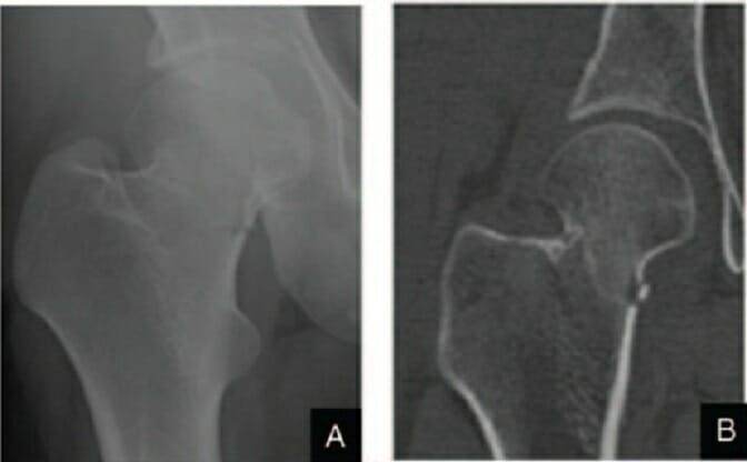
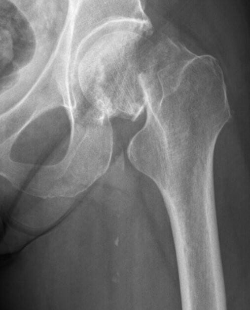
The diagnosis of a hip fracture Fracture A fracture is a disruption of the cortex of any bone and periosteum and is commonly due to mechanical stress after an injury or accident. Open fractures due to trauma can be a medical emergency. Fractures are frequently associated with automobile accidents, workplace injuries, and trauma. Overview of Bone Fractures is made clinically and with diagnostic imaging. A hip fracture Fracture A fracture is a disruption of the cortex of any bone and periosteum and is commonly due to mechanical stress after an injury or accident. Open fractures due to trauma can be a medical emergency. Fractures are frequently associated with automobile accidents, workplace injuries, and trauma. Overview of Bone Fractures may be complicated by a dislocation depending on the degree of adduction Adduction Examination of the Upper Limbs at the time of impact, especially in motor vehicle accidents Motor Vehicle Accidents Spinal Cord Injuries.
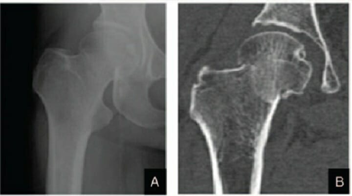
  • X-ray X-ray Penetrating electromagnetic radiation emitted when the inner orbital electrons of an atom are excited and release radiant energy. X-ray wavelengths range from 1 pm to 10 nm. Hard x-rays are the higher energy, shorter wavelength x-rays. Soft x-rays or grenz rays are less energetic and longer in wavelength. The short wavelength end of the x-ray spectrum overlaps the gamma rays wavelength range. The distinction between gamma rays and x-rays is based on their radiation source. Pulmonary Function Tests: An anteroposterior (AP) X-ray X-ray Penetrating electromagnetic radiation emitted when the inner orbital electrons of an atom are excited and release radiant energy. X-ray wavelengths range from 1 pm to 10 nm. Hard x-rays are the higher energy, shorter wavelength x-rays. Soft x-rays or grenz rays are less energetic and longer in wavelength. The short wavelength end of the x-ray spectrum overlaps the gamma rays wavelength range. The distinction between gamma rays and x-rays is based on their radiation source. Pulmonary Function Tests of the hip is ideal.
    • Fracture Fracture A fracture is a disruption of the cortex of any bone and periosteum and is commonly due to mechanical stress after an injury or accident. Open fractures due to trauma can be a medical emergency. Fractures are frequently associated with automobile accidents, workplace injuries, and trauma. Overview of Bone Fractures description:
      • Location of fracture Fracture A fracture is a disruption of the cortex of any bone and periosteum and is commonly due to mechanical stress after an injury or accident. Open fractures due to trauma can be a medical emergency. Fractures are frequently associated with automobile accidents, workplace injuries, and trauma. Overview of Bone Fractures lines (extra or intracapsular)
      • Complete or incomplete
      • Displacement Displacement The process by which an emotional or behavioral response that is appropriate for one situation appears in another situation for which it is inappropriate. Defense Mechanisms
    • Secondary findings:
      • Increased opacity Opacity Imaging of the Lungs and Pleura of soft tissues ( inflammation Inflammation Inflammation is a complex set of responses to infection and injury involving leukocytes as the principal cellular mediators in the body’s defense against pathogenic organisms. Inflammation is also seen as a response to tissue injury in the process of wound healing. The 5 cardinal signs of inflammation are pain, heat, redness, swelling, and loss of function. Inflammation and edema Edema Edema is a condition in which excess serous fluid accumulates in the body cavity or interstitial space of connective tissues. Edema is a symptom observed in several medical conditions. It can be categorized into 2 types, namely, peripheral (in the extremities) and internal (in an organ or body cavity). Edema)
      • Periosteal reaction/callus formation
  • CT/MRI:
    • MRI is the gold standard.
    • Best visualization of pelvic and femoral anatomy 
    • CT is the best alternative if MRI is not available.
    • Unstable patients Unstable Patients Blunt Chest Trauma should NOT be taken for a scan.
  • Laboratory tests:
    • CBC
    • Serum chemistry panel with renal function (creatinine and BUN)
    • PT and PTT
    • Type and cross for possible transfusion

Management

Definitive management depends on the type of fracture Fracture A fracture is a disruption of the cortex of any bone and periosteum and is commonly due to mechanical stress after an injury or accident. Open fractures due to trauma can be a medical emergency. Fractures are frequently associated with automobile accidents, workplace injuries, and trauma. Overview of Bone Fractures and requires consultation with an orthopedic surgeon.
Treatment

  • Goals of treatment:
    • Stable fixation that allows for immediate weight bearing
    • Maximization of the potential for return to the pre-fracture level of mobility
    • Minimization of the potential for implant failure (if present)
  • Initial management:
  • Nonoperative management:
    • Patients Patients Individuals participating in the health care system for the purpose of receiving therapeutic, diagnostic, or preventive procedures. Clinician–Patient Relationship at the end stage of a terminal illness 
  • Orthopedic surgical management:
    • Definitive procedure depends on the fracture Fracture A fracture is a disruption of the cortex of any bone and periosteum and is commonly due to mechanical stress after an injury or accident. Open fractures due to trauma can be a medical emergency. Fractures are frequently associated with automobile accidents, workplace injuries, and trauma. Overview of Bone Fractures type (extracapsular/intracapsular).
    • Open reduction internal fixation is usually performed:
      • Improved outcomes over conservative management
      • May be performed with a regional nerve block
  • Oncology consultation:
    • If the fracture Fracture A fracture is a disruption of the cortex of any bone and periosteum and is commonly due to mechanical stress after an injury or accident. Open fractures due to trauma can be a medical emergency. Fractures are frequently associated with automobile accidents, workplace injuries, and trauma. Overview of Bone Fractures is pathological in origin
    • Palliative procedures may be indicated in patients Patients Individuals participating in the health care system for the purpose of receiving therapeutic, diagnostic, or preventive procedures. Clinician–Patient Relationship with bone Bone Bone is a compact type of hardened connective tissue composed of bone cells, membranes, an extracellular mineralized matrix, and central bone marrow. The 2 primary types of bone are compact and spongy. Bones: Structure and Types malignancy Malignancy Hemothorax:
      • Restore mobility 
      • Reduce pain Pain An unpleasant sensation induced by noxious stimuli which are detected by nerve endings of nociceptive neurons. Pain: Types and Pathways 
  • Rehabilitation care:
    • Deep vein thrombosis Thrombosis Formation and development of a thrombus or blood clot in the blood vessel. Epidemic Typhus ( DVT DVT Deep vein thrombosis (DVT) usually occurs in the deep veins of the lower extremities. The affected veins include the femoral, popliteal, iliofemoral, and pelvic veins. Proximal DVT is more likely to cause a pulmonary embolism (PE) and is generally considered more serious. Deep Vein Thrombosis) prophylaxis Prophylaxis Cephalosporins:
    • Postoperative blood transfusion may be needed if Hb Hb The oxygen-carrying proteins of erythrocytes. They are found in all vertebrates and some invertebrates. The number of globin subunits in the hemoglobin quaternary structure differs between species. Structures range from monomeric to a variety of multimeric arrangements. Gas Exchange < 8 g/dL
    • Early mobilization is highly encouraged.
    • Intensive physical therapy Physical Therapy Becker Muscular Dystrophy to restore previous mobility
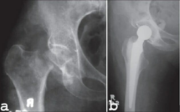
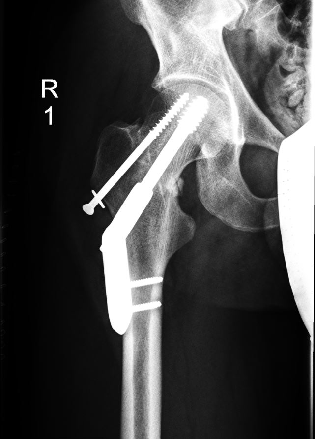
Unspecified displaced fracture of the right femur with surgical repair

Unspecified displaced fracture of the right femur with surgical repair

Image: “Hip fractures with DHS 1” by Prof. Dr. med. Ralf Puls. License: CC BY 3.0

Complications

  • Infection
  • Nonunion: 1%–30%. Rates are lower with intertrochanteric fractures.
  • Chronic pain Chronic pain Aching sensation that persists for more than a few months. It may or may not be associated with trauma or disease, and may persist after the initial injury has healed. Its localization, character, and timing are more vague than with acute pain. Pain Management
  • AVN:
    • Treatment is controversial.
    • May require surgical total hip arthroplasty Arthroplasty Surgical reconstruction of a joint to relieve pain or restore motion. Osteoarthritis
  • Post-traumatic osteoarthritis Osteoarthritis Osteoarthritis (OA) is the most common form of arthritis, and is due to cartilage destruction and changes of the subchondral bone. The risk of developing this disorder increases with age, obesity, and repetitive joint use or trauma. Patients develop gradual joint pain, stiffness lasting < 30 minutes, and decreased range of motion. Osteoarthritis
  • DVT DVT Deep vein thrombosis (DVT) usually occurs in the deep veins of the lower extremities. The affected veins include the femoral, popliteal, iliofemoral, and pelvic veins. Proximal DVT is more likely to cause a pulmonary embolism (PE) and is generally considered more serious. Deep Vein Thrombosis: 
    • Blood clots can form in the arm Arm The arm, or “upper arm” in common usage, is the region of the upper limb that extends from the shoulder to the elbow joint and connects inferiorly to the forearm through the cubital fossa. It is divided into 2 fascial compartments (anterior and posterior). Arm: Anatomy or leg Leg The lower leg, or just “leg” in anatomical terms, is the part of the lower limb between the knee and the ankle joint. The bony structure is composed of the tibia and fibula bones, and the muscles of the leg are grouped into the anterior, lateral, and posterior compartments by extensions of fascia. Leg: Anatomy veins Veins Veins are tubular collections of cells, which transport deoxygenated blood and waste from the capillary beds back to the heart. Veins are classified into 3 types: small veins/venules, medium veins, and large veins. Each type contains 3 primary layers: tunica intima, tunica media, and tunica adventitia. Veins: Histology after a period of immobility and decreased activity. 
    • Anticoagulants Anticoagulants Anticoagulants are drugs that retard or interrupt the coagulation cascade. The primary classes of available anticoagulants include heparins, vitamin K-dependent antagonists (e.g., warfarin), direct thrombin inhibitors, and factor Xa inhibitors. Anticoagulants are indicated if the patient is at high risk for DVT DVT Deep vein thrombosis (DVT) usually occurs in the deep veins of the lower extremities. The affected veins include the femoral, popliteal, iliofemoral, and pelvic veins. Proximal DVT is more likely to cause a pulmonary embolism (PE) and is generally considered more serious. Deep Vein Thrombosis.
  • Fat embolism Fat embolism Blocking of a blood vessel by fat deposits in the circulation. It is often seen after fractures of large bones or after administration of corticosteroids. Nonthrombotic Embolism:
    • Possibly due to the introduction of fat globules into the systemic circulation Circulation The movement of the blood as it is pumped through the cardiovascular system. ABCDE Assessment after a fracture Fracture A fracture is a disruption of the cortex of any bone and periosteum and is commonly due to mechanical stress after an injury or accident. Open fractures due to trauma can be a medical emergency. Fractures are frequently associated with automobile accidents, workplace injuries, and trauma. Overview of Bone Fractures
    • Clinical presentation:
      • Neurological abnormalities
      • Petechial rash Rash Rocky Mountain Spotted Fever
      • Hypoxia Hypoxia Sub-optimal oxygen levels in the ambient air of living organisms. Ischemic Cell Damage may mimic the presentation of pulmonary embolism Pulmonary Embolism Pulmonary embolism (PE) is a potentially fatal condition that occurs as a result of intraluminal obstruction of the main pulmonary artery or its branches. The causative factors include thrombi, air, amniotic fluid, and fat. In PE, gas exchange is impaired due to the decreased return of deoxygenated blood to the lungs. Pulmonary Embolism.
    • Consequences are similar to those in thrombotic embolism.
    • Treatment is supportive.
  • Delirium Delirium Delirium is a medical condition characterized by acute disturbances in attention and awareness. Symptoms may fluctuate during the course of a day and involve memory deficits and disorientation. Delirium in the elderly

Prognosis Prognosis A prediction of the probable outcome of a disease based on a individual’s condition and the usual course of the disease as seen in similar situations. Non-Hodgkin Lymphomas

  • 40%–60% of patients Patients Individuals participating in the health care system for the purpose of receiving therapeutic, diagnostic, or preventive procedures. Clinician–Patient Relationship regain baseline mobility.
  • 20%–60% of previously independent patients Patients Individuals participating in the health care system for the purpose of receiving therapeutic, diagnostic, or preventive procedures. Clinician–Patient Relationship become dependent for at least 1 activity.
  • Mortality Mortality All deaths reported in a given population. Measures of Health Status:
    • 10% at 1 month
    • 30% at 1 year

Differential Diagnosis

  • Osteoarthritis Osteoarthritis Osteoarthritis (OA) is the most common form of arthritis, and is due to cartilage destruction and changes of the subchondral bone. The risk of developing this disorder increases with age, obesity, and repetitive joint use or trauma. Patients develop gradual joint pain, stiffness lasting < 30 minutes, and decreased range of motion. Osteoarthritis of the hip: the most common form of arthritis Arthritis Acute or chronic inflammation of joints. Osteoarthritis due to cartilage Cartilage Cartilage is a type of connective tissue derived from embryonic mesenchyme that is responsible for structural support, resilience, and the smoothness of physical actions. Perichondrium (connective tissue membrane surrounding cartilage) compensates for the absence of vasculature in cartilage by providing nutrition and support. Cartilage: Histology destruction and changes of the subchondral bone Subchondral Bone Osteochondritis Dissecans. The risk of developing osteoarthritis Osteoarthritis Osteoarthritis (OA) is the most common form of arthritis, and is due to cartilage destruction and changes of the subchondral bone. The risk of developing this disorder increases with age, obesity, and repetitive joint use or trauma. Patients develop gradual joint pain, stiffness lasting < 30 minutes, and decreased range of motion. Osteoarthritis of the hip increases with age, obesity Obesity Obesity is a condition associated with excess body weight, specifically with the deposition of excessive adipose tissue. Obesity is considered a global epidemic. Major influences come from the western diet and sedentary lifestyles, but the exact mechanisms likely include a mixture of genetic and environmental factors. Obesity, repetitive joint use, or trauma.
  • Acute hip dislocation: uncoupling of the femoral head from the acetabulum, usually due to trauma. Patients Patients Individuals participating in the health care system for the purpose of receiving therapeutic, diagnostic, or preventive procedures. Clinician–Patient Relationship often present after an injury or accident. Diagnosis is made clinically and based on an X-ray X-ray Penetrating electromagnetic radiation emitted when the inner orbital electrons of an atom are excited and release radiant energy. X-ray wavelengths range from 1 pm to 10 nm. Hard x-rays are the higher energy, shorter wavelength x-rays. Soft x-rays or grenz rays are less energetic and longer in wavelength. The short wavelength end of the x-ray spectrum overlaps the gamma rays wavelength range. The distinction between gamma rays and x-rays is based on their radiation source. Pulmonary Function Tests. Management is with reduction or surgery.
  • Pelvic fracture Fracture A fracture is a disruption of the cortex of any bone and periosteum and is commonly due to mechanical stress after an injury or accident. Open fractures due to trauma can be a medical emergency. Fractures are frequently associated with automobile accidents, workplace injuries, and trauma. Overview of Bone Fractures: a disruption in the cortex of the pelvic bone Pelvic bone Bones that constitute each half of the pelvic girdle in vertebrates, formed by fusion of the ilium; ischium; and pubic bone. Pelvis: Anatomy due to high-energy trauma or osteoporosis Osteoporosis Osteoporosis refers to a decrease in bone mass and density leading to an increased number of fractures. There are 2 forms of osteoporosis: primary, which is commonly postmenopausal or senile; and secondary, which is a manifestation of immobilization, underlying medical disorders, or long-term use of certain medications. Osteoporosis. Patients Patients Individuals participating in the health care system for the purpose of receiving therapeutic, diagnostic, or preventive procedures. Clinician–Patient Relationship usually present with limb length discrepancy Length Discrepancy Blount’s Disease, intense pain Pain An unpleasant sensation induced by noxious stimuli which are detected by nerve endings of nociceptive neurons. Pain: Types and Pathways on palpation Palpation Application of fingers with light pressure to the surface of the body to determine consistency of parts beneath in physical diagnosis; includes palpation for determining the outlines of organs. Dermatologic Examination, and mechanical instability. Diagnosis is made clinically and confirmed with diagnostic imaging. Management may be nonsurgical in fragility fractures Fragility fractures Pelvic fractures are a disruption in the cortex of a pelvic bone involving iliac wing fractures, acetabular fractures, or those causing loss of integrity of the pelvic ring (the sacrum and the 2 innominate bones). Patients often present with a history of trauma or a fall, limb length discrepancy, intense pain on palpation, and mechanical instability. Pelvic Fractures due to osteoporosis Osteoporosis Osteoporosis refers to a decrease in bone mass and density leading to an increased number of fractures. There are 2 forms of osteoporosis: primary, which is commonly postmenopausal or senile; and secondary, which is a manifestation of immobilization, underlying medical disorders, or long-term use of certain medications. Osteoporosis, and surgical in patients Patients Individuals participating in the health care system for the purpose of receiving therapeutic, diagnostic, or preventive procedures. Clinician–Patient Relationship who have undergone trauma.
  • Osteosarcoma Osteosarcoma Osteosarcoma is a primary malignant tumor of the bone characterized by the production of osteoid or immature bone by the tumor cells. The disease is most common in children and young adults and most frequently affects growth plates of the long bones, although it can involve any bone. Osteosarcoma: a primary malignant tumor Tumor Inflammation of the bone Bone Bone is a compact type of hardened connective tissue composed of bone cells, membranes, an extracellular mineralized matrix, and central bone marrow. The 2 primary types of bone are compact and spongy. Bones: Structure and Types characterized by the production of osteoid Osteoid Bones: Development and Ossification or immature bone Bone Bone is a compact type of hardened connective tissue composed of bone cells, membranes, an extracellular mineralized matrix, and central bone marrow. The 2 primary types of bone are compact and spongy. Bones: Structure and Types by tumor Tumor Inflammation cells. Osteosarcoma Osteosarcoma Osteosarcoma is a primary malignant tumor of the bone characterized by the production of osteoid or immature bone by the tumor cells. The disease is most common in children and young adults and most frequently affects growth plates of the long bones, although it can involve any bone. Osteosarcoma is most common in children and young adults and most frequently affects the growth plates Growth Plates The area between the epiphysis and the diaphysis within which bone growth occurs. Osteosarcoma of long bones Long bones Length greater than width. Bones: Structure and Types; however, any bone Bone Bone is a compact type of hardened connective tissue composed of bone cells, membranes, an extracellular mineralized matrix, and central bone marrow. The 2 primary types of bone are compact and spongy. Bones: Structure and Types can be involved. Osteosarcoma Osteosarcoma Osteosarcoma is a primary malignant tumor of the bone characterized by the production of osteoid or immature bone by the tumor cells. The disease is most common in children and young adults and most frequently affects growth plates of the long bones, although it can involve any bone. Osteosarcoma can present with pain Pain An unpleasant sensation induced by noxious stimuli which are detected by nerve endings of nociceptive neurons. Pain: Types and Pathways, swelling Swelling Inflammation, a palpable mass Mass Three-dimensional lesion that occupies a space within the breast Imaging of the Breast, and, sometimes, a pathologic fracture Pathologic Fracture Chondrosarcoma. Diagnosis is established with imaging studies and biopsy Biopsy Removal and pathologic examination of specimens from the living body. Ewing Sarcoma. Treatment involves systemic chemotherapy Chemotherapy Osteosarcoma and surgical resection.

References

  1. Emmerson, B.R., et al. (2021). Hip fracture overview. StatPearls. Retrieved June 8, 2021, from http://www.ncbi.nlm.nih.gov/books/NBK557514/
  2. Kazley, J., Bagchi, K. (2021). Femoral neck fractures. StatPearls. Retrieved June 8, 2021, from http://www.ncbi.nlm.nih.gov/books/NBK537347/
  3. Papachristos, I.V., Giannoudis, P.V. (2020). Overview of classification and surgical management of hip fractures. Orthopaedics and Trauma. 34(2), 56–63. https://doi.org/10.1016/j.mporth.2020.01.001
  4. Foster, K.W. (2021). Overview of common hip fractures in adults. UpToDate. Retrieved June 9, 2021, from https://www.uptodate.com/contents/overview-of-common-hip-fractures-in-adults

Create your free account or log in to continue reading!

Sign up now and get free access to Lecturio with concept pages, medical videos, and questions for your medical education.

User Reviews

Details