Advertisement

Advertisement

Advertisement

Advertisement

Heme Metabolism

Heme is an iron-containing porphyrin (which is made of 4 pyrrole groups), synthesized mostly in the bone marrow Bone marrow The soft tissue filling the cavities of bones. Bone marrow exists in two types, yellow and red. Yellow marrow is found in the large cavities of large bones and consists mostly of fat cells and a few primitive blood cells. Red marrow is a hematopoietic tissue and is the site of production of erythrocytes and granular leukocytes. Bone marrow is made up of a framework of connective tissue containing branching fibers with the frame being filled with marrow cells. Bone Marrow: Composition and Hematopoiesis and the liver Liver The liver is the largest gland in the human body. The liver is found in the superior right quadrant of the abdomen and weighs approximately 1.5 kilograms. Its main functions are detoxification, metabolism, nutrient storage (e.g., iron and vitamins), synthesis of coagulation factors, formation of bile, filtration, and storage of blood. Liver: Anatomy. Heme is a component of many crucial substances, including cytochromes, myoglobin Myoglobin A conjugated protein which is the oxygen-transporting pigment of muscle. It is made up of one globin polypeptide chain and one heme group. Rhabdomyolysis, and hemoglobin. Biologic functions include the transportation of gases (e.g., O2), and electron transfer. Biosynthesis Biosynthesis The biosynthesis of peptides and proteins on ribosomes, directed by messenger RNA, via transfer RNA that is charged with standard proteinogenic amino acids. Virology of heme is an 8-step process initiated by the synthesis Synthesis Polymerase Chain Reaction (PCR) of aminolevulinic acid. Iron Iron A metallic element with atomic symbol fe, atomic number 26, and atomic weight 55. 85. It is an essential constituent of hemoglobins; cytochromes; and iron-binding proteins. It plays a role in cellular redox reactions and in the transport of oxygen. Trace Elements availability affects heme production, as the last step involves insertion of ferrous ion. Iron Iron A metallic element with atomic symbol fe, atomic number 26, and atomic weight 55. 85. It is an essential constituent of hemoglobins; cytochromes; and iron-binding proteins. It plays a role in cellular redox reactions and in the transport of oxygen. Trace Elements is obtained from the diet and from the breakdown of heme-containing products. In the process of catabolism, heme is converted into bile Bile An emulsifying agent produced in the liver and secreted into the duodenum. Its composition includes bile acids and salts; cholesterol; and electrolytes. It aids digestion of fats in the duodenum. Gallbladder and Biliary Tract: Anatomy pigments, out of which bilirubin is excreted. Mutations involving the enzymes Enzymes Enzymes are complex protein biocatalysts that accelerate chemical reactions without being consumed by them. Due to the body's constant metabolic needs, the absence of enzymes would make life unsustainable, as reactions would occur too slowly without these molecules. Basics of Enzymes in heme synthesis Synthesis Polymerase Chain Reaction (PCR) lead to a group of disorders known as porphyrias Porphyrias Porphyrias are a group of metabolic disorders caused by a disturbance in the synthesis of heme. In most cases, porphyria is caused by a hereditary enzyme defect. The disease patterns differ depending on the affected enzyme, and the variants of porphyria can be clinically differentiated between acute and nonacute forms. Porphyrias, and a defect in the catabolism of heme causes hyperbilirubinemias.
Last updated: Jan 31, 2024

Editorial responsibility: Stanley Oiseth, Lindsay Jones, Evelin Maza

Advertisement

Advertisement

Advertisement

Advertisement

Advertisement

Advertisement

Structure and Function of Heme

Structure

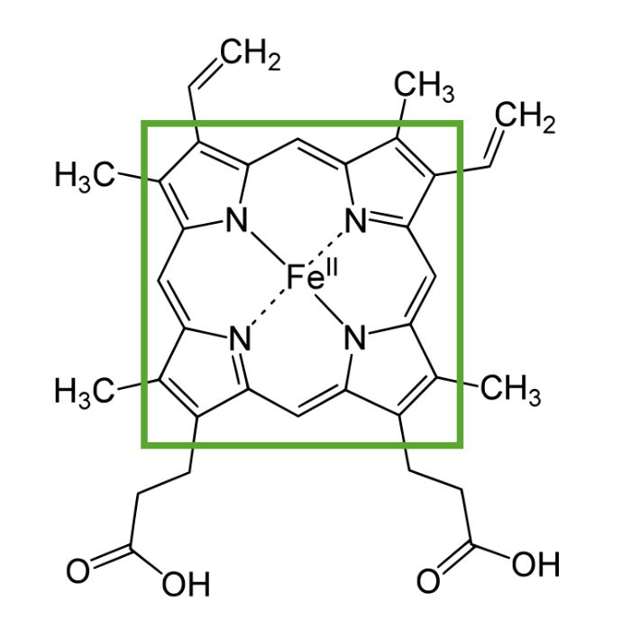
  • Heme is a flat, planar structure containing a porphyrin ring with a ferrous atom in the center (ferroprotoporphyrin).
  • Heme is present in:
    • Hemoglobin
    • Myoglobin Myoglobin A conjugated protein which is the oxygen-transporting pigment of muscle. It is made up of one globin polypeptide chain and one heme group. Rhabdomyolysis
    • Cytochromes
    • Peroxidase
    • Catalase Catalase An oxidoreductase that catalyzes the conversion of hydrogen peroxide to water and oxygen. It is present in many animal cells. A deficiency of this enzyme results in acatalasia. Nocardia/Nocardiosis
    • Tryptophan pyrrolase
    • Nitric oxide Nitric Oxide A free radical gas produced endogenously by a variety of mammalian cells, synthesized from arginine by nitric oxide synthase. Nitric oxide is one of the endothelium-dependent relaxing factors released by the vascular endothelium and mediates vasodilation. It also inhibits platelet aggregation, induces disaggregation of aggregated platelets, and inhibits platelet adhesion to the vascular endothelium. Nitric oxide activates cytosolic guanylate cyclase and thus elevates intracellular levels of cyclic gmp. Pulmonary Hypertension Drugs (NO) synthase
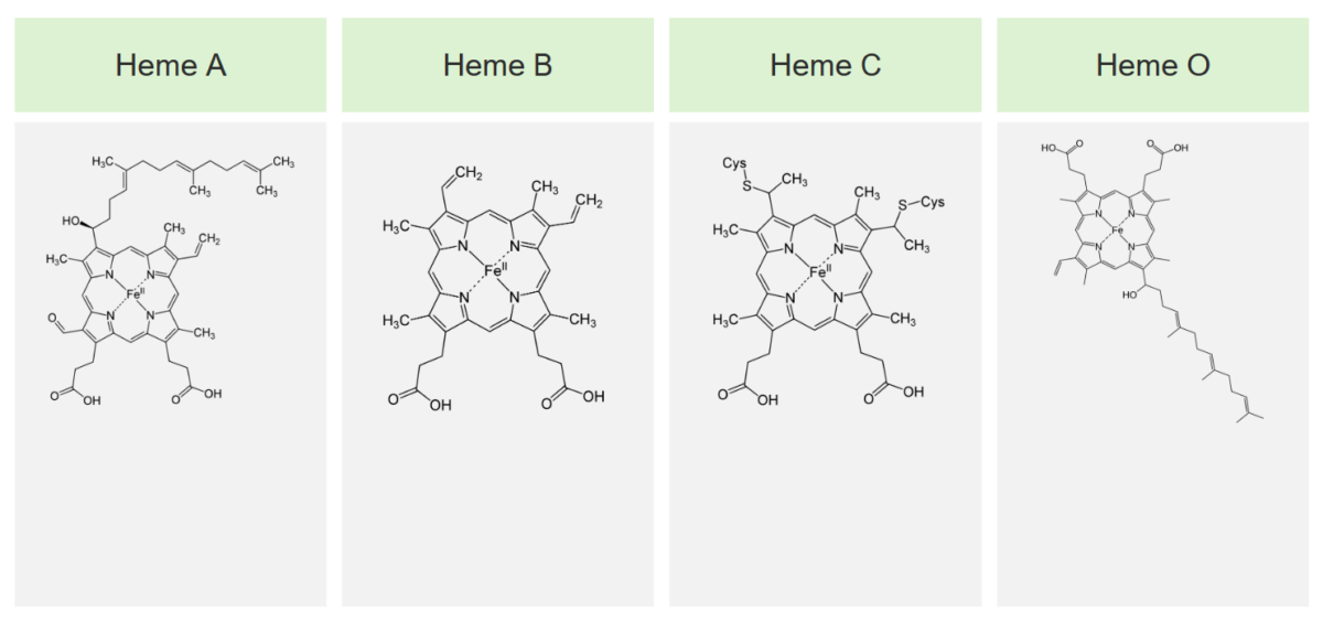
  • The 4 main types of heme are:
    • Heme A: part of complex IV Complex IV Cyanide Poisoning of electron transport system 
    • Heme B:
    • Heme C: present in cytochrome c proteins Proteins Linear polypeptides that are synthesized on ribosomes and may be further modified, crosslinked, cleaved, or assembled into complex proteins with several subunits. The specific sequence of amino acids determines the shape the polypeptide will take, during protein folding, and the function of the protein. Energy Homeostasis and links via cysteines
    • Heme O: functions in bacterial oxidases

Function

  • Transport of O2 from the lungs Lungs Lungs are the main organs of the respiratory system. Lungs are paired viscera located in the thoracic cavity and are composed of spongy tissue. The primary function of the lungs is to oxygenate blood and eliminate CO2. Lungs: Anatomy to the tissues
  • In cytochromes, oxidation and reduction of iron Iron A metallic element with atomic symbol fe, atomic number 26, and atomic weight 55. 85. It is an essential constituent of hemoglobins; cytochromes; and iron-binding proteins. It plays a role in cellular redox reactions and in the transport of oxygen. Trace Elements, which is essential in the electron transport chain Electron transport chain The electron transport chain (ETC) sends electrons through a series of proteins, which generate an electrochemical proton gradient that produces energy in the form of adenosine triphosphate (ATP). Electron Transport Chain (ETC)

Related videos

Biosynthesis of Heme

Heme is synthesized in the normoblasts, but not in the mature erythrocytes Erythrocytes Erythrocytes, or red blood cells (RBCs), are the most abundant cells in the blood. While erythrocytes in the fetus are initially produced in the yolk sac then the liver, the bone marrow eventually becomes the main site of production. Erythrocytes: Histology. The biosynthesis Biosynthesis The biosynthesis of peptides and proteins on ribosomes, directed by messenger RNA, via transfer RNA that is charged with standard proteinogenic amino acids. Virology of heme takes place in 8 steps.

Step 1

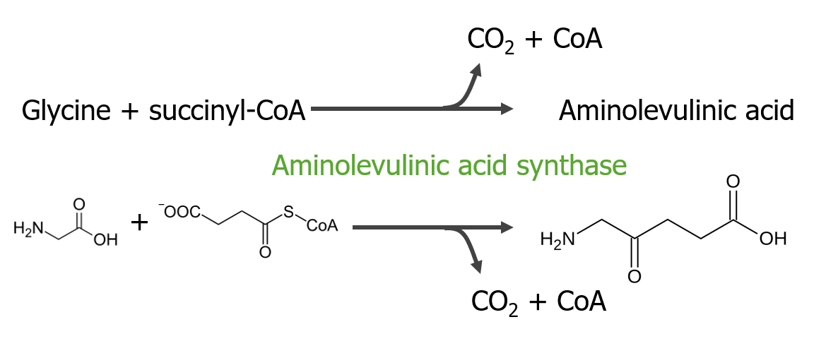
Step 1 is the synthesis Synthesis Polymerase Chain Reaction (PCR) of aminolevulinic acid.

  • Succinyl coenzyme A Succinyl coenzyme A Citric Acid Cycle (CoA) and glycine Glycine A non-essential amino acid. It is found primarily in gelatin and silk fibroin and used therapeutically as a nutrient. It is also a fast inhibitory neurotransmitter. Synthesis of Nonessential Amino Acids condense (in the presence of pyridoxal phosphate Phosphate Inorganic salts of phosphoric acid. Electrolytes) forming delta-aminolevulinic acid:
    • Enzyme: aminolevulinic acid synthase (rate-limiting step of the pathway)
    • Site: mitochondria Mitochondria Semiautonomous, self-reproducing organelles that occur in the cytoplasm of all cells of most, but not all, eukaryotes. Each mitochondrion is surrounded by a double limiting membrane. The inner membrane is highly invaginated, and its projections are called cristae. Mitochondria are the sites of the reactions of oxidative phosphorylation, which result in the formation of ATP. They contain distinctive ribosomes, transfer RNAs; amino Acyl tRNA synthetases; and elongation and termination factors. Mitochondria depend upon genes within the nucleus of the cells in which they reside for many essential messenger RNAs. Mitochondria are believed to have arisen from aerobic bacteria that established a symbiotic relationship with primitive protoeukaryotes. The Cell: Organelles 
    • Process controlled by presence of Fe²⁺-binding proteins Proteins Linear polypeptides that are synthesized on ribosomes and may be further modified, crosslinked, cleaved, or assembled into complex proteins with several subunits. The specific sequence of amino acids determines the shape the polypeptide will take, during protein folding, and the function of the protein. Energy Homeostasis
  • Because of the involvement of pyridoxal phosphate Phosphate Inorganic salts of phosphoric acid. Electrolytes (an active form of vitamin B6) in delta-aminolevulinic acid synthesis Synthesis Polymerase Chain Reaction (PCR), anemia Anemia Anemia is a condition in which individuals have low Hb levels, which can arise from various causes. Anemia is accompanied by a reduced number of RBCs and may manifest with fatigue, shortness of breath, pallor, and weakness. Subtypes are classified by the size of RBCs, chronicity, and etiology. Anemia: Overview and Types is a manifestation of pyridoxine deficiency. 
  • Mutations in ALAS2 ( gene Gene A category of nucleic acid sequences that function as units of heredity and which code for the basic instructions for the development, reproduction, and maintenance of organisms. Basic Terms of Genetics for erythroid aminolevulinic acid synthase) cause X-linked X-linked Genetic diseases that are linked to gene mutations on the X chromosome in humans or the X chromosome in other species. Included here are animal models of human X-linked diseases. Common Variable Immunodeficiency (CVID) sideroblastic anemia Sideroblastic anemia Sideroblastic anemias are a heterogeneous group of bone marrow disorders characterized by abnormal iron accumulation in the mitochondria of erythroid precursors. The accumulated iron appears as granules in a ringlike distribution around the nucleus, giving rise to the characteristic morphological feature of a ring sideroblast. Sideroblastic Anemia ( iron Iron A metallic element with atomic symbol fe, atomic number 26, and atomic weight 55. 85. It is an essential constituent of hemoglobins; cytochromes; and iron-binding proteins. It plays a role in cellular redox reactions and in the transport of oxygen. Trace Elements builds up because of reduced production of heme).
Step one of heme metabolism

Step 1 of heme metabolism

Image by Lecturio.

Step 2

Step 2 is the formation of porphobilinogen (PBG).
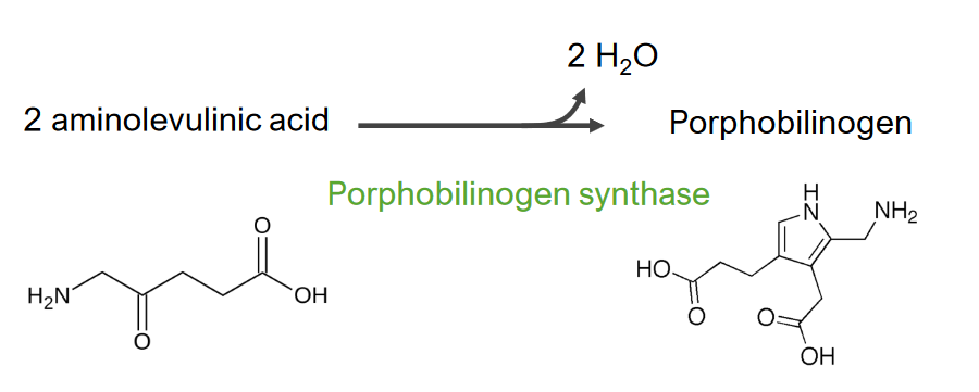
  • Moving out to the cytosol Cytosol A cell’s cytoskeleton is a network of intracellular protein fibers that provides structural support, anchors organelles, and aids intra- and extracellular movement. The Cell: Cytosol and Cytoskeleton, 2 molecules of aminolevulinic acid condense to form PBG (a pyrrole), removing 2 molecules of water in the process:
    • Enzyme: aminolevulinic acid dehydratase or PBG synthase 
    • Site: cytosol Cytosol A cell’s cytoskeleton is a network of intracellular protein fibers that provides structural support, anchors organelles, and aids intra- and extracellular movement. The Cell: Cytosol and Cytoskeleton
    • Aminolevulinic acid dehydratase is sensitive to Mg2+ and pH pH The quantitative measurement of the acidity or basicity of a solution. Acid-Base Balance and is easily inactivated by heavy metals.
  • This step is affected by lead poisoning Lead poisoning Poisoning that results from chronic or acute ingestion, injection, inhalation, or skin absorption of lead or lead compounds. Metal Poisoning (Lead, Arsenic, Iron). (Aminolevulinic acid dehydratase is inhibited by lead.)
  • Deficiency of the enzyme, a rare genetic disease, leads to aminolevulinic acid dehydratase porphyria.
Step 2 of heme metabolism

Step 2 of heme metabolism
Formation of porphobilinogen

Image by Lecturio.

Step 3

Step 3 is the formation of hydroxymethylbilane ( HMB HMB Excessive menstrual blood loss (objectively defined as > 80 mL blood loss/cycle). Can be based on heavy flow, as determined by the patient Abnormal Uterine Bleeding).
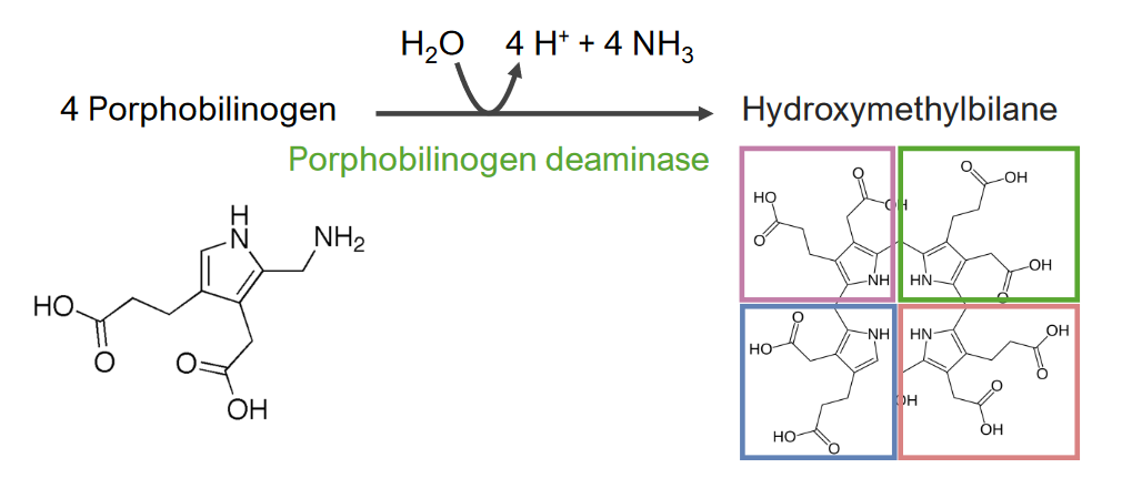
  • 4 molecules of PBG condense to form HMB HMB Excessive menstrual blood loss (objectively defined as > 80 mL blood loss/cycle). Can be based on heavy flow, as determined by the patient Abnormal Uterine Bleeding, a linear tetrapyrrole.
    • Enzyme: PBG deaminase/ HMB HMB Excessive menstrual blood loss (objectively defined as > 80 mL blood loss/cycle). Can be based on heavy flow, as determined by the patient Abnormal Uterine Bleeding synthase
    • Site: cytosol Cytosol A cell’s cytoskeleton is a network of intracellular protein fibers that provides structural support, anchors organelles, and aids intra- and extracellular movement. The Cell: Cytosol and Cytoskeleton
    • Molecules are joined through their amine rings.
  • Reduced activity of the enzyme results in intermittent porphyria (where there is accumulation of porphyrin precursors).
Step 3 of heme metabolism

Step 3 of heme metabolism:
Formation of hydroxymethylbilane

Image by Lecturio.

Step 4

Step 4 is the formation of uroporphyrinogen (UPG).
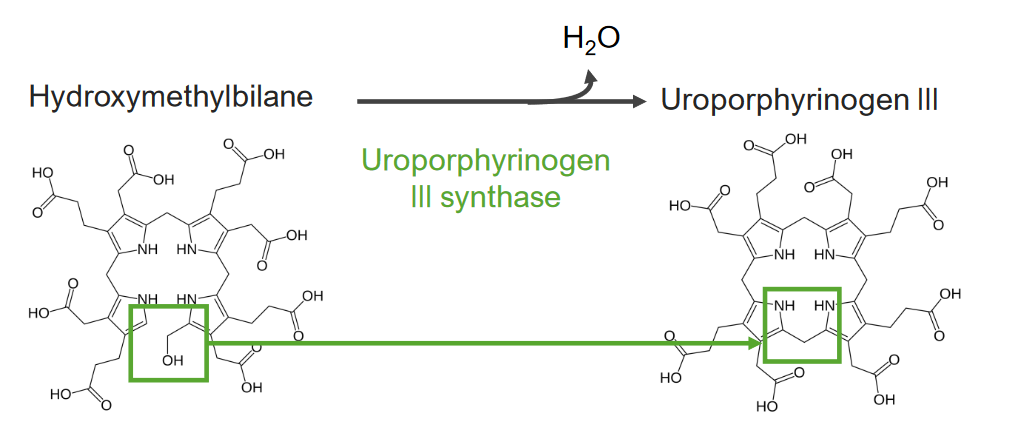
  • HMB HMB Excessive menstrual blood loss (objectively defined as > 80 mL blood loss/cycle). Can be based on heavy flow, as determined by the patient Abnormal Uterine Bleeding (a linear compound) is converted to UPG III:
    • Enzyme: UPG III synthase (UROS)
    • Site: cytosol Cytosol A cell’s cytoskeleton is a network of intracellular protein fibers that provides structural support, anchors organelles, and aids intra- and extracellular movement. The Cell: Cytosol and Cytoskeleton
    • Cyclization of the linear HMB HMB Excessive menstrual blood loss (objectively defined as > 80 mL blood loss/cycle). Can be based on heavy flow, as determined by the patient Abnormal Uterine Bleeding forms UPG III, the 1st cyclic intermediate of the pathway.
    • HMB HMB Excessive menstrual blood loss (objectively defined as > 80 mL blood loss/cycle). Can be based on heavy flow, as determined by the patient Abnormal Uterine Bleeding can also spontaneously cyclize to UPG I (nonphysiologic), requiring UROS to convert to UPG III.
  • Enzyme deficiency is seen in the autosomal recessive Autosomal recessive Autosomal inheritance, both dominant and recessive, refers to the transmission of genes from the 22 autosomal chromosomes. Autosomal recessive diseases are only expressed when 2 copies of the recessive allele are inherited. Autosomal Recessive and Autosomal Dominant Inheritance disorder congenital erythropoietic porphyria Congenital erythropoietic porphyria An autosomal recessive porphyria that is due to a deficiency of uroporphyrinogen III synthase in the bone marrow; also known as congenital erythropoietic porphyria. This disease is characterized by splenomegaly; anemia; photosensitivity; cutaneous lesions; accumulation of hydroxymethylbilane; and increased excretion of uroporphyrins and coproporphyrins. Porphyrias.
Step 4 of heme metabolism

Step 4 of heme metabolism:
Formation of uroporphyrinogen

Image by Lecturio.

Step 5

Step 5 is the synthesis Synthesis Polymerase Chain Reaction (PCR) of coproporphyrinogen (CPG) III.
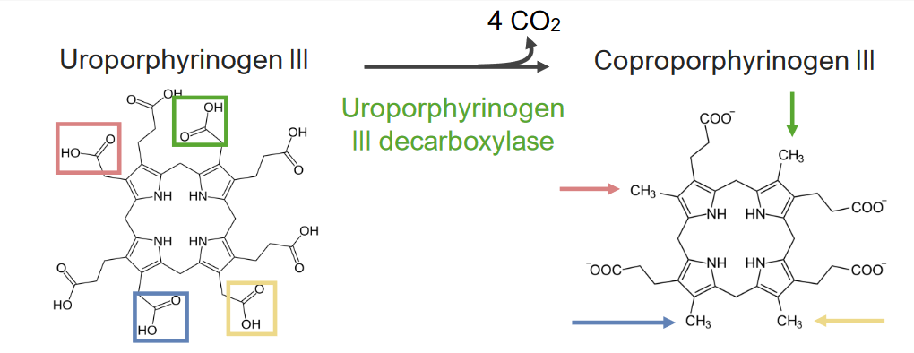
  • Involves the decarboxylation Decarboxylation The removal of a carboxyl group, usually in the form of carbon dioxide, from a chemical compound. Catabolism of Amino Acids of UPG III to CPG III with the elimination Elimination The initial damage and destruction of tumor cells by innate and adaptive immunity. Completion of the phase means no cancer growth. Cancer Immunotherapy of 4 CO2 molecules:
    • Acetate groups are decarboxylated to methyl groups.
    • Enzyme: uroporphyrinogen decarboxylase (UROD)
    • Site: cytosol Cytosol A cell’s cytoskeleton is a network of intracellular protein fibers that provides structural support, anchors organelles, and aids intra- and extracellular movement. The Cell: Cytosol and Cytoskeleton 
  • UROD mutations cause familial porphyria cutanea tarda Porphyria cutanea tarda An autosomal dominant or acquired porphyria due to a deficiency of uroporphyrinogen decarboxylase in the liver. It is characterized by photosensitivity and cutaneous lesions with little or no neurologic symptoms. Type I is the acquired form and is strongly associated with liver diseases and hepatic toxicities caused by alcohol or estrogenic steroids. Type II is the familial form. Porphyrias and hepatoerythropoietic porphyria.
Step 5 of heme metabolism

Step 5 of heme metabolism:
Formation of coproporphyrinogen III

Image by Lecturio.

Step 6

Step 6 is the synthesis Synthesis Polymerase Chain Reaction (PCR) of protoporphyrinogen (PPG).
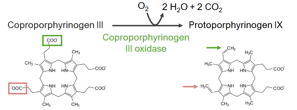
  • Oxidation of CPG III to PPG IX:
    • Molecular oxygen is required for this reaction.
    • 2 propionic side chains are decarboxylated to vinyl groups. 
    • Enzyme: CPG oxidase Oxidase Neisseria (CPOX)
    • Site: mitochondria Mitochondria Semiautonomous, self-reproducing organelles that occur in the cytoplasm of all cells of most, but not all, eukaryotes. Each mitochondrion is surrounded by a double limiting membrane. The inner membrane is highly invaginated, and its projections are called cristae. Mitochondria are the sites of the reactions of oxidative phosphorylation, which result in the formation of ATP. They contain distinctive ribosomes, transfer RNAs; amino Acyl tRNA synthetases; and elongation and termination factors. Mitochondria depend upon genes within the nucleus of the cells in which they reside for many essential messenger RNAs. Mitochondria are believed to have arisen from aerobic bacteria that established a symbiotic relationship with primitive protoeukaryotes. The Cell: Organelles
  • Reduced amount of CPG III oxidase Oxidase Neisseria leads to hereditary coproporphyria Hereditary coproporphyria An autosomal dominant porphyria that is due to a deficiency of coproporphyrinogen oxidase in the liver, the sixth enzyme in the 8-enzyme biosynthetic pathway of heme. Clinical features include both neurological symptoms and cutaneous lesions. Patients excrete increased levels of porphyrin precursors, 5-aminolevulinate and coproporphyrins. Porphyrias.
Step 6 of heme metabolism

Step 6 of heme metabolism:
Synthesis of protoporphyrinogen

Image by Lecturio.

Step 7

Step 7 is the generation of protoporphyrin (PP).
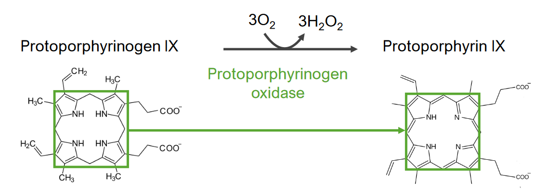
  • PPG IX is converted to PP IX by oxidation:
    • Methylene bridges are oxidized to methenyl bridges.
    • Enzyme: PPG oxidase Oxidase Neisseria (PPOX)
    • Site: mitochondria Mitochondria Semiautonomous, self-reproducing organelles that occur in the cytoplasm of all cells of most, but not all, eukaryotes. Each mitochondrion is surrounded by a double limiting membrane. The inner membrane is highly invaginated, and its projections are called cristae. Mitochondria are the sites of the reactions of oxidative phosphorylation, which result in the formation of ATP. They contain distinctive ribosomes, transfer RNAs; amino Acyl tRNA synthetases; and elongation and termination factors. Mitochondria depend upon genes within the nucleus of the cells in which they reside for many essential messenger RNAs. Mitochondria are believed to have arisen from aerobic bacteria that established a symbiotic relationship with primitive protoeukaryotes. The Cell: Organelles
  • Variegate porphyria Variegate porphyria An autosomal dominant porphyria that is due to a deficiency of protoporphyrinogen oxidase in the liver, the seventh enzyme in the 8-enzyme biosynthetic pathway of heme. Clinical features include both neurological symptoms and cutaneous lesions. Patients excrete increased levels of porphyrin precursors, coproporphyrins and protoporphyrinogen. Porphyrias is caused by mutations in PPOX.
Step 7 of heme metabolism

Step 7 of heme metabolism:
Generation of protoporphyrin from protoporphyrinogen IX

Image by Lecturio.

Step 8

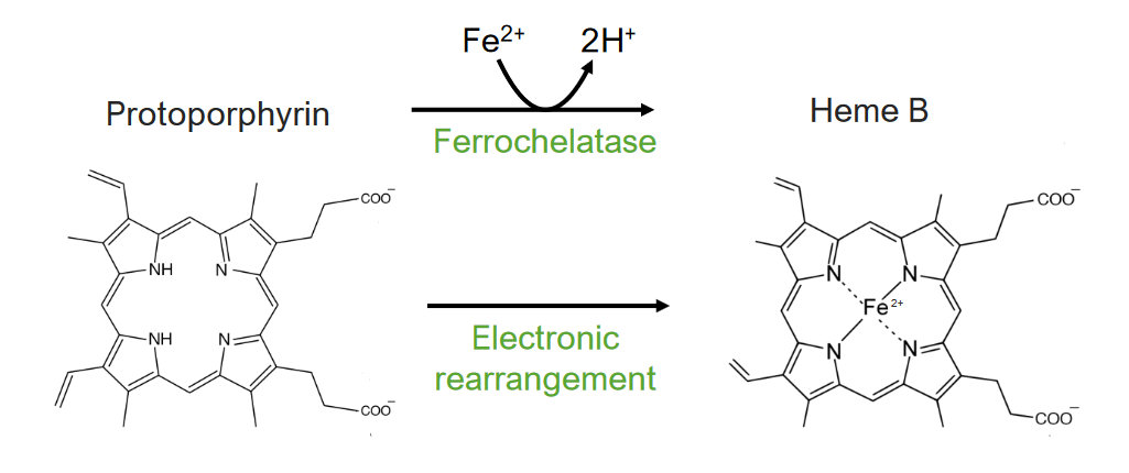
Step 8 is the generation of heme.

  • Attachment of ferrous ion to PP:
    • The ferrous ion is inserted in the middle of the porphyrin ring.
    • Enzyme: ferrochelatase (FECH)/heme synthase 
    • Enzyme also facilitates chelation of PP with Zn, forming Zn PP.
    • Site: mitochondria Mitochondria Semiautonomous, self-reproducing organelles that occur in the cytoplasm of all cells of most, but not all, eukaryotes. Each mitochondrion is surrounded by a double limiting membrane. The inner membrane is highly invaginated, and its projections are called cristae. Mitochondria are the sites of the reactions of oxidative phosphorylation, which result in the formation of ATP. They contain distinctive ribosomes, transfer RNAs; amino Acyl tRNA synthetases; and elongation and termination factors. Mitochondria depend upon genes within the nucleus of the cells in which they reside for many essential messenger RNAs. Mitochondria are believed to have arisen from aerobic bacteria that established a symbiotic relationship with primitive protoeukaryotes. The Cell: Organelles
  • Mutations affecting FECH can lead to erythropoietic protoporphyria Erythropoietic protoporphyria An autosomal dominant porphyria that is due to a deficiency of ferrochelatase (heme synthetase) in both the liver and the bone marrow, the last enzyme in the 8-enzyme biosynthetic pathway of heme. Clinical features include mainly neurological symptoms, rarely cutaneous lesions, and elevated levels of protoporphyrin and coproporphyrins in the feces. Porphyrias (EPP).
  • Lead also inhibits FECH.
The final step of heme metabolism

The 8th and final step of heme metabolism:
Formation of heme

Image by Lecturio.

Summary of heme synthesis Synthesis Polymerase Chain Reaction (PCR)

Table: Steps of heme synthesis Synthesis Polymerase Chain Reaction (PCR)
Step Site of process Enzyme Disease associated with enzyme gene Gene A category of nucleic acid sequences that function as units of heredity and which code for the basic instructions for the development, reproduction, and maintenance of organisms. Basic Terms of Genetics mutations
1. Synthesis Synthesis Polymerase Chain Reaction (PCR) of aminolevulinic acid Mitochondria Mitochondria Semiautonomous, self-reproducing organelles that occur in the cytoplasm of all cells of most, but not all, eukaryotes. Each mitochondrion is surrounded by a double limiting membrane. The inner membrane is highly invaginated, and its projections are called cristae. Mitochondria are the sites of the reactions of oxidative phosphorylation, which result in the formation of ATP. They contain distinctive ribosomes, transfer RNAs; amino Acyl tRNA synthetases; and elongation and termination factors. Mitochondria depend upon genes within the nucleus of the cells in which they reside for many essential messenger RNAs. Mitochondria are believed to have arisen from aerobic bacteria that established a symbiotic relationship with primitive protoeukaryotes. The Cell: Organelles Aminolevulinic acid synthase
  • X-linked X-linked Genetic diseases that are linked to gene mutations on the X chromosome in humans or the X chromosome in other species. Included here are animal models of human X-linked diseases. Common Variable Immunodeficiency (CVID) sideroblastic anemia Sideroblastic anemia Sideroblastic anemias are a heterogeneous group of bone marrow disorders characterized by abnormal iron accumulation in the mitochondria of erythroid precursors. The accumulated iron appears as granules in a ringlike distribution around the nucleus, giving rise to the characteristic morphological feature of a ring sideroblast. Sideroblastic Anemia (associated with aminolevulinic acid synthase 2 loss-of-function mutations)
  • X-linked protoporphyria X-linked protoporphyria Porphyrias (associated with aminolevulinic acid synthase 2 gain-of function mutations)
2. Formation of porphobilinogen (PBG) Cytosol Cytosol A cell’s cytoskeleton is a network of intracellular protein fibers that provides structural support, anchors organelles, and aids intra- and extracellular movement. The Cell: Cytosol and Cytoskeleton Aminolevulinic acid dehydratase or PBG synthase Aminolevulinic acid dehydratase porphyria
3. Formation of hydroxymethylbilane ( HMB HMB Excessive menstrual blood loss (objectively defined as > 80 mL blood loss/cycle). Can be based on heavy flow, as determined by the patient Abnormal Uterine Bleeding) PBG deaminase/ HMB HMB Excessive menstrual blood loss (objectively defined as > 80 mL blood loss/cycle). Can be based on heavy flow, as determined by the patient Abnormal Uterine Bleeding synthase Acute intermittent porphyria Acute intermittent porphyria An autosomal dominant porphyria that is due to a deficiency of hydroxymethylbilane synthase in the liver, the third enzyme in the 8-enzyme biosynthetic pathway of heme. Clinical features are recurrent and life-threatening neurologic disturbances, abdominal pain, and elevated level of aminolevulinic acid and porphobilinogen in the urine. Porphyrias
4. Formation of uroporphyrinogen (UPG) UPG III synthase Congenital erythropoietic porphyria Congenital erythropoietic porphyria An autosomal recessive porphyria that is due to a deficiency of uroporphyrinogen III synthase in the bone marrow; also known as congenital erythropoietic porphyria. This disease is characterized by splenomegaly; anemia; photosensitivity; cutaneous lesions; accumulation of hydroxymethylbilane; and increased excretion of uroporphyrins and coproporphyrins. Porphyrias
5. Synthesis Synthesis Polymerase Chain Reaction (PCR) of coproporphyrinogen (CPG) III UPG decarboxylase Porphyria cutanea tarda Porphyria cutanea tarda An autosomal dominant or acquired porphyria due to a deficiency of uroporphyrinogen decarboxylase in the liver. It is characterized by photosensitivity and cutaneous lesions with little or no neurologic symptoms. Type I is the acquired form and is strongly associated with liver diseases and hepatic toxicities caused by alcohol or estrogenic steroids. Type II is the familial form. Porphyrias and hepatoerythropoietic porphyria
6. Synthesis Synthesis Polymerase Chain Reaction (PCR) of protoporphyrinogen (PPG) Mitochondria Mitochondria Semiautonomous, self-reproducing organelles that occur in the cytoplasm of all cells of most, but not all, eukaryotes. Each mitochondrion is surrounded by a double limiting membrane. The inner membrane is highly invaginated, and its projections are called cristae. Mitochondria are the sites of the reactions of oxidative phosphorylation, which result in the formation of ATP. They contain distinctive ribosomes, transfer RNAs; amino Acyl tRNA synthetases; and elongation and termination factors. Mitochondria depend upon genes within the nucleus of the cells in which they reside for many essential messenger RNAs. Mitochondria are believed to have arisen from aerobic bacteria that established a symbiotic relationship with primitive protoeukaryotes. The Cell: Organelles CPG oxidase Oxidase Neisseria Hereditary coproporphyria Hereditary coproporphyria An autosomal dominant porphyria that is due to a deficiency of coproporphyrinogen oxidase in the liver, the sixth enzyme in the 8-enzyme biosynthetic pathway of heme. Clinical features include both neurological symptoms and cutaneous lesions. Patients excrete increased levels of porphyrin precursors, 5-aminolevulinate and coproporphyrins. Porphyrias
7. Generation of protoporphyrin (PP) Protoporphyrinogen oxidase Oxidase Neisseria Variegate porphyria Variegate porphyria An autosomal dominant porphyria that is due to a deficiency of protoporphyrinogen oxidase in the liver, the seventh enzyme in the 8-enzyme biosynthetic pathway of heme. Clinical features include both neurological symptoms and cutaneous lesions. Patients excrete increased levels of porphyrin precursors, coproporphyrins and protoporphyrinogen. Porphyrias
8. Generation of heme Ferrochelatase/heme synthase Erythropoietic protoporphyria Erythropoietic protoporphyria An autosomal dominant porphyria that is due to a deficiency of ferrochelatase (heme synthetase) in both the liver and the bone marrow, the last enzyme in the 8-enzyme biosynthetic pathway of heme. Clinical features include mainly neurological symptoms, rarely cutaneous lesions, and elevated levels of protoporphyrin and coproporphyrins in the feces. Porphyrias
ALA and PBG are porphyrin precursors
Heme synthesis

Heme synthesis:
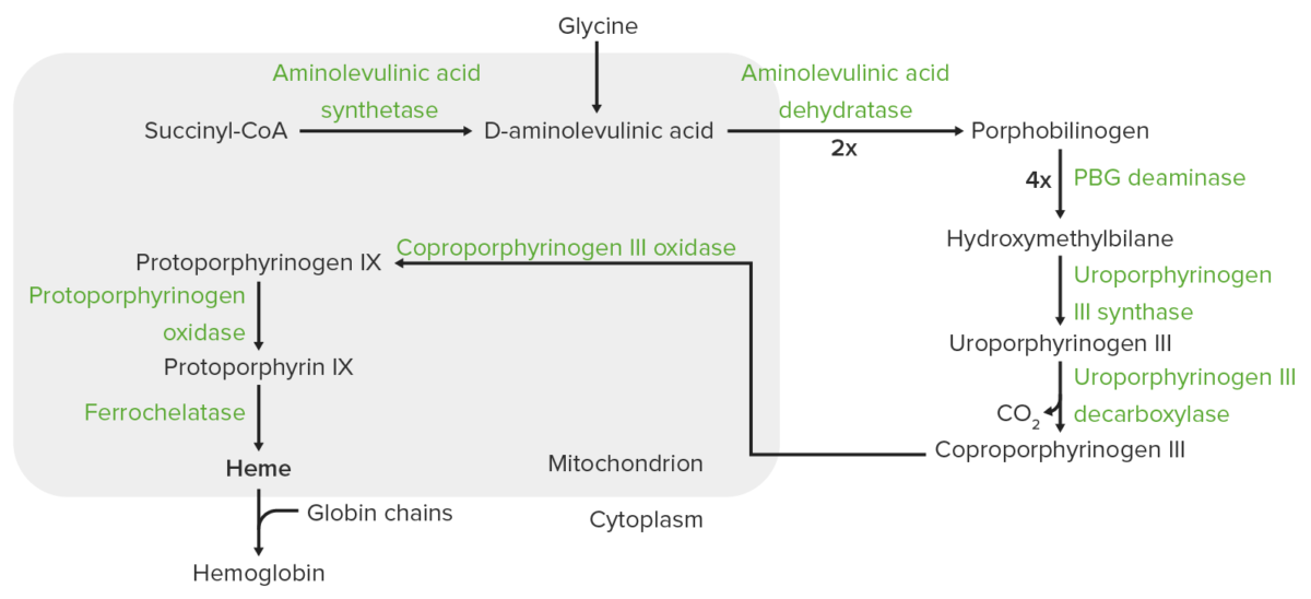
The process of heme synthesis takes place in the mitochondria and cytoplasm.
In the mitochondria, succinyl coenzyme A (CoA) combines with glycine to form aminolevulinic acid.
This reaction is catalyzed by aminolevulinic acid synthase. The aminolevulinic acid exits to the cytoplasm, where 2 aminolevulinic acid molecules condense to produce porphobilinogen (PBG). The subsequent steps lead to the formation of coproporphyrinogen III, which is transported back to the mitochondria. Oxidase facilitates conversion of coproporphyrinogen III to protoporphyrinogen IX, which then is converted to protoporphyrin IX. Ferrous iron is inserted into protoporphyrin IX, forming heme (catalyzed by ferrochelatase).

Image by Lecturio.

Regulation of Heme Synthesis

  • Synthesis Synthesis Polymerase Chain Reaction (PCR) of heme mostly occurs in the bone marrow Bone marrow The soft tissue filling the cavities of bones. Bone marrow exists in two types, yellow and red. Yellow marrow is found in the large cavities of large bones and consists mostly of fat cells and a few primitive blood cells. Red marrow is a hematopoietic tissue and is the site of production of erythrocytes and granular leukocytes. Bone marrow is made up of a framework of connective tissue containing branching fibers with the frame being filled with marrow cells. Bone Marrow: Composition and Hematopoiesis (> 80%) and the liver Liver The liver is the largest gland in the human body. The liver is found in the superior right quadrant of the abdomen and weighs approximately 1.5 kilograms. Its main functions are detoxification, metabolism, nutrient storage (e.g., iron and vitamins), synthesis of coagulation factors, formation of bile, filtration, and storage of blood. Liver: Anatomy.
  • Rate of synthesis Synthesis Polymerase Chain Reaction (PCR) depends on the expression of ALAS gene Gene A category of nucleic acid sequences that function as units of heredity and which code for the basic instructions for the development, reproduction, and maintenance of organisms. Basic Terms of Genetics (for aminolevulinic acid synthase):
    • ALAS2 regulates aminolevulinic acid synthase in the erythrocyte.
    • ALAS1 has a housekeeping role in providing heme to nonerythroid cells.
  • Other factors:
    • Iron Iron A metallic element with atomic symbol fe, atomic number 26, and atomic weight 55. 85. It is an essential constituent of hemoglobins; cytochromes; and iron-binding proteins. It plays a role in cellular redox reactions and in the transport of oxygen. Trace Elements availability (↓ iron Iron A metallic element with atomic symbol fe, atomic number 26, and atomic weight 55. 85. It is an essential constituent of hemoglobins; cytochromes; and iron-binding proteins. It plays a role in cellular redox reactions and in the transport of oxygen. Trace Elements, ↓ ALAS2 translation Translation Translation is the process of synthesizing a protein from a messenger RNA (mRNA) transcript. This process is divided into three primary stages: initiation, elongation, and termination. Translation is catalyzed by structures known as ribosomes, which are large complexes of proteins and ribosomal RNA (rRNA). Stages and Regulation of Translation)
    • Heme synthesis Synthesis Polymerase Chain Reaction (PCR) has to be coordinated with globin synthesis Synthesis Polymerase Chain Reaction (PCR) to produce hemoglobin.
  • ↓ Heme synthesis Synthesis Polymerase Chain Reaction (PCR):
    • Excess heme exerts a negative feedback Negative feedback Hypothalamic and Pituitary Hormones on ALAS gene Gene A category of nucleic acid sequences that function as units of heredity and which code for the basic instructions for the development, reproduction, and maintenance of organisms. Basic Terms of Genetics expression → ↓ synthesis Synthesis Polymerase Chain Reaction (PCR) of ALA synthase 
    • Hemin:
      • Produced when ferrous iron Iron A metallic element with atomic symbol fe, atomic number 26, and atomic weight 55. 85. It is an essential constituent of hemoglobins; cytochromes; and iron-binding proteins. It plays a role in cellular redox reactions and in the transport of oxygen. Trace Elements is oxidized to ferric iron Iron A metallic element with atomic symbol fe, atomic number 26, and atomic weight 55. 85. It is an essential constituent of hemoglobins; cytochromes; and iron-binding proteins. It plays a role in cellular redox reactions and in the transport of oxygen. Trace Elements in the presence of excess free heme
      • ↓ Aminolevulinic acid synthase synthesis Synthesis Polymerase Chain Reaction (PCR) and mitochondrial transport 
    • ↑ Cellular concentration of glucose Glucose A primary source of energy for living organisms. It is naturally occurring and is found in fruits and other parts of plants in its free state. It is used therapeutically in fluid and nutrient replacement. Lactose Intolerance:
      • Prevents induction of aminolevulinic acid synthase 
      • The basis for the administration of glucose Glucose A primary source of energy for living organisms. It is naturally occurring and is found in fruits and other parts of plants in its free state. It is used therapeutically in fluid and nutrient replacement. Lactose Intolerance in an acute attack of porphyria
  • ↑ Heme synthesis Synthesis Polymerase Chain Reaction (PCR):
  • Drugs and other agents:
    • Lead inhibits the steps catalyzed by ferrochelatase and aminolevulinic acid dehydratase. 
    • Isoniazid Isoniazid Antibacterial agent used primarily as a tuberculostatic. It remains the treatment of choice for tuberculosis. Antimycobacterial Drugs decreases the availability of pyridoxal phosphate Phosphate Inorganic salts of phosphoric acid. Electrolytes.
    • Drugs that are metabolized via cytochrome P450 Cytochrome P450 A superfamily of hundreds of closely related hemeproteins found throughout the phylogenetic spectrum, from animals, plants, fungi, to bacteria. They include numerous complex monooxygenases (mixed function oxygenases). In animals, these p450 enzymes serve two major functions: (1) biosynthesis of steroids, fatty acids, and bile acids; (2) metabolism of endogenous and a wide variety of exogenous substrates, such as toxins and drugs (biotransformation). They are classified, according to their sequence similarities rather than functions, into cyp gene families (>40% homology) and subfamilies (>59% homology). For example, enzymes from the cyp1, cyp2, and cyp3 gene families are responsible for most drug metabolism. Drug-Induced Liver Injury (which contains heme), such as barbiturates Barbiturates A class of chemicals derived from barbituric acid or thiobarbituric acid. Many of these are gaba modulators used as hypnotics and sedatives, as anesthetics, or as anticonvulsants. Intravenous Anesthetics, induce heme synthesis Synthesis Polymerase Chain Reaction (PCR).

Catabolism of Heme

Heme breaks down, resulting in bile Bile An emulsifying agent produced in the liver and secreted into the duodenum. Its composition includes bile acids and salts; cholesterol; and electrolytes. It aids digestion of fats in the duodenum. Gallbladder and Biliary Tract: Anatomy pigments as the end products, with bilirubin excreted through the bile Bile An emulsifying agent produced in the liver and secreted into the duodenum. Its composition includes bile acids and salts; cholesterol; and electrolytes. It aids digestion of fats in the duodenum. Gallbladder and Biliary Tract: Anatomy. The steps of heme catabolism are:
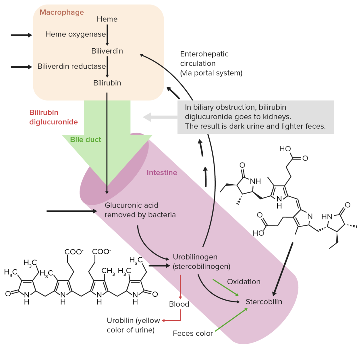
  • Degradation begins in the macrophages Macrophages The relatively long-lived phagocytic cell of mammalian tissues that are derived from blood monocytes. Main types are peritoneal macrophages; alveolar macrophages; histiocytes; kupffer cells of the liver; and osteoclasts. They may further differentiate within chronic inflammatory lesions to epithelioid cells or may fuse to form foreign body giant cells or langhans giant cells. Innate Immunity: Phagocytes and Antigen Presentation of the spleen Spleen The spleen is the largest lymphoid organ in the body, located in the LUQ of the abdomen, superior to the left kidney and posterior to the stomach at the level of the 9th-11th ribs just below the diaphragm. The spleen is highly vascular and acts as an important blood filter, cleansing the blood of pathogens and damaged erythrocytes. Spleen: Anatomy, which remove senescent and damaged erythrocytes Erythrocytes Erythrocytes, or red blood cells (RBCs), are the most abundant cells in the blood. While erythrocytes in the fetus are initially produced in the yolk sac then the liver, the bone marrow eventually becomes the main site of production. Erythrocytes: Histology from the circulation Circulation The movement of the blood as it is pumped through the cardiovascular system. ABCDE Assessment.
  • Erythrocytes Erythrocytes Erythrocytes, or red blood cells (RBCs), are the most abundant cells in the blood. While erythrocytes in the fetus are initially produced in the yolk sac then the liver, the bone marrow eventually becomes the main site of production. Erythrocytes: Histology are engulfed by the reticuloendothelial system and release hemoglobin when lysed.
    • Heme is oxidized to biliverdin by heme oxygenase. 
    • Biliverdin is then reduced to bilirubin by biliverdin reductase.
  • Bilirubin is transported to the liver Liver The liver is the largest gland in the human body. The liver is found in the superior right quadrant of the abdomen and weighs approximately 1.5 kilograms. Its main functions are detoxification, metabolism, nutrient storage (e.g., iron and vitamins), synthesis of coagulation factors, formation of bile, filtration, and storage of blood. Liver: Anatomy and is conjugated with glucuronic acid.
  • Conjugated bilirubin is transported to the small intestine Small intestine The small intestine is the longest part of the GI tract, extending from the pyloric orifice of the stomach to the ileocecal junction. The small intestine is the major organ responsible for chemical digestion and absorption of nutrients. It is divided into 3 segments: the duodenum, the jejunum, and the ileum. Small Intestine: Anatomy through the bile Bile An emulsifying agent produced in the liver and secreted into the duodenum. Its composition includes bile acids and salts; cholesterol; and electrolytes. It aids digestion of fats in the duodenum. Gallbladder and Biliary Tract: Anatomy duct and then to the large intestine Large intestine The large intestines constitute the last portion of the digestive system. The large intestine consists of the cecum, appendix, colon (with ascending, transverse, descending, and sigmoid segments), rectum, and anal canal. The primary function of the colon is to remove water and compact the stool prior to expulsion from the body via the rectum and anal canal. Colon, Cecum, and Appendix: Anatomy.
  • Bilirubin is converted to urobilinogen via bacterial reduction:
    • A small fraction is reabsorbed and undergoes renal excretion. (Urobilin causes the yellow color of urine.)
    • In the large intestine Large intestine The large intestines constitute the last portion of the digestive system. The large intestine consists of the cecum, appendix, colon (with ascending, transverse, descending, and sigmoid segments), rectum, and anal canal. The primary function of the colon is to remove water and compact the stool prior to expulsion from the body via the rectum and anal canal. Colon, Cecum, and Appendix: Anatomy, urobilinogen is converted to stercobilinogen, which is further oxidized to stercobilin. (Stercobilin causes the brown color of feces.)
  • Enterohepatic circulation Enterohepatic Circulation Recycling through liver by excretion in bile, reabsorption from intestines (intestinal reabsorption) into portal circulation, passage back into liver, and re-excretion in bile. Pharmacokinetics and Pharmacodynamics is the reabsorption of urobilinogen from the intestine and return to the liver Liver The liver is the largest gland in the human body. The liver is found in the superior right quadrant of the abdomen and weighs approximately 1.5 kilograms. Its main functions are detoxification, metabolism, nutrient storage (e.g., iron and vitamins), synthesis of coagulation factors, formation of bile, filtration, and storage of blood. Liver: Anatomy by the portal blood.
Normal extrahepatic circulation of bilirubin

Normal extrahepatic circulation of bilirubin

Image by Lecturio. License: CC BY-NC-SA 4.0

Iron Metabolism

Iron absorption Iron absorption Digestion and Absorption and transport

Iron absorption Iron absorption Digestion and Absorption:

  • Sources of iron Iron A metallic element with atomic symbol fe, atomic number 26, and atomic weight 55. 85. It is an essential constituent of hemoglobins; cytochromes; and iron-binding proteins. It plays a role in cellular redox reactions and in the transport of oxygen. Trace Elements:
    • Food/diet
    • Breakdown of iron-containing products (e.g., hemoglobin)
    • Release from reticuloendothelial stores
  • Dietary iron Iron A metallic element with atomic symbol fe, atomic number 26, and atomic weight 55. 85. It is an essential constituent of hemoglobins; cytochromes; and iron-binding proteins. It plays a role in cellular redox reactions and in the transport of oxygen. Trace Elements is absorbed by the enterocytes of the duodenum Duodenum The shortest and widest portion of the small intestine adjacent to the pylorus of the stomach. It is named for having the length equal to about the width of 12 fingers. Small Intestine: Anatomy and proximal jejunum Jejunum The middle portion of the small intestine, between duodenum and ileum. It represents about 2/5 of the remaining portion of the small intestine below duodenum. Small Intestine: Anatomy.
    • Iron Iron A metallic element with atomic symbol fe, atomic number 26, and atomic weight 55. 85. It is an essential constituent of hemoglobins; cytochromes; and iron-binding proteins. It plays a role in cellular redox reactions and in the transport of oxygen. Trace Elements in the ferric state (Fe³⁺) is reduced to the ferrous state (Fe²⁺) by ferrireductase present on the surface of enterocytes. 
    • Ascorbic acid favors reduction of ferric iron Iron A metallic element with atomic symbol fe, atomic number 26, and atomic weight 55. 85. It is an essential constituent of hemoglobins; cytochromes; and iron-binding proteins. It plays a role in cellular redox reactions and in the transport of oxygen. Trace Elements to the ferrous state. 
    • Divalent metal transporter (DMT1) transports Fe²⁺ (not Fe³⁺) from the apical surface of enterocytes to the interior of the cell. 
    • Other transporters include endosomes and heme transporter.
    • For iron Iron A metallic element with atomic symbol fe, atomic number 26, and atomic weight 55. 85. It is an essential constituent of hemoglobins; cytochromes; and iron-binding proteins. It plays a role in cellular redox reactions and in the transport of oxygen. Trace Elements to reach the circulation Circulation The movement of the blood as it is pumped through the cardiovascular system. ABCDE Assessment, ferroportin helps export iron Iron A metallic element with atomic symbol fe, atomic number 26, and atomic weight 55. 85. It is an essential constituent of hemoglobins; cytochromes; and iron-binding proteins. It plays a role in cellular redox reactions and in the transport of oxygen. Trace Elements from the intestinal cell. 

Iron Iron A metallic element with atomic symbol fe, atomic number 26, and atomic weight 55. 85. It is an essential constituent of hemoglobins; cytochromes; and iron-binding proteins. It plays a role in cellular redox reactions and in the transport of oxygen. Trace Elements transport:
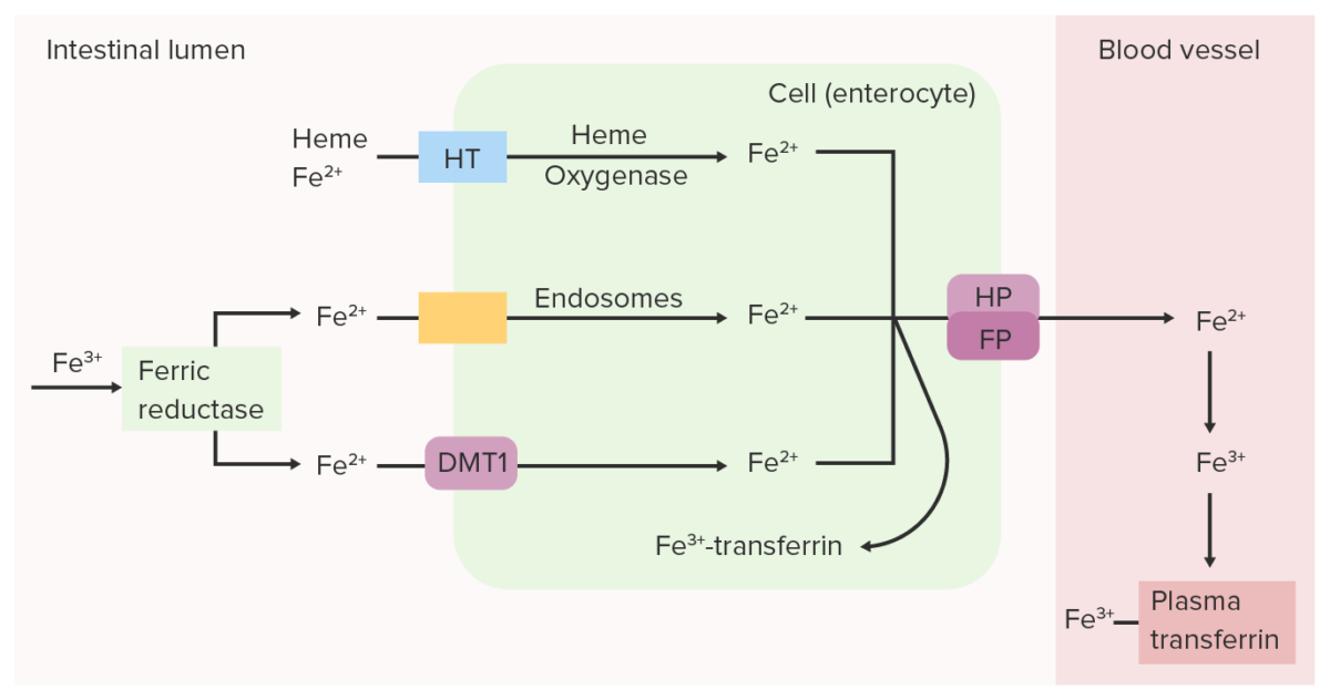
  • In order for ferroportin to transport the iron Iron A metallic element with atomic symbol fe, atomic number 26, and atomic weight 55. 85. It is an essential constituent of hemoglobins; cytochromes; and iron-binding proteins. It plays a role in cellular redox reactions and in the transport of oxygen. Trace Elements out of the cell, Fe³⁺ is needed to bind BIND Hyperbilirubinemia of the Newborn with transferrin (in circulation Circulation The movement of the blood as it is pumped through the cardiovascular system. ABCDE Assessment). 
  • Fe²⁺ is oxidized to Fe³⁺ with the aid of hephaestin (a copper-containing membrane protein that has ferroxidase activity) .
  • Fe³⁺ goes into circulation Circulation The movement of the blood as it is pumped through the cardiovascular system. ABCDE Assessment bound to transferrin ( iron Iron A metallic element with atomic symbol fe, atomic number 26, and atomic weight 55. 85. It is an essential constituent of hemoglobins; cytochromes; and iron-binding proteins. It plays a role in cellular redox reactions and in the transport of oxygen. Trace Elements transport protein synthesized in the liver Liver The liver is the largest gland in the human body. The liver is found in the superior right quadrant of the abdomen and weighs approximately 1.5 kilograms. Its main functions are detoxification, metabolism, nutrient storage (e.g., iron and vitamins), synthesis of coagulation factors, formation of bile, filtration, and storage of blood. Liver: Anatomy).
  • Iron Iron A metallic element with atomic symbol fe, atomic number 26, and atomic weight 55. 85. It is an essential constituent of hemoglobins; cytochromes; and iron-binding proteins. It plays a role in cellular redox reactions and in the transport of oxygen. Trace Elements is then transported to the tissues.
  • Transferrin binds to transferrin receptors Receptors Receptors are proteins located either on the surface of or within a cell that can bind to signaling molecules known as ligands (e.g., hormones) and cause some type of response within the cell. Receptors, which are expressed significantly in cells with high iron Iron A metallic element with atomic symbol fe, atomic number 26, and atomic weight 55. 85. It is an essential constituent of hemoglobins; cytochromes; and iron-binding proteins. It plays a role in cellular redox reactions and in the transport of oxygen. Trace Elements demands (e.g., erythroid marrow).
Intestinal ferric

Intestinal ferric (Fe3+) reductase reduces Fe3+ (ferric) to Fe2+ (ferrous). Fe2+ is transported from the lumen into the intestinal epithelial cell through divalent metal transporter 1 (DMT1), heme transporter (HT) and/or endosomes. Fe2+ can be converted back to Fe3+ and bound to transferrin within the intestinal cell or can be transported into the blood by ferroportin (FP) and hephaestin (HP). Oxidized iron (Fe3+), which binds to plasma transferrin, is carried through the circulation to the tissues.

Image by Lecturio.
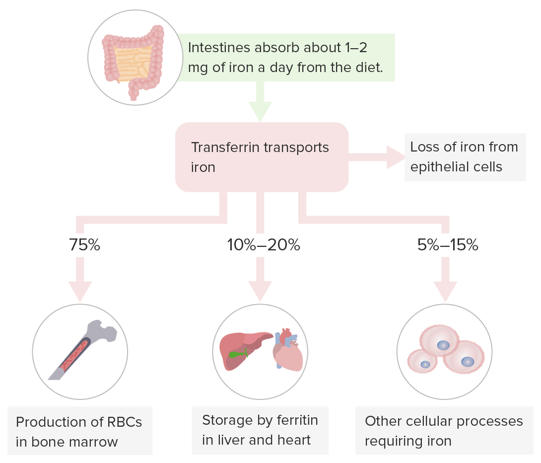
Iron Iron A metallic element with atomic symbol fe, atomic number 26, and atomic weight 55. 85. It is an essential constituent of hemoglobins; cytochromes; and iron-binding proteins. It plays a role in cellular redox reactions and in the transport of oxygen. Trace Elements storage

  • Transferrin carries iron Iron A metallic element with atomic symbol fe, atomic number 26, and atomic weight 55. 85. It is an essential constituent of hemoglobins; cytochromes; and iron-binding proteins. It plays a role in cellular redox reactions and in the transport of oxygen. Trace Elements for:
    • Hematopoiesis Hematopoiesis The development and formation of various types of blood cells. Hematopoiesis can take place in the bone marrow (medullary) or outside the bone marrow (extramedullary hematopoiesis). Bone Marrow: Composition and Hematopoiesis in the bone marrow Bone marrow The soft tissue filling the cavities of bones. Bone marrow exists in two types, yellow and red. Yellow marrow is found in the large cavities of large bones and consists mostly of fat cells and a few primitive blood cells. Red marrow is a hematopoietic tissue and is the site of production of erythrocytes and granular leukocytes. Bone marrow is made up of a framework of connective tissue containing branching fibers with the frame being filled with marrow cells. Bone Marrow: Composition and Hematopoiesis
    • Iron Iron A metallic element with atomic symbol fe, atomic number 26, and atomic weight 55. 85. It is an essential constituent of hemoglobins; cytochromes; and iron-binding proteins. It plays a role in cellular redox reactions and in the transport of oxygen. Trace Elements storage in the liver Liver The liver is the largest gland in the human body. The liver is found in the superior right quadrant of the abdomen and weighs approximately 1.5 kilograms. Its main functions are detoxification, metabolism, nutrient storage (e.g., iron and vitamins), synthesis of coagulation factors, formation of bile, filtration, and storage of blood. Liver: Anatomy (primary storage site) and other organs
    • Cellular processes requiring iron Iron A metallic element with atomic symbol fe, atomic number 26, and atomic weight 55. 85. It is an essential constituent of hemoglobins; cytochromes; and iron-binding proteins. It plays a role in cellular redox reactions and in the transport of oxygen. Trace Elements 
  • Storage forms of iron Iron A metallic element with atomic symbol fe, atomic number 26, and atomic weight 55. 85. It is an essential constituent of hemoglobins; cytochromes; and iron-binding proteins. It plays a role in cellular redox reactions and in the transport of oxygen. Trace Elements:
    • Ferritin Ferritin Iron-containing proteins that are widely distributed in animals, plants, and microorganisms. Their major function is to store iron in a nontoxic bioavailable form. Each ferritin molecule consists of ferric iron in a hollow protein shell (apoferritins) made of 24 subunits of various sequences depending on the species and tissue types. Hereditary Hemochromatosis:
      • Main iron Iron A metallic element with atomic symbol fe, atomic number 26, and atomic weight 55. 85. It is an essential constituent of hemoglobins; cytochromes; and iron-binding proteins. It plays a role in cellular redox reactions and in the transport of oxygen. Trace Elements storage protein
      • 4500 atoms of iron Iron A metallic element with atomic symbol fe, atomic number 26, and atomic weight 55. 85. It is an essential constituent of hemoglobins; cytochromes; and iron-binding proteins. It plays a role in cellular redox reactions and in the transport of oxygen. Trace Elements (when fully loaded)
  • Hemosiderin: ↑ iron Iron A metallic element with atomic symbol fe, atomic number 26, and atomic weight 55. 85. It is an essential constituent of hemoglobins; cytochromes; and iron-binding proteins. It plays a role in cellular redox reactions and in the transport of oxygen. Trace Elements ferritin Ferritin Iron-containing proteins that are widely distributed in animals, plants, and microorganisms. Their major function is to store iron in a nontoxic bioavailable form. Each ferritin molecule consists of ferric iron in a hollow protein shell (apoferritins) made of 24 subunits of various sequences depending on the species and tissue types. Hereditary Hemochromatosis forms hemosiderin granules (hemosiderin pigment = aggregates of ferritin Ferritin Iron-containing proteins that are widely distributed in animals, plants, and microorganisms. Their major function is to store iron in a nontoxic bioavailable form. Each ferritin molecule consists of ferric iron in a hollow protein shell (apoferritins) made of 24 subunits of various sequences depending on the species and tissue types. Hereditary Hemochromatosis micelles Micelles Particles consisting of aggregates of molecules held loosely together by secondary bonds. The surface of micelles are usually comprised of amphiphatic compounds that are oriented in a way that minimizes the energy of interaction between the micelle and its environment. Liquids that contain large numbers of suspended micelles are referred to as emulsions. Malabsorption and Maldigestion)
Iron storage

Storage of iron:
Transferrin carries iron for hematopoiesis in the bone marrow, iron storage in the liver (primary storage site) and other organs, and cellular processes requiring iron.
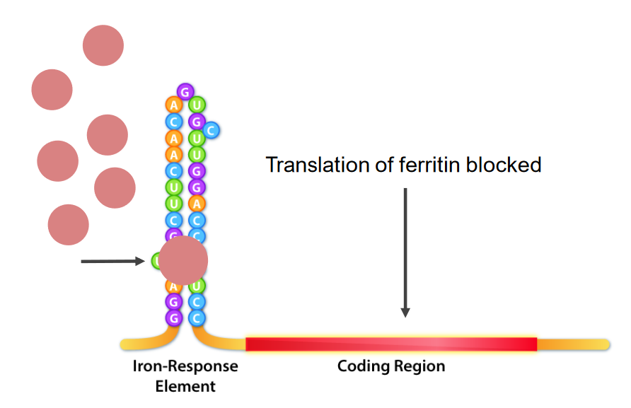
Image by Lecturio.

Iron Iron A metallic element with atomic symbol fe, atomic number 26, and atomic weight 55. 85. It is an essential constituent of hemoglobins; cytochromes; and iron-binding proteins. It plays a role in cellular redox reactions and in the transport of oxygen. Trace Elements regulation

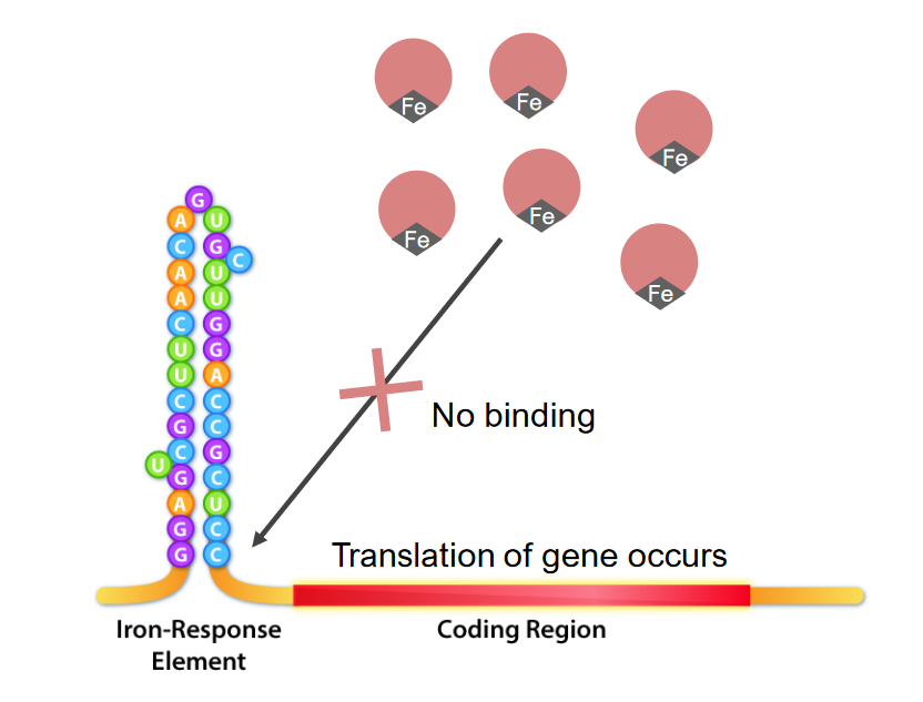
  • Regulation at the molecular level via the iron Iron A metallic element with atomic symbol fe, atomic number 26, and atomic weight 55. 85. It is an essential constituent of hemoglobins; cytochromes; and iron-binding proteins. It plays a role in cellular redox reactions and in the transport of oxygen. Trace Elements response element (IRE) and IRE binding proteins Proteins Linear polypeptides that are synthesized on ribosomes and may be further modified, crosslinked, cleaved, or assembled into complex proteins with several subunits. The specific sequence of amino acids determines the shape the polypeptide will take, during protein folding, and the function of the protein. Energy Homeostasis (IRBPs) or iron Iron A metallic element with atomic symbol fe, atomic number 26, and atomic weight 55. 85. It is an essential constituent of hemoglobins; cytochromes; and iron-binding proteins. It plays a role in cellular redox reactions and in the transport of oxygen. Trace Elements regulatory proteins Regulatory proteins Proteins and Peptides (IRPs):
    • Involved in posttranscriptional regulation of iron-related genes Genes A category of nucleic acid sequences that function as units of heredity and which code for the basic instructions for the development, reproduction, and maintenance of organisms. DNA Types and Structure
    • Controls cellular iron Iron A metallic element with atomic symbol fe, atomic number 26, and atomic weight 55. 85. It is an essential constituent of hemoglobins; cytochromes; and iron-binding proteins. It plays a role in cellular redox reactions and in the transport of oxygen. Trace Elements uptake, storage and release
    • IRE: part of the untranslated region (UTR) of the mRNA mRNA RNA sequences that serve as templates for protein synthesis. Bacterial mRNAs are generally primary transcripts in that they do not require post-transcriptional processing. Eukaryotic mRNA is synthesized in the nucleus and must be exported to the cytoplasm for translation. Most eukaryotic mRNAs have a sequence of polyadenylic acid at the 3′ end, referred to as the poly(a) tail. The function of this tail is not known for certain, but it may play a role in the export of mature mRNA from the nucleus as well as in helping stabilize some mRNA molecules by retarding their degradation in the cytoplasm. RNA Types and Structure of the target genes Genes A category of nucleic acid sequences that function as units of heredity and which code for the basic instructions for the development, reproduction, and maintenance of organisms. DNA Types and Structure (e.g., ferritin Ferritin Iron-containing proteins that are widely distributed in animals, plants, and microorganisms. Their major function is to store iron in a nontoxic bioavailable form. Each ferritin molecule consists of ferric iron in a hollow protein shell (apoferritins) made of 24 subunits of various sequences depending on the species and tissue types. Hereditary Hemochromatosis, transferrin receptor Receptor Receptors are proteins located either on the surface of or within a cell that can bind to signaling molecules known as ligands (e.g., hormones) and cause some type of response within the cell. Receptors, and other iron-metabolizing proteins Proteins Linear polypeptides that are synthesized on ribosomes and may be further modified, crosslinked, cleaved, or assembled into complex proteins with several subunits. The specific sequence of amino acids determines the shape the polypeptide will take, during protein folding, and the function of the protein. Energy Homeostasis)
      • Ferritin Ferritin Iron-containing proteins that are widely distributed in animals, plants, and microorganisms. Their major function is to store iron in a nontoxic bioavailable form. Each ferritin molecule consists of ferric iron in a hollow protein shell (apoferritins) made of 24 subunits of various sequences depending on the species and tissue types. Hereditary Hemochromatosis and ferroportin have the IREs in the 5′ UTR.
      • Transferrin receptor Receptor Receptors are proteins located either on the surface of or within a cell that can bind to signaling molecules known as ligands (e.g., hormones) and cause some type of response within the cell. Receptors (TfR) has IREs in the 3′ UTR (where binding of IRBP protects from nuclease degradation).
    • IRBPs: either a translational enhancer or a translational inhibitor, binding to iron Iron A metallic element with atomic symbol fe, atomic number 26, and atomic weight 55. 85. It is an essential constituent of hemoglobins; cytochromes; and iron-binding proteins. It plays a role in cellular redox reactions and in the transport of oxygen. Trace Elements or IRE depending on the required regulation
  • In iron Iron A metallic element with atomic symbol fe, atomic number 26, and atomic weight 55. 85. It is an essential constituent of hemoglobins; cytochromes; and iron-binding proteins. It plays a role in cellular redox reactions and in the transport of oxygen. Trace Elements deficiency (low iron Iron A metallic element with atomic symbol fe, atomic number 26, and atomic weight 55. 85. It is an essential constituent of hemoglobins; cytochromes; and iron-binding proteins. It plays a role in cellular redox reactions and in the transport of oxygen. Trace Elements):
    • Ferritin Ferritin Iron-containing proteins that are widely distributed in animals, plants, and microorganisms. Their major function is to store iron in a nontoxic bioavailable form. Each ferritin molecule consists of ferric iron in a hollow protein shell (apoferritins) made of 24 subunits of various sequences depending on the species and tissue types. Hereditary Hemochromatosis:
      • Ferritin Ferritin Iron-containing proteins that are widely distributed in animals, plants, and microorganisms. Their major function is to store iron in a nontoxic bioavailable form. Each ferritin molecule consists of ferric iron in a hollow protein shell (apoferritins) made of 24 subunits of various sequences depending on the species and tissue types. Hereditary Hemochromatosis or iron Iron A metallic element with atomic symbol fe, atomic number 26, and atomic weight 55. 85. It is an essential constituent of hemoglobins; cytochromes; and iron-binding proteins. It plays a role in cellular redox reactions and in the transport of oxygen. Trace Elements for storage is not needed, as there is low iron Iron A metallic element with atomic symbol fe, atomic number 26, and atomic weight 55. 85. It is an essential constituent of hemoglobins; cytochromes; and iron-binding proteins. It plays a role in cellular redox reactions and in the transport of oxygen. Trace Elements for cell usage. 
      • IRBPs bind BIND Hyperbilirubinemia of the Newborn ferritin Ferritin Iron-containing proteins that are widely distributed in animals, plants, and microorganisms. Their major function is to store iron in a nontoxic bioavailable form. Each ferritin molecule consists of ferric iron in a hollow protein shell (apoferritins) made of 24 subunits of various sequences depending on the species and tissue types. Hereditary Hemochromatosis IREs at the 5′ UTR, and interrupt initiation of translation Initiation of Translation A process of genetic translation whereby the formation of a peptide chain is started. It includes assembly of the ribosome components, the messenger RNA coding for the polypeptide to be made, initiator tRNA, and peptide initiation factors; and placement of the first amino acid in the peptide chain. The details and components of this process are unique for prokaryotic protein biosynthesis and eukaryotic protein biosynthesis. Stages and Regulation of Translation.
      • Less ferritin Ferritin Iron-containing proteins that are widely distributed in animals, plants, and microorganisms. Their major function is to store iron in a nontoxic bioavailable form. Each ferritin molecule consists of ferric iron in a hollow protein shell (apoferritins) made of 24 subunits of various sequences depending on the species and tissue types. Hereditary Hemochromatosis frees up iron Iron A metallic element with atomic symbol fe, atomic number 26, and atomic weight 55. 85. It is an essential constituent of hemoglobins; cytochromes; and iron-binding proteins. It plays a role in cellular redox reactions and in the transport of oxygen. Trace Elements for the cells.
    • TfR:
      • Since transferrin is needed, IRBPs bind BIND Hyperbilirubinemia of the Newborn IREs of TfR and produce a different effect.
      • IRBP binding to IREs on the 3′ UTR stabilizes the mRNA mRNA RNA sequences that serve as templates for protein synthesis. Bacterial mRNAs are generally primary transcripts in that they do not require post-transcriptional processing. Eukaryotic mRNA is synthesized in the nucleus and must be exported to the cytoplasm for translation. Most eukaryotic mRNAs have a sequence of polyadenylic acid at the 3′ end, referred to as the poly(a) tail. The function of this tail is not known for certain, but it may play a role in the export of mature mRNA from the nucleus as well as in helping stabilize some mRNA molecules by retarding their degradation in the cytoplasm. RNA Types and Structure (protecting from endonucleases).
      • This biding allows increased translation Translation Translation is the process of synthesizing a protein from a messenger RNA (mRNA) transcript. This process is divided into three primary stages: initiation, elongation, and termination. Translation is catalyzed by structures known as ribosomes, which are large complexes of proteins and ribosomal RNA (rRNA). Stages and Regulation of Translation of TfR, enhancing iron absorption Iron absorption Digestion and Absorption.
  • In iron Iron A metallic element with atomic symbol fe, atomic number 26, and atomic weight 55. 85. It is an essential constituent of hemoglobins; cytochromes; and iron-binding proteins. It plays a role in cellular redox reactions and in the transport of oxygen. Trace Elements abundance (high iron Iron A metallic element with atomic symbol fe, atomic number 26, and atomic weight 55. 85. It is an essential constituent of hemoglobins; cytochromes; and iron-binding proteins. It plays a role in cellular redox reactions and in the transport of oxygen. Trace Elements):
    • Ferritin Ferritin Iron-containing proteins that are widely distributed in animals, plants, and microorganisms. Their major function is to store iron in a nontoxic bioavailable form. Each ferritin molecule consists of ferric iron in a hollow protein shell (apoferritins) made of 24 subunits of various sequences depending on the species and tissue types. Hereditary Hemochromatosis:
      • Ferritin Ferritin Iron-containing proteins that are widely distributed in animals, plants, and microorganisms. Their major function is to store iron in a nontoxic bioavailable form. Each ferritin molecule consists of ferric iron in a hollow protein shell (apoferritins) made of 24 subunits of various sequences depending on the species and tissue types. Hereditary Hemochromatosis is needed to bind BIND Hyperbilirubinemia of the Newborn excess iron Iron A metallic element with atomic symbol fe, atomic number 26, and atomic weight 55. 85. It is an essential constituent of hemoglobins; cytochromes; and iron-binding proteins. It plays a role in cellular redox reactions and in the transport of oxygen. Trace Elements.
      • Iron Iron A metallic element with atomic symbol fe, atomic number 26, and atomic weight 55. 85. It is an essential constituent of hemoglobins; cytochromes; and iron-binding proteins. It plays a role in cellular redox reactions and in the transport of oxygen. Trace Elements binds the IRBPs (preventing IREs to be bound), allowing translation Translation Translation is the process of synthesizing a protein from a messenger RNA (mRNA) transcript. This process is divided into three primary stages: initiation, elongation, and termination. Translation is catalyzed by structures known as ribosomes, which are large complexes of proteins and ribosomal RNA (rRNA). Stages and Regulation of Translation of ferritin Ferritin Iron-containing proteins that are widely distributed in animals, plants, and microorganisms. Their major function is to store iron in a nontoxic bioavailable form. Each ferritin molecule consists of ferric iron in a hollow protein shell (apoferritins) made of 24 subunits of various sequences depending on the species and tissue types. Hereditary Hemochromatosis.
    • TfR:
      • Increased iron Iron A metallic element with atomic symbol fe, atomic number 26, and atomic weight 55. 85. It is an essential constituent of hemoglobins; cytochromes; and iron-binding proteins. It plays a role in cellular redox reactions and in the transport of oxygen. Trace Elements binds the IRBPs.
      • Dissociation Dissociation Defense Mechanisms of IRBPs from the IREs at the 3′ UTR exposes the transcripts to endonucleases, increasing mRNA mRNA RNA sequences that serve as templates for protein synthesis. Bacterial mRNAs are generally primary transcripts in that they do not require post-transcriptional processing. Eukaryotic mRNA is synthesized in the nucleus and must be exported to the cytoplasm for translation. Most eukaryotic mRNAs have a sequence of polyadenylic acid at the 3′ end, referred to as the poly(a) tail. The function of this tail is not known for certain, but it may play a role in the export of mature mRNA from the nucleus as well as in helping stabilize some mRNA molecules by retarding their degradation in the cytoplasm. RNA Types and Structure degradation.
      • Thus, iron absorption Iron absorption Digestion and Absorption will be inhibited.

Regulation of iron Iron A metallic element with atomic symbol fe, atomic number 26, and atomic weight 55. 85. It is an essential constituent of hemoglobins; cytochromes; and iron-binding proteins. It plays a role in cellular redox reactions and in the transport of oxygen. Trace Elements availability

Certain conditions require a decrease or increase in iron absorption Iron absorption Digestion and Absorption and circulating iron Iron A metallic element with atomic symbol fe, atomic number 26, and atomic weight 55. 85. It is an essential constituent of hemoglobins; cytochromes; and iron-binding proteins. It plays a role in cellular redox reactions and in the transport of oxygen. Trace Elements, a pathway regulated by hepcidin Hepcidin Forms of hepcidin, a cationic amphipathic peptide synthesized in the liver as a prepropeptide which is first processed into prohepcidin and then into the biologically active hepcidin forms, including in human the 20-, 22-, and 25-amino acid residue peptide forms. Hepcidin acts as a homeostatic regulators of iron metabolism and also possesses antimicrobial activity. Hereditary Hemochromatosis:

  • Liver-derived peptide regulating the plasma Plasma The residual portion of blood that is left after removal of blood cells by centrifugation without prior blood coagulation. Transfusion Products iron Iron A metallic element with atomic symbol fe, atomic number 26, and atomic weight 55. 85. It is an essential constituent of hemoglobins; cytochromes; and iron-binding proteins. It plays a role in cellular redox reactions and in the transport of oxygen. Trace Elements concentration
  • Actions (through binding ferroportin):
    • Inhibits intestinal iron Iron A metallic element with atomic symbol fe, atomic number 26, and atomic weight 55. 85. It is an essential constituent of hemoglobins; cytochromes; and iron-binding proteins. It plays a role in cellular redox reactions and in the transport of oxygen. Trace Elements uptake
    • Inhibits release of iron Iron A metallic element with atomic symbol fe, atomic number 26, and atomic weight 55. 85. It is an essential constituent of hemoglobins; cytochromes; and iron-binding proteins. It plays a role in cellular redox reactions and in the transport of oxygen. Trace Elements from macrophages Macrophages The relatively long-lived phagocytic cell of mammalian tissues that are derived from blood monocytes. Main types are peritoneal macrophages; alveolar macrophages; histiocytes; kupffer cells of the liver; and osteoclasts. They may further differentiate within chronic inflammatory lesions to epithelioid cells or may fuse to form foreign body giant cells or langhans giant cells. Innate Immunity: Phagocytes and Antigen Presentation with old RBCs RBCs Erythrocytes, or red blood cells (RBCs), are the most abundant cells in the blood. While erythrocytes in the fetus are initially produced in the yolk sac then the liver, the bone marrow eventually becomes the main site of production. Erythrocytes: Histology
  • Iron Iron A metallic element with atomic symbol fe, atomic number 26, and atomic weight 55. 85. It is an essential constituent of hemoglobins; cytochromes; and iron-binding proteins. It plays a role in cellular redox reactions and in the transport of oxygen. Trace Elements sensing mediated by different proteins Proteins Linear polypeptides that are synthesized on ribosomes and may be further modified, crosslinked, cleaved, or assembled into complex proteins with several subunits. The specific sequence of amino acids determines the shape the polypeptide will take, during protein folding, and the function of the protein. Energy Homeostasis:
    • HFE (hereditary iron Iron A metallic element with atomic symbol fe, atomic number 26, and atomic weight 55. 85. It is an essential constituent of hemoglobins; cytochromes; and iron-binding proteins. It plays a role in cellular redox reactions and in the transport of oxygen. Trace Elements) protein
    • TfR2
    • Hemojuvelin Hemojuvelin Hereditary Hemochromatosis
  • Affected by:
    • Iron Iron A metallic element with atomic symbol fe, atomic number 26, and atomic weight 55. 85. It is an essential constituent of hemoglobins; cytochromes; and iron-binding proteins. It plays a role in cellular redox reactions and in the transport of oxygen. Trace Elements: ↑ hepcidin Hepcidin Forms of hepcidin, a cationic amphipathic peptide synthesized in the liver as a prepropeptide which is first processed into prohepcidin and then into the biologically active hepcidin forms, including in human the 20-, 22-, and 25-amino acid residue peptide forms. Hepcidin acts as a homeostatic regulators of iron metabolism and also possesses antimicrobial activity. Hereditary Hemochromatosis to reduce iron Iron A metallic element with atomic symbol fe, atomic number 26, and atomic weight 55. 85. It is an essential constituent of hemoglobins; cytochromes; and iron-binding proteins. It plays a role in cellular redox reactions and in the transport of oxygen. Trace Elements
    • Inflammation Inflammation Inflammation is a complex set of responses to infection and injury involving leukocytes as the principal cellular mediators in the body’s defense against pathogenic organisms. Inflammation is also seen as a response to tissue injury in the process of wound healing. The 5 cardinal signs of inflammation are pain, heat, redness, swelling, and loss of function. Inflammation: ↑ hepcidin Hepcidin Forms of hepcidin, a cationic amphipathic peptide synthesized in the liver as a prepropeptide which is first processed into prohepcidin and then into the biologically active hepcidin forms, including in human the 20-, 22-, and 25-amino acid residue peptide forms. Hepcidin acts as a homeostatic regulators of iron metabolism and also possesses antimicrobial activity. Hereditary Hemochromatosis to limit Limit A value (e.g., pressure or time) that should not be exceeded and which is specified by the operator to protect the lung Invasive Mechanical Ventilation iron Iron A metallic element with atomic symbol fe, atomic number 26, and atomic weight 55. 85. It is an essential constituent of hemoglobins; cytochromes; and iron-binding proteins. It plays a role in cellular redox reactions and in the transport of oxygen. Trace Elements availability to microorganisms
    • Erythropoietin Erythropoietin Glycoprotein hormone, secreted chiefly by the kidney in the adult and the liver in the fetus, that acts on erythroid stem cells of the bone marrow to stimulate proliferation and differentiation. Erythrocytes: Histology: ↓ hepcidin Hepcidin Forms of hepcidin, a cationic amphipathic peptide synthesized in the liver as a prepropeptide which is first processed into prohepcidin and then into the biologically active hepcidin forms, including in human the 20-, 22-, and 25-amino acid residue peptide forms. Hepcidin acts as a homeostatic regulators of iron metabolism and also possesses antimicrobial activity. Hereditary Hemochromatosis to increase iron Iron A metallic element with atomic symbol fe, atomic number 26, and atomic weight 55. 85. It is an essential constituent of hemoglobins; cytochromes; and iron-binding proteins. It plays a role in cellular redox reactions and in the transport of oxygen. Trace Elements for hematopoiesis Hematopoiesis The development and formation of various types of blood cells. Hematopoiesis can take place in the bone marrow (medullary) or outside the bone marrow (extramedullary hematopoiesis). Bone Marrow: Composition and Hematopoiesis

Clinical Relevance

  • Hereditary hemochromatosis Hemochromatosis A disorder of iron metabolism characterized by a triad of hemosiderosis; liver cirrhosis; and diabetes mellitus. It is caused by massive iron deposits in parenchymal cells that may develop after a prolonged increase of iron absorption. Hereditary Hemochromatosis: autosomal recessive Autosomal recessive Autosomal inheritance, both dominant and recessive, refers to the transmission of genes from the 22 autosomal chromosomes. Autosomal recessive diseases are only expressed when 2 copies of the recessive allele are inherited. Autosomal Recessive and Autosomal Dominant Inheritance disorder most often associated with HFE gene Gene A category of nucleic acid sequences that function as units of heredity and which code for the basic instructions for the development, reproduction, and maintenance of organisms. Basic Terms of Genetics mutations. There is increased iron Iron A metallic element with atomic symbol fe, atomic number 26, and atomic weight 55. 85. It is an essential constituent of hemoglobins; cytochromes; and iron-binding proteins. It plays a role in cellular redox reactions and in the transport of oxygen. Trace Elements intestinal absorption Absorption Absorption involves the uptake of nutrient molecules and their transfer from the lumen of the GI tract across the enterocytes and into the interstitial space, where they can be taken up in the venous or lymphatic circulation. Digestion and Absorption and iron Iron A metallic element with atomic symbol fe, atomic number 26, and atomic weight 55. 85. It is an essential constituent of hemoglobins; cytochromes; and iron-binding proteins. It plays a role in cellular redox reactions and in the transport of oxygen. Trace Elements deposition in several organs, such as the liver Liver The liver is the largest gland in the human body. The liver is found in the superior right quadrant of the abdomen and weighs approximately 1.5 kilograms. Its main functions are detoxification, metabolism, nutrient storage (e.g., iron and vitamins), synthesis of coagulation factors, formation of bile, filtration, and storage of blood. Liver: Anatomy, heart, skin Skin The skin, also referred to as the integumentary system, is the largest organ of the body. The skin is primarily composed of the epidermis (outer layer) and dermis (deep layer). The epidermis is primarily composed of keratinocytes that undergo rapid turnover, while the dermis contains dense layers of connective tissue. Skin: Structure and Functions and pancreas Pancreas The pancreas lies mostly posterior to the stomach and extends across the posterior abdominal wall from the duodenum on the right to the spleen on the left. This organ has both exocrine and endocrine tissue. Pancreas: Anatomy. Clinical presentation includes the triad of cirrhosis Cirrhosis Cirrhosis is a late stage of hepatic parenchymal necrosis and scarring (fibrosis) most commonly due to hepatitis C infection and alcoholic liver disease. Patients may present with jaundice, ascites, and hepatosplenomegaly. Cirrhosis can also cause complications such as hepatic encephalopathy, portal hypertension, portal vein thrombosis, and hepatorenal syndrome. Cirrhosis, diabetes Diabetes Diabetes mellitus (DM) is a metabolic disease characterized by hyperglycemia and dysfunction of the regulation of glucose metabolism by insulin. Type 1 DM is diagnosed mostly in children and young adults as the result of autoimmune destruction of β cells in the pancreas and the resulting lack of insulin. Type 2 DM has a significant association with obesity and is characterized by insulin resistance. Diabetes Mellitus, and skin Skin The skin, also referred to as the integumentary system, is the largest organ of the body. The skin is primarily composed of the epidermis (outer layer) and dermis (deep layer). The epidermis is primarily composed of keratinocytes that undergo rapid turnover, while the dermis contains dense layers of connective tissue. Skin: Structure and Functions bronzing. Diagnosis consists of iron studies Iron Studies Iron Deficiency Anemia showing transferrin and ferritin Ferritin Iron-containing proteins that are widely distributed in animals, plants, and microorganisms. Their major function is to store iron in a nontoxic bioavailable form. Each ferritin molecule consists of ferric iron in a hollow protein shell (apoferritins) made of 24 subunits of various sequences depending on the species and tissue types. Hereditary Hemochromatosis elevation. Genetic screening Genetic Screening Physical Examination of the Newborn is recommended for family members. Management requires phlebotomy Phlebotomy The techniques used to draw blood from a vein for diagnostic purposes or for treatment of certain blood disorders such as erythrocytosis, hemochromatosis, polycythemia vera, and porphyria cutanea tarda. Hereditary Hemochromatosis (or iron Iron A metallic element with atomic symbol fe, atomic number 26, and atomic weight 55. 85. It is an essential constituent of hemoglobins; cytochromes; and iron-binding proteins. It plays a role in cellular redox reactions and in the transport of oxygen. Trace Elements chelation therapy in some cases) to prevent disease progression. The presence of hepatic fibrosis Hepatic fibrosis Autosomal Recessive Polycystic Kidney Disease (ARPKD) is a poor prognostic factor.
  • Porphyrias Porphyrias Porphyrias are a group of metabolic disorders caused by a disturbance in the synthesis of heme. In most cases, porphyria is caused by a hereditary enzyme defect. The disease patterns differ depending on the affected enzyme, and the variants of porphyria can be clinically differentiated between acute and nonacute forms. Porphyrias: group of metabolic disorders caused by a disturbance in the synthesis Synthesis Polymerase Chain Reaction (PCR) of heme. In most cases, porphyria is caused by a hereditary enzyme defect. The disease patterns differ depending on the affected enzyme, and the variants of porphyria can be clinically differentiated between acute and nonacute forms. Individuals with porphyria present with photosensitive skin Skin The skin, also referred to as the integumentary system, is the largest organ of the body. The skin is primarily composed of the epidermis (outer layer) and dermis (deep layer). The epidermis is primarily composed of keratinocytes that undergo rapid turnover, while the dermis contains dense layers of connective tissue. Skin: Structure and Functions eruptions and sometimes systemic symptoms such as abdominal pain Abdominal Pain Acute Abdomen and neuropathy Neuropathy Leprosy. Porphyrias Porphyrias Porphyrias are a group of metabolic disorders caused by a disturbance in the synthesis of heme. In most cases, porphyria is caused by a hereditary enzyme defect. The disease patterns differ depending on the affected enzyme, and the variants of porphyria can be clinically differentiated between acute and nonacute forms. Porphyrias are managed by avoiding triggers, such as sun exposure and consumption of alcohol. When flares occur, therapy is targeted toward symptom relief. 
  • Jaundice Jaundice Jaundice is the abnormal yellowing of the skin and/or sclera caused by the accumulation of bilirubin. Hyperbilirubinemia is caused by either an increase in bilirubin production or a decrease in the hepatic uptake, conjugation, or excretion of bilirubin. Jaundice: abnormal yellowing of the skin Skin The skin, also referred to as the integumentary system, is the largest organ of the body. The skin is primarily composed of the epidermis (outer layer) and dermis (deep layer). The epidermis is primarily composed of keratinocytes that undergo rapid turnover, while the dermis contains dense layers of connective tissue. Skin: Structure and Functions and/or sclera Sclera The white, opaque, fibrous, outer tunic of the eyeball, covering it entirely excepting the segment covered anteriorly by the cornea. It is essentially avascular but contains apertures for vessels, lymphatics, and nerves. Eye: Anatomy caused by the accumulation of bilirubin. Hyperbilirubinemia Hyperbilirubinemia A condition characterized by an abnormal increase of bilirubin in the blood, which may result in jaundice. Bilirubin, a breakdown product of heme, is normally excreted in the bile or further catabolized before excretion in the urine. Jaundice is caused by either an increase in bilirubin production or a decrease in the hepatic uptake, conjugation Conjugation A parasexual process in bacteria; algae; fungi; and ciliate eukaryota for achieving exchange of chromosome material during fusion of two cells. In bacteria, this is a unidirectional transfer of genetic material; in protozoa it is a bi-directional exchange. In algae and fungi, it is a form of sexual reproduction, with the union of male and female gametes. Bacteriology, or excretion of bilirubin. Etiologies often involve the liver Liver The liver is the largest gland in the human body. The liver is found in the superior right quadrant of the abdomen and weighs approximately 1.5 kilograms. Its main functions are detoxification, metabolism, nutrient storage (e.g., iron and vitamins), synthesis of coagulation factors, formation of bile, filtration, and storage of blood. Liver: Anatomy and can be prehepatic, intrahepatic, or posthepatic. Other symptoms of hyperbilirubinemia Hyperbilirubinemia A condition characterized by an abnormal increase of bilirubin in the blood, which may result in jaundice. Bilirubin, a breakdown product of heme, is normally excreted in the bile or further catabolized before excretion in the urine. Jaundice include pruritus Pruritus An intense itching sensation that produces the urge to rub or scratch the skin to obtain relief. Atopic Dermatitis (Eczema), pale stools, and darkened urine. The diagnosis is made on the basis of liver function tests Liver function tests Liver function tests, also known as hepatic function panels, are one of the most commonly performed screening blood tests. Such tests are also used to detect, evaluate, and monitor acute and chronic liver diseases. Liver Function Tests and imaging. Management is focused on treatment of the underlying condition.

References

  1. Anderson, K., Sood, G. (2020). Porphyrias: An overview. UpToDate. Retrieved October 26, 2021, from https://www.uptodate.com/contents/porphyrias-an-overview
  2. Chung, J., Chen, C., Paw, B. (2012). Heme metabolism and erythropoiesis. Current Opinions in Hematology. https://www.ncbi.nlm.nih.gov/pmc/articles/PMC4086261/
  3. Ogun, A.S., Joy, N.V., Valentine M. (2021). Biochemistry, heme synthesis. StatPearls. Retrieved October 26, 2021, from https://www.ncbi.nlm.nih.gov/books/NBK537329/ 
  4. Porteus, M., Mantanona, T. (2018). Blood. Chapter 14 of Janson, L.W., Tischler, M.E. (Eds.), The Big Picture: Medical Biochemistry. McGraw-Hill. https://accessmedicine.mhmedical.com/content.aspx?bookid=2355&sectionid=185845059
  5. Zhang, D.-L., Ghosh, M.C., Rouault, T.A. (2014). The physiological functions of iron regulatory proteins in iron homeostasis—an update. Front Pharmacol 5:124. https://doi.org/10.3389/fphar.2014.00124 
  6. Zhou, Z.D., Tan, E.K. (2017). Iron regulatory protein (IRP)-iron responsive element (IRE) signaling pathway in human neurodegenerative diseases. Molecular Neurodegeneration 12:75. https://doi.org/10.1186/s13024-017-0218-4 

Create your free account or log in to continue reading!

Sign up now and get free access to Lecturio with concept pages, medical videos, and questions for your medical education.

User Reviews

Details