Advertisement

Advertisement

Advertisement

Advertisement

Hand and Wrist Pain

Hand Hand The hand constitutes the distal part of the upper limb and provides the fine, precise movements needed in activities of daily living. It consists of 5 metacarpal bones and 14 phalanges, as well as numerous muscles innervated by the median and ulnar nerves. Hand: Anatomy and wrist pain Pain An unpleasant sensation induced by noxious stimuli which are detected by nerve endings of nociceptive neurons. Pain: Types and Pathways is very common among the general population. Up to 30% of adults will have hand Hand The hand constitutes the distal part of the upper limb and provides the fine, precise movements needed in activities of daily living. It consists of 5 metacarpal bones and 14 phalanges, as well as numerous muscles innervated by the median and ulnar nerves. Hand: Anatomy pain Pain An unpleasant sensation induced by noxious stimuli which are detected by nerve endings of nociceptive neurons. Pain: Types and Pathways during their lives, and half of all cases progress to chronic pain Chronic pain Aching sensation that persists for more than a few months. It may or may not be associated with trauma or disease, and may persist after the initial injury has healed. Its localization, character, and timing are more vague than with acute pain. Pain Management. The causes of hand Hand The hand constitutes the distal part of the upper limb and provides the fine, precise movements needed in activities of daily living. It consists of 5 metacarpal bones and 14 phalanges, as well as numerous muscles innervated by the median and ulnar nerves. Hand: Anatomy and wrist pain Pain An unpleasant sensation induced by noxious stimuli which are detected by nerve endings of nociceptive neurons. Pain: Types and Pathways can be classified into mechanical (e.g., fractures, inflammation Inflammation Inflammation is a complex set of responses to infection and injury involving leukocytes as the principal cellular mediators in the body's defense against pathogenic organisms. Inflammation is also seen as a response to tissue injury in the process of wound healing. The 5 cardinal signs of inflammation are pain, heat, redness, swelling, and loss of function. Inflammation, ligament tear), neurologic (e.g., nerve entrapment), and systemic (e.g., autoimmune arthritis Arthritis Acute or chronic inflammation of joints. Osteoarthritis). Reaching an accurate diagnosis involves following an evidence-based systematic approach. Treatment includes reducing stress on the ligaments, pain Pain An unpleasant sensation induced by noxious stimuli which are detected by nerve endings of nociceptive neurons. Pain: Types and Pathways management, and surgery.

Last updated: Nov 21, 2022

Editorial responsibility: Stanley Oiseth, Lindsay Jones, Evelin Maza

Advertisement

Advertisement

Advertisement

Advertisement

Advertisement

Advertisement

Advertisement

Advertisement

Overview

History

Up to 70% of cases of hand Hand The hand constitutes the distal part of the upper limb and provides the fine, precise movements needed in activities of daily living. It consists of 5 metacarpal bones and 14 phalanges, as well as numerous muscles innervated by the median and ulnar nerves. Hand: Anatomy or wrist pain Pain An unpleasant sensation induced by noxious stimuli which are detected by nerve endings of nociceptive neurons. Pain: Types and Pathways can be determined by history alone.
  • Systemic associations may stem from systemic illness.
  • Recent trauma can lead to acute injuries and even fractures (e.g., fall on outstretched hand Fall On Outstretched Hand Distal Radius Fractures → scaphoid fracture Fracture A fracture is a disruption of the cortex of any bone and periosteum and is commonly due to mechanical stress after an injury or accident. Open fractures due to trauma can be a medical emergency. Fractures are frequently associated with automobile accidents, workplace injuries, and trauma. Overview of Bone Fractures, lunate dislocation, or scapholunate dissociation Dissociation Defense Mechanisms)
  • Location and description of pain Pain An unpleasant sensation induced by noxious stimuli which are detected by nerve endings of nociceptive neurons. Pain: Types and Pathways
  • Painful movements
  • Patient occupation and sports/recreation

Physical examination

  • Inspection Inspection Dermatologic Examination
  • Range of motion Range of motion The distance and direction to which a bone joint can be extended. Range of motion is a function of the condition of the joints, muscles, and connective tissues involved. Joint flexibility can be improved through appropriate muscle strength exercises. Examination of the Upper Limbs: can easily identify tendon rupture
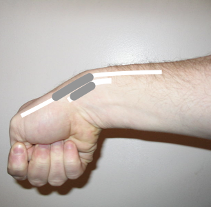
  • Examine the snuffbox to detect scaphoid fracture Fracture A fracture is a disruption of the cortex of any bone and periosteum and is commonly due to mechanical stress after an injury or accident. Open fractures due to trauma can be a medical emergency. Fractures are frequently associated with automobile accidents, workplace injuries, and trauma. Overview of Bone Fractures or De Quervain’s tenosynovitis
  • Neurovascular exam
  • Finkelstein’s test:
    • Differentiation between De Quervain’s tenosynovitis and arthritis Arthritis Acute or chronic inflammation of joints. Osteoarthritis of the first metacarpal
    • Instruct the patient to deviate the wrist to the ulnar aspect while grasping the thumb (against examiner’s resistance Resistance Physiologically, the opposition to flow of air caused by the forces of friction. As a part of pulmonary function testing, it is the ratio of driving pressure to the rate of air flow. Ventilation: Mechanics of Breathing).
    • Pain Pain An unpleasant sensation induced by noxious stimuli which are detected by nerve endings of nociceptive neurons. Pain: Types and Pathways over the radial styloid is suggestive of tenosynovitis.

Diagnostic workup

  • X-ray X-ray Penetrating electromagnetic radiation emitted when the inner orbital electrons of an atom are excited and release radiant energy. X-ray wavelengths range from 1 pm to 10 nm. Hard x-rays are the higher energy, shorter wavelength x-rays. Soft x-rays or grenz rays are less energetic and longer in wavelength. The short wavelength end of the x-ray spectrum overlaps the gamma rays wavelength range. The distinction between gamma rays and x-rays is based on their radiation source. Pulmonary Function Tests:
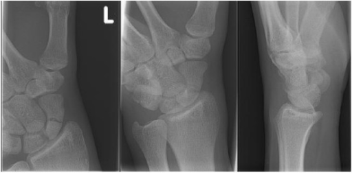
    • Radiography is often negative in acute fractures.
    • Delayed radiographic images can confirm the diagnosis in retrospect.
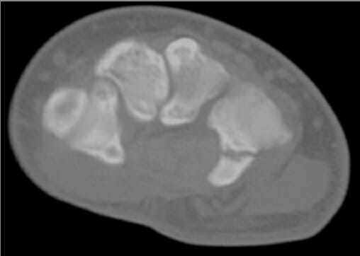
  • Computed tomography (CT) scan: confirms nonunion Nonunion Hip Fractures of a fracture Fracture A fracture is a disruption of the cortex of any bone and periosteum and is commonly due to mechanical stress after an injury or accident. Open fractures due to trauma can be a medical emergency. Fractures are frequently associated with automobile accidents, workplace injuries, and trauma. Overview of Bone Fractures, avascular necrosis Avascular Necrosis Hip Fractures, and acute fractures
  • Magnetic resonance imaging (MRI): superior to other imaging modalities in the visualization of soft tissues such as ligaments and tendons 
  • Nerve conduction studies: diagnostic study for compressive neuropathy Neuropathy Leprosy (i.e., carpal tunnel Carpal Tunnel The carpal tunnel is formed by the transverse carpal ligament (flexor retinaculum) superiorly and the carpal bones inferiorly. Carpal Tunnel Syndrome syndrome/cubital tunnel syndrome)
  • Laboratory evaluation: ordered in cases where systemic causes are suspected
    • CBC
    • Autoimmune assays
    • Renal and liver function tests Liver function tests Liver function tests, also known as hepatic function panels, are one of the most commonly performed screening blood tests. Such tests are also used to detect, evaluate, and monitor acute and chronic liver diseases. Liver Function Tests

Related videos

Differential Diagnosis

Mechanical causes

  • Fractures: history of recent trauma and point-tenderness over a bony structure(s)
    • Scaphoid fracture Fracture A fracture is a disruption of the cortex of any bone and periosteum and is commonly due to mechanical stress after an injury or accident. Open fractures due to trauma can be a medical emergency. Fractures are frequently associated with automobile accidents, workplace injuries, and trauma. Overview of Bone Fractures
      • Most commonly fractured carpal bone Bone Bone is a compact type of hardened connective tissue composed of bone cells, membranes, an extracellular mineralized matrix, and central bone marrow. The 2 primary types of bone are compact and spongy. Bones: Structure and Types
      • History of falling onto an outstretched hand Hand The hand constitutes the distal part of the upper limb and provides the fine, precise movements needed in activities of daily living. It consists of 5 metacarpal bones and 14 phalanges, as well as numerous muscles innervated by the median and ulnar nerves. Hand: Anatomy
      • Tenderness upon palpation Palpation Application of fingers with light pressure to the surface of the body to determine consistency of parts beneath in physical diagnosis; includes palpation for determining the outlines of organs. Dermatologic Examination of snuffbox
      • Fracture Fracture A fracture is a disruption of the cortex of any bone and periosteum and is commonly due to mechanical stress after an injury or accident. Open fractures due to trauma can be a medical emergency. Fractures are frequently associated with automobile accidents, workplace injuries, and trauma. Overview of Bone Fractures may not be immediately apparent in X-ray X-ray Penetrating electromagnetic radiation emitted when the inner orbital electrons of an atom are excited and release radiant energy. X-ray wavelengths range from 1 pm to 10 nm. Hard x-rays are the higher energy, shorter wavelength x-rays. Soft x-rays or grenz rays are less energetic and longer in wavelength. The short wavelength end of the x-ray spectrum overlaps the gamma rays wavelength range. The distinction between gamma rays and x-rays is based on their radiation source. Pulmonary Function Tests.
      • To confirm the diagnosis early, perform a CT scan or MRI, or repeat X-rays X-rays X-rays are high-energy particles of electromagnetic radiation used in the medical field for the generation of anatomical images. X-rays are projected through the body of a patient and onto a film, and this technique is called conventional or projectional radiography. X-rays within 2 weeks.
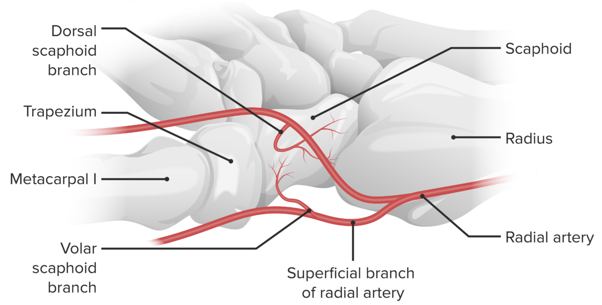
      • High risk of nonunion Nonunion Hip Fractures and avascular necrosis Avascular Necrosis Hip Fractures (osteonecrosis) due to retrograde blood supply from a branch of the radial artery Radial Artery The direct continuation of the brachial trunk, originating at the bifurcation of the brachial artery opposite the neck of the radius. Its branches may be divided into three groups corresponding to the three regions in which the vessel is situated, the forearm, wrist, and hand. Forearm: Anatomy
      • Management includes thumb spica splint for 2 weeks (then repeat X-ray X-ray Penetrating electromagnetic radiation emitted when the inner orbital electrons of an atom are excited and release radiant energy. X-ray wavelengths range from 1 pm to 10 nm. Hard x-rays are the higher energy, shorter wavelength x-rays. Soft x-rays or grenz rays are less energetic and longer in wavelength. The short wavelength end of the x-ray spectrum overlaps the gamma rays wavelength range. The distinction between gamma rays and x-rays is based on their radiation source. Pulmonary Function Tests), immobilization Immobilization Delirium with a cast for non-displaced fractures, or surgical intervention for displaced fractures.
    • 5th metacarpal fracture Fracture A fracture is a disruption of the cortex of any bone and periosteum and is commonly due to mechanical stress after an injury or accident. Open fractures due to trauma can be a medical emergency. Fractures are frequently associated with automobile accidents, workplace injuries, and trauma. Overview of Bone Fractures (boxer’s fracture Fracture A fracture is a disruption of the cortex of any bone and periosteum and is commonly due to mechanical stress after an injury or accident. Open fractures due to trauma can be a medical emergency. Fractures are frequently associated with automobile accidents, workplace injuries, and trauma. Overview of Bone Fractures): classic injury that occurs from striking an object with a closed fist
    • Hook of hamate fracture Fracture A fracture is a disruption of the cortex of any bone and periosteum and is commonly due to mechanical stress after an injury or accident. Open fractures due to trauma can be a medical emergency. Fractures are frequently associated with automobile accidents, workplace injuries, and trauma. Overview of Bone Fractures: may cause ulnar nerve injury Ulnar Nerve Injury Peripheral Nerve Injuries in the Upper Extremity
  • Dislocations:
  • De Quervain’s tenosynovitis:
    • Entrapment tendonitis Tendonitis Reactive Arthritis or tenosynovitis of the abductor pollicis longus Abductor pollicis longus Forearm: Anatomy tendon and the extensor pollicis brevis Extensor pollicis brevis Forearm: Anatomy tendon
    • Patient presents with pain Pain An unpleasant sensation induced by noxious stimuli which are detected by nerve endings of nociceptive neurons. Pain: Types and Pathways over the distal radius Radius The outer shorter of the two bones of the forearm, lying parallel to the ulna and partially revolving around it. Forearm: Anatomy.
    • Classically affects new mothers who hold their infants with the thumb outstretched (abducted and extended), but a history of overuse is not necessary.
    • Diagnosis:
      • Finkelstein’s test: With the forearm Forearm The forearm is the region of the upper limb between the elbow and the wrist. The term “forearm” is used in anatomy to distinguish this area from the arm, a term that is commonly used to describe the entire upper limb. The forearm consists of 2 long bones (the radius and the ulna), the interosseous membrane, and multiple arteries, nerves, and muscles. Forearm: Anatomy in neutral position, place it on a desk or table with the hand Hand The hand constitutes the distal part of the upper limb and provides the fine, precise movements needed in activities of daily living. It consists of 5 metacarpal bones and 14 phalanges, as well as numerous muscles innervated by the median and ulnar nerves. Hand: Anatomy hanging off the edge. The physician will perform passive and slow ulnar deviation of the hand Hand The hand constitutes the distal part of the upper limb and provides the fine, precise movements needed in activities of daily living. It consists of 5 metacarpal bones and 14 phalanges, as well as numerous muscles innervated by the median and ulnar nerves. Hand: Anatomy, extend the thumb, and flex it into the palm. The test is positive if the patient reports aggravation of pain Pain An unpleasant sensation induced by noxious stimuli which are detected by nerve endings of nociceptive neurons. Pain: Types and Pathways over the radial styloid process when asked to radially deviate against the examiner’s resistance Resistance Physiologically, the opposition to flow of air caused by the forces of friction. As a part of pulmonary function testing, it is the ratio of driving pressure to the rate of air flow. Ventilation: Mechanics of Breathing.
      • Ultrasonography can reveal synovial thickening.
    • Treatment includes:
      • Splints (adherence is inconsistent)
      • Rest (i.e., immobilization Immobilization Delirium to restrict aggravating movements)
      • Steroid injection.
Hand and wrist pain

The scaphoid’s blood supply:
The scaphoid’s blood supply is primarily received from the lateral and distal branches of the radial artery via the palmar and dorsal branches. The palmar and dorsal branches provide an “abundant” supply of blood to the middle and distal portions of the bone, while the proximal portion receives the retrograde flow. The dorsal branch supplies the majority of the middle and distal portions, while the palmar branch supplies only the distal ⅓ of the bone.
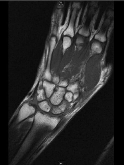
Image by Lecturio.

Neurologic causes

  • Carpal tunnel Carpal Tunnel The carpal tunnel is formed by the transverse carpal ligament (flexor retinaculum) superiorly and the carpal bones inferiorly. Carpal Tunnel Syndrome syndrome:
    • A peripheral mononeuropathy Mononeuropathy Disease or trauma involving a single peripheral nerve in isolation, or out of proportion to evidence of diffuse peripheral nerve dysfunction. Mononeuropathy multiplex refers to a condition characterized by multiple isolated nerve injuries. Mononeuropathies may result from a wide variety of causes, including ischemia; traumatic injury; compression; connective tissue diseases; cumulative trauma disorders; and other conditions. Mononeuropathy and Plexopathy affecting the hand Hand The hand constitutes the distal part of the upper limb and provides the fine, precise movements needed in activities of daily living. It consists of 5 metacarpal bones and 14 phalanges, as well as numerous muscles innervated by the median and ulnar nerves. Hand: Anatomy/wrist caused by compression Compression Blunt Chest Trauma of the median nerve Median Nerve A major nerve of the upper extremity. In humans, the fibers of the median nerve originate in the lower cervical and upper thoracic spinal cord (usually C6 to T1), travel via the brachial plexus, and supply sensory and motor innervation to parts of the forearm and hand. Cubital Fossa: Anatomy within the carpal tunnel Carpal Tunnel The carpal tunnel is formed by the transverse carpal ligament (flexor retinaculum) superiorly and the carpal bones inferiorly. Carpal Tunnel Syndrome deep to the flexor retinaculum Flexor Retinaculum Ankle Joint: Anatomy (aka transverse carpal ligament)
    • Presents with sensory Sensory Neurons which conduct nerve impulses to the central nervous system. Nervous System: Histology disturbances (e.g., pain Pain An unpleasant sensation induced by noxious stimuli which are detected by nerve endings of nociceptive neurons. Pain: Types and Pathways, tingling Tingling Posterior Cord Syndrome, numbness) and/or weakness of the palmar surface of the thumb, index finger, and middle finger and the radial half of the ring finger
  • Cubital tunnel syndrome:
    • A peripheral mononeuropathy Mononeuropathy Disease or trauma involving a single peripheral nerve in isolation, or out of proportion to evidence of diffuse peripheral nerve dysfunction. Mononeuropathy multiplex refers to a condition characterized by multiple isolated nerve injuries. Mononeuropathies may result from a wide variety of causes, including ischemia; traumatic injury; compression; connective tissue diseases; cumulative trauma disorders; and other conditions. Mononeuropathy and Plexopathy affecting the hand Hand The hand constitutes the distal part of the upper limb and provides the fine, precise movements needed in activities of daily living. It consists of 5 metacarpal bones and 14 phalanges, as well as numerous muscles innervated by the median and ulnar nerves. Hand: Anatomy/wrist caused by compression Compression Blunt Chest Trauma of the ulnar nerve Ulnar Nerve A major nerve of the upper extremity. In humans, the fibers of the ulnar nerve originate in the lower cervical and upper thoracic spinal cord (usually C7 to T1), travel via the medial cord of the brachial plexus, and supply sensory and motor innervation to parts of the hand and forearm. Axilla and Brachial Plexus: Anatomy within the cubital tunnel at the medial elbow
    • Presents with sensory Sensory Neurons which conduct nerve impulses to the central nervous system. Nervous System: Histology disturbances (e.g., pain Pain An unpleasant sensation induced by noxious stimuli which are detected by nerve endings of nociceptive neurons. Pain: Types and Pathways, tingling Tingling Posterior Cord Syndrome, numbness) and/or weakness of the palmar surface of the ulnar half of the ring finger and pinky finger
  • Thoracic outlet Thoracic Outlet Thoracic Outlet Syndrome compression Compression Blunt Chest Trauma syndrome:
    • Group of conditions that involve the compression Compression Blunt Chest Trauma of neurovascular structures, such as the subclavian vessels or brachial plexus Brachial Plexus The large network of nerve fibers which distributes the innervation of the upper extremity. The brachial plexus extends from the neck into the axilla. In humans, the nerves of the plexus usually originate from the lower cervical and the first thoracic spinal cord segments (c5-c8 and T1), but variations are not uncommon. Peripheral Nerve Injuries in the Cervicothoracic Region, as they pass from the lower neck Neck The part of a human or animal body connecting the head to the rest of the body. Peritonsillar Abscess into the axilla Axilla The axilla is a pyramid-shaped space located between the upper thorax and the arm. The axilla has a base, an apex, and 4 walls (anterior, medial, lateral, posterior). The base of the pyramid is made up of the axillary skin. The apex is the axillary inlet, located between the 1st rib, superior border of the scapula, and clavicle. Axilla and Brachial Plexus: Anatomy
    • The thoracic outlet Thoracic Outlet Thoracic Outlet Syndrome consists of the scalene muscles, first rib, and clavicle Clavicle A bone on the ventral side of the shoulder girdle, which in humans is commonly called the collar bone. Clavicle Fracture
    • May be caused by trauma, tumors, or the presence of a cervical rib
    • Results in neurovascular compromise that may affect sensation and/or strength of the wrist and hand Hand The hand constitutes the distal part of the upper limb and provides the fine, precise movements needed in activities of daily living. It consists of 5 metacarpal bones and 14 phalanges, as well as numerous muscles innervated by the median and ulnar nerves. Hand: Anatomy

Systemic causes

  • Amyloidosis Amyloidosis Amyloidosis is a disease caused by abnormal extracellular tissue deposition of fibrils composed of various misfolded low-molecular-weight protein subunits. These proteins are frequently byproducts of other pathological processes (e.g., multiple myeloma). Amyloidosis:
  • Leukemia: the unregulated proliferation of white blood cells that can result in hand Hand The hand constitutes the distal part of the upper limb and provides the fine, precise movements needed in activities of daily living. It consists of 5 metacarpal bones and 14 phalanges, as well as numerous muscles innervated by the median and ulnar nerves. Hand: Anatomy pain Pain An unpleasant sensation induced by noxious stimuli which are detected by nerve endings of nociceptive neurons. Pain: Types and Pathways due to bone Bone Bone is a compact type of hardened connective tissue composed of bone cells, membranes, an extracellular mineralized matrix, and central bone marrow. The 2 primary types of bone are compact and spongy. Bones: Structure and Types invasion and metastasis Metastasis The transfer of a neoplasm from one organ or part of the body to another remote from the primary site. Grading, Staging, and Metastasis
  • Multiple myeloma Multiple myeloma Multiple myeloma (MM) is a malignant condition of plasma cells (activated B lymphocytes) primarily seen in the elderly. Monoclonal proliferation of plasma cells results in cytokine-driven osteoclastic activity and excessive secretion of IgG antibodies. Multiple Myeloma: can result in hand Hand The hand constitutes the distal part of the upper limb and provides the fine, precise movements needed in activities of daily living. It consists of 5 metacarpal bones and 14 phalanges, as well as numerous muscles innervated by the median and ulnar nerves. Hand: Anatomy pain Pain An unpleasant sensation induced by noxious stimuli which are detected by nerve endings of nociceptive neurons. Pain: Types and Pathways due to lytic changes 
  • Osteomyelitis Osteomyelitis Osteomyelitis is an infection of the bone that results from the spread of microorganisms from the blood (hematogenous), nearby infected tissue, or open wounds (non-hematogenous). Infections are most commonly caused by Staphylococcus aureus. Osteomyelitis: an infection of the bone Bone Bone is a compact type of hardened connective tissue composed of bone cells, membranes, an extracellular mineralized matrix, and central bone marrow. The 2 primary types of bone are compact and spongy. Bones: Structure and Types most commonly caused by Staphylococcus aureus Staphylococcus aureus Potentially pathogenic bacteria found in nasal membranes, skin, hair follicles, and perineum of warm-blooded animals. They may cause a wide range of infections and intoxications. Brain Abscess
  • Peripheral neuropathy Neuropathy Leprosy:
    • Peripheral nerve damage that presents as:
      • Distal sensory Sensory Neurons which conduct nerve impulses to the central nervous system. Nervous System: Histology loss
      • Paresthesias Paresthesias Subjective cutaneous sensations (e.g., cold, warmth, tingling, pressure, etc.) that are experienced spontaneously in the absence of stimulation. Posterior Cord Syndrome
      • Dysesthesias
      • Weakness 
    • May be caused by systemic conditions, such as diabetes Diabetes Diabetes mellitus (DM) is a metabolic disease characterized by hyperglycemia and dysfunction of the regulation of glucose metabolism by insulin. Type 1 DM is diagnosed mostly in children and young adults as the result of autoimmune destruction of β cells in the pancreas and the resulting lack of insulin. Type 2 DM has a significant association with obesity and is characterized by insulin resistance. Diabetes Mellitus mellitus or vitamin deficiency
  • Complex regional pain Regional Pain Complex Regional Pain Syndrome (CRPS) syndrome ( CRPS CRPS Complex regional pain syndrome (CRPS) is a chronic regional neuropathic pain condition characterized by excruciating pain (out of proportion to apparent tissue damage or inciting trauma), paresthesia, allodynia, temperature abnormalities, skin discoloration, edema, reduced range of motion, and bone demineralization. Complex Regional Pain Syndrome (CRPS)): a neuropathic/ nociplastic pain Nociplastic pain Altered nociception despite no clear evidence of actual or threatened tissue damage causing the activation of peripheral nociceptors or evidence for disease or lesion of the somatosensory system causing the pain. Pain Management condition characterized by pain Pain An unpleasant sensation induced by noxious stimuli which are detected by nerve endings of nociceptive neurons. Pain: Types and Pathways that is more severe or has lasted longer than would be expected from the causative injury
  • Rheumatologic disorders: Inflammation Inflammation Inflammation is a complex set of responses to infection and injury involving leukocytes as the principal cellular mediators in the body’s defense against pathogenic organisms. Inflammation is also seen as a response to tissue injury in the process of wound healing. The 5 cardinal signs of inflammation are pain, heat, redness, swelling, and loss of function. Inflammation of the synovium is responsible for the pain Pain An unpleasant sensation induced by noxious stimuli which are detected by nerve endings of nociceptive neurons. Pain: Types and Pathways that the patient may experience.
    • Rheumatoid arthritis Arthritis Acute or chronic inflammation of joints. Osteoarthritis: an inflammatory polyarthritis Polyarthritis Rheumatoid Arthritis that presents with pain Pain An unpleasant sensation induced by noxious stimuli which are detected by nerve endings of nociceptive neurons. Pain: Types and Pathways in joints that are warm and “boggy” to touch; associated with characteristic bony deformities
    • Systemic lupus erythematosus Systemic lupus erythematosus Systemic lupus erythematosus (SLE) is a chronic autoimmune, inflammatory condition that causes immune-complex deposition in organs, resulting in systemic manifestations. Women, particularly those of African American descent, are more commonly affected. Systemic Lupus Erythematosus ( SLE SLE Systemic lupus erythematosus (SLE) is a chronic autoimmune, inflammatory condition that causes immune-complex deposition in organs, resulting in systemic manifestations. Women, particularly those of African American descent, are more commonly affected. Systemic Lupus Erythematosus): a chronic inflammatory condition characterized by clinical involvement of the skin Skin The skin, also referred to as the integumentary system, is the largest organ of the body. The skin is primarily composed of the epidermis (outer layer) and dermis (deep layer). The epidermis is primarily composed of keratinocytes that undergo rapid turnover, while the dermis contains dense layers of connective tissue. Skin: Structure and Functions, joints, kidneys Kidneys The kidneys are a pair of bean-shaped organs located retroperitoneally against the posterior wall of the abdomen on either side of the spine. As part of the urinary tract, the kidneys are responsible for blood filtration and excretion of water-soluble waste in the urine. Kidneys: Anatomy, blood cells, and central nervous system Central nervous system The main information-processing organs of the nervous system, consisting of the brain, spinal cord, and meninges. Nervous System: Anatomy, Structure, and Classification
    • Raynaud’s phenomenon: an exaggerated vascular response of the fingers to cold temperatures or emotional stress that results in sequential Sequential Computed Tomography (CT) digital ischemia Ischemia A hypoperfusion of the blood through an organ or tissue caused by a pathologic constriction or obstruction of its blood vessels, or an absence of blood circulation. Ischemic Cell Damage, hypoxia Hypoxia Sub-optimal oxygen levels in the ambient air of living organisms. Ischemic Cell Damage, and hyperemia
  • Osteoarthritis Osteoarthritis Osteoarthritis (OA) is the most common form of arthritis, and is due to cartilage destruction and changes of the subchondral bone. The risk of developing this disorder increases with age, obesity, and repetitive joint use or trauma. Patients develop gradual joint pain, stiffness lasting < 30 minutes, and decreased range of motion. Osteoarthritis: the most common form of arthritis Arthritis Acute or chronic inflammation of joints. Osteoarthritis, due to progressive wasting of articular cartilage Cartilage Cartilage is a type of connective tissue derived from embryonic mesenchyme that is responsible for structural support, resilience, and the smoothness of physical actions. Perichondrium (connective tissue membrane surrounding cartilage) compensates for the absence of vasculature in cartilage by providing nutrition and support. Cartilage: Histology and surrounding joint structures with minimal inflammation Inflammation Inflammation is a complex set of responses to infection and injury involving leukocytes as the principal cellular mediators in the body’s defense against pathogenic organisms. Inflammation is also seen as a response to tissue injury in the process of wound healing. The 5 cardinal signs of inflammation are pain, heat, redness, swelling, and loss of function. Inflammation

Management

Initially, diagnose and treat the underlying condition. General principles of hand Hand The hand constitutes the distal part of the upper limb and provides the fine, precise movements needed in activities of daily living. It consists of 5 metacarpal bones and 14 phalanges, as well as numerous muscles innervated by the median and ulnar nerves. Hand: Anatomy and wrist pain Pain An unpleasant sensation induced by noxious stimuli which are detected by nerve endings of nociceptive neurons. Pain: Types and Pathways management are carried out according to the RICE principle, as follows:

  • Rest:
    • Immobilization Immobilization Delirium of the involved hand Hand The hand constitutes the distal part of the upper limb and provides the fine, precise movements needed in activities of daily living. It consists of 5 metacarpal bones and 14 phalanges, as well as numerous muscles innervated by the median and ulnar nerves. Hand: Anatomy as appropriate to the condition (i.e., fracture Fracture A fracture is a disruption of the cortex of any bone and periosteum and is commonly due to mechanical stress after an injury or accident. Open fractures due to trauma can be a medical emergency. Fractures are frequently associated with automobile accidents, workplace injuries, and trauma. Overview of Bone Fractures, tendinopathy)
    • Allows for scar Scar Dermatologic Examination tissue to form and unite the separated parts of a structure to avoid any further damage
    • Below-elbow cast is most commonly used.
  • Ice application:
    • Works by inducing vasoconstriction Vasoconstriction The physiological narrowing of blood vessels by contraction of the vascular smooth muscle. Vascular Resistance, Flow, and Mean Arterial Pressure 
    • Reduces inflammation Inflammation Inflammation is a complex set of responses to infection and injury involving leukocytes as the principal cellular mediators in the body’s defense against pathogenic organisms. Inflammation is also seen as a response to tissue injury in the process of wound healing. The 5 cardinal signs of inflammation are pain, heat, redness, swelling, and loss of function. Inflammation and limits hematoma Hematoma A collection of blood outside the blood vessels. Hematoma can be localized in an organ, space, or tissue. Intussusception formation
  • Compression Compression Blunt Chest Trauma:
    • Reduces the amount of fluid or blood accumulated in edema Edema Edema is a condition in which excess serous fluid accumulates in the body cavity or interstitial space of connective tissues. Edema is a symptom observed in several medical conditions. It can be categorized into 2 types, namely, peripheral (in the extremities) and internal (in an organ or body cavity). Edema and hematomas
  • Elevate the limb:
    • Decreases hydrostatic pressure Hydrostatic pressure The pressure due to the weight of fluid. Edema
    • Improves drainage and reduces edema Edema Edema is a condition in which excess serous fluid accumulates in the body cavity or interstitial space of connective tissues. Edema is a symptom observed in several medical conditions. It can be categorized into 2 types, namely, peripheral (in the extremities) and internal (in an organ or body cavity). Edema

Clinical Relevance

The following are possible etiologies of hand Hand The hand constitutes the distal part of the upper limb and provides the fine, precise movements needed in activities of daily living. It consists of 5 metacarpal bones and 14 phalanges, as well as numerous muscles innervated by the median and ulnar nerves. Hand: Anatomy and wrist pain Pain An unpleasant sensation induced by noxious stimuli which are detected by nerve endings of nociceptive neurons. Pain: Types and Pathways:

  • Carpal tunnel Carpal Tunnel The carpal tunnel is formed by the transverse carpal ligament (flexor retinaculum) superiorly and the carpal bones inferiorly. Carpal Tunnel Syndrome syndrome: The carpal tunnel Carpal Tunnel The carpal tunnel is formed by the transverse carpal ligament (flexor retinaculum) superiorly and the carpal bones inferiorly. Carpal Tunnel Syndrome is formed anteriorly by the flexor retinaculum Flexor Retinaculum Ankle Joint: Anatomy and posteriorly by the carpal bones Carpal bones The eight bones of the wrist: scaphoid bone; lunate bone; triquetrum bone; pisiform bone; trapezium bone; trapezoid bone; capitate bone; and hamate bone. Carpal Tunnel Syndrome. The median nerve Median Nerve A major nerve of the upper extremity. In humans, the fibers of the median nerve originate in the lower cervical and upper thoracic spinal cord (usually C6 to T1), travel via the brachial plexus, and supply sensory and motor innervation to parts of the forearm and hand. Cubital Fossa: Anatomy and the tendons of the flexor digitorum longus Flexor digitorum longus Leg: Anatomy travel to the fingers through the carpal tunnel Carpal Tunnel The carpal tunnel is formed by the transverse carpal ligament (flexor retinaculum) superiorly and the carpal bones inferiorly. Carpal Tunnel Syndrome. Carpal tunnel Carpal Tunnel The carpal tunnel is formed by the transverse carpal ligament (flexor retinaculum) superiorly and the carpal bones inferiorly. Carpal Tunnel Syndrome syndrome occurs from narrowing of the tunnel and subsequent compression Compression Blunt Chest Trauma of the structures within it, particularly the median nerve Median Nerve A major nerve of the upper extremity. In humans, the fibers of the median nerve originate in the lower cervical and upper thoracic spinal cord (usually C6 to T1), travel via the brachial plexus, and supply sensory and motor innervation to parts of the forearm and hand. Cubital Fossa: Anatomy.
  • Ulnar nerve injury Ulnar Nerve Injury Peripheral Nerve Injuries in the Upper Extremity: The ulnar nerve Ulnar Nerve A major nerve of the upper extremity. In humans, the fibers of the ulnar nerve originate in the lower cervical and upper thoracic spinal cord (usually C7 to T1), travel via the medial cord of the brachial plexus, and supply sensory and motor innervation to parts of the hand and forearm. Axilla and Brachial Plexus: Anatomy may be injured in multiple positions, including a fractured or dislocated hook of the hamate. Features include a loss of sensation in the fourth and fifth fingers of the hand Hand The hand constitutes the distal part of the upper limb and provides the fine, precise movements needed in activities of daily living. It consists of 5 metacarpal bones and 14 phalanges, as well as numerous muscles innervated by the median and ulnar nerves. Hand: Anatomy, weakness that worsens with physical activity, and a claw hand Hand The hand constitutes the distal part of the upper limb and provides the fine, precise movements needed in activities of daily living. It consists of 5 metacarpal bones and 14 phalanges, as well as numerous muscles innervated by the median and ulnar nerves. Hand: Anatomy
  • Thoracic outlet Thoracic Outlet Thoracic Outlet Syndrome syndrome: Thoracic outlet Thoracic Outlet Thoracic Outlet Syndrome syndrome is caused by compression Compression Blunt Chest Trauma of the neurovascular structures at the thoracic outlet Thoracic Outlet Thoracic Outlet Syndrome, especially those passing through the inter-scalene triangle. Signs and symptoms in the patient are produced according to the structures involved (i.e., neurogenic, arterial, and venous symptoms). The syndrome is classified on the basis of the structures involved.
  • Osteoarthritis Osteoarthritis Osteoarthritis (OA) is the most common form of arthritis, and is due to cartilage destruction and changes of the subchondral bone. The risk of developing this disorder increases with age, obesity, and repetitive joint use or trauma. Patients develop gradual joint pain, stiffness lasting < 30 minutes, and decreased range of motion. Osteoarthritis: degenerative disorder of the articular cartilage Cartilage Cartilage is a type of connective tissue derived from embryonic mesenchyme that is responsible for structural support, resilience, and the smoothness of physical actions. Perichondrium (connective tissue membrane surrounding cartilage) compensates for the absence of vasculature in cartilage by providing nutrition and support. Cartilage: Histology, along with the subchondral bone Subchondral Bone Osteochondritis Dissecans and other joint structures. It is the most common type of joint disease and is the leading cause of disability Disability Determination of the degree of a physical, mental, or emotional handicap. The diagnosis is applied to legal qualification for benefits and income under disability insurance and to eligibility for social security and workman’s compensation benefits. ABCDE Assessment in older adults. The main risk factors for osteoarthritis Osteoarthritis Osteoarthritis (OA) is the most common form of arthritis, and is due to cartilage destruction and changes of the subchondral bone. The risk of developing this disorder increases with age, obesity, and repetitive joint use or trauma. Patients develop gradual joint pain, stiffness lasting < 30 minutes, and decreased range of motion. Osteoarthritis are a family history Family History Adult Health Maintenance of the disease, female gender Gender Gender Dysphoria, past trauma to the involved joint, aging, and obesity Obesity Obesity is a condition associated with excess body weight, specifically with the deposition of excessive adipose tissue. Obesity is considered a global epidemic. Major influences come from the western diet and sedentary lifestyles, but the exact mechanisms likely include a mixture of genetic and environmental factors. Obesity.
  • Rheumatoid arthritis Arthritis Acute or chronic inflammation of joints. Osteoarthritis: chronic inflammatory systemic disease that proceeds progressively in stages. The basis of the disease is an inflammation Inflammation Inflammation is a complex set of responses to infection and injury involving leukocytes as the principal cellular mediators in the body’s defense against pathogenic organisms. Inflammation is also seen as a response to tissue injury in the process of wound healing. The 5 cardinal signs of inflammation are pain, heat, redness, swelling, and loss of function. Inflammation of the synovial membrane Synovial Membrane The inner membrane of a joint capsule surrounding a freely movable joint. It is loosely attached to the external fibrous capsule and secretes synovial fluid. Hip Joint: Anatomy (i.e., the inner layer of the joint capsule Capsule An envelope of loose gel surrounding a bacterial cell which is associated with the virulence of pathogenic bacteria. Some capsules have a well-defined border, whereas others form a slime layer that trails off into the medium. Most capsules consist of relatively simple polysaccharides but there are some bacteria whose capsules are made of polypeptides. Bacteroides). Attributed to synovitis Synovitis Inflammation of the synovial membrane. Rheumatoid Arthritis, rheumatoid arthritis Arthritis Acute or chronic inflammation of joints. Osteoarthritis can lead to secondary diseases such as arthritis Arthritis Acute or chronic inflammation of joints. Osteoarthritis, bursitis, or tenosynovitis.
  • Systemic lupus erythematosus Systemic lupus erythematosus Systemic lupus erythematosus (SLE) is a chronic autoimmune, inflammatory condition that causes immune-complex deposition in organs, resulting in systemic manifestations. Women, particularly those of African American descent, are more commonly affected. Systemic Lupus Erythematosus: an autoimmune disease with wide variation in signs and symptoms. Symptoms range from mild conditions to severe organ manifestations, which sometimes makes the diagnosis difficult. Systemic lupus erythematosus Systemic lupus erythematosus Systemic lupus erythematosus (SLE) is a chronic autoimmune, inflammatory condition that causes immune-complex deposition in organs, resulting in systemic manifestations. Women, particularly those of African American descent, are more commonly affected. Systemic Lupus Erythematosus may affect all joints of the human body, so patients Patients Individuals participating in the health care system for the purpose of receiving therapeutic, diagnostic, or preventive procedures. Clinician–Patient Relationship may present with joint pain Pain An unpleasant sensation induced by noxious stimuli which are detected by nerve endings of nociceptive neurons. Pain: Types and Pathways, including wrist/ hand Hand The hand constitutes the distal part of the upper limb and provides the fine, precise movements needed in activities of daily living. It consists of 5 metacarpal bones and 14 phalanges, as well as numerous muscles innervated by the median and ulnar nerves. Hand: Anatomy pain Pain An unpleasant sensation induced by noxious stimuli which are detected by nerve endings of nociceptive neurons. Pain: Types and Pathways.

Create your free account or log in to continue reading!

Sign up now and get free access to Lecturio with concept pages, medical videos, and questions for your medical education.

User Reviews

Details