Gynecomastia

Gynecomastia is a benign Benign Fibroadenoma proliferation of male breast glandular ductal tissue, usually bilateral, caused by increased estrogen Estrogen Compounds that interact with estrogen receptors in target tissues to bring about the effects similar to those of estradiol. Estrogens stimulate the female reproductive organs, and the development of secondary female sex characteristics. Estrogenic chemicals include natural, synthetic, steroidal, or non-steroidal compounds. Ovaries: Anatomy activity, decreased testosterone Testosterone A potent androgenic steroid and major product secreted by the leydig cells of the testis. Its production is stimulated by luteinizing hormone from the pituitary gland. In turn, testosterone exerts feedback control of the pituitary LH and FSH secretion. Depending on the tissues, testosterone can be further converted to dihydrotestosterone or estradiol. Androgens and Antiandrogens activity, or medications. The condition is common and physiological in neonates, adolescent boys, and elderly men. Some cases are pathological or secondary to chronic liver Liver The liver is the largest gland in the human body. The liver is found in the superior right quadrant of the abdomen and weighs approximately 1.5 kilograms. Its main functions are detoxification, metabolism, nutrient storage (e.g., iron and vitamins), synthesis of coagulation factors, formation of bile, filtration, and storage of blood. Liver: Anatomy disease, kidney disease, or hyperthyroidism Hyperthyroidism Hypersecretion of thyroid hormones from the thyroid gland. Elevated levels of thyroid hormones increase basal metabolic rate. Thyrotoxicosis and Hyperthyroidism. The majority of cases are asymptomatic and do not need treatment.

Last updated: Nov 20, 2024

Editorial responsibility: Stanley Oiseth, Lindsay Jones, Evelin Maza

Overview

Asymptomatic gynecomastia is common enough that it could almost be considered a normal condition, with a trimodal age distribution.

Gynecomastia is classified into 3 major groups: physiological, pathological, and idiopathic Idiopathic Dermatomyositis.

Physiological gynecomastia

  • Neonates
    • Due to elevated estrogen Estrogen Compounds that interact with estrogen receptors in target tissues to bring about the effects similar to those of estradiol. Estrogens stimulate the female reproductive organs, and the development of secondary female sex characteristics. Estrogenic chemicals include natural, synthetic, steroidal, or non-steroidal compounds. Ovaries: Anatomy concentrations in fetal blood
    • Typically regresses by the age of 3 weeks
    • Occurs in 60%90% of neonates
  • Adolescent boys
    • Due to a transient increase in estradiol Estradiol The 17-beta-isomer of estradiol, an aromatized C18 steroid with hydroxyl group at 3-beta- and 17-beta-position. Estradiol-17-beta is the most potent form of mammalian estrogenic steroids. Noncontraceptive Estrogen and Progestins concentration at the onset of puberty Puberty Puberty is a complex series of physical, psychosocial, and cognitive transitions usually experienced by adolescents (11-19 years of age). Puberty is marked by a growth in stature and the development of secondary sexual characteristics, achievement of fertility, and changes in most body systems. Puberty
    • Typically regresses after 18 months of puberty Puberty Puberty is a complex series of physical, psychosocial, and cognitive transitions usually experienced by adolescents (11-19 years of age). Puberty is marked by a growth in stature and the development of secondary sexual characteristics, achievement of fertility, and changes in most body systems. Puberty
    • Occurs in 50% of male teenagers
    • Not common after the age of 17 years
  • Older men 
    • Due to increased body fat content, which is responsible for the increased conversion of androgens Androgens Androgens are naturally occurring steroid hormones responsible for development and maintenance of the male sex characteristics, including penile, scrotal, and clitoral growth, development of sexual hair, deepening of the voice, and musculoskeletal growth. Androgens and Antiandrogens to estrogens
    • Occurs in up to 70% of men aged 5069 years

Pathological gynecomastia

  • Medications: estimated to cause approximately 10%25% of all cases of gynecomastia
    • Definitely associated with gynecomastia: estrogens, cimetidine Cimetidine A histamine congener, it competitively inhibits histamine binding to histamine h2 receptors. Cimetidine has a range of pharmacological actions. It inhibits gastric acid secretion, as well as pepsin and gastrin output. Antihistamines, ketoconazole Ketoconazole Broad spectrum antifungal agent used for long periods at high doses, especially in immunosuppressed patients. Azoles, growth hormone, gonadotropins, antiandrogen therapies, and 5-alpha-reductase inhibitors
    • Probably associated with gynecomastia: first-generation and atypical antipsychotics Atypical Antipsychotics Antiemetics in adults, calcium Calcium A basic element found in nearly all tissues. It is a member of the alkaline earth family of metals with the atomic symbol ca, atomic number 20, and atomic weight 40. Calcium is the most abundant mineral in the body and combines with phosphorus to form calcium phosphate in the bones and teeth. It is essential for the normal functioning of nerves and muscles and plays a role in blood coagulation (as factor IV) and in many enzymatic processes. Electrolytes channel blockers, omeprazole Omeprazole A 4-methoxy-3, 5-dimethylpyridyl, 5-methoxybenzimidazole derivative of timoprazole that is used in the therapy of stomach ulcers and zollinger-ellison syndrome. The drug inhibits an h(+)-k(+)-exchanging ATPase which is found in gastric parietal cells. Gastric Acid Drugs, HIV HIV Anti-HIV Drugs drugs, alkylating agents, anabolic steroids Steroids A group of polycyclic compounds closely related biochemically to terpenes. They include cholesterol, numerous hormones, precursors of certain vitamins, bile acids, alcohols (sterols), and certain natural drugs and poisons. Steroids have a common nucleus, a fused, reduced 17-carbon atom ring system, cyclopentanoperhydrophenanthrene. Most steroids also have two methyl groups and an aliphatic side-chain attached to the nucleus. Benign Liver Tumors, alcohol, and opioids Opioids Opiates are drugs that are derived from the sap of the opium poppy. Opiates have been used since antiquity for the relief of acute severe pain. Opioids are synthetic opiates with properties that are substantially similar to those of opiates. Opioid Analgesics
    • Many other medications have been reported to be associated with gynecomastia but good evidence is lacking.
    • Certain herbal supplements, such as those containing phytoestrogens, have been associated with gynecomastia.
  • Metabolic disorders: liver Liver The liver is the largest gland in the human body. The liver is found in the superior right quadrant of the abdomen and weighs approximately 1.5 kilograms. Its main functions are detoxification, metabolism, nutrient storage (e.g., iron and vitamins), synthesis of coagulation factors, formation of bile, filtration, and storage of blood. Liver: Anatomy disease, cirrhosis Cirrhosis Cirrhosis is a late stage of hepatic parenchymal necrosis and scarring (fibrosis) most commonly due to hepatitis C infection and alcoholic liver disease. Patients may present with jaundice, ascites, and hepatosplenomegaly. Cirrhosis can also cause complications such as hepatic encephalopathy, portal hypertension, portal vein thrombosis, and hepatorenal syndrome. Cirrhosis, type 1 Type 1 Spinal Muscular Atrophy diabetes Diabetes Diabetes mellitus (DM) is a metabolic disease characterized by hyperglycemia and dysfunction of the regulation of glucose metabolism by insulin. Type 1 DM is diagnosed mostly in children and young adults as the result of autoimmune destruction of β cells in the pancreas and the resulting lack of insulin. Type 2 DM has a significant association with obesity and is characterized by insulin resistance. Diabetes Mellitus, hyperthyroidism Hyperthyroidism Hypersecretion of thyroid hormones from the thyroid gland. Elevated levels of thyroid hormones increase basal metabolic rate. Thyrotoxicosis and Hyperthyroidism, chronic renal disease
  • Disorders of sex Sex The totality of characteristics of reproductive structure, functions, phenotype, and genotype, differentiating the male from the female organism. Gender Dysphoria development (DSDs):
    • Klinefelter syndrome Klinefelter syndrome Klinefelter syndrome is a chromosomal aneuploidy characterized by the presence of 1 or more extra X chromosomes in a male karyotype, most commonly leading to karyotype 47,XXY. Klinefelter syndrome is associated with decreased levels of testosterone and is the most common cause of congenital hypogonadism. Klinefelter Syndrome
    • Androgen insensitivity syndrome Androgen insensitivity syndrome Androgen insensitivity syndrome (AIS) is an X-linked recessive condition in which a genetic mutation affects the function of androgen receptors, resulting in complete (CAIS), partial (PAIS), or mild (MAIS) resistance to testosterone. All individuals with AIS have a 46,XY karyotype; however, phenotypes vary and include phenotypic female, virilized female, undervirilized male, and phenotypic male individuals. Androgen Insensitivity Syndrome
  • Tumors:
    • Testicular tumors: germ cell, Leydig cell, Sertoli cell, sex Sex The totality of characteristics of reproductive structure, functions, phenotype, and genotype, differentiating the male from the female organism. Gender Dysphoria cord
    • Adrenal tumors: adenoma or carcinoma
  • Systemic Conditions:
    • Malnutrition Malnutrition Malnutrition is a clinical state caused by an imbalance or deficiency of calories and/or micronutrients and macronutrients. The 2 main manifestations of acute severe malnutrition are marasmus (total caloric insufficiency) and kwashiorkor (protein malnutrition with characteristic edema). Malnutrition in children in resource-limited countries and Refeeding: nutritional deficiencies and subsequent refeeding can alter hormone levels, resulting in gynecomastia.

Idiopathic Idiopathic Dermatomyositis gynecomastia

Mnemonic

To recall the causes of gynecomastia, remember CODES:
  • C: Cirrhosis
  • O: Obesity
  • D: Digoxin
  • E: Estrogen
  • S: Spironolactone

Clinical Presentation

  • Most patients Patients Individuals participating in the health care system for the purpose of receiving therapeutic, diagnostic, or preventive procedures. Clinician–Patient Relationship are asymptomatic.
  • Clinical features:
    • Concentric with the nipple Nipple The conic organs which usually give outlet to milk from the mammary glands. Examination of the Breast, usually bilateral and firm
    • Can present with tender areolar complex
  • Clinical history:
    • Family history Family History Adult Health Maintenance of gynecomastia
    • Age of onset and duration of the condition
    • History of mumps Mumps Mumps is caused by a single-stranded, linear, negative-sense RNA virus of the family Paramyxoviridae. Mumps is typically a disease of childhood, which manifests initially with fever, muscle pain, headache, poor appetite, and a general feeling of malaise, and is classically followed by parotitis. Mumps Virus/Mumps (if affected by mumps Mumps Mumps is caused by a single-stranded, linear, negative-sense RNA virus of the family Paramyxoviridae. Mumps is typically a disease of childhood, which manifests initially with fever, muscle pain, headache, poor appetite, and a general feeling of malaise, and is classically followed by parotitis. Mumps Virus/Mumps orchitis Orchitis Inflammation of a testis. It has many features of epididymitis, such as swollen scrotum; pain; pyuria; and fever. It is usually related to infections in the urinary tract, which likely spread to the epididymis and then the testis through either the vas deferens or the lymphatics of the spermatic cord. Epididymitis and Orchitis, with subsequent impairment in the production of testosterone Testosterone A potent androgenic steroid and major product secreted by the leydig cells of the testis. Its production is stimulated by luteinizing hormone from the pituitary gland. In turn, testosterone exerts feedback control of the pituitary LH and FSH secretion. Depending on the tissues, testosterone can be further converted to dihydrotestosterone or estradiol. Androgens and Antiandrogens by Leydig cells Leydig Cells Steroid-producing cells in the interstitial tissue of the testis. They are under the regulation of pituitary hormones; luteinizing hormone; or interstitial cell-stimulating hormone. Testosterone is the major androgen (androgens) produced. Testicles: Anatomy), testicular trauma, alcohol use, drug use
    • Any recent changes in nipple Nipple The conic organs which usually give outlet to milk from the mammary glands. Examination of the Breast size and any pain Pain An unpleasant sensation induced by noxious stimuli which are detected by nerve endings of nociceptive neurons. Pain: Types and Pathways or discharge from the nipples
    • History of sexual dysfunction Sexual dysfunction Physiological disturbances in normal sexual performance in either the male or the female. Sexual Physiology, infertility Infertility Infertility is the inability to conceive in the context of regular intercourse. The most common causes of infertility in women are related to ovulatory dysfunction or tubal obstruction, whereas, in men, abnormal sperm is a common cause. Infertility, or hypogonadism Hypogonadism Hypogonadism is a condition characterized by reduced or no sex hormone production by the testes or ovaries. Hypogonadism can result from primary (hypergonadotropic) or secondary (hypogonadotropic) failure. Symptoms include infertility, increased risk of osteoporosis, erectile dysfunction, decreased libido, and regression (or absence) of secondary sexual characteristics. Hypogonadism
  • Consider malignancy Malignancy Hemothorax if:

Diagnosis

  • Diagnosis is made via relevant history and physical examination:
  • Laboratory:
    • Serum levels of testosterone Testosterone A potent androgenic steroid and major product secreted by the leydig cells of the testis. Its production is stimulated by luteinizing hormone from the pituitary gland. In turn, testosterone exerts feedback control of the pituitary LH and FSH secretion. Depending on the tissues, testosterone can be further converted to dihydrotestosterone or estradiol. Androgens and Antiandrogens, estradiol Estradiol The 17-beta-isomer of estradiol, an aromatized C18 steroid with hydroxyl group at 3-beta- and 17-beta-position. Estradiol-17-beta is the most potent form of mammalian estrogenic steroids. Noncontraceptive Estrogen and Progestins, luteinizing hormone, and human chorionic gonadotropin (hCG) → to help identify pathological causes
    • Luteinizing hormone and follicle-stimulating hormone ( FSH FSH A major gonadotropin secreted by the adenohypophysis. Follicle-stimulating hormone stimulates gametogenesis and the supporting cells such as the ovarian granulosa cells, the testicular sertoli cells, and leydig cells. Fsh consists of two noncovalently linked subunits, alpha and beta. Within a species, the alpha subunit is common in the three pituitary glycoprotein hormones (TSH, LH, and FSH), but the beta subunit is unique and confers its biological specificity. Menstrual Cycle) levels → to exclude hypogonadism Hypogonadism Hypogonadism is a condition characterized by reduced or no sex hormone production by the testes or ovaries. Hypogonadism can result from primary (hypergonadotropic) or secondary (hypogonadotropic) failure. Symptoms include infertility, increased risk of osteoporosis, erectile dysfunction, decreased libido, and regression (or absence) of secondary sexual characteristics. Hypogonadism
    • Liver function tests Liver function tests Liver function tests, also known as hepatic function panels, are one of the most commonly performed screening blood tests. Such tests are also used to detect, evaluate, and monitor acute and chronic liver diseases. Liver Function Tests, renal function tests, and thyroid Thyroid The thyroid gland is one of the largest endocrine glands in the human body. The thyroid gland is a highly vascular, brownish-red gland located in the visceral compartment of the anterior region of the neck. Thyroid Gland: Anatomy hormone assay → to identify reversible or treatable causes of gynecomastia
    • Biopsy Biopsy Removal and pathologic examination of specimens from the living body. Ewing Sarcoma: if suspicious for malignancy Malignancy Hemothorax
    • Histology of gynecomastia: extensive ductal epithelial hyperplasia Hyperplasia An increase in the number of cells in a tissue or organ without tumor formation. It differs from hypertrophy, which is an increase in bulk without an increase in the number of cells. Cellular Adaptation in the early stages, rarely with lobular proliferation; later stages show fibrosis Fibrosis Any pathological condition where fibrous connective tissue invades any organ, usually as a consequence of inflammation or other injury. Bronchiolitis Obliterans
  • Imaging:
    • Mammography Mammography Radiographic examination of the breast. Breast Cancer Screening and breast ultrasonography with biopsy Biopsy Removal and pathologic examination of specimens from the living body. Ewing Sarcoma: in case of suspicion of breast cancer Breast cancer Breast cancer is a disease characterized by malignant transformation of the epithelial cells of the breast. Breast cancer is the most common form of cancer and 2nd most common cause of cancer-related death among women. Breast Cancer
    • Testicular ultrasound: may be indicated to exclude testicular tumors
    • Abdominal computed tomography (CT) scans or ultrasonography: to exclude liver Liver The liver is the largest gland in the human body. The liver is found in the superior right quadrant of the abdomen and weighs approximately 1.5 kilograms. Its main functions are detoxification, metabolism, nutrient storage (e.g., iron and vitamins), synthesis of coagulation factors, formation of bile, filtration, and storage of blood. Liver: Anatomy tumors and/or liver Liver The liver is the largest gland in the human body. The liver is found in the superior right quadrant of the abdomen and weighs approximately 1.5 kilograms. Its main functions are detoxification, metabolism, nutrient storage (e.g., iron and vitamins), synthesis of coagulation factors, formation of bile, filtration, and storage of blood. Liver: Anatomy cirrhosis Cirrhosis Cirrhosis is a late stage of hepatic parenchymal necrosis and scarring (fibrosis) most commonly due to hepatitis C infection and alcoholic liver disease. Patients may present with jaundice, ascites, and hepatosplenomegaly. Cirrhosis can also cause complications such as hepatic encephalopathy, portal hypertension, portal vein thrombosis, and hepatorenal syndrome. Cirrhosis
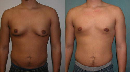
Gynecomastia

Physiological gynecomastia in an adolescent male

Image: “Adolescent Gynecomastia corrected by surgery protocol” by David Andrew Copeland, Dr. Mordcai Blau. License: GNU Free Documentation License

Management

  • Treatment of underlying cause:
    • Discontinue the offending drug
    • Treat the underlying medical condition
  • Observation:
    • Indicated in physiological gynecomastia throughout age period
    • Indicated in pathological gynecomastia < 6 months of onset
    • Mammography Mammography Radiographic examination of the breast. Breast Cancer Screening and biopsy Biopsy Removal and pathologic examination of specimens from the living body. Ewing Sarcoma if clinical suspicion of malignancy Malignancy Hemothorax
  • Medical therapy:
    • Testosterone Testosterone A potent androgenic steroid and major product secreted by the leydig cells of the testis. Its production is stimulated by luteinizing hormone from the pituitary gland. In turn, testosterone exerts feedback control of the pituitary LH and FSH secretion. Depending on the tissues, testosterone can be further converted to dihydrotestosterone or estradiol. Androgens and Antiandrogens replacement: in patients Patients Individuals participating in the health care system for the purpose of receiving therapeutic, diagnostic, or preventive procedures. Clinician–Patient Relationship with hypogonadism Hypogonadism Hypogonadism is a condition characterized by reduced or no sex hormone production by the testes or ovaries. Hypogonadism can result from primary (hypergonadotropic) or secondary (hypogonadotropic) failure. Symptoms include infertility, increased risk of osteoporosis, erectile dysfunction, decreased libido, and regression (or absence) of secondary sexual characteristics. Hypogonadism
    • Selective estrogen Estrogen Compounds that interact with estrogen receptors in target tissues to bring about the effects similar to those of estradiol. Estrogens stimulate the female reproductive organs, and the development of secondary female sex characteristics. Estrogenic chemicals include natural, synthetic, steroidal, or non-steroidal compounds. Ovaries: Anatomy receptor Receptor Receptors are proteins located either on the surface of or within a cell that can bind to signaling molecules known as ligands (e.g., hormones) and cause some type of response within the cell. Receptors modulators (e.g., tamoxifen Tamoxifen One of the selective estrogen receptor modulators with tissue-specific activities. Tamoxifen acts as an anti-estrogen (inhibiting agent) in the mammary tissue, but as an estrogen (stimulating agent) in cholesterol metabolism, bone density, and cell proliferation in the endometrium. Antiestrogens): for severe physiological gynecomastia or idiopathic Idiopathic Dermatomyositis gynecomastia lasting longer than 3 months
  • Surgery: Subcutaneous mastectomy Mastectomy Surgical procedure to remove one or both breasts. Fat Necrosis of the Breast is indicated for cosmesis in gynecomastia lasting longer than one year.

Differential Diagnosis

The following conditions are differential diagnoses for gynecomastia:

  • Hypogonadism Hypogonadism Hypogonadism is a condition characterized by reduced or no sex hormone production by the testes or ovaries. Hypogonadism can result from primary (hypergonadotropic) or secondary (hypogonadotropic) failure. Symptoms include infertility, increased risk of osteoporosis, erectile dysfunction, decreased libido, and regression (or absence) of secondary sexual characteristics. Hypogonadism: a condition characterized by decreased sex Sex The totality of characteristics of reproductive structure, functions, phenotype, and genotype, differentiating the male from the female organism. Gender Dysphoria steroid production in the gonads Gonads The gamete-producing glands, ovary or testis. Hormones: Overview and Types. In men, this can be the result of primary or secondary testicular failure due to pituitary Pituitary A small, unpaired gland situated in the sella turcica. It is connected to the hypothalamus by a short stalk which is called the infundibulum. Hormones: Overview and Types or hypothalamic disorders.
  • Breast cancer Breast cancer Breast cancer is a disease characterized by malignant transformation of the epithelial cells of the breast. Breast cancer is the most common form of cancer and 2nd most common cause of cancer-related death among women. Breast Cancer: a malignancy Malignancy Hemothorax that originates in breast tissue; 1% of breast cancer Breast cancer Breast cancer is a disease characterized by malignant transformation of the epithelial cells of the breast. Breast cancer is the most common form of cancer and 2nd most common cause of cancer-related death among women. Breast Cancer cases occur in men. This diagnosis should be considered if the mass Mass Three-dimensional lesion that occupies a space within the breast Imaging of the Breast is unilateral, hard, fixed, associated with skin Skin The skin, also referred to as the integumentary system, is the largest organ of the body. The skin is primarily composed of the epidermis (outer layer) and dermis (deep layer). The epidermis is primarily composed of keratinocytes that undergo rapid turnover, while the dermis contains dense layers of connective tissue. Skin: Structure and Functions dimpling, ulcerative, not concentric with the nipple Nipple The conic organs which usually give outlet to milk from the mammary glands. Examination of the Breast, or associated with bloody discharge, or if axillary lymphadenopathy Axillary Lymphadenopathy Lymphadenopathy is present.
  • Mastitis Mastitis Mastitis is inflammation of the breast tissue with or without infection. The most common form of mastitis is associated with lactation in the first few weeks after birth. Non-lactational mastitis includes periductal mastitis and idiopathic granulomatous mastitis (IGM). Mastitis: inflammation Inflammation Inflammation is a complex set of responses to infection and injury involving leukocytes as the principal cellular mediators in the body’s defense against pathogenic organisms. Inflammation is also seen as a response to tissue injury in the process of wound healing. The 5 cardinal signs of inflammation are pain, heat, redness, swelling, and loss of function. Inflammation of the mammary gland Mammary gland Glandular tissue in the breast of human that is under the influence of hormones such as estrogens; progestins; and prolactin. In women, after parturition, the mammary glands secrete milk for the nourishment of the young. Breasts: Anatomy tissue, which can be lactational or non-lactational. Mostly affects women of childbearing age within the first 6 weeks of lactation Lactation The processes of milk secretion by the maternal mammary glands after parturition. The proliferation of the mammary glandular tissue, milk synthesis, and milk expulsion or let down are regulated by the interactions of several hormones including estradiol; progesterone; prolactin; and oxytocin. Breastfeeding.
  • Lipoma Lipoma A lipoma is a benign neoplasm of fat cells (adipocytes) and the most common soft tissue tumor in adults. The etiology is unknown, but obesity is a predisposing factor; genetics also play a role, with multiple lipomas occurring in various inherited disorders. Lipoma: a benign Benign Fibroadenoma soft tissue Soft Tissue Soft Tissue Abscess tumor Tumor Inflammation that is composed of mature adipocytes Adipocytes Cells in the body that store fats, usually in the form of triglycerides. White adipocytes are the predominant type and found mostly in the abdominal cavity and subcutaneous tissue. Brown adipocytes are thermogenic cells that can be found in newborns of some species and hibernating mammals. Adipose Tissue: Histology. Lipoma Lipoma A lipoma is a benign neoplasm of fat cells (adipocytes) and the most common soft tissue tumor in adults. The etiology is unknown, but obesity is a predisposing factor; genetics also play a role, with multiple lipomas occurring in various inherited disorders. Lipoma usually presents as a subcutaneous, soft mass Mass Three-dimensional lesion that occupies a space within the breast Imaging of the Breast that is mobile and painless.
  • Pseudogynecomastia: when the male breast area has excess adipose (fat) tissue behind, around, and under the nipples. The condition is usually due to obesity Obesity Obesity is a condition associated with excess body weight, specifically with the deposition of excessive adipose tissue. Obesity is considered a global epidemic. Major influences come from the western diet and sedentary lifestyles, but the exact mechanisms likely include a mixture of genetic and environmental factors. Obesity.

References

  1. Johnson, R. E., & Murad, M. H. (2009). Gynecomastia: pathophysiology, evaluation, and management. Mayo Clinic proceedings, 84(11), 1010–1015. https://doi.org/10.1016/S0025-6196(11)60671-X
  2. Braunstein, G. D., & Anawalt, B. D. (2024). Clinical features, diagnosis, and evaluation of gynecomastia in adults. UpToDate. Retrieved November 19, 2024, from https://www.uptodate.com/contents/clinical-features-diagnosis-and-evaluation-of-gynecomastia-in-adults
  3. Hoda SA, Brogi E, Koerner FC, Rosen PP. Rosen’s Breast Pathology, 4th Edition. Lippincott Williams & Wilkins.
  4. Nuttall, F. Q., Warrier, R. S., & Gannon, M. C. (2015). Gynecomastia and drugs: a critical evaluation of the literature. European journal of clinical pharmacology, 71(5), 569–578. https://doi.org/10.1007/s00228-015-1835-x 
  5. Deepinder F,  Braunstein G. Drug-induced gynecomastia: An evidence-based review. Expert Opinion on Drug Safety. 2012;11(5):779-95. doi: 10.1517/14740338.2012.712109

Create your free account or log in to continue reading!

Sign up now and get free access to Lecturio with concept pages, medical videos, and questions for your medical education.

User Reviews

Details