Advertisement

Advertisement

Advertisement

Advertisement

Gluteal Region: Anatomy

The gluteal region is located posterior to the pelvic girdle and extends distally into the upper leg Leg The lower leg, or just "leg" in anatomical terms, is the part of the lower limb between the knee and the ankle joint. The bony structure is composed of the tibia and fibula bones, and the muscles of the leg are grouped into the anterior, lateral, and posterior compartments by extensions of fascia. Leg: Anatomy as the posterior thigh Thigh The thigh is the region of the lower limb found between the hip and the knee joint. There is a single bone in the thigh called the femur, which is surrounded by large muscles grouped into 3 fascial compartments. Thigh: Anatomy. The gluteal region consists of the gluteal muscles and several clinically important arteries Arteries Arteries are tubular collections of cells that transport oxygenated blood and nutrients from the heart to the tissues of the body. The blood passes through the arteries in order of decreasing luminal diameter, starting in the largest artery (the aorta) and ending in the small arterioles. Arteries are classified into 3 types: large elastic arteries, medium muscular arteries, and small arteries and arterioles. Arteries: Histology, veins Veins Veins are tubular collections of cells, which transport deoxygenated blood and waste from the capillary beds back to the heart. Veins are classified into 3 types: small veins/venules, medium veins, and large veins. Each type contains 3 primary layers: tunica intima, tunica media, and tunica adventitia. Veins: Histology, and nerves. The muscles of the gluteal region help to move the hip joint Hip joint The hip joint is a ball-and-socket joint formed by the head of the femur and the acetabulum of the pelvis. The hip joint is the most stable joint in the body and is supported by a very strong capsule and several ligaments, allowing the joint to sustain forces that can be multiple times the total body weight. Hip Joint: Anatomy during walking, running, standing, and sitting and are specialized for bearing weight and maintaining the horizontal balance of the pelvis Pelvis The pelvis consists of the bony pelvic girdle, the muscular and ligamentous pelvic floor, and the pelvic cavity, which contains viscera, vessels, and multiple nerves and muscles. The pelvic girdle, composed of 2 "hip" bones and the sacrum, is a ring-like bony structure of the axial skeleton that links the vertebral column with the lower extremities. Pelvis: Anatomy.

Last updated: Nov 19, 2024

Editorial responsibility: Stanley Oiseth, Lindsay Jones, Evelin Maza

Advertisement

Advertisement

Advertisement

Advertisement

Advertisement

Advertisement

Components and Boundaries of the Gluteal Region

Components

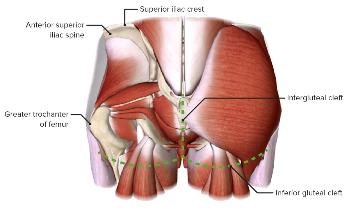
The gluteal region is the area posterior to the pelvic girdle between the iliac crest and the gluteal fold. The region comprises the following:

Muscle groups: 

Nerves: 

  • Sciatic
  • Superior and inferior gluteal
  • Posterior femoral cutaneous
  • Pudendal

Vessels: 

  • Superior and inferior gluteal arteries Arteries Arteries are tubular collections of cells that transport oxygenated blood and nutrients from the heart to the tissues of the body. The blood passes through the arteries in order of decreasing luminal diameter, starting in the largest artery (the aorta) and ending in the small arterioles. Arteries are classified into 3 types: large elastic arteries, medium muscular arteries, and small arteries and arterioles. Arteries: Histology (branches of the internal iliac artery) 
  • Superior and inferior gluteal veins Veins Veins are tubular collections of cells, which transport deoxygenated blood and waste from the capillary beds back to the heart. Veins are classified into 3 types: small veins/venules, medium veins, and large veins. Each type contains 3 primary layers: tunica intima, tunica media, and tunica adventitia. Veins: Histology (drain into the internal iliac vein)

Foramina:  

  • Greater and lesser sciatic foramina of the pelvis Pelvis The pelvis consists of the bony pelvic girdle, the muscular and ligamentous pelvic floor, and the pelvic cavity, which contains viscera, vessels, and multiple nerves and muscles. The pelvic girdle, composed of 2 “hip” bones and the sacrum, is a ring-like bony structure of the axial skeleton that links the vertebral column with the lower extremities. Pelvis: Anatomy (formed by the sacrospinous and sacrotuberous ligaments)

Boundaries

Boundaries of the gluteal region

Boundaries of the gluteal region

Image by BioDigital, edited by Lecturio.
Related videos

Gluteal Muscles

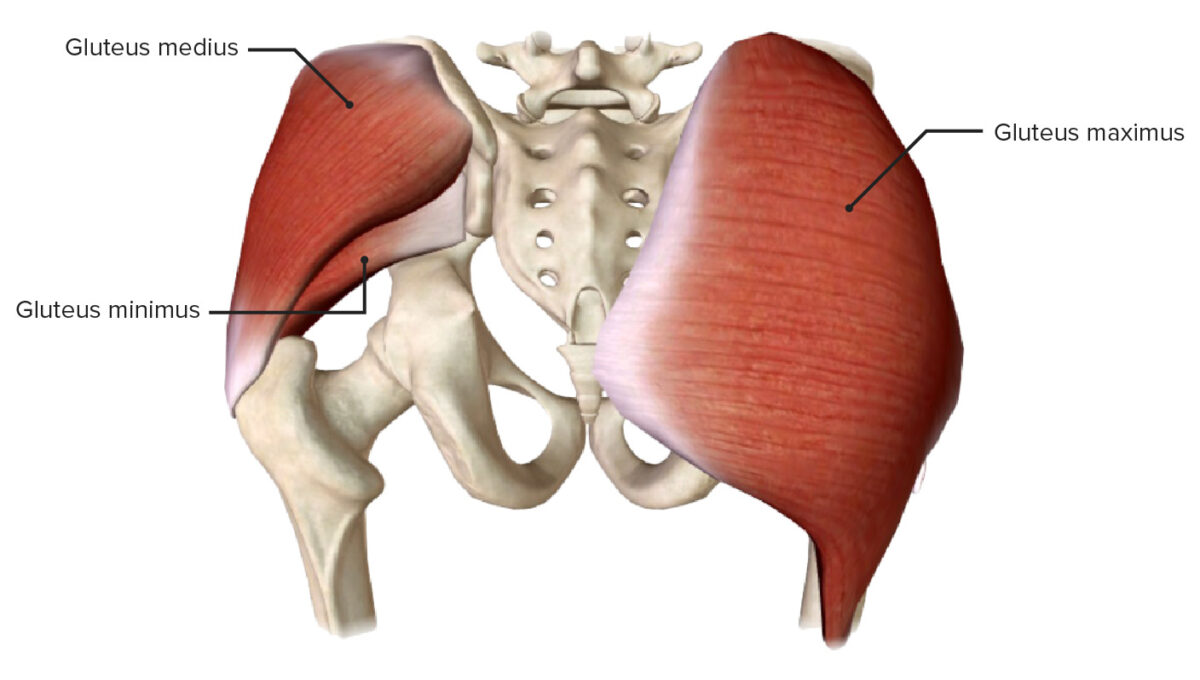
The gluteal muscles can be divided into 2 groups that are responsible for the main movements of the hip joint Hip joint The hip joint is a ball-and-socket joint formed by the head of the femur and the acetabulum of the pelvis. The hip joint is the most stable joint in the body and is supported by a very strong capsule and several ligaments, allowing the joint to sustain forces that can be multiple times the total body weight. Hip Joint: Anatomy

Superficial gluteal muscles

Muscle Origin Insertion Nerve supply Function
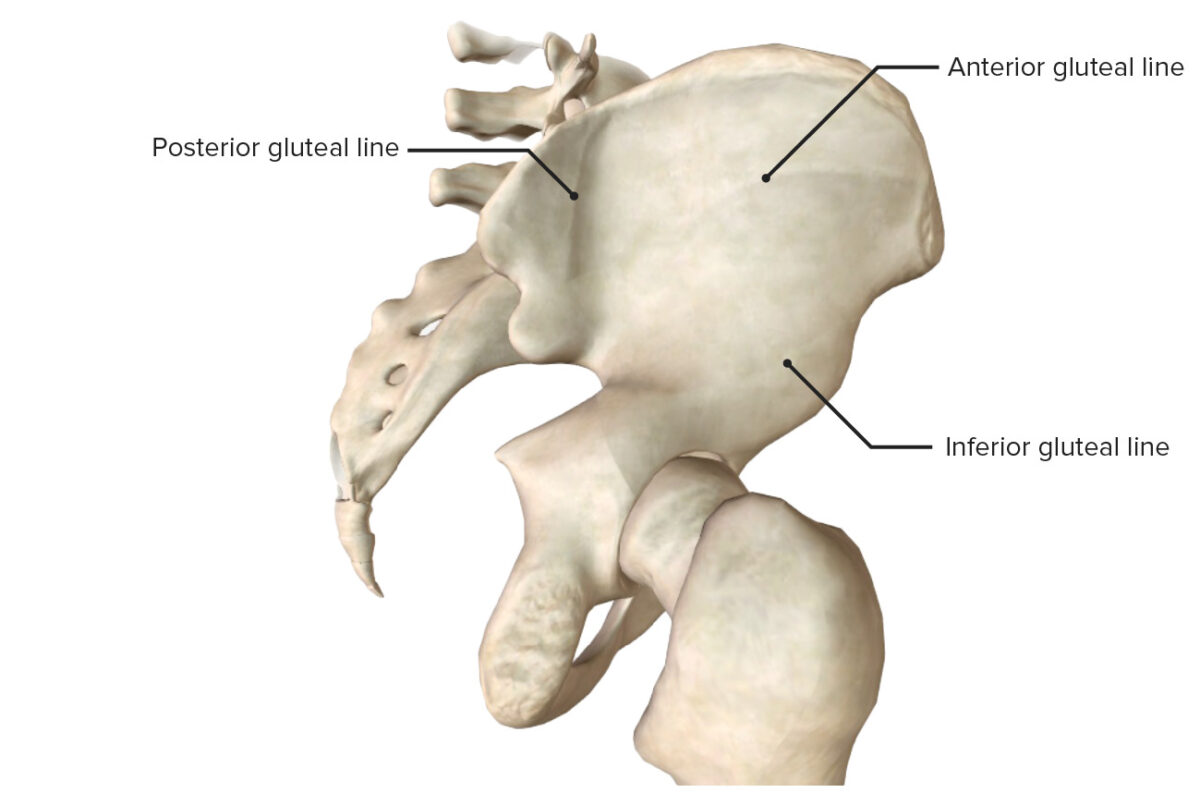
Gluteus maximus Ilium posterior to the posterior gluteal line, posterior sacrum Sacrum Five fused vertebrae forming a triangle-shaped structure at the back of the pelvis. It articulates superiorly with the lumbar vertebrae, inferiorly with the coccyx, and anteriorly with the ilium of the pelvis. The sacrum strengthens and stabilizes the pelvis. Vertebral Column: Anatomy and coccyx Coccyx The last bone in the vertebral column in tailless primates considered to be a vestigial tail-bone consisting of three to five fused vertebrae. Vertebral Column: Anatomy, and sacrotuberous ligament Iliotibial tract Iliotibial tract Thigh: Anatomy (75%) and gluteal tuberosity (25%) Inferior gluteal nerve ( S1 S1 Heart Sounds, S2 S2 Heart Sounds)
  • Extends the hip
  • Assists lateral rotation Rotation Motion of an object in which either one or more points on a line are fixed. It is also the motion of a particle about a fixed point. X-rays
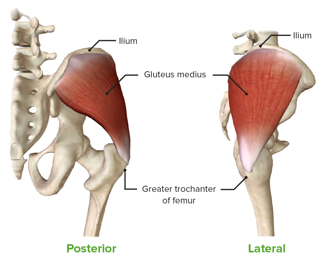
Gluteus medius External ilium between the anterior and posterior gluteal lines Greater trochanter of the femur Superior gluteal nerve (L4, L5, S1 S1 Heart Sounds)
  • Abducts and medially rotates the hip
  • Keeps the pelvis Pelvis The pelvis consists of the bony pelvic girdle, the muscular and ligamentous pelvic floor, and the pelvic cavity, which contains viscera, vessels, and multiple nerves and muscles. The pelvic girdle, composed of 2 “hip” bones and the sacrum, is a ring-like bony structure of the axial skeleton that links the vertebral column with the lower extremities. Pelvis: Anatomy level when the opposite limb is off the ground (swing phase)
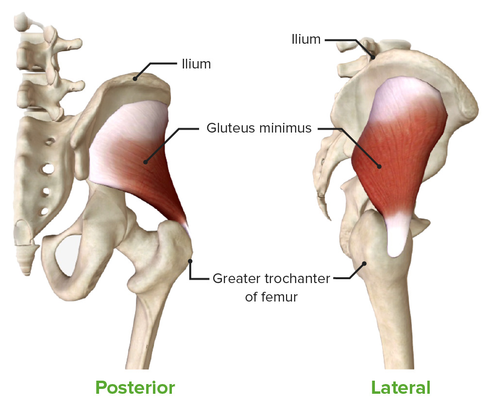
Gluteus minimus External ilium between the anterior and inferior gluteal lines Greater trochanter of the femur
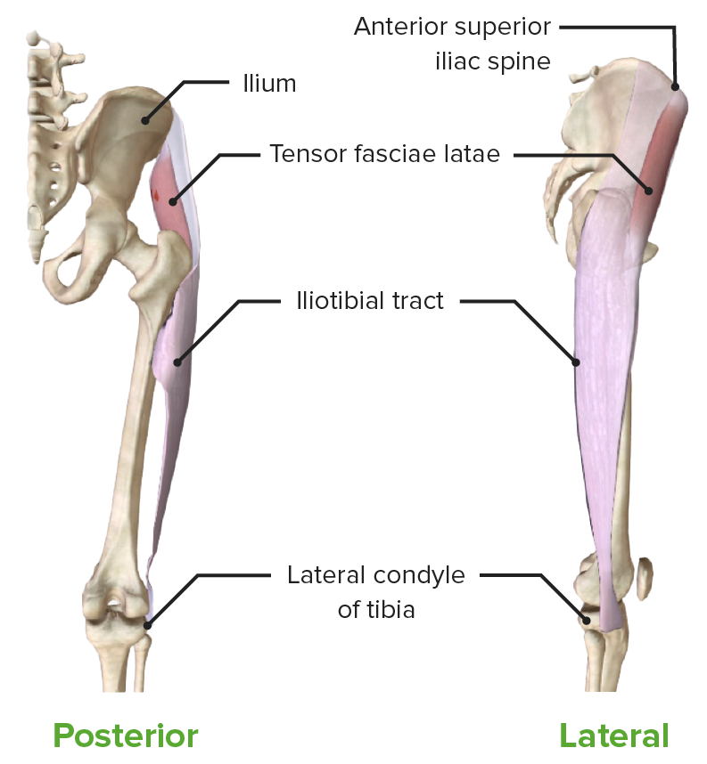
Tensor fasciae latae Anterior superior iliac spine Anterior Superior Iliac Spine Chronic Apophyseal Injury Iliotibial tract Iliotibial tract Thigh: Anatomy to the lateral condyle of the tibia Tibia The second longest bone of the skeleton. It is located on the medial side of the lower leg, articulating with the fibula laterally, the talus distally, and the femur proximally. Knee Joint: Anatomy
  • Flexes the hip
  • Stabilizes the knee joint Knee joint The knee joint is made up of the articulations between the femur, tibia, and patella bones, and is one of the largest and most complex joints of the human body. The knee is classified as a synovial hinge joint, which primarily allows for flexion and extension with a more limited degree of translation and rotation. Knee Joint: Anatomy

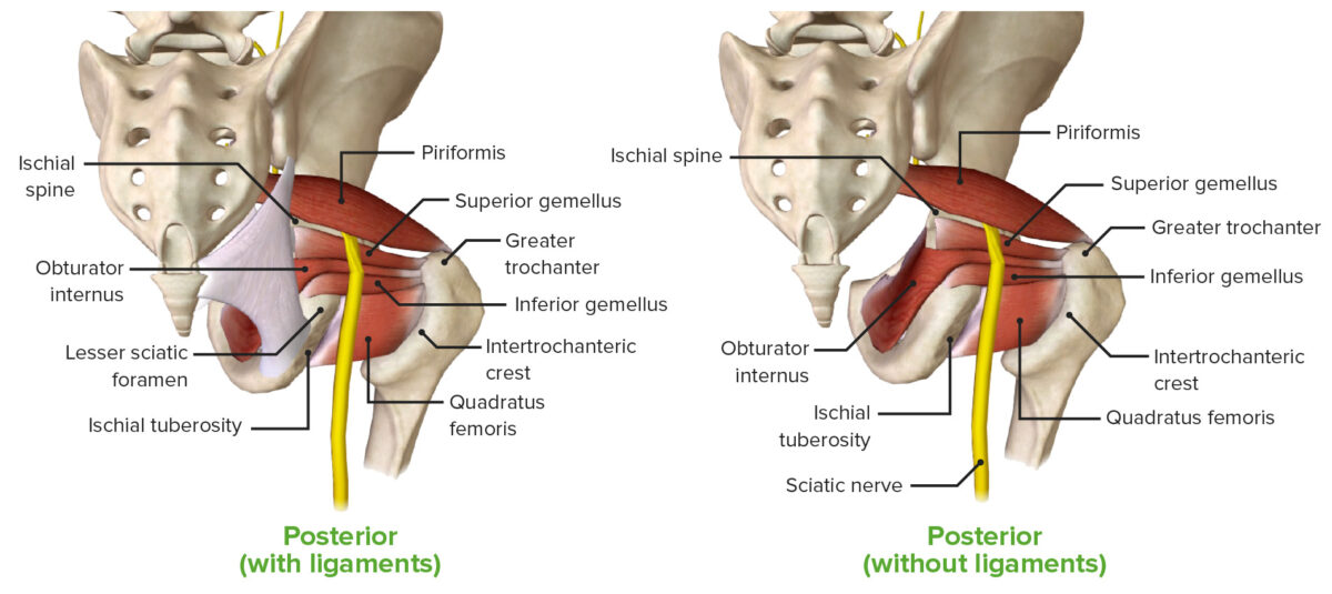
Deep gluteal muscles

Muscle Origin Insertion Nerve supply Function
Piriformis Piriformis Vagina, Vulva, and Pelvic Floor: Anatomy
  • Anterior surface of the sacrum Sacrum Five fused vertebrae forming a triangle-shaped structure at the back of the pelvis. It articulates superiorly with the lumbar vertebrae, inferiorly with the coccyx, and anteriorly with the ilium of the pelvis. The sacrum strengthens and stabilizes the pelvis. Vertebral Column: Anatomy
Greater trochanter (superior surface) Anterior rami of S1 S1 Heart Sounds
Gemelli
  • Superior: ischial spine Spine The human spine, or vertebral column, is the most important anatomical and functional axis of the human body. It consists of 7 cervical vertebrae, 12 thoracic vertebrae, and 5 lumbar vertebrae and is limited cranially by the skull and caudally by the sacrum. Vertebral Column: Anatomy
  • Inferior: ischial tuberosity Ischial Tuberosity Chronic Apophyseal Injury
Greater trochanter (medial surface)
Obturator internus Obturator internus Vagina, Vulva, and Pelvic Floor: Anatomy
  • Pelvic surface of the ilium, ischium, and obturator membrane
Greater trochanter (medial surface)
Quadratus femoris Intertrochanteric crest N to the quadratus femoris (L5, S1 S1 Heart Sounds)
  • Lateral rotation Rotation Motion of an object in which either one or more points on a line are fixed. It is also the motion of a particle about a fixed point. X-rays 
  • Holds the head of the femur Head of the femur The hemispheric articular surface at the upper extremity of the thigh bone. Hip Joint: Anatomy within the acetabulum

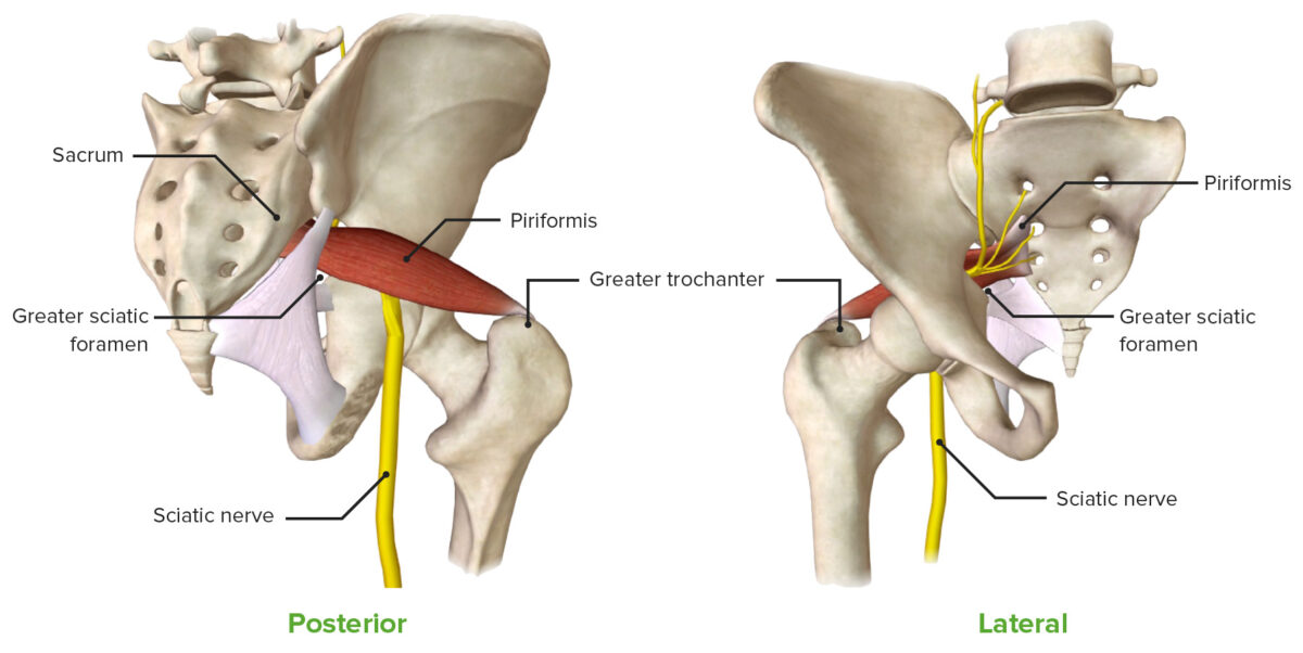
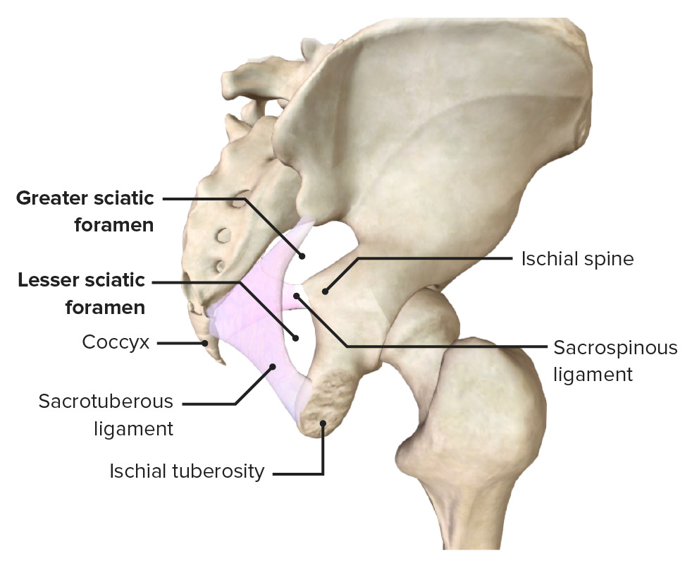
Sciatic Foramina

The superior and inferior foramina are formed by the following ligaments inserted into the bony pelvis Pelvis The pelvis consists of the bony pelvic girdle, the muscular and ligamentous pelvic floor, and the pelvic cavity, which contains viscera, vessels, and multiple nerves and muscles. The pelvic girdle, composed of 2 “hip” bones and the sacrum, is a ring-like bony structure of the axial skeleton that links the vertebral column with the lower extremities. Pelvis: Anatomy:

  • Sacrospinous ligament: extends from the lateral edge of the sacrum Sacrum Five fused vertebrae forming a triangle-shaped structure at the back of the pelvis. It articulates superiorly with the lumbar vertebrae, inferiorly with the coccyx, and anteriorly with the ilium of the pelvis. The sacrum strengthens and stabilizes the pelvis. Vertebral Column: Anatomy and coccyx Coccyx The last bone in the vertebral column in tailless primates considered to be a vestigial tail-bone consisting of three to five fused vertebrae. Vertebral Column: Anatomy to the ischial spine Spine The human spine, or vertebral column, is the most important anatomical and functional axis of the human body. It consists of 7 cervical vertebrae, 12 thoracic vertebrae, and 5 lumbar vertebrae and is limited cranially by the skull and caudally by the sacrum. Vertebral Column: Anatomy
  • Sacrotuberous ligament: extends from the lateral edge of the sacrum Sacrum Five fused vertebrae forming a triangle-shaped structure at the back of the pelvis. It articulates superiorly with the lumbar vertebrae, inferiorly with the coccyx, and anteriorly with the ilium of the pelvis. The sacrum strengthens and stabilizes the pelvis. Vertebral Column: Anatomy and coccyx Coccyx The last bone in the vertebral column in tailless primates considered to be a vestigial tail-bone consisting of three to five fused vertebrae. Vertebral Column: Anatomy to the ischial tuberosity Ischial Tuberosity Chronic Apophyseal Injury

The sacrospinous and sacrotuberous ligaments create the following foramina, or passageways:

Greater sciatic foramen Lesser sciatic foramen
Boundaries
  • Superior: anterior sacroiliac ligament
  • Inferior: sacrospinous ligament and the ischial spine Spine The human spine, or vertebral column, is the most important anatomical and functional axis of the human body. It consists of 7 cervical vertebrae, 12 thoracic vertebrae, and 5 lumbar vertebrae and is limited cranially by the skull and caudally by the sacrum. Vertebral Column: Anatomy
  • Anterolateral: greater sciatic notch of the ilium
  • Posteromedial: sacrotuberous ligament
  • Superior: sacrospinous ligament and the ischial spine Spine The human spine, or vertebral column, is the most important anatomical and functional axis of the human body. It consists of 7 cervical vertebrae, 12 thoracic vertebrae, and 5 lumbar vertebrae and is limited cranially by the skull and caudally by the sacrum. Vertebral Column: Anatomy
  • Anterior: ischial tuberosity Ischial Tuberosity Chronic Apophyseal Injury
  • Posterior: sacrotuberous ligament
Contents
Sciatic foramina

The greater and lesser sciatic foramina are created by the spaces between the sacrospinous and sacrotuberous ligaments.

Image by BioDigital, edited by Lecturio.
Gluteal Vessels

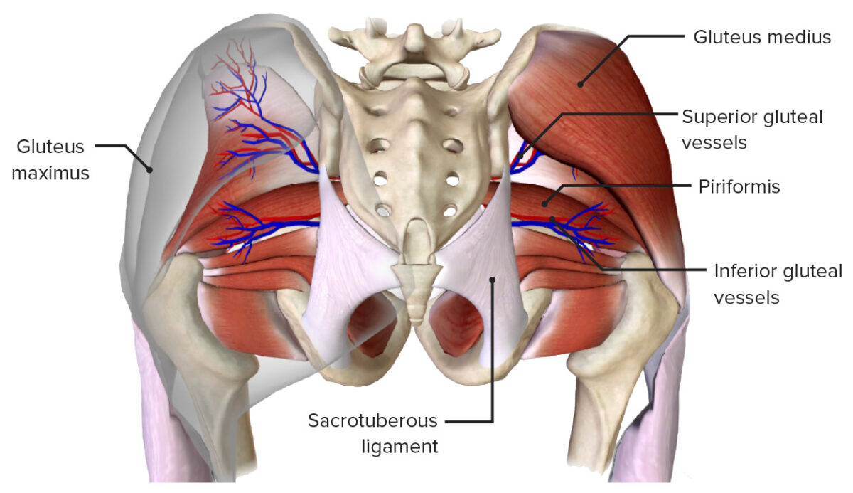
Gluteal arteries Arteries Arteries are tubular collections of cells that transport oxygenated blood and nutrients from the heart to the tissues of the body. The blood passes through the arteries in order of decreasing luminal diameter, starting in the largest artery (the aorta) and ending in the small arterioles. Arteries are classified into 3 types: large elastic arteries, medium muscular arteries, and small arteries and arterioles. Arteries: Histology

Two branches drain from the internal iliac arteries Arteries Arteries are tubular collections of cells that transport oxygenated blood and nutrients from the heart to the tissues of the body. The blood passes through the arteries in order of decreasing luminal diameter, starting in the largest artery (the aorta) and ending in the small arterioles. Arteries are classified into 3 types: large elastic arteries, medium muscular arteries, and small arteries and arterioles. Arteries: Histology:

  • Superior gluteal artery:
    • The largest branch of the internal iliac
    • Goes through the greater sciatic foramen and the suprapiriform foramen
    • Supplies the gluteus medius, minimus, tensor fasciae latae, and piriformis Piriformis Vagina, Vulva, and Pelvic Floor: Anatomy muscles in the gluteal region
    • Also supplies the skin Skin The skin, also referred to as the integumentary system, is the largest organ of the body. The skin is primarily composed of the epidermis (outer layer) and dermis (deep layer). The epidermis is primarily composed of keratinocytes that undergo rapid turnover, while the dermis contains dense layers of connective tissue. Skin: Structure and Functions over the sacrum Sacrum Five fused vertebrae forming a triangle-shaped structure at the back of the pelvis. It articulates superiorly with the lumbar vertebrae, inferiorly with the coccyx, and anteriorly with the ilium of the pelvis. The sacrum strengthens and stabilizes the pelvis. Vertebral Column: Anatomy and hip joint Hip joint The hip joint is a ball-and-socket joint formed by the head of the femur and the acetabulum of the pelvis. The hip joint is the most stable joint in the body and is supported by a very strong capsule and several ligaments, allowing the joint to sustain forces that can be multiple times the total body weight. Hip Joint: Anatomy
  • Inferior gluteal artery:
    • Goes through the greater sciatic foramen and the infrapiriform foramen
    • Supplies the gluteus maximus, obturator internus Obturator internus Vagina, Vulva, and Pelvic Floor: Anatomy, and quadratus femoris muscles in the gluteal region
    • Also supplies the sciatic nerve, pelvic floor Pelvic floor Soft tissue formed mainly by the pelvic diaphragm, which is composed of the two levator ani and two coccygeus muscles. The pelvic diaphragm lies just below the pelvic aperture (outlet) and separates the pelvic cavity from the perineum. It extends between the pubic bone anteriorly and the coccyx posteriorly. Vagina, Vulva, and Pelvic Floor: Anatomy, and skin Skin The skin, also referred to as the integumentary system, is the largest organ of the body. The skin is primarily composed of the epidermis (outer layer) and dermis (deep layer). The epidermis is primarily composed of keratinocytes that undergo rapid turnover, while the dermis contains dense layers of connective tissue. Skin: Structure and Functions of the gluteal and thigh Thigh The thigh is the region of the lower limb found between the hip and the knee joint. There is a single bone in the thigh called the femur, which is surrounded by large muscles grouped into 3 fascial compartments. Thigh: Anatomy regions

Gluteal veins Veins Veins are tubular collections of cells, which transport deoxygenated blood and waste from the capillary beds back to the heart. Veins are classified into 3 types: small veins/venules, medium veins, and large veins. Each type contains 3 primary layers: tunica intima, tunica media, and tunica adventitia. Veins: Histology

  • The veins Veins Veins are tubular collections of cells, which transport deoxygenated blood and waste from the capillary beds back to the heart. Veins are classified into 3 types: small veins/venules, medium veins, and large veins. Each type contains 3 primary layers: tunica intima, tunica media, and tunica adventitia. Veins: Histology of the gluteal regions accompany the gluteal arteries Arteries Arteries are tubular collections of cells that transport oxygenated blood and nutrients from the heart to the tissues of the body. The blood passes through the arteries in order of decreasing luminal diameter, starting in the largest artery (the aorta) and ending in the small arterioles. Arteries are classified into 3 types: large elastic arteries, medium muscular arteries, and small arteries and arterioles. Arteries: Histology and are named accordingly: superior and inferior gluteal veins Veins Veins are tubular collections of cells, which transport deoxygenated blood and waste from the capillary beds back to the heart. Veins are classified into 3 types: small veins/venules, medium veins, and large veins. Each type contains 3 primary layers: tunica intima, tunica media, and tunica adventitia. Veins: Histology.
  • Drain into the internal iliac vein
Gluteal vessels

The gluteal vessels emerging through the suprapiriform foramen and infrapiriform foramina

Image by BioDigital, edited by Lecturio.
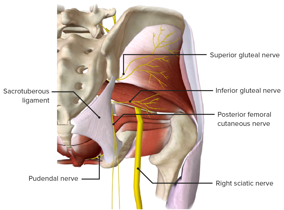
Gluteal Nerves

Nerve Origin Muscles supplied
Sciatic Anterior and posterior divisions of the nerve roots L4-S3
  • Muscles of the posterior compartment of the leg Leg The lower leg, or just “leg” in anatomical terms, is the part of the lower limb between the knee and the ankle joint. The bony structure is composed of the tibia and fibula bones, and the muscles of the leg are grouped into the anterior, lateral, and posterior compartments by extensions of fascia. Leg: Anatomy
  • Muscles of the sole of the foot Foot The foot is the terminal portion of the lower limb, whose primary function is to bear weight and facilitate locomotion. The foot comprises 26 bones, including the tarsal bones, metatarsal bones, and phalanges. The bones of the foot form longitudinal and transverse arches and are supported by various muscles, ligaments, and tendons. Foot: Anatomy
  • Muscles in the anterior and lateral compartments of the leg Leg The lower leg, or just “leg” in anatomical terms, is the part of the lower limb between the knee and the ankle joint. The bony structure is composed of the tibia and fibula bones, and the muscles of the leg are grouped into the anterior, lateral, and posterior compartments by extensions of fascia. Leg: Anatomy
  • Innervates no muscles in the gluteal region
Superior gluteal L4-S1 ( Sacral plexus Sacral plexus Pelvis: Anatomy)
  • Gluteus medius
  • Gluteus minimus
  • Tensor fasciae latae
Inferior gluteal L5-S2 ( Sacral plexus Sacral plexus Pelvis: Anatomy)
  • Gluteus maximus
Posterior femoral cutaneous S1-S3 ( Sacral plexus Sacral plexus Pelvis: Anatomy)
  • Skin Skin The skin, also referred to as the integumentary system, is the largest organ of the body. The skin is primarily composed of the epidermis (outer layer) and dermis (deep layer). The epidermis is primarily composed of keratinocytes that undergo rapid turnover, while the dermis contains dense layers of connective tissue. Skin: Structure and Functions over the lower and lateral parts of the gluteus maximus
  • Skin Skin The skin, also referred to as the integumentary system, is the largest organ of the body. The skin is primarily composed of the epidermis (outer layer) and dermis (deep layer). The epidermis is primarily composed of keratinocytes that undergo rapid turnover, while the dermis contains dense layers of connective tissue. Skin: Structure and Functions of the posterior and medial thigh Thigh The thigh is the region of the lower limb found between the hip and the knee joint. There is a single bone in the thigh called the femur, which is surrounded by large muscles grouped into 3 fascial compartments. Thigh: Anatomy
Pudendal S2-S4 (Pudendal plexus)
  • Muscles of the pelvic floor Pelvic floor Soft tissue formed mainly by the pelvic diaphragm, which is composed of the two levator ani and two coccygeus muscles. The pelvic diaphragm lies just below the pelvic aperture (outlet) and separates the pelvic cavity from the perineum. It extends between the pubic bone anteriorly and the coccyx posteriorly. Vagina, Vulva, and Pelvic Floor: Anatomy
  • Cutaneous perineal branches 
Sacral plexus Sacral plexus Pelvis: Anatomy  L4-S4 (direct branches)
Gluteal nerves

The deep layer of the gluteal region, featuring the nerves of the gluteal region

Image by BioDigital, edited by Lecturio.
Clinical Relevance

The following are clinically relevant to the gluteal region:

  • Intramuscular injections: The superolateral region of the gluteal region is relatively free of nerves and vessels and is often used for intramuscular injections.
  • Trendelenburg gait Trendelenburg Gait Legg-Calvé-Perthes Disease: abnormal gait Gait Manner or style of walking. Neurological Examination secondary to the weakness of the hip abductors, primarily the gluteus medius and gluteus minimus muscles, which are essential to maintaining the balance of the pelvis Pelvis The pelvis consists of the bony pelvic girdle, the muscular and ligamentous pelvic floor, and the pelvic cavity, which contains viscera, vessels, and multiple nerves and muscles. The pelvic girdle, composed of 2 “hip” bones and the sacrum, is a ring-like bony structure of the axial skeleton that links the vertebral column with the lower extremities. Pelvis: Anatomy during the gait Gait Manner or style of walking. Neurological Examination cycle. Weakness of the hip abductors causes a drop of the contralateral pelvis Pelvis The pelvis consists of the bony pelvic girdle, the muscular and ligamentous pelvic floor, and the pelvic cavity, which contains viscera, vessels, and multiple nerves and muscles. The pelvic girdle, composed of 2 “hip” bones and the sacrum, is a ring-like bony structure of the axial skeleton that links the vertebral column with the lower extremities. Pelvis: Anatomy while walking, or a Trendelenburg gait Trendelenburg Gait Legg-Calvé-Perthes Disease
  • Piriformis Piriformis Vagina, Vulva, and Pelvic Floor: Anatomy syndrome: Also called deep gluteal syndrome or wallet neuritis, piriformis Piriformis Vagina, Vulva, and Pelvic Floor: Anatomy syndrome is characterized by a combination of symptoms involving the hip, buttock, and upper thigh Thigh The thigh is the region of the lower limb found between the hip and the knee joint. There is a single bone in the thigh called the femur, which is surrounded by large muscles grouped into 3 fascial compartments. Thigh: Anatomy. Described as peripheral neuritis of the sciatic nerve and may be related to irritation of the same at the level of the piriformis Piriformis Vagina, Vulva, and Pelvic Floor: Anatomy muscle. May be caused by trauma, hematoma Hematoma A collection of blood outside the blood vessels. Hematoma can be localized in an organ, space, or tissue. Intussusception, excessive sitting, and anatomic variations of the muscle and nerve. 
  • Superior gluteal nerve palsy Palsy paralysis of an area of the body, thus incapable of voluntary movement Cranial Nerve Palsies: occurs secondary to a peripheral injury of the superior gluteal nerve leading to motor Motor Neurons which send impulses peripherally to activate muscles or secretory cells. Nervous System: Histology loss, specifically involving the gluteus medius and minimus. Superior gluteal nerve palsy Palsy paralysis of an area of the body, thus incapable of voluntary movement Cranial Nerve Palsies manifests as a Trendelenburg gait Trendelenburg Gait Legg-Calvé-Perthes Disease. The most common cause is an iatrogenic Iatrogenic Any adverse condition in a patient occurring as the result of treatment by a physician, surgeon, or other health professional, especially infections acquired by a patient during the course of treatment. Anterior Cord Syndrome injury during hip surgery and intramuscular injection. 
  • Lesions of the inferior gluteal nerve: most commonly occur through iatrogenic Iatrogenic Any adverse condition in a patient occurring as the result of treatment by a physician, surgeon, or other health professional, especially infections acquired by a patient during the course of treatment. Anterior Cord Syndrome injuries (e.g., surgery), trauma, hernias, or pelvic tumors. May lead to a functional deficiency of the gluteus maximus muscle, causing a “gluteus maximus lurch.” Patients Patients Individuals participating in the health care system for the purpose of receiving therapeutic, diagnostic, or preventive procedures. Clinician–Patient Relationship present with difficulties walking up stairs or standing up from a chair.

References

  1. Drake, R.L., Vogl, A.W., & Mitchell, A.W.M. (2014). Gray’s Anatomy for Students (3rd ed.). Churchill Livingstone.

Create your free account or log in to continue reading!

Sign up now and get free access to Lecturio with concept pages, medical videos, and questions for your medical education.

User Reviews

Details