Nervous System: Anatomy, Structure, and Classification

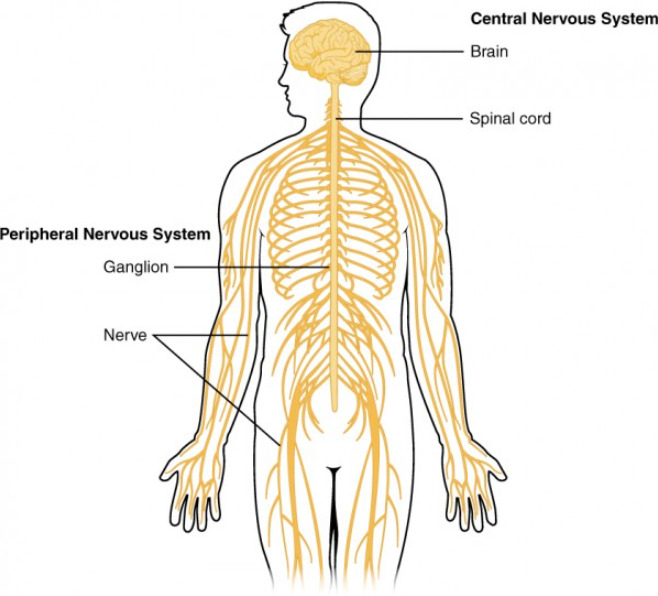
The components of the human nervous system weigh about 2.04 kg (4.5 lb) in total, which accounts for approximately 3% of the entire body weight. The nervous system is a small and complex system that consists of an intricate network of neural cells (or neurons Neurons The basic cellular units of nervous tissue. Each neuron consists of a body, an axon, and dendrites. Their purpose is to receive, conduct, and transmit impulses in the nervous system. Nervous System: Histology) and even more glial cells Glial Cells The non-neuronal cells of the nervous system. They not only provide physical support, but also respond to injury, regulate the ionic and chemical composition of the extracellular milieu, participate in the blood-brain barrier and blood-retinal barrier, form the myelin insulation of nervous pathways, guide neuronal migration during development, and exchange metabolites with neurons. Neuroglia have high-affinity transmitter uptake systems, voltage-dependent and transmitter-gated ion channels, and can release transmitters, but their role in signaling (as in many other functions) is unclear. Nervous System: Histology (for support and insulation). The system is divided according to its anatomical components as well as its functional characteristics. The brain and spinal cord Spinal cord The spinal cord is the major conduction pathway connecting the brain to the body; it is part of the CNS. In cross section, the spinal cord is divided into an H-shaped area of gray matter (consisting of synapsing neuronal cell bodies) and a surrounding area of white matter (consisting of ascending and descending tracts of myelinated axons). Spinal Cord: Anatomy are referred to as the central nervous system, and the branches of nerves from these structures are referred to as the peripheral nervous system.

Last updated: Nov 19, 2024

Editorial responsibility: Stanley Oiseth, Lindsay Jones, Evelin Maza

Overview

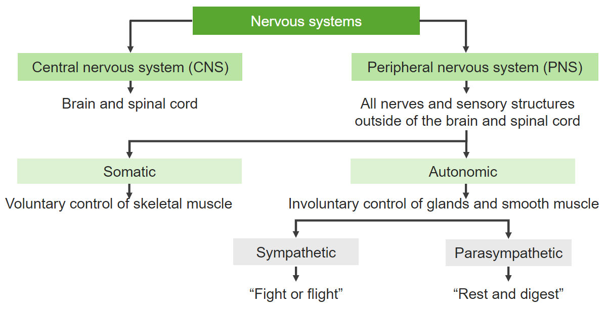
The nervous system is separated into the central and peripheral nervous systems. In addition, the sympathetic, parasympathetic, and enteric nervous systems act as parts of the autonomic nervous system Autonomic nervous system The ANS is a component of the peripheral nervous system that uses both afferent (sensory) and efferent (effector) neurons, which control the functioning of the internal organs and involuntary processes via connections with the CNS. The ANS consists of the sympathetic and parasympathetic nervous systems. Autonomic Nervous System: Anatomy.
Anatomical subdivision

  • CNS: brain and spinal cord Spinal cord The spinal cord is the major conduction pathway connecting the brain to the body; it is part of the CNS. In cross section, the spinal cord is divided into an H-shaped area of gray matter (consisting of synapsing neuronal cell bodies) and a surrounding area of white matter (consisting of ascending and descending tracts of myelinated axons). Spinal Cord: Anatomy
  • Peripheral nervous system (PNS):
    • Somatic nervous system:
    • ANS ANS The ans is a component of the peripheral nervous system that uses both afferent (sensory) and efferent (effector) neurons, which control the functioning of the internal organs and involuntary processes via connections with the CNS. The ans consists of the sympathetic and parasympathetic nervous systems. Autonomic Nervous System: Anatomy:
      • Sympathetic system Sympathetic system The thoracolumbar division of the autonomic nervous system. Sympathetic preganglionic fibers originate in neurons of the intermediolateral column of the spinal cord and project to the paravertebral and prevertebral ganglia, which in turn project to target organs. The sympathetic nervous system mediates the body’s response to stressful situations, i.e., the fight or flight reactions. It often acts reciprocally to the parasympathetic system. Autonomic Nervous System: Anatomy
      • Parasympathetic system Parasympathetic system The craniosacral division of the autonomic nervous system. The cell bodies of the parasympathetic preganglionic fibers are in brain stem nuclei and in the sacral spinal cord. They synapse in cranial autonomic ganglia or in terminal ganglia near target organs. The parasympathetic nervous system generally acts to conserve resources and restore homeostasis, often with effects reciprocal to the sympathetic nervous system. Autonomic Nervous System: Anatomy
      • Enteric nervous system Enteric nervous system Two ganglionated neural plexuses in the gut wall which form one of the three major divisions of the autonomic nervous system. The enteric nervous system innervates the gastrointestinal tract, the pancreas, and the gallbladder. It contains sensory neurons, interneurons, and motor neurons. Thus the circuitry can autonomously sense the tension and the chemical environment in the gut and regulate blood vessel tone, motility, secretions, and fluid transport. The system is itself governed by the central nervous system and receives both parasympathetic and sympathetic innervation. Autonomic Nervous System: Anatomy

Functional subdivision

Central Nervous System

The CNS is composed of the brain, brain stem Brain Stem The brain stem is a stalk-like structure that connects the cerebrum with the spinal cord and consists of the midbrain, pons, and medulla oblongata. It also plays a critical role in the control of cardiovascular and respiratory function, consciousness, and the sleep-wake cycle. Brain Stem: Anatomy, cerebellum Cerebellum The cerebellum, Latin for “little brain,” is located in the posterior cranial fossa, dorsal to the pons and midbrain, and its principal role is in the coordination of movements. The cerebellum consists of 3 lobes on either side of its 2 hemispheres and is connected in the middle by the vermis. Cerebellum: Anatomy, and spinal cord Spinal cord The spinal cord is the major conduction pathway connecting the brain to the body; it is part of the CNS. In cross section, the spinal cord is divided into an H-shaped area of gray matter (consisting of synapsing neuronal cell bodies) and a surrounding area of white matter (consisting of ascending and descending tracts of myelinated axons). Spinal Cord: Anatomy. Each of these structures is covered by meninges Meninges The brain and the spinal cord are enveloped by 3 overlapping layers of connective tissue called the meninges. The layers are, from the most external layer to the most internal layer, the dura mater, arachnoid mater, and pia mater. Between these layers are 3 potential spaces called the epidural, subdural, and subarachnoid spaces. Meninges: Anatomy and CSF.

Composition

The CNS is contained within the skull Skull The skull (cranium) is the skeletal structure of the head supporting the face and forming a protective cavity for the brain. The skull consists of 22 bones divided into the viscerocranium (facial skeleton) and the neurocranium. Skull: Anatomy and spinal column Spinal column The human spine, or vertebral column, is the most important anatomical and functional axis of the human body. It consists of 7 cervical vertebrae, 12 thoracic vertebrae, and 5 lumbar vertebrae and is limited cranially by the skull and caudally by the sacrum. Vertebral Column: Anatomy. The components of this system include:
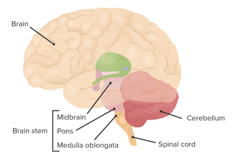
  • Cerebral cortex Cerebral cortex The cerebral cortex is the largest and most developed part of the human brain and CNS. Occupying the upper part of the cranial cavity, the cerebral cortex has 4 lobes and is divided into 2 hemispheres that are joined centrally by the corpus callosum. Cerebral Cortex: Anatomy
  • Spinal cord Spinal cord The spinal cord is the major conduction pathway connecting the brain to the body; it is part of the CNS. In cross section, the spinal cord is divided into an H-shaped area of gray matter (consisting of synapsing neuronal cell bodies) and a surrounding area of white matter (consisting of ascending and descending tracts of myelinated axons). Spinal Cord: Anatomy
  • Brain stem Brain Stem The brain stem is a stalk-like structure that connects the cerebrum with the spinal cord and consists of the midbrain, pons, and medulla oblongata. It also plays a critical role in the control of cardiovascular and respiratory function, consciousness, and the sleep-wake cycle. Brain Stem: Anatomy
  • Cerebellum Cerebellum The cerebellum, Latin for “little brain,” is located in the posterior cranial fossa, dorsal to the pons and midbrain, and its principal role is in the coordination of movements. The cerebellum consists of 3 lobes on either side of its 2 hemispheres and is connected in the middle by the vermis. Cerebellum: Anatomy

Protective covering

The meninges Meninges The brain and the spinal cord are enveloped by 3 overlapping layers of connective tissue called the meninges. The layers are, from the most external layer to the most internal layer, the dura mater, arachnoid mater, and pia mater. Between these layers are 3 potential spaces called the epidural, subdural, and subarachnoid spaces. Meninges: Anatomy provide a protective covering to the brain and spinal cord Spinal cord The spinal cord is the major conduction pathway connecting the brain to the body; it is part of the CNS. In cross section, the spinal cord is divided into an H-shaped area of gray matter (consisting of synapsing neuronal cell bodies) and a surrounding area of white matter (consisting of ascending and descending tracts of myelinated axons). Spinal Cord: Anatomy. The meninges Meninges The brain and the spinal cord are enveloped by 3 overlapping layers of connective tissue called the meninges. The layers are, from the most external layer to the most internal layer, the dura mater, arachnoid mater, and pia mater. Between these layers are 3 potential spaces called the epidural, subdural, and subarachnoid spaces. Meninges: Anatomy are composed of:

  • Dura mater Dura mater The outermost of the three meninges, a fibrous membrane of connective tissue that covers the brain and the spinal cord. Meninges: Anatomy
  • Arachnoid mater Arachnoid mater A delicate membrane enveloping the brain and spinal cord. It lies between the pia mater and the dura mater. It is separated from the pia mater by the subarachnoid cavity which is filled with cerebrospinal fluid. Meninges: Anatomy
  • Pia mater Pia mater The innermost layer of the three meninges covering the brain and spinal cord. It is the fine vascular membrane that lies under the arachnoid and the dura mater. Meninges: Anatomy

Cerebrospinal fluid Cerebrospinal Fluid A watery fluid that is continuously produced in the choroid plexus and circulates around the surface of the brain; spinal cord; and in the cerebral ventricles. Ventricular System: Anatomy

  • Clear, colorless liquid containing a small amount of protein
  • Provides buoyancy and protection for the brain
  • Regulates chemical concentration of neurotransmitters and metabolic waste

Related videos

Peripheral Nervous System

The PNS is composed of neural components that extend from the aspects of the CNS. The 2 divisions of the PNS are the somatic nervous system (SNS) and the ANS ANS The ans is a component of the peripheral nervous system that uses both afferent (sensory) and efferent (effector) neurons, which control the functioning of the internal organs and involuntary processes via connections with the CNS. The ans consists of the sympathetic and parasympathetic nervous systems. Autonomic Nervous System: Anatomy.

Somatic nervous system

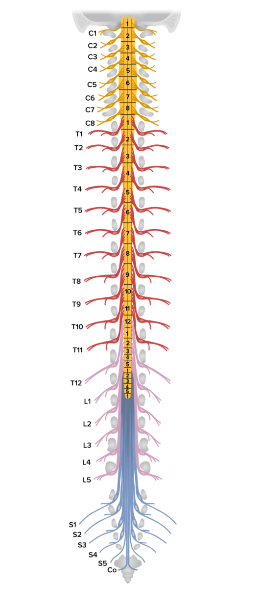
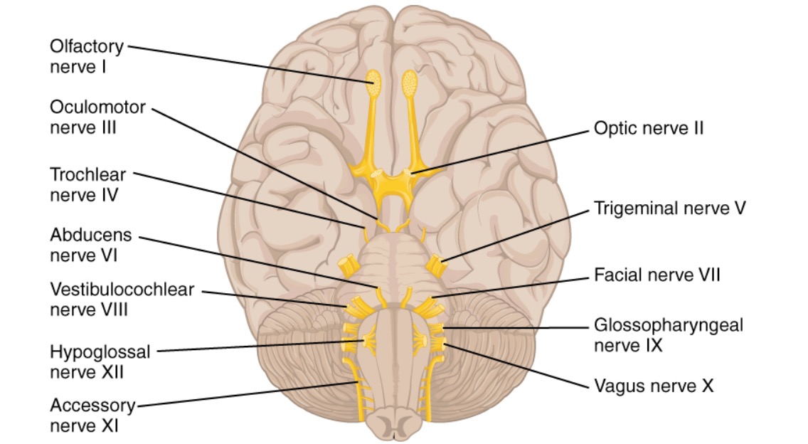
The SNS is composed of cranial nerves Cranial nerves There are 12 pairs of cranial nerves (CNs), which run from the brain to various parts of the head, neck, and trunk. The CNs can be sensory or motor or both. The CNs are named and numbered in Roman numerals according to their location, from the front to the back of the brain. The 12 Cranial Nerves: Overview and Functions and spinal nerves Spinal nerves The 31 paired peripheral nerves formed by the union of the dorsal and ventral spinal roots from each spinal cord segment. The spinal nerve plexuses and the spinal roots are also included. Spinal Cord: Anatomy that are located outside of the CNS. There are 12 cranial and 31 spinal nerve pairs:
  • 8 cervical
  • 12 thoracic
  • 5 lumbar
  • 5 sacral
  • 1 coccygeal

The somatic nervous system is composed of:

Anatomical landmarks of the spinal cord general structure of the nervous system

Anatomical landmarks of the spinal cord

Image by Lecturio.

The somatic nervous system is involved in:

  • Voluntary muscular movement
  • Reflex arcs: 
    • Involuntary
    • Monosynaptic or polysynaptic, according to the number of synapses

Autonomic nervous system Autonomic nervous system The ANS is a component of the peripheral nervous system that uses both afferent (sensory) and efferent (effector) neurons, which control the functioning of the internal organs and involuntary processes via connections with the CNS. The ANS consists of the sympathetic and parasympathetic nervous systems. Autonomic Nervous System: Anatomy

The ANS ANS The ans is a component of the peripheral nervous system that uses both afferent (sensory) and efferent (effector) neurons, which control the functioning of the internal organs and involuntary processes via connections with the CNS. The ans consists of the sympathetic and parasympathetic nervous systems. Autonomic Nervous System: Anatomy monitors and controls the functions of internal organs. The ANS ANS The ans is a component of the peripheral nervous system that uses both afferent (sensory) and efferent (effector) neurons, which control the functioning of the internal organs and involuntary processes via connections with the CNS. The ans consists of the sympathetic and parasympathetic nervous systems. Autonomic Nervous System: Anatomy is composed of the parasympathetic and sympathetic nervous systems. The nerve fibers Nerve Fibers Slender processes of neurons, including the axons and their glial envelopes (myelin sheath). Nerve fibers conduct nerve impulses to and from the central nervous system. Nervous System: Histology of the ANS ANS The ans is a component of the peripheral nervous system that uses both afferent (sensory) and efferent (effector) neurons, which control the functioning of the internal organs and involuntary processes via connections with the CNS. The ans consists of the sympathetic and parasympathetic nervous systems. Autonomic Nervous System: Anatomy originate in the CNS and synapse Synapse The junction between 2 neurons is called a synapse. The synapse allows a neuron to pass an electrical or chemical signal to another neuron or target effector cell. Synapses and Neurotransmission with ganglia in the periphery.

  • Sympathetic nervous system: fight-or-flight response
  • Parasympathetic nervous system: rest-and-digest processes

Clinical Relevance

  • Upper motor Motor Neurons which send impulses peripherally to activate muscles or secretory cells. Nervous System: Histology neuron syndrome: collection of deficits that can occur after damage to an upper motor Motor Neurons which send impulses peripherally to activate muscles or secretory cells. Nervous System: Histology neuron (e.g., cerebrovascular accident Cerebrovascular accident An ischemic stroke (also known as cerebrovascular accident) is an acute neurologic injury that occurs as a result of brain ischemia; this condition may be due to cerebral blood vessel occlusion by thrombosis or embolism, or rarely due to systemic hypoperfusion. Ischemic Stroke). Symptoms include weakness, decreased motor Motor Neurons which send impulses peripherally to activate muscles or secretory cells. Nervous System: Histology control, altered muscle tone Muscle tone The state of activity or tension of a muscle beyond that related to its physical properties, that is, its active resistance to stretch. In skeletal muscle, tonus is dependent upon efferent innervation. Skeletal Muscle Contraction, a decrease or increase in the baseline level of muscle activity, decreased endurance, exaggerated deep tendon reflexes Deep Tendon Reflexes Neurological Examination, spasticity Spasticity Spinal Disk Herniation, and clonus Clonus Ehrlichiosis and Anaplasmosis
  • Lower motor neuron Lower Motor Neuron Motor Neuron Lesions syndrome: collection of deficits that can occur after damage to a lower motor neuron Lower Motor Neuron Motor Neuron Lesions (e.g., trauma or impingement). Signs and symptoms may include paralysis or paresis Paresis A general term referring to a mild to moderate degree of muscular weakness, occasionally used as a synonym for paralysis (severe or complete loss of motor function). In the older literature, paresis often referred specifically to paretic neurosyphilis. ‘general paresis’ and ‘general paralysis’ may still carry that connotation. Bilateral lower extremity paresis is referred to as paraparesis. Spinal Disk Herniation, muscle atrophy Atrophy Decrease in the size of a cell, tissue, organ, or multiple organs, associated with a variety of pathological conditions such as abnormal cellular changes, ischemia, malnutrition, or hormonal changes. Cellular Adaptation, areflexia Areflexia Duchenne Muscular Dystrophy, and fibrillations. 
  • Amyotrophic lateral sclerosis Sclerosis A pathological process consisting of hardening or fibrosis of an anatomical structure, often a vessel or a nerve. Wilms Tumor ( ALS ALS Amyotrophic lateral sclerosis (ALS), also known as Lou Gehrig’s disease, is a sporadic or inherited neurodegenerative disease of upper motor neurons (UMNs) and lower motor neurons (LMNs). ALS is the most common progressive motor neuron disease in North America, primarily affecting men and individuals of Caucasian ethnicity. Amyotrophic Lateral Sclerosis): also known as Lou Gehrig disease. Amyotrophic lateral sclerosis Sclerosis A pathological process consisting of hardening or fibrosis of an anatomical structure, often a vessel or a nerve. Wilms Tumor is a sporadic Sporadic Selective IgA Deficiency or inherited neurodegenerative disease of the upper motor Motor Neurons which send impulses peripherally to activate muscles or secretory cells. Nervous System: Histology neurons Neurons The basic cellular units of nervous tissue. Each neuron consists of a body, an axon, and dendrites. Their purpose is to receive, conduct, and transmit impulses in the nervous system. Nervous System: Histology (UMNs) and the lower motor Motor Neurons which send impulses peripherally to activate muscles or secretory cells. Nervous System: Histology neurons Neurons The basic cellular units of nervous tissue. Each neuron consists of a body, an axon, and dendrites. Their purpose is to receive, conduct, and transmit impulses in the nervous system. Nervous System: Histology (LMNs). This disease is characterized by the coexistence of UMN and LMN signs and symptoms that lead to weakness and difficulty speaking and swallowing Swallowing The act of taking solids and liquids into the gastrointestinal tract through the mouth and throat. Gastrointestinal Motility. The diagnosis is made clinically. Management is supportive and symptomatic, with a poor prognosis Prognosis A prediction of the probable outcome of a disease based on a individual’s condition and the usual course of the disease as seen in similar situations. Non-Hodgkin Lymphomas and low 5-year survival.

References

  1. Brotman, R.G., Moreno-Escobar, M.C., Joseph, J., Pawar, G. (2021). Amyotrophic lateral sclerosis. StatPearls. http://www.ncbi.nlm.nih.gov/books/NBK556151/
  2. Huppert L.A., & Dyster T.G. (Eds.). (2021). Diseases & pathophysiology in neurology. In: Huppert’s Notes: Pathophysiology and Clinical Pearls for Internal Medicine. McGraw-Hill.
  3. Brown, R.H., Jr. (2018). Amyotrophic lateral sclerosis and other motor neuron diseases. In: Harrison’s Principles of Internal Medicine, 20th ed. McGraw-Hill. 
  4. Elman, L.B., McCluskey, L. (2021). Clinical features of amyotrophic lateral sclerosis and other forms of motor neuron disease. UpToDate. https://www.uptodate.com/contents/clinical-features-of-amyotrophic-lateral-sclerosis-and-other-forms-of-motor-neuron-disease
  5. Ratti, E. (2015). Motor Neuron Diseases. DeckerMed Medicine. Retrieved August 8, 2021, from http://dx.doi.org/10.2310/PSYCH.6266

Create your free account or log in to continue reading!

Sign up now and get free access to Lecturio with concept pages, medical videos, and questions for your medical education.

User Reviews

Details