Advertisement

Advertisement

Advertisement

Advertisement

Forearm: Anatomy

The forearm is the region of the upper limb between the elbow and the wrist. The term “forearm” is used in anatomy to distinguish this area from the arm Arm The arm, or "upper arm" in common usage, is the region of the upper limb that extends from the shoulder to the elbow joint and connects inferiorly to the forearm through the cubital fossa. It is divided into 2 fascial compartments (anterior and posterior). Arm: Anatomy, a term that is commonly used to describe the entire upper limb. The forearm consists of 2 long bones Long bones Length greater than width. Bones: Structure and Types (the radius and the ulna), the interosseous membrane, and multiple arteries Arteries Arteries are tubular collections of cells that transport oxygenated blood and nutrients from the heart to the tissues of the body. The blood passes through the arteries in order of decreasing luminal diameter, starting in the largest artery (the aorta) and ending in the small arterioles. Arteries are classified into 3 types: large elastic arteries, medium muscular arteries, and small arteries and arterioles. Arteries: Histology, nerves, and muscles. The muscles are grouped into 2 compartments: anterior and posterior. The function of these muscles is flexion Flexion Examination of the Upper Limbs and extension Extension Examination of the Upper Limbs of the wrist and fingers, while also contributing to flexion Flexion Examination of the Upper Limbs of the elbow.

Last updated: Nov 19, 2024

Editorial responsibility: Stanley Oiseth, Lindsay Jones, Evelin Maza

Advertisement

Advertisement

Advertisement

Advertisement

Advertisement

Advertisement

Bones and Articulations of the Forearm

Bones

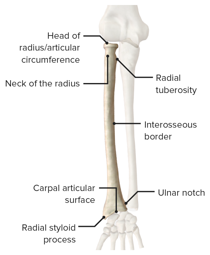
The forearm contains 2 long bones Long bones Length greater than width. Bones: Structure and Types

  • Radius
    • Located on the lateral side of the forearm
    • Articulates proximally with the humerus Humerus Bone in humans and primates extending from the shoulder joint to the elbow joint. Arm: Anatomy and ulna
    • Articulates distally with the ulna, scaphoid, and lunate bones
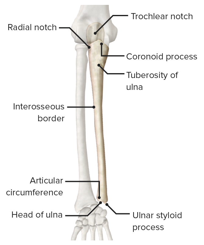
  • Ulna
    • Located on the medial side of the forearm
    • Tapers gradually from proximal to distal
    • Articulates proximally with the humerus Humerus Bone in humans and primates extending from the shoulder joint to the elbow joint. Arm: Anatomy and radius
    • Articulates distally with the radius and the articular disc of the wrist (triangular fibrocartilage Fibrocartilage A type of cartilage whose matrix contains large bundles of collagen type I. Fibrocartilage is typically found in the intervertebral disk; pubic symphysis; tibial menisci; and articular discs in synovial joints. Cartilage: Histology complex (TFCC))

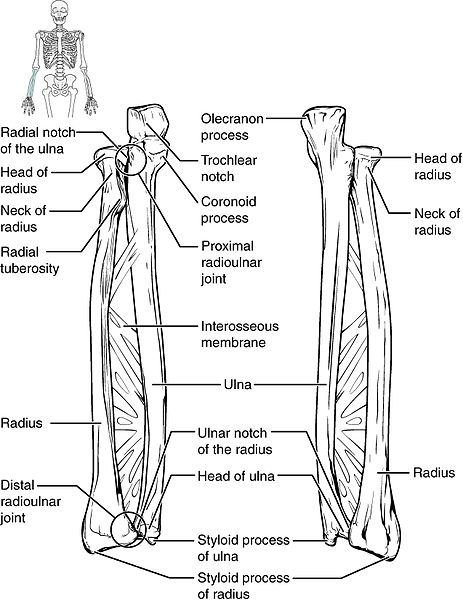
The bones of the forearm, the radius laterally and ulna medially, are held together by the fibrous Fibrous Fibrocystic Change interosseous membrane.
Joints

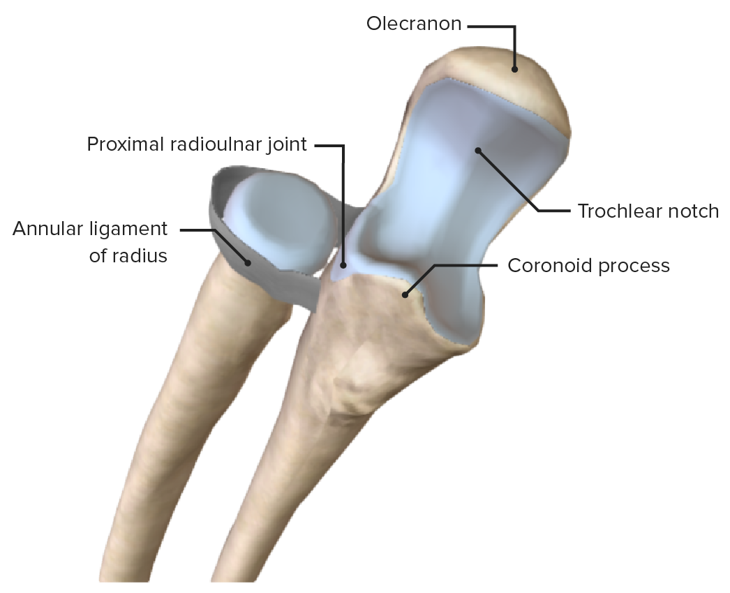
  • Proximal radioulnar joint
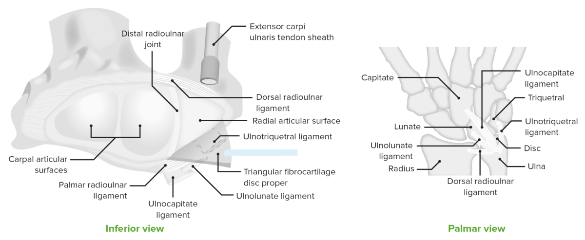
  • Distal radioulnar Distal radioulnar Wrist Joint: Anatomy joint
    • Pivot joint
    • Between the head of the ulna and the ulnar notch of the distal radius
    • Uniaxial, allows for the movements of pronation-supination
    • Supported by the TFCC, a major ligamentous stabilizer of the distal radioulnar Distal radioulnar Wrist Joint: Anatomy joint and ulnar carpus that cushions the forces transmitted through the ulnar side of the wrist. Consists of:
      • Central articular disc
      • Meniscus homolog
      • Ulnar collateral ligament Ulnar collateral ligament The internal lateral ligament that travels from the medial aspect of the elbow uniting the distal aspect of the humerus to the proximal aspect of the ulna. Elbow Joint: Anatomy
      • Dorsal and volar radioulnar ligaments
      • Extensor carpi ulnaris subsheath
  • Interosseous membrane
    • Fibrous Fibrous Fibrocystic Change tissue that creates the radioulnar syndesmosis
    • Spans the space between the radius and ulna and transfers forces during pronation Pronation Applies to movements of the forearm in turning the palm backward or downward. When referring to the foot, a combination of eversion and abduction movements in the tarsal and metatarsal joints (turning the foot up and in toward the midline of the body). Examination of the Upper Limbs and supination Supination Applies to movements of the forearm in turning the palm forward or upward. When referring to the foot, a combination of adduction and inversion movements of the foot. Examination of the Upper Limbs
Inferior view of the distal radio-ulnar joint

Distal radio-ulnar joint

Image by Lecturio.
Muscles of the Anterior Compartment of the Forearm

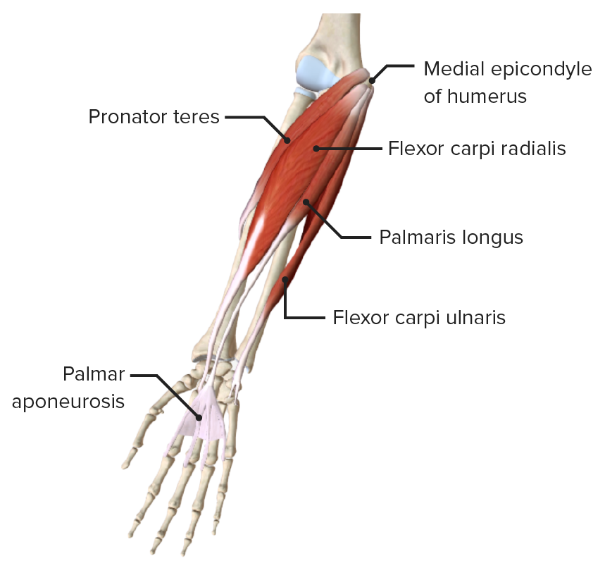
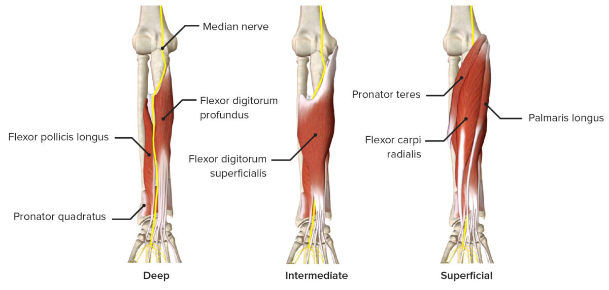
The muscles of the anterior compartment of the forearm are often separated into superficial, intermediate, and deep layers.

Superficial layer

Muscle Origin Insertion Innervation Function
Pronator teres Middle of lateral surface of radius Median nerve Median Nerve A major nerve of the upper extremity. In humans, the fibers of the median nerve originate in the lower cervical and upper thoracic spinal cord (usually C6 to T1), travel via the brachial plexus, and supply sensory and motor innervation to parts of the forearm and hand. Cubital Fossa: Anatomy (C7) Pronates and flexes forearm
Flexor carpi radialis Medial epicondyle Medial epicondyle Arm: Anatomy of humerus Humerus Bone in humans and primates extending from the shoulder joint to the elbow joint. Arm: Anatomy Base of 2nd metacarpal Flexes and abducts wrist
Palmaris longus Flexor retinaculum Flexor Retinaculum Ankle Joint: Anatomy and palmar aponeurosis Palmar aponeurosis Hand: Anatomy Flexes wrist and tenses palmar aponeurosis Palmar aponeurosis Hand: Anatomy
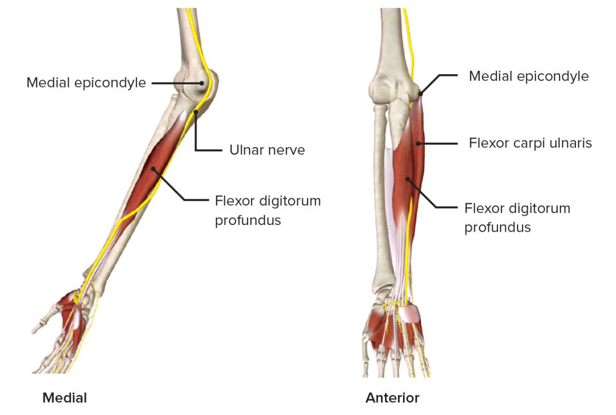
Flexor carpi ulnaris Olecranon Olecranon A prominent projection of the ulna that articulates with the humerus and forms the outer protuberance of the elbow joint. Arm: Anatomy and posterior ulna Pisiform, hook of hamate, 5th metacarpal Ulnar nerve Ulnar Nerve A major nerve of the upper extremity. In humans, the fibers of the ulnar nerve originate in the lower cervical and upper thoracic spinal cord (usually C7 to T1), travel via the medial cord of the brachial plexus, and supply sensory and motor innervation to parts of the hand and forearm. Axilla and Brachial Plexus: Anatomy (C8) Flexes and adducts wrist
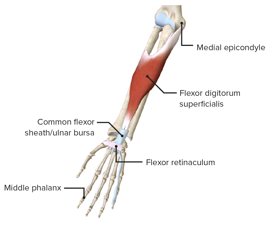
Anterior view of the right forearm, featuring the muscles of the superficial layer of the anterior compartment

Anterior view of the right forearm, featuring the muscles of the superficial layer of the anterior compartment

Image by BioDigital, edited by Lecturio
Intermediate layer

Muscle Origin Insertion Innervation Function
Flexor digitorum superficialis Middle phalanges Phalanges Bones that make up the skeleton of the fingers, consisting of two for the thumb, and three for each of the other fingers. Hand: Anatomy of medial 4 digits Median nerve Median Nerve A major nerve of the upper extremity. In humans, the fibers of the median nerve originate in the lower cervical and upper thoracic spinal cord (usually C6 to T1), travel via the brachial plexus, and supply sensory and motor innervation to parts of the forearm and hand. Cubital Fossa: Anatomy (C7, C8, T1)
  • Flexes wrist
  • Flexes proximal interphalangeal joint
Anterior view of the right forearm, featuring the only muscle of the intermediate layer of the anterior compartment

Anterior view of the right forearm, featuring the muscles of the intermediate layer of the anterior compartment

Image by BioDigital, edited by Lecturio

Deep layer

Muscle Origin Insertion Innervation Function
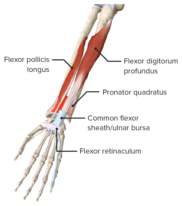
Flexor digitorum profundus Proximal surfaces of medial and anterior surfaces of ulna and interosseous membrane Middle phalanges Phalanges Bones that make up the skeleton of the fingers, consisting of two for the thumb, and three for each of the other fingers. Hand: Anatomy of medial 4 digits
  • Digits 2–3: median nerve Median Nerve A major nerve of the upper extremity. In humans, the fibers of the median nerve originate in the lower cervical and upper thoracic spinal cord (usually C6 to T1), travel via the brachial plexus, and supply sensory and motor innervation to parts of the forearm and hand. Cubital Fossa: Anatomy (C7, C8, T1)
  • Digits 4–5: ulnar nerve Ulnar Nerve A major nerve of the upper extremity. In humans, the fibers of the ulnar nerve originate in the lower cervical and upper thoracic spinal cord (usually C7 to T1), travel via the medial cord of the brachial plexus, and supply sensory and motor innervation to parts of the hand and forearm. Axilla and Brachial Plexus: Anatomy (T1)
  • Flexes wrist
  • Flexes distal interphalangeal joint
Flexor pollicis longus Anterior surface of radius and interosseous membrane Distal phalanx of 1st digit Anterior interosseous nerve Anterior Interosseous Nerve Supracondylar Fracture, branch of median (C8)
  • Flexes wrist
  • Flexes interphalangeal and metacarpo- phalangeal joints of 1st digit
Pronator quadratus Distal quarter of ulna Distal quarter of radius Pronates forearm
Anterior view of the right forearm, featuring the muscles of the deep layer of the anterior compartment

Anterior view of the right forearm, featuring the muscles of the deep layer of the anterior compartment

Image by BioDigital, edited by Lecturio

Muscles of the Posterior Compartment of the Forearm

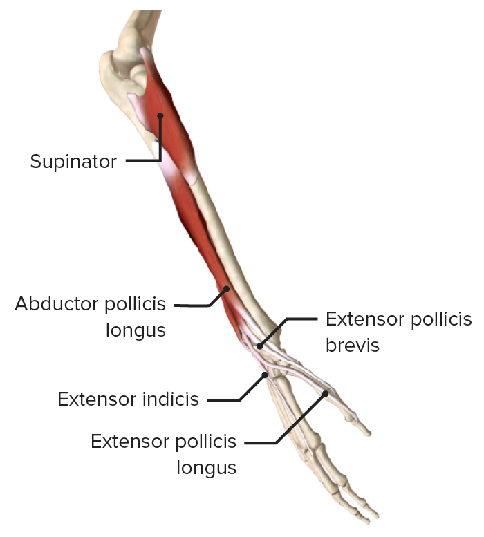
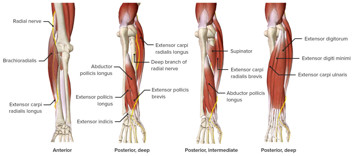
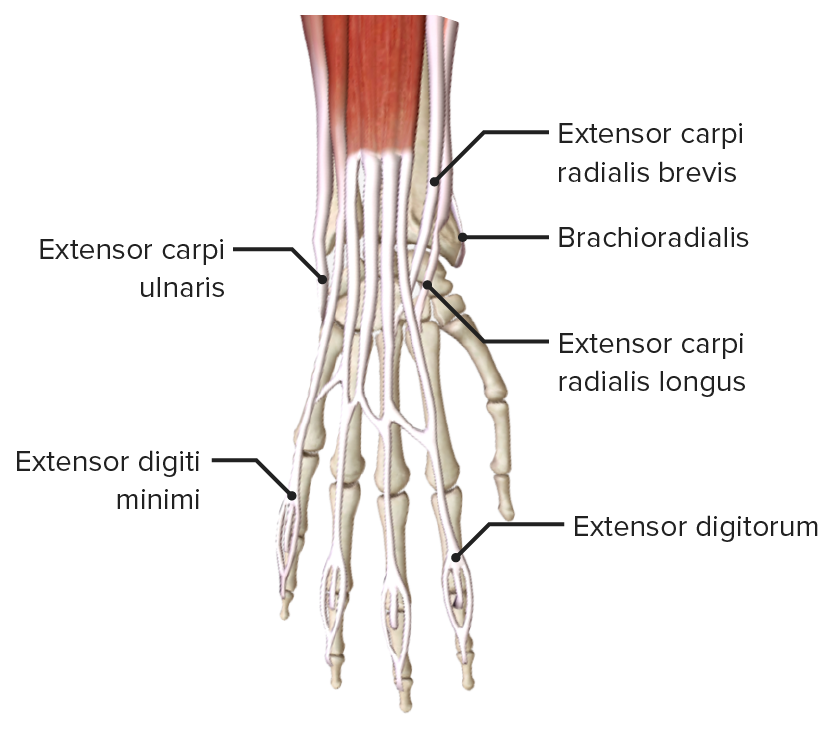
The muscles of the posterior compartment of the forearm are separated into superficial and deep layers.

Superficial layer

Muscle Origin Insertion Innervation Function
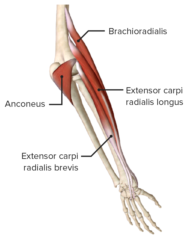
Brachio-radialis Proximal ⅔ of lateral supracondylar ridge Lateral surface of distal radius and pre-styloid process Radial nerve Radial Nerve A major nerve of the upper extremity. In humans the fibers of the radial nerve originate in the lower cervical and upper thoracic spinal cord (usually C5 to T1), travel via the posterior cord of the brachial plexus, and supply motor innervation to extensor muscles of the arm and cutaneous sensory fibers to extensor regions of the arm and hand. Axilla and Brachial Plexus: Anatomy (C6) Weak flexor of elbow, strong flexor when midpronated
Extensor carpi radialis longus Lateral supracondylar ridge Dorsal aspect of 2nd metacarpal Radial nerve Radial Nerve A major nerve of the upper extremity. In humans the fibers of the radial nerve originate in the lower cervical and upper thoracic spinal cord (usually C5 to T1), travel via the posterior cord of the brachial plexus, and supply motor innervation to extensor muscles of the arm and cutaneous sensory fibers to extensor regions of the arm and hand. Axilla and Brachial Plexus: Anatomy (C6, C7) Extend and abduct wrist
Extensor carpi radialis brevis Lateral epicondyle Lateral epicondyle Arm: Anatomy (common extensor origin) Dorsal aspect of 3rd metacarpal Deep branch of radial nerve Radial Nerve A major nerve of the upper extremity. In humans the fibers of the radial nerve originate in the lower cervical and upper thoracic spinal cord (usually C5 to T1), travel via the posterior cord of the brachial plexus, and supply motor innervation to extensor muscles of the arm and cutaneous sensory fibers to extensor regions of the arm and hand. Axilla and Brachial Plexus: Anatomy (C7)
Extensor digitorum Lateral epicondyle Lateral epicondyle Arm: Anatomy (common extensor origin) Extensor expansion of medial 4 digits Posterior interosseous nerve (C7; from deep radial nerve Radial Nerve A major nerve of the upper extremity. In humans the fibers of the radial nerve originate in the lower cervical and upper thoracic spinal cord (usually C5 to T1), travel via the posterior cord of the brachial plexus, and supply motor innervation to extensor muscles of the arm and cutaneous sensory fibers to extensor regions of the arm and hand. Axilla and Brachial Plexus: Anatomy)
Extensor digiti minimi Extensor expansion of 5th digit
Extensor carpi ulnaris Lateral epicondyle Lateral epicondyle Arm: Anatomy and posterior surface of ulna Dorsal aspect of 5th metacarpal Extends and adducts wrist
Anconeus Anconeus Elbow Joint: Anatomy Inserts on the posterior aspect of lateral epicondyle Lateral epicondyle Arm: Anatomy Lateral surface of the olecranon Olecranon A prominent projection of the ulna that articulates with the humerus and forms the outer protuberance of the elbow joint. Arm: Anatomy Radial nerve Radial Nerve A major nerve of the upper extremity. In humans the fibers of the radial nerve originate in the lower cervical and upper thoracic spinal cord (usually C5 to T1), travel via the posterior cord of the brachial plexus, and supply motor innervation to extensor muscles of the arm and cutaneous sensory fibers to extensor regions of the arm and hand. Axilla and Brachial Plexus: Anatomy (C7, C8) Extends the elbow

Deep layer

Muscle Origin Insertion Innervation Function
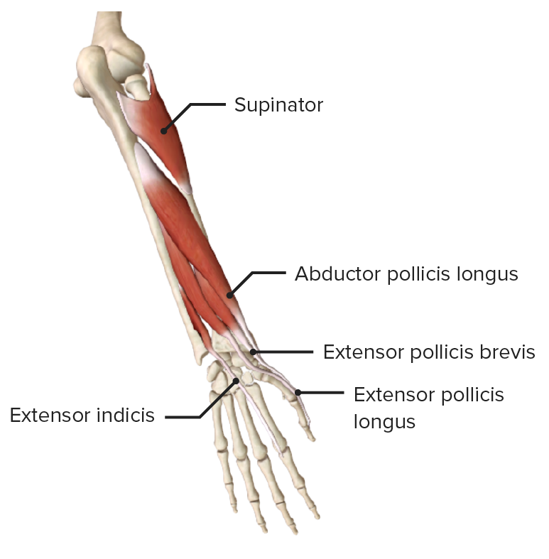
Abductor pollicis longus Posterior surface of radius and ulna and interosseous membrane Metacarpal of 1st digit Posterior interosseous nerve (C8) from deep radial nerve Radial Nerve A major nerve of the upper extremity. In humans the fibers of the radial nerve originate in the lower cervical and upper thoracic spinal cord (usually C5 to T1), travel via the posterior cord of the brachial plexus, and supply motor innervation to extensor muscles of the arm and cutaneous sensory fibers to extensor regions of the arm and hand. Axilla and Brachial Plexus: Anatomy
  • Extends wrist
  • Abducts thumb and extends it at carpometacarpal Carpometacarpal The articulations between the carpal bones and the metacarpal bones. Wrist Joint: Anatomy joint
Extensor pollicis longus Posterior surface of ulna and interosseous membrane Dorsal surface of distal phalanx of 1st digit
  • Extends wrist
  • Extends distal (extensor pollicis longus (EPL))/proximal (extensor pollicis brevis (EPB)) phalanx of thumb at interphalangeal joint
  • Extends metacarpophalangeal and carpometacarpal Carpometacarpal The articulations between the carpal bones and the metacarpal bones. Wrist Joint: Anatomy joints
Extensor pollicis brevis Posterior surface of radius and interosseous membrane Dorsal surface of proximal phalanx of 1st digit
Supinator Lateral epicondyle Lateral epicondyle Arm: Anatomy of humerus Humerus Bone in humans and primates extending from the shoulder joint to the elbow joint. Arm: Anatomy, supinator fossa, and proximal ulna Posterior/lateral/anterior surfaces of proximal radius Deep branch of radial nerve Radial Nerve A major nerve of the upper extremity. In humans the fibers of the radial nerve originate in the lower cervical and upper thoracic spinal cord (usually C5 to T1), travel via the posterior cord of the brachial plexus, and supply motor innervation to extensor muscles of the arm and cutaneous sensory fibers to extensor regions of the arm and hand. Axilla and Brachial Plexus: Anatomy (C8) Supination Supination Applies to movements of the forearm in turning the palm forward or upward. When referring to the foot, a combination of adduction and inversion movements of the foot. Examination of the Upper Limbs of forearm
Extensor indicis Posterior surface of ulna and interosseous membrane Extensor expansion of 2nd digit Posterior interosseous nerve (C8) from deep radial nerve Radial Nerve A major nerve of the upper extremity. In humans the fibers of the radial nerve originate in the lower cervical and upper thoracic spinal cord (usually C5 to T1), travel via the posterior cord of the brachial plexus, and supply motor innervation to extensor muscles of the arm and cutaneous sensory fibers to extensor regions of the arm and hand. Axilla and Brachial Plexus: Anatomy
  • Extends wrist
  • Extends 2nd digit (independent)

Vessels of the Forearm

Arteries Arteries Arteries are tubular collections of cells that transport oxygenated blood and nutrients from the heart to the tissues of the body. The blood passes through the arteries in order of decreasing luminal diameter, starting in the largest artery (the aorta) and ending in the small arterioles. Arteries are classified into 3 types: large elastic arteries, medium muscular arteries, and small arteries and arterioles. Arteries: Histology

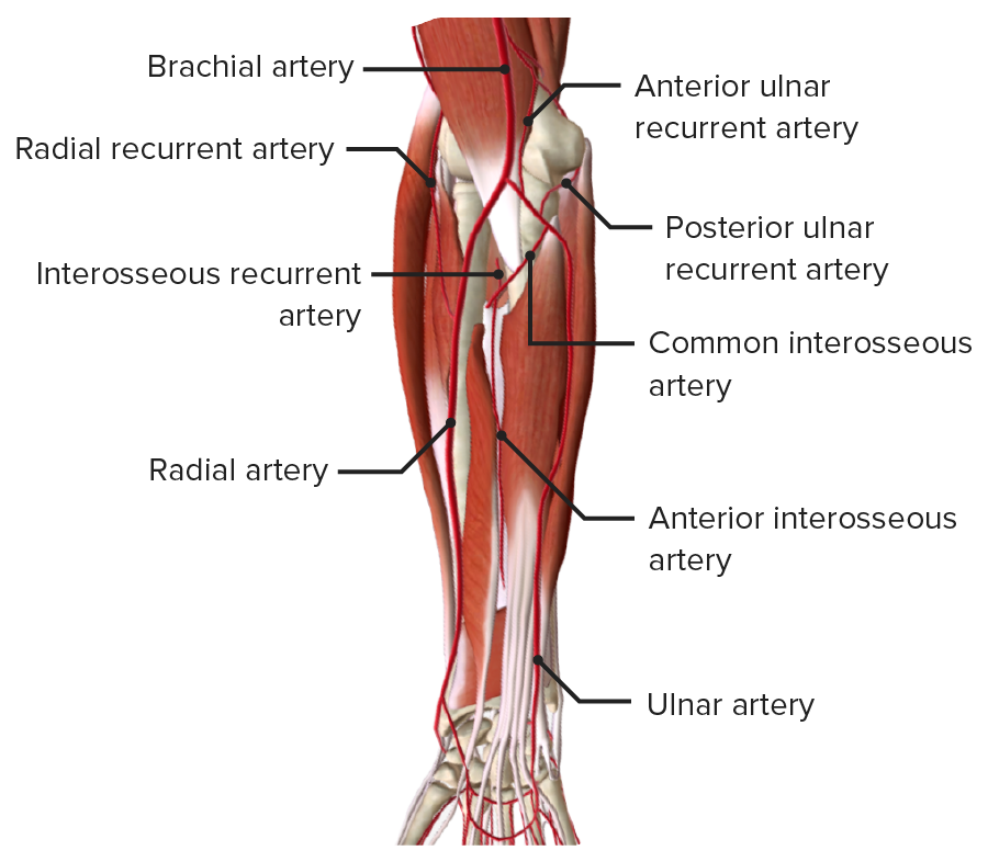
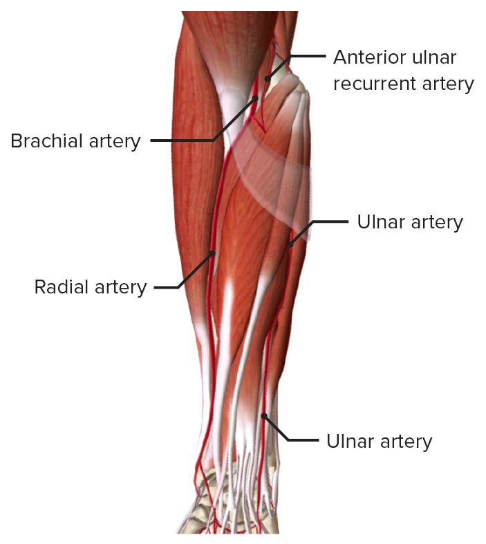
The 2 main arteries Arteries Arteries are tubular collections of cells that transport oxygenated blood and nutrients from the heart to the tissues of the body. The blood passes through the arteries in order of decreasing luminal diameter, starting in the largest artery (the aorta) and ending in the small arterioles. Arteries are classified into 3 types: large elastic arteries, medium muscular arteries, and small arteries and arterioles. Arteries: Histology of the forearm, the radial and ulnar arteries Arteries Arteries are tubular collections of cells that transport oxygenated blood and nutrients from the heart to the tissues of the body. The blood passes through the arteries in order of decreasing luminal diameter, starting in the largest artery (the aorta) and ending in the small arterioles. Arteries are classified into 3 types: large elastic arteries, medium muscular arteries, and small arteries and arterioles. Arteries: Histology, are branches of the brachial artery Brachial Artery The continuation of the axillary artery; it branches into the radial and ulnar arteries. Cubital Fossa: Anatomy. These 2 arteries Arteries Arteries are tubular collections of cells that transport oxygenated blood and nutrients from the heart to the tissues of the body. The blood passes through the arteries in order of decreasing luminal diameter, starting in the largest artery (the aorta) and ending in the small arterioles. Arteries are classified into 3 types: large elastic arteries, medium muscular arteries, and small arteries and arterioles. Arteries: Histology join in the hand Hand The hand constitutes the distal part of the upper limb and provides the fine, precise movements needed in activities of daily living. It consists of 5 metacarpal bones and 14 phalanges, as well as numerous muscles innervated by the median and ulnar nerves. Hand: Anatomy, forming an anastomosis, the superficial and deep palmar arteries Arteries Arteries are tubular collections of cells that transport oxygenated blood and nutrients from the heart to the tissues of the body. The blood passes through the arteries in order of decreasing luminal diameter, starting in the largest artery (the aorta) and ending in the small arterioles. Arteries are classified into 3 types: large elastic arteries, medium muscular arteries, and small arteries and arterioles. Arteries: Histology.
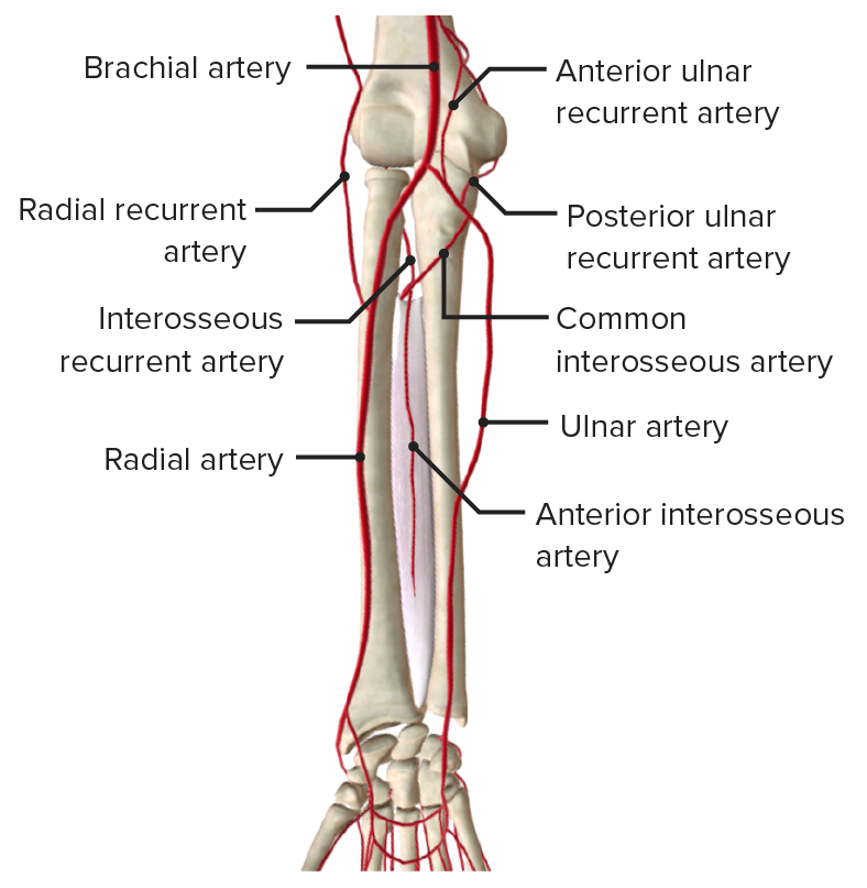
  • Radial artery
    • Supplies the posterior lateral forearm
    • Gives off important branches and contributions:
      • Radial recurrent artery: anastomoses around the elbow
      • Palmar carpal branch: supplies the carpal bones Carpal bones The eight bones of the wrist: scaphoid bone; lunate bone; triquetrum bone; pisiform bone; trapezium bone; trapezoid bone; capitate bone; and hamate bone. Carpal Tunnel Syndrome
      • Contributes to the superficial and deep palmar arches of the hand Hand The hand constitutes the distal part of the upper limb and provides the fine, precise movements needed in activities of daily living. It consists of 5 metacarpal bones and 14 phalanges, as well as numerous muscles innervated by the median and ulnar nerves. Hand: Anatomy 
  • Ulnar artery
    • Supplies the anterior medial forearm and interosseous membrane
    • Gives off important branches and contributions:
      • Ulnar recurrent artery: anastomoses around the elbow
      • Common interosseous artery: gives off anterior and posterior interosseous arteries Arteries Arteries are tubular collections of cells that transport oxygenated blood and nutrients from the heart to the tissues of the body. The blood passes through the arteries in order of decreasing luminal diameter, starting in the largest artery (the aorta) and ending in the small arterioles. Arteries are classified into 3 types: large elastic arteries, medium muscular arteries, and small arteries and arterioles. Arteries: Histology
      • Dorsal and palmar carpal branches: supply the carpal bones Carpal bones The eight bones of the wrist: scaphoid bone; lunate bone; triquetrum bone; pisiform bone; trapezium bone; trapezoid bone; capitate bone; and hamate bone. Carpal Tunnel Syndrome
      • Contributes to the superficial and deep palmar arches of the hand Hand The hand constitutes the distal part of the upper limb and provides the fine, precise movements needed in activities of daily living. It consists of 5 metacarpal bones and 14 phalanges, as well as numerous muscles innervated by the median and ulnar nerves. Hand: Anatomy

Veins Veins Veins are tubular collections of cells, which transport deoxygenated blood and waste from the capillary beds back to the heart. Veins are classified into 3 types: small veins/venules, medium veins, and large veins. Each type contains 3 primary layers: tunica intima, tunica media, and tunica adventitia. Veins: Histology

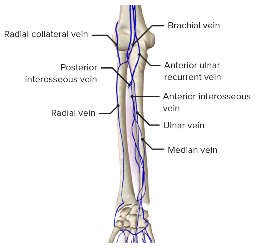
The veins Veins Veins are tubular collections of cells, which transport deoxygenated blood and waste from the capillary beds back to the heart. Veins are classified into 3 types: small veins/venules, medium veins, and large veins. Each type contains 3 primary layers: tunica intima, tunica media, and tunica adventitia. Veins: Histology of the forearm are divided into superficial and deep:
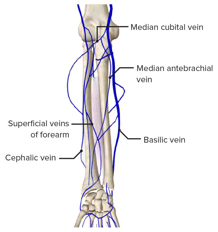
  • Superficial veins Veins Veins are tubular collections of cells, which transport deoxygenated blood and waste from the capillary beds back to the heart. Veins are classified into 3 types: small veins/venules, medium veins, and large veins. Each type contains 3 primary layers: tunica intima, tunica media, and tunica adventitia. Veins: Histology
    • Cephalic: lateral forearm, connected to basilic via the median cubital vein
    • Basilic: medial forearm, connects with the median cubital vein and median antebrachial vein
    • Median antebrachial vein: within subcutaneous tissue Subcutaneous tissue Loose connective tissue lying under the dermis, which binds skin loosely to subjacent tissues. It may contain a pad of adipocytes, which vary in number according to the area of the body and vary in size according to the nutritional state. Soft Tissue Abscess, most commonly terminates in the median cubital vein
  • Deep veins Veins Veins are tubular collections of cells, which transport deoxygenated blood and waste from the capillary beds back to the heart. Veins are classified into 3 types: small veins/venules, medium veins, and large veins. Each type contains 3 primary layers: tunica intima, tunica media, and tunica adventitia. Veins: Histology
    • Radial and ulnar: communicate with superficial veins Veins Veins are tubular collections of cells, which transport deoxygenated blood and waste from the capillary beds back to the heart. Veins are classified into 3 types: small veins/venules, medium veins, and large veins. Each type contains 3 primary layers: tunica intima, tunica media, and tunica adventitia. Veins: Histology via perforator veins Veins Veins are tubular collections of cells, which transport deoxygenated blood and waste from the capillary beds back to the heart. Veins are classified into 3 types: small veins/venules, medium veins, and large veins. Each type contains 3 primary layers: tunica intima, tunica media, and tunica adventitia. Veins: Histology, joined by transverse branches

Innervation of the Forearm

The median, ulnar, and radial nerves pass through the forearm and innervate the muscles of both the anterior and posterior compartments.

The 3 main motor Motor Neurons which send impulses peripherally to activate muscles or secretory cells. Nervous System: Histology nerves of the forearm innervate the following muscles:

  • Median: all muscles of the anterior or flexor compartment of the forearm (except for the 2 muscles supplied by the ulnar nerve Ulnar Nerve A major nerve of the upper extremity. In humans, the fibers of the ulnar nerve originate in the lower cervical and upper thoracic spinal cord (usually C7 to T1), travel via the medial cord of the brachial plexus, and supply sensory and motor innervation to parts of the hand and forearm. Axilla and Brachial Plexus: Anatomy)
  • Ulnar: flexor carpi ulnaris and medial half of the flexor digitorum profundus in the anterior or flexor compartment
  • Radial: all muscles of the posterior or extensor compartment of the forearm

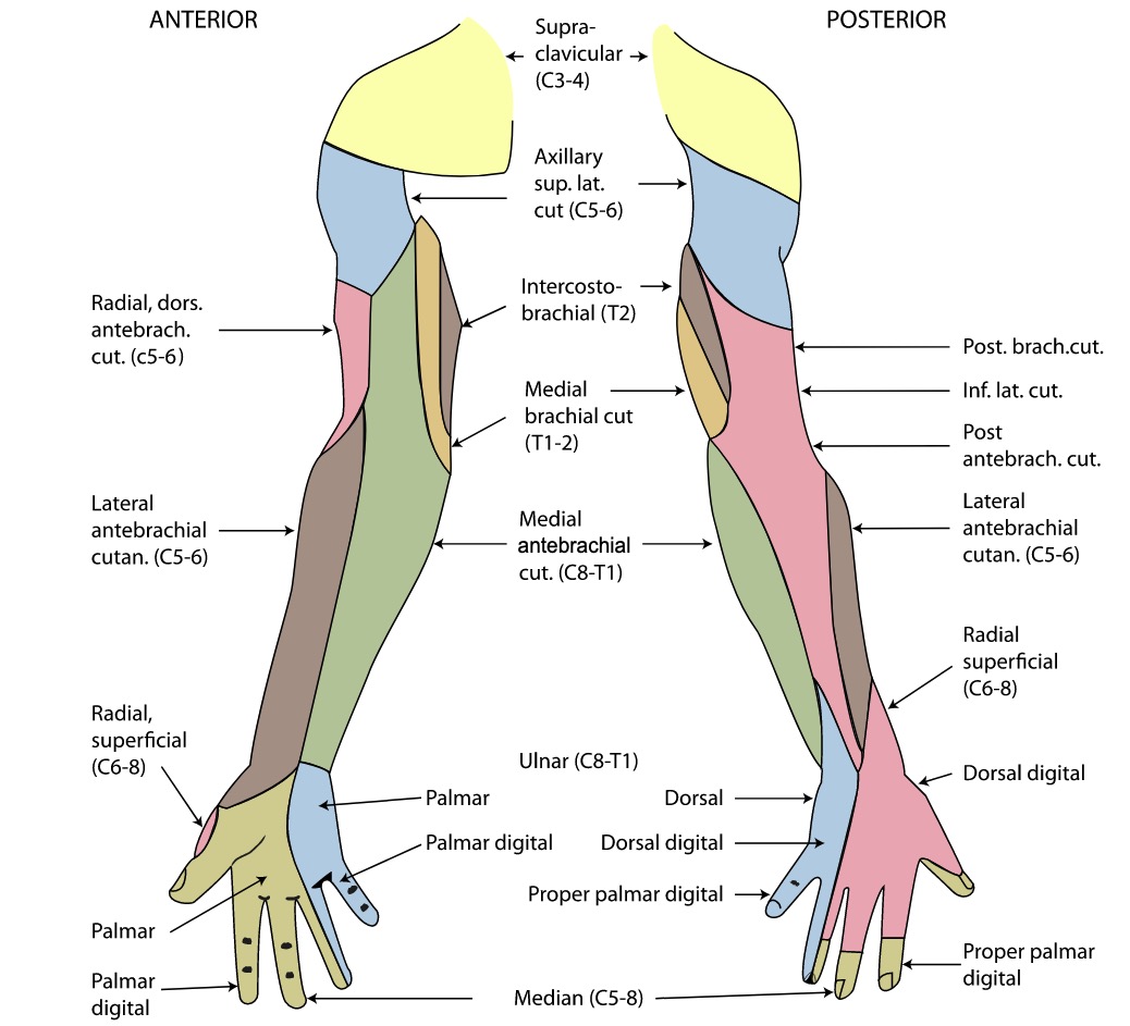
There are 3 main sensory Sensory Neurons which conduct nerve impulses to the central nervous system. Nervous System: Histology nerves of the forearm:
  • Lateral antebrachial cutaneous nerve (from the musculocutaneous nerve Musculocutaneous Nerve A major nerve of the upper extremity. The fibers of the musculocutaneous nerve originate in the lower cervical spinal cord (usually C5 to C7), travel via the lateral cord of the brachial plexus, and supply sensory and motor innervation to the upper arm, elbow, and forearm. Axilla and Brachial Plexus: Anatomy): innervates the skin Skin The skin, also referred to as the integumentary system, is the largest organ of the body. The skin is primarily composed of the epidermis (outer layer) and dermis (deep layer). The epidermis is primarily composed of keratinocytes that undergo rapid turnover, while the dermis contains dense layers of connective tissue. Skin: Structure and Functions over the lateral arm Arm The arm, or “upper arm” in common usage, is the region of the upper limb that extends from the shoulder to the elbow joint and connects inferiorly to the forearm through the cubital fossa. It is divided into 2 fascial compartments (anterior and posterior). Arm: Anatomy
  • Medial antebrachial cutaneous nerve (from the medial cord of the brachial plexus Brachial Plexus The large network of nerve fibers which distributes the innervation of the upper extremity. The brachial plexus extends from the neck into the axilla. In humans, the nerves of the plexus usually originate from the lower cervical and the first thoracic spinal cord segments (c5-c8 and T1), but variations are not uncommon. Peripheral Nerve Injuries in the Cervicothoracic Region): innervates the skin Skin The skin, also referred to as the integumentary system, is the largest organ of the body. The skin is primarily composed of the epidermis (outer layer) and dermis (deep layer). The epidermis is primarily composed of keratinocytes that undergo rapid turnover, while the dermis contains dense layers of connective tissue. Skin: Structure and Functions over the medial forearm
  • Inferior or posterior lateral cutaneous nerve (from the radial nerve Radial Nerve A major nerve of the upper extremity. In humans the fibers of the radial nerve originate in the lower cervical and upper thoracic spinal cord (usually C5 to T1), travel via the posterior cord of the brachial plexus, and supply motor innervation to extensor muscles of the arm and cutaneous sensory fibers to extensor regions of the arm and hand. Axilla and Brachial Plexus: Anatomy): innervates the skin Skin The skin, also referred to as the integumentary system, is the largest organ of the body. The skin is primarily composed of the epidermis (outer layer) and dermis (deep layer). The epidermis is primarily composed of keratinocytes that undergo rapid turnover, while the dermis contains dense layers of connective tissue. Skin: Structure and Functions over the posterior arm Arm The arm, or “upper arm” in common usage, is the region of the upper limb that extends from the shoulder to the elbow joint and connects inferiorly to the forearm through the cubital fossa. It is divided into 2 fascial compartments (anterior and posterior). Arm: Anatomy

Clinical Relevance

The following are common clinical conditions related to the forearm:

  • Distal radius fractures Distal radius fractures Distal radius fractures are one of the most common fractures encountered in practice and are often associated with falling onto an outstretched hand. These fractures are most frequently seen in older individuals, especially women. Distal Radius Fractures: common fractures of the arm Arm The arm, or “upper arm” in common usage, is the region of the upper limb that extends from the shoulder to the elbow joint and connects inferiorly to the forearm through the cubital fossa. It is divided into 2 fascial compartments (anterior and posterior). Arm: Anatomy, with a bimodal distribution Bimodal distribution Measures of Central Tendency and Dispersion. May be caused by low-energy falls or high-energy trauma in younger patients Patients Individuals participating in the health care system for the purpose of receiving therapeutic, diagnostic, or preventive procedures. Clinician–Patient Relationship. More common in women over the age of 50 with osteoporosis Osteoporosis Osteoporosis refers to a decrease in bone mass and density leading to an increased number of fractures. There are 2 forms of osteoporosis: primary, which is commonly postmenopausal or senile; and secondary, which is a manifestation of immobilization, underlying medical disorders, or long-term use of certain medications. Osteoporosis.
  • Triangular fibrocartilage Fibrocartilage A type of cartilage whose matrix contains large bundles of collagen type I. Fibrocartilage is typically found in the intervertebral disk; pubic symphysis; tibial menisci; and articular discs in synovial joints. Cartilage: Histology complex injuries: The TFCC is the primary ligamentous stabilizer of the distal radioulnar Distal radioulnar Wrist Joint: Anatomy joint. The TFCC can be injured acutely with a fall on an outstretched hand Hand The hand constitutes the distal part of the upper limb and provides the fine, precise movements needed in activities of daily living. It consists of 5 metacarpal bones and 14 phalanges, as well as numerous muscles innervated by the median and ulnar nerves. Hand: Anatomy or chronically secondary to overuse injuries.
  • Peripheral nerve injuries in the upper extremity Peripheral nerve injuries in the upper extremity Peripheral nerve damage affecting the upper extremities is a common occupational injury and also occurs in individuals who participate in recreational sports. Injuries can affect the axillary, musculocutaneous, median, ulnar, or radial nerves. Peripheral Nerve Injuries in the Upper Extremity:
    • Median nerve Median Nerve A major nerve of the upper extremity. In humans, the fibers of the median nerve originate in the lower cervical and upper thoracic spinal cord (usually C6 to T1), travel via the brachial plexus, and supply sensory and motor innervation to parts of the forearm and hand. Cubital Fossa: Anatomy neuropathy Neuropathy Leprosy in the forearm: The most common location of median nerve Median Nerve A major nerve of the upper extremity. In humans, the fibers of the median nerve originate in the lower cervical and upper thoracic spinal cord (usually C6 to T1), travel via the brachial plexus, and supply sensory and motor innervation to parts of the forearm and hand. Cubital Fossa: Anatomy mononeuropathy Mononeuropathy Disease or trauma involving a single peripheral nerve in isolation, or out of proportion to evidence of diffuse peripheral nerve dysfunction. Mononeuropathy multiplex refers to a condition characterized by multiple isolated nerve injuries. Mononeuropathies may result from a wide variety of causes, including ischemia; traumatic injury; compression; connective tissue diseases; cumulative trauma disorders; and other conditions. Mononeuropathy and Plexopathy is the carpal tunnel Carpal Tunnel The carpal tunnel is formed by the transverse carpal ligament (flexor retinaculum) superiorly and the carpal bones inferiorly. Carpal Tunnel Syndrome. Two less common median nerve Median Nerve A major nerve of the upper extremity. In humans, the fibers of the median nerve originate in the lower cervical and upper thoracic spinal cord (usually C6 to T1), travel via the brachial plexus, and supply sensory and motor innervation to parts of the forearm and hand. Cubital Fossa: Anatomy entrapments found in the proximal forearm include pronator syndrome Pronator Syndrome Mononeuropathy and Plexopathy and anterior interosseous syndrome.
      • Carpal tunnel Carpal Tunnel The carpal tunnel is formed by the transverse carpal ligament (flexor retinaculum) superiorly and the carpal bones inferiorly. Carpal Tunnel Syndrome syndrome (CTS): The most common compressive neuropathy Neuropathy Leprosy is often precipitated by repetitive vibration Vibration A continuing periodic change in displacement with respect to a fixed reference. Neurological Examination and motions and is associated with diabetes Diabetes Diabetes mellitus (DM) is a metabolic disease characterized by hyperglycemia and dysfunction of the regulation of glucose metabolism by insulin. Type 1 DM is diagnosed mostly in children and young adults as the result of autoimmune destruction of β cells in the pancreas and the resulting lack of insulin. Type 2 DM has a significant association with obesity and is characterized by insulin resistance. Diabetes Mellitus, thyroid Thyroid The thyroid gland is one of the largest endocrine glands in the human body. The thyroid gland is a highly vascular, brownish-red gland located in the visceral compartment of the anterior region of the neck. Thyroid Gland: Anatomy disease, rheumatoid arthritis Arthritis Acute or chronic inflammation of joints. Osteoarthritis, and pregnancy Pregnancy The status during which female mammals carry their developing young (embryos or fetuses) in utero before birth, beginning from fertilization to birth. Pregnancy: Diagnosis, Physiology, and Care.
    • Radial nerve Radial Nerve A major nerve of the upper extremity. In humans the fibers of the radial nerve originate in the lower cervical and upper thoracic spinal cord (usually C5 to T1), travel via the posterior cord of the brachial plexus, and supply motor innervation to extensor muscles of the arm and cutaneous sensory fibers to extensor regions of the arm and hand. Axilla and Brachial Plexus: Anatomy neuropathy Neuropathy Leprosy: The radial nerve Radial Nerve A major nerve of the upper extremity. In humans the fibers of the radial nerve originate in the lower cervical and upper thoracic spinal cord (usually C5 to T1), travel via the posterior cord of the brachial plexus, and supply motor innervation to extensor muscles of the arm and cutaneous sensory fibers to extensor regions of the arm and hand. Axilla and Brachial Plexus: Anatomy can become injured in the forearm through its branch, the posterior interosseous nerve (PIN), causing supinator syndrome and radial tunnel syndrome. In addition, the radial superficial nerve (RSN) can be injured at the distal forearm, and is called cheiralgia paresthetica or “handcuff neuropathy Neuropathy Leprosy.”
    • Ulnar nerve Ulnar Nerve A major nerve of the upper extremity. In humans, the fibers of the ulnar nerve originate in the lower cervical and upper thoracic spinal cord (usually C7 to T1), travel via the medial cord of the brachial plexus, and supply sensory and motor innervation to parts of the hand and forearm. Axilla and Brachial Plexus: Anatomy neuropathy Neuropathy Leprosy: The ulnar nerve Ulnar Nerve A major nerve of the upper extremity. In humans, the fibers of the ulnar nerve originate in the lower cervical and upper thoracic spinal cord (usually C7 to T1), travel via the medial cord of the brachial plexus, and supply sensory and motor innervation to parts of the hand and forearm. Axilla and Brachial Plexus: Anatomy may be injured at the posterior medial elbow secondary to direct trauma Direct Trauma Toddler’s Fractures to the medial elbow, lacerations, fractures of the distal humerus Humerus Bone in humans and primates extending from the shoulder joint to the elbow joint. Arm: Anatomy, or entrapment of the nerve.
      • Cubital tunnel syndrome: compressive neuropathy Neuropathy Leprosy of the ulnar nerve Ulnar Nerve A major nerve of the upper extremity. In humans, the fibers of the ulnar nerve originate in the lower cervical and upper thoracic spinal cord (usually C7 to T1), travel via the medial cord of the brachial plexus, and supply sensory and motor innervation to parts of the hand and forearm. Axilla and Brachial Plexus: Anatomy at the elbow; the 2nd-most common compressive neuropathy Neuropathy Leprosy. Cubital tunnel syndrome can present with interosseous muscle atrophy Atrophy Decrease in the size of a cell, tissue, organ, or multiple organs, associated with a variety of pathological conditions such as abnormal cellular changes, ischemia, malnutrition, or hormonal changes. Cellular Adaptation and a “claw hand Hand The hand constitutes the distal part of the upper limb and provides the fine, precise movements needed in activities of daily living. It consists of 5 metacarpal bones and 14 phalanges, as well as numerous muscles innervated by the median and ulnar nerves. Hand: Anatomy.”  
      • Guyon’s canal Guyon’s canal Wrist Joint: Anatomy neuropathy Neuropathy Leprosy: compression Compression Blunt Chest Trauma of the ulnar nerve Ulnar Nerve A major nerve of the upper extremity. In humans, the fibers of the ulnar nerve originate in the lower cervical and upper thoracic spinal cord (usually C7 to T1), travel via the medial cord of the brachial plexus, and supply sensory and motor innervation to parts of the hand and forearm. Axilla and Brachial Plexus: Anatomy at the distal forearm/wrist at Guyon’s canal Guyon’s canal Wrist Joint: Anatomy. The condition is sometimes called “handlebar palsy Palsy paralysis of an area of the body, thus incapable of voluntary movement Cranial Nerve Palsies,” as it is seen in cyclists.

References

  1. Drake, R.L., Vogl, A.W., & Mitchell, A.W.M. (2014). Gray’s Anatomy for Students (3rd ed.). Philadelphia, PA:  Churchill Livingstone.

Create your free account or log in to continue reading!

Sign up now and get free access to Lecturio with concept pages, medical videos, and questions for your medical education.

User Reviews

Details