Advertisement

Advertisement

Advertisement

Advertisement

Foot Deformities

Foot Foot The foot is the terminal portion of the lower limb, whose primary function is to bear weight and facilitate locomotion. The foot comprises 26 bones, including the tarsal bones, metatarsal bones, and phalanges. The bones of the foot form longitudinal and transverse arches and are supported by various muscles, ligaments, and tendons. Foot: Anatomy deformities in children include congenital Congenital Chorioretinitis or acquired malformations of the feet. Two common examples are talipes equinovarus, commonly known as clubfoot, and metatarsus adductus, also called metatarsus varus. Depending on their etiology, foot Foot The foot is the terminal portion of the lower limb, whose primary function is to bear weight and facilitate locomotion. The foot comprises 26 bones, including the tarsal bones, metatarsal bones, and phalanges. The bones of the foot form longitudinal and transverse arches and are supported by various muscles, ligaments, and tendons. Foot: Anatomy deformities can be self-limiting Self-Limiting Meningitis in Children or may require surgical correction. Early detection and recognition are crucial for proper treatment.

Last updated: Feb 7, 2023

Editorial responsibility: Stanley Oiseth, Lindsay Jones, Evelin Maza

Advertisement

Advertisement

Advertisement

Advertisement

Advertisement

Advertisement

Clubfoot (Talipes Equinovarus)

Definition

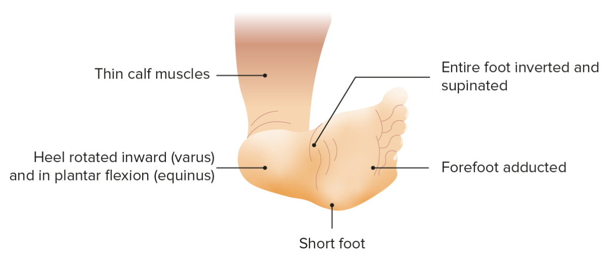
Clubfoot, also called talipes equinovarus, is a complex condition with a plantar flexed foot Foot The foot is the terminal portion of the lower limb, whose primary function is to bear weight and facilitate locomotion. The foot comprises 26 bones, including the tarsal bones, metatarsal bones, and phalanges. The bones of the foot form longitudinal and transverse arches and are supported by various muscles, ligaments, and tendons. Foot: Anatomy (equinus), adductus of the forefoot, and an inversion deformity Deformity Examination of the Upper Limbs of the heel (varus).

Epidemiology

  • Strong familial tendency
  • 1–3 per 1,000 live births:
    • Highest in Polynesian population: 7 per 1,000 live births
    • Lowest in Asian population: 0.6 per 1,000 live births
  • Male-to-female ratio: 2:1

Etiology

Etiology is debated, as the majority of infants with clubfoot have no identifiable syndromic, genetic, or extrinsic cause.

  • Positional, or postural, clubfoot: normal foot Foot The foot is the terminal portion of the lower limb, whose primary function is to bear weight and facilitate locomotion. The foot comprises 26 bones, including the tarsal bones, metatarsal bones, and phalanges. The bones of the foot form longitudinal and transverse arches and are supported by various muscles, ligaments, and tendons. Foot: Anatomy held in deforming position during gestation; usually easily correctable
  • Congenital Congenital Chorioretinitis: most common type; usually treated with casting and bracing
  • Syndromic: clubfoot associated with other congenital Congenital Chorioretinitis syndromes
  • Acquired: seen in patients Patients Individuals participating in the health care system for the purpose of receiving therapeutic, diagnostic, or preventive procedures. Clinician–Patient Relationship with polio and cerebral palsy Palsy paralysis of an area of the body, thus incapable of voluntary movement Cranial Nerve Palsies
  • Associated with arthrogryposis multiplex congenita, myelodysplasia Myelodysplasia Non-Hodgkin Lymphomas, spina bifida, and maternal cigarette use

Classification

There are multiple classification systems for talipes equinovarus:
  • Dimeglio: measures and scores 4 elements
    1. Equinus in sagittal plane Sagittal plane Anterior Abdominal Wall: Anatomy
    2. Varus deviation in frontal Frontal The bone that forms the frontal aspect of the skull. Its flat part forms the forehead, articulating inferiorly with the nasal bone and the cheek bone on each side of the face. Skull: Anatomy plane
    3. Derotation around talus Talus The second largest of the tarsal bones. It articulates with the tibia and fibula to form the ankle joint. Ankle Joint: Anatomy of calcaneo-forefoot (CFF) block 
    4. Adduction Adduction Examination of the Upper Limbs of forefoot on hindfoot in horizontal plane
  • Pirani: 6 “signs” scored 0, 0.5, or 1 based on severity
    1. Medial crease
    2. Curved lateral border
    3. Lateral head talus Talus The second largest of the tarsal bones. It articulates with the tibia and fibula to form the ankle joint. Ankle Joint: Anatomy
    4. Posterior crease
    5. Empty heel
    6. Rigid equinus

Clinical presentation

  • Mid- and forefoot are adducted (medially deviated).
  • Equinovarus:
    • Plantar flexion Flexion Examination of the Upper Limbs of foot Foot The foot is the terminal portion of the lower limb, whose primary function is to bear weight and facilitate locomotion. The foot comprises 26 bones, including the tarsal bones, metatarsal bones, and phalanges. The bones of the foot form longitudinal and transverse arches and are supported by various muscles, ligaments, and tendons. Foot: Anatomy at ankle (equinus): dorsiflexion beyond 90° not possible. 
    • Inversion deformity Deformity Examination of the Upper Limbs of heel (varus); hindfoot supinated
  • Calf is smaller at presentation (and will remain smaller throughout life).
  • Generally painless
Clubfoot clinical presentation

Talipes equinovarus (clubfoot)
Foot deformity featuring plantar flexion, medial rotation of the forefoot and inward facing sole
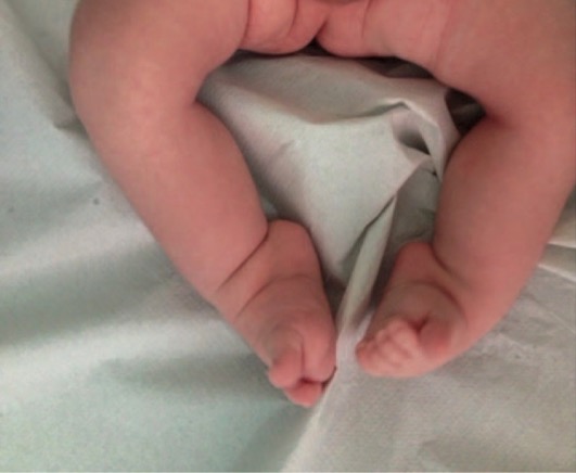
Image by Lecturio.

Diagnosis

Management

Refer to a pediatric orthopedic surgeon soon after birth for prompt initiation of treatment.
  • Initial:
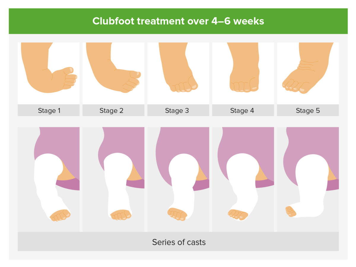
    • Manipulation of foot Foot The foot is the terminal portion of the lower limb, whose primary function is to bear weight and facilitate locomotion. The foot comprises 26 bones, including the tarsal bones, metatarsal bones, and phalanges. The bones of the foot form longitudinal and transverse arches and are supported by various muscles, ligaments, and tendons. Foot: Anatomy to stretch contracted tissues followed by serial casting (Ponseti method)
    • Correction occurs rapidly if instituted shortly after birth.
  • If equinus remains, surgery may be required:
    • Percutaneous Achilles tenotomy, to achieve full correction
    • Further surgical correction may be needed if foot Foot The foot is the terminal portion of the lower limb, whose primary function is to bear weight and facilitate locomotion. The foot comprises 26 bones, including the tarsal bones, metatarsal bones, and phalanges. The bones of the foot form longitudinal and transverse arches and are supported by various muscles, ligaments, and tendons. Foot: Anatomy is rigid and resistant to correction.
  • Prognosis Prognosis A prediction of the probable outcome of a disease based on a individual’s condition and the usual course of the disease as seen in similar situations. Non-Hodgkin Lymphomas is excellent if treatment is initiated soon after birth.
Management of clubfoot

Ponseti method
Consecutive splinting with casts made following the Ponseti method allow for gradual correction of talipes equinovarus (clubfoot).

Image by Lecturio.

Complications

  • Persistent limp
    • Due to child learning to walk on side and top of foot Foot The foot is the terminal portion of the lower limb, whose primary function is to bear weight and facilitate locomotion. The foot comprises 26 bones, including the tarsal bones, metatarsal bones, and phalanges. The bones of the foot form longitudinal and transverse arches and are supported by various muscles, ligaments, and tendons. Foot: Anatomy
    • Occurs when condition is recognized late or not addressed properly
  • Residual loss of range of motion Range of motion The distance and direction to which a bone joint can be extended. Range of motion is a function of the condition of the joints, muscles, and connective tissues involved. Joint flexibility can be improved through appropriate muscle strength exercises. Examination of the Upper Limbs and strength

Mnemonic for talipes equinovarus of clubfoot

CAVE:

  • Cavus
  • Adductus
  • Varus
  • Equinus

Metatarsus Adductus (Metatarsus Varus)

Definition

Metatarsus adductus is a common congenital Congenital Chorioretinitis foot Foot The foot is the terminal portion of the lower limb, whose primary function is to bear weight and facilitate locomotion. The foot comprises 26 bones, including the tarsal bones, metatarsal bones, and phalanges. The bones of the foot form longitudinal and transverse arches and are supported by various muscles, ligaments, and tendons. Foot: Anatomy deformity Deformity Examination of the Upper Limbs that presents with adduction Adduction Examination of the Upper Limbs, or inward deviation of the forefoot (at the tarsometatarsal joint Tarsometatarsal Joint Foot: Anatomy), with normal hindfoot alignment.

Epidemiology

  • Most common foot Foot The foot is the terminal portion of the lower limb, whose primary function is to bear weight and facilitate locomotion. The foot comprises 26 bones, including the tarsal bones, metatarsal bones, and phalanges. The bones of the foot form longitudinal and transverse arches and are supported by various muscles, ligaments, and tendons. Foot: Anatomy deformity Deformity Examination of the Upper Limbs in infants
  • Male-to-female ratio: 1:1
  • Up to 50% of cases are bilateral.
  • More common in firstborn children and twins
  • Associated with developmental hip dysplasia and torticollis Torticollis A symptom, not a disease, of a twisted neck. In most instances, the head is tipped toward one side and the chin rotated toward the other. The involuntary muscle contractions in the neck region of patients with torticollis can be due to congenital defects, trauma, inflammation, tumors, and neurological or other factors. Cranial Nerve Palsies

Etiology

The exact cause is unknown, although 1 proposed etiology is the mechanical effect of a small uterine space.
Classification

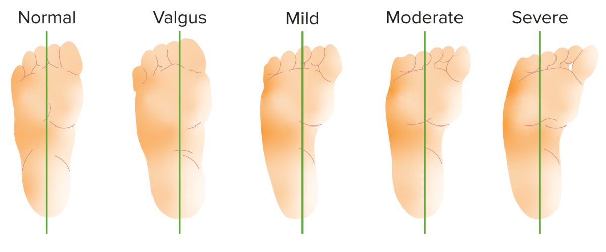
  • Bleck classification by heel bisection: 
    • Normal: heel bisector line through 2nd and 3rd toe interspace
    • Mild: through 3rd toe
    • Moderate: between 3rd and 4th toe web space
    • Severe: through 4th and 5th toe web space
  • Berg classification:
    • Simple
    • Complex
    • Skew foot Foot The foot is the terminal portion of the lower limb, whose primary function is to bear weight and facilitate locomotion. The foot comprises 26 bones, including the tarsal bones, metatarsal bones, and phalanges. The bones of the foot form longitudinal and transverse arches and are supported by various muscles, ligaments, and tendons. Foot: Anatomy
    • Serpentine foot Foot The foot is the terminal portion of the lower limb, whose primary function is to bear weight and facilitate locomotion. The foot comprises 26 bones, including the tarsal bones, metatarsal bones, and phalanges. The bones of the foot form longitudinal and transverse arches and are supported by various muscles, ligaments, and tendons. Foot: Anatomy (complex skew foot Foot The foot is the terminal portion of the lower limb, whose primary function is to bear weight and facilitate locomotion. The foot comprises 26 bones, including the tarsal bones, metatarsal bones, and phalanges. The bones of the foot form longitudinal and transverse arches and are supported by various muscles, ligaments, and tendons. Foot: Anatomy)
Bleck classification of metatarsus adductus

Bleck classification of metatarsus adductus by heel bisector method
This method is used for classifying the severity of metatarsus adductus deformity. Normal heel bisector line through the 2nd and 3rd toe web space, mild heel bisector line through the 3rd toe, moderate heel bisector through 3rd and 4th toe web space, and severe heel bisector through the 4th and 5th toe web space.
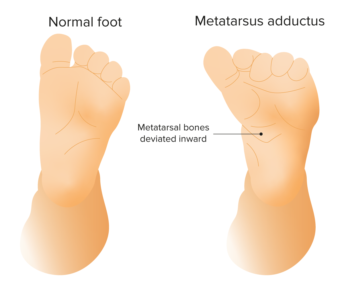
Image by Lecturio.

Clinical presentation

  • Medial deviation of forefoot at tarsometatarsal joint Tarsometatarsal Joint Foot: Anatomy
  • Normal hindfoot
  • Forefoot can be “stretched” into neutral position.
  • Ankle with normal range of motion Range of motion The distance and direction to which a bone joint can be extended. Range of motion is a function of the condition of the joints, muscles, and connective tissues involved. Joint flexibility can be improved through appropriate muscle strength exercises. Examination of the Upper Limbs
  • Painless
Metatarsus adductus

Metatarsus adductus: common congenital foot deformity featuring medial deviation of the forefoot at the tarsometatarsal joint

Image by Lecturio.

Diagnosis

  • Parents notice intoeing in first 1–2 years of life.
  • Can be detected prenatally by ultrasound

Management

  • Benign Benign Fibroadenoma condition that resolves spontaneously or with nonoperative therapy in most cases before the age of 4
  • Passive stretching or no therapy depending on severity
  • Rigid deformity Deformity Examination of the Upper Limbs will require serial casting and evaluation by pediatric orthopedic surgeon.

Prognosis Prognosis A prediction of the probable outcome of a disease based on a individual’s condition and the usual course of the disease as seen in similar situations. Non-Hodgkin Lymphomas

Prognosis Prognosis A prediction of the probable outcome of a disease based on a individual’s condition and the usual course of the disease as seen in similar situations. Non-Hodgkin Lymphomas is excellent, if treatment is started early.

Clinical Relevance

The following conditions may accompany talipes equinovarus and metatarsus adductus:

  • Developmental hip dysplasia (DHD): congenital Congenital Chorioretinitis orthopedic condition characterized by abnormal development of acetabulum due to luxation of femoral head from hip joint Hip joint The hip joint is a ball-and-socket joint formed by the head of the femur and the acetabulum of the pelvis. The hip joint is the most stable joint in the body and is supported by a very strong capsule and several ligaments, allowing the joint to sustain forces that can be multiple times the total body weight. Hip Joint: Anatomy. Developmental hip dysplasia has been associated with talipes equinovarus and metatarsus adductus; therefore, careful hip examination is essential in these patients Patients Individuals participating in the health care system for the purpose of receiving therapeutic, diagnostic, or preventive procedures. Clinician–Patient Relationship.
  • Intoeing: sometimes referred to as being “ pigeon-toed Pigeon-Toed Blount’s Disease”; a medial rotational variation where feet or toes point toward the midline during gait Gait Manner or style of walking. Neurological Examination. Most common causes are metatarsus adductus of foot Foot The foot is the terminal portion of the lower limb, whose primary function is to bear weight and facilitate locomotion. The foot comprises 26 bones, including the tarsal bones, metatarsal bones, and phalanges. The bones of the foot form longitudinal and transverse arches and are supported by various muscles, ligaments, and tendons. Foot: Anatomy, femoral anteversion of hip, and tibial torsion. Intoeing that is painful, associated with limb length discrepancy Length Discrepancy Blount’s Disease, and associated with delayed developmental milestones Developmental milestones Developmental milestones are the skills or abilities that most children are able to perform when they reach a certain age. Understanding the appropriate milestones and at what age they are reached helps clinicians identify symptoms of delayed development. Developmental milestones are divided into 5 important domains: gross motor, fine motor, language, social, and cognitive. Developmental Milestones and Normal Growth (possible cerebral palsy Palsy paralysis of an area of the body, thus incapable of voluntary movement Cranial Nerve Palsies) or a family history Family History Adult Health Maintenance of skeletal dysplasias requires further workup and orthopedic evaluation.

References

  1. Winell, J. J., & Davidson, R. S. (2020). The foot and toes. In R. M. Kliegman MD, J. W. St Geme MD, N. J. Blum MD, Shah, Samir S., MD, MSCE, Tasker, Robert C., MBBS, MD & Wilson, Karen M., MD, MPH (Eds.), Nelson textbook of pediatrics. Retrieved January 29, 2021, from https://www.clinicalkey.es/#!/content/3-s2.0-B9780323529501006945
  2. Green, A., M.D. (2020). The pediatric foot and ankle. Pediatric Clinics of North America, 67(1), 169-183. doi:http://dx.doi.org/10.1016/j.pcl.2019.09.007
  3. Ricco, A. I., Richards, B. S., & Herring, J. A. (2014). Disorders of the foot. In J. A. Herring MD (Ed.), Tachdjian’s pediatric orthopaedics (pp. 761-883). Retrieved January 19, 2021, from https://www.clinicalkey.es/#!/content/3-s2.0-B9781437715491000234

Create your free account or log in to continue reading!

Sign up now and get free access to Lecturio with concept pages, medical videos, and questions for your medical education.

User Reviews

Details