The elbow is the synovial hinge joint between the humerus Humerus Bone in humans and primates extending from the shoulder joint to the elbow joint. Arm: Anatomy in the upper arm Upper Arm The arm, or "upper arm" in common usage, is the region of the upper limb that extends from the shoulder to the elbow joint and connects inferiorly to the forearm through the cubital fossa. It is divided into 2 fascial compartments (anterior and posterior). Arm: Anatomy and the radius Radius The outer shorter of the two bones of the forearm, lying parallel to the ulna and partially revolving around it. Forearm: Anatomy and ulna Ulna The inner and longer bone of the forearm. Forearm: Anatomy in the forearm Forearm The forearm is the region of the upper limb between the elbow and the wrist. The term "forearm" is used in anatomy to distinguish this area from the arm, a term that is commonly used to describe the entire upper limb. The forearm consists of 2 long bones (the radius and the ulna), the interosseous membrane, and multiple arteries, nerves, and muscles. Forearm: Anatomy. The elbow consists of 3 joints, which form a functional unit enclosed within a single articular capsule Capsule An envelope of loose gel surrounding a bacterial cell which is associated with the virulence of pathogenic bacteria. Some capsules have a well-defined border, whereas others form a slime layer that trails off into the medium. Most capsules consist of relatively simple polysaccharides but there are some bacteria whose capsules are made of polypeptides. Bacteroides. The elbow is the link between the powerful motions of the shoulder and the intricate fine-motor function of the hand Hand The hand constitutes the distal part of the upper limb and provides the fine, precise movements needed in activities of daily living. It consists of 5 metacarpal bones and 14 phalanges, as well as numerous muscles innervated by the median and ulnar nerves. Hand: Anatomy. To provide that link, the motions of the elbow include extension Extension Examination of the Upper Limbs and flexion Flexion Examination of the Upper Limbs as well as pronation Pronation Applies to movements of the forearm in turning the palm backward or downward. When referring to the foot, a combination of eversion and abduction movements in the tarsal and metatarsal joints (turning the foot up and in toward the midline of the body). Examination of the Upper Limbs and supination Supination Applies to movements of the forearm in turning the palm forward or upward. When referring to the foot, a combination of adduction and inversion movements of the foot. Examination of the Upper Limbs of the forearm Forearm The forearm is the region of the upper limb between the elbow and the wrist. The term "forearm" is used in anatomy to distinguish this area from the arm, a term that is commonly used to describe the entire upper limb. The forearm consists of 2 long bones (the radius and the ulna), the interosseous membrane, and multiple arteries, nerves, and muscles. Forearm: Anatomy.
Last updated: Nov 19, 2024
The elbow joint consists of 3 separate articulations enclosed in a single capsule Capsule An envelope of loose gel surrounding a bacterial cell which is associated with the virulence of pathogenic bacteria. Some capsules have a well-defined border, whereas others form a slime layer that trails off into the medium. Most capsules consist of relatively simple polysaccharides but there are some bacteria whose capsules are made of polypeptides. Bacteroides:
| Joint | Articular surfaces | Type | Function |
|---|---|---|---|
| Humeroulnar joint |
|
Simple hinge-joint |
|
| Humeroradial joint |
|
Limited ball-and-socket joint | Limited pronation-supination in semiflexion |
| Proximal radioulnar joint Proximal radioulnar joint Forearm: Anatomy |
|
Pivot joint | Pronation-supination in any degree of flexion-extension |
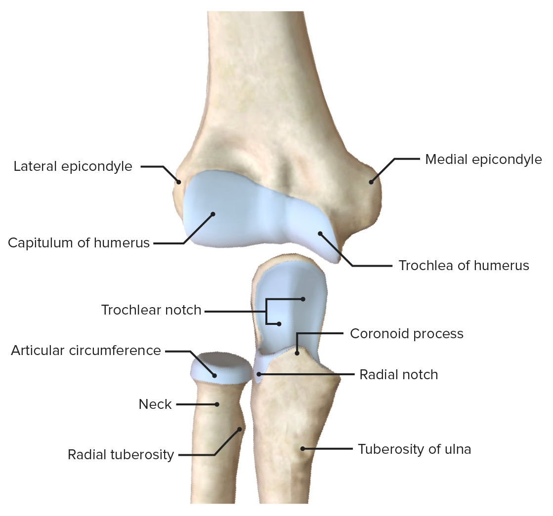
Articular surfaces of the elbow joint
Image by BioDigital, edited by Lecturio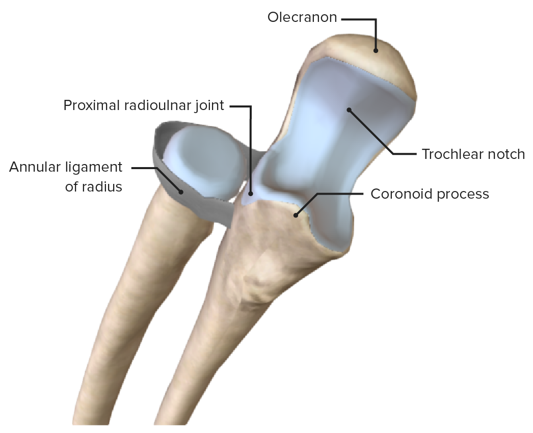
Proximal radioulnar joint, featuring its main supporting ligament, the annular ligament of the radius
Image by BioDigital, edited by Lecturio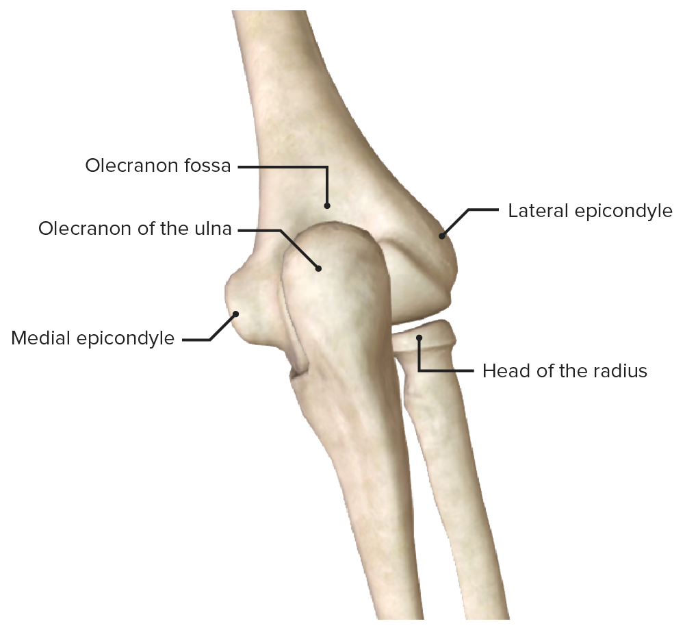
Posterior view of the elbow (humeroulnar joint), featuring the olecranon
Image by BioDigital, edited by Lecturio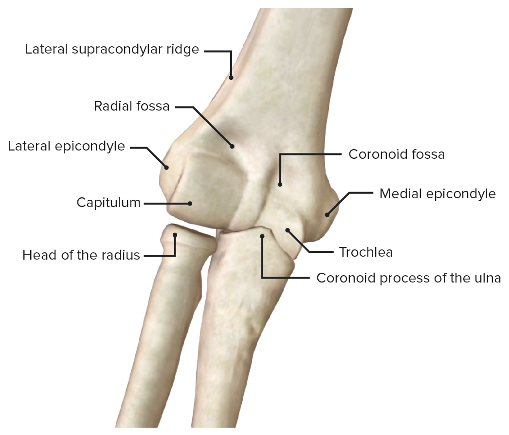
Anterior view of the elbow (humeroulnar joint), featuring the articular surfaces
Image by BioDigital, edited by LecturioThe elbow capsule Capsule An envelope of loose gel surrounding a bacterial cell which is associated with the virulence of pathogenic bacteria. Some capsules have a well-defined border, whereas others form a slime layer that trails off into the medium. Most capsules consist of relatively simple polysaccharides but there are some bacteria whose capsules are made of polypeptides. Bacteroides is supported by the ligaments of the elbow, especially the radial (lateral) and ulnar (medial) collateral ligaments.
| Ligament | Attachments | Function |
|---|---|---|
| Radial collateral ligament |
|
Stabilizes the elbow joint against varus stress |
| Ulnar collateral ligament |
|
Stabilizes the humeroulnar joint against valgus stress |
| Annular ligament Annular Ligament Radial Head Subluxation (Nursemaid’s Elbow) of the radius Radius The outer shorter of the two bones of the forearm, lying parallel to the ulna and partially revolving around it. Forearm: Anatomy | Anterior-posterior margins of the radial notch | Surrounds and anchors the radial head to the radial notch of the ulna Ulna The inner and longer bone of the forearm. Forearm: Anatomy |
| Interosseous membrane Interosseous Membrane A sheet of fibrous connective tissue rich in collagen often linking two parallel bony structures forming a syndesmosis type joint. It provides longitudinal stability, tensile strength, and weight distribution/transfer and may allow limited movement in syndesmoses. Forearm: Anatomy | Interosseous margins of the radius Radius The outer shorter of the two bones of the forearm, lying parallel to the ulna and partially revolving around it. Forearm: Anatomy and the ulna Ulna The inner and longer bone of the forearm. Forearm: Anatomy |
|
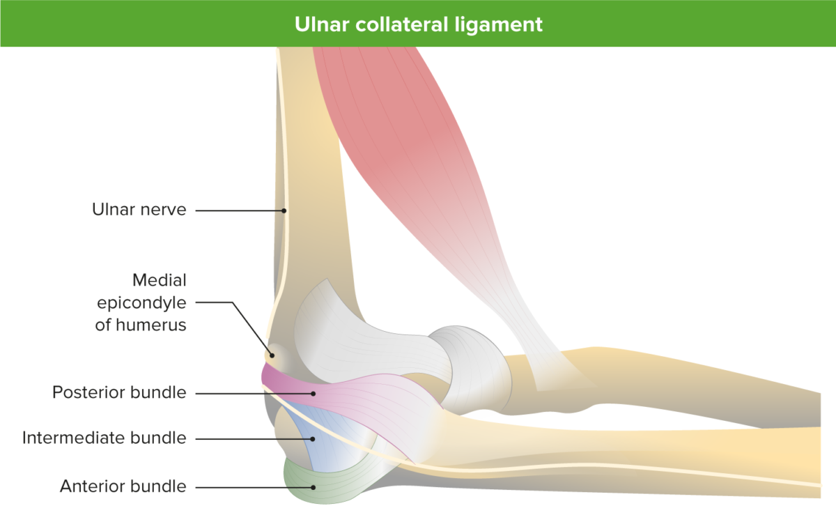
Medial view of the elbow illustrating the 3 bands of the medial ulnar collateral ligament
Image by Lecturio.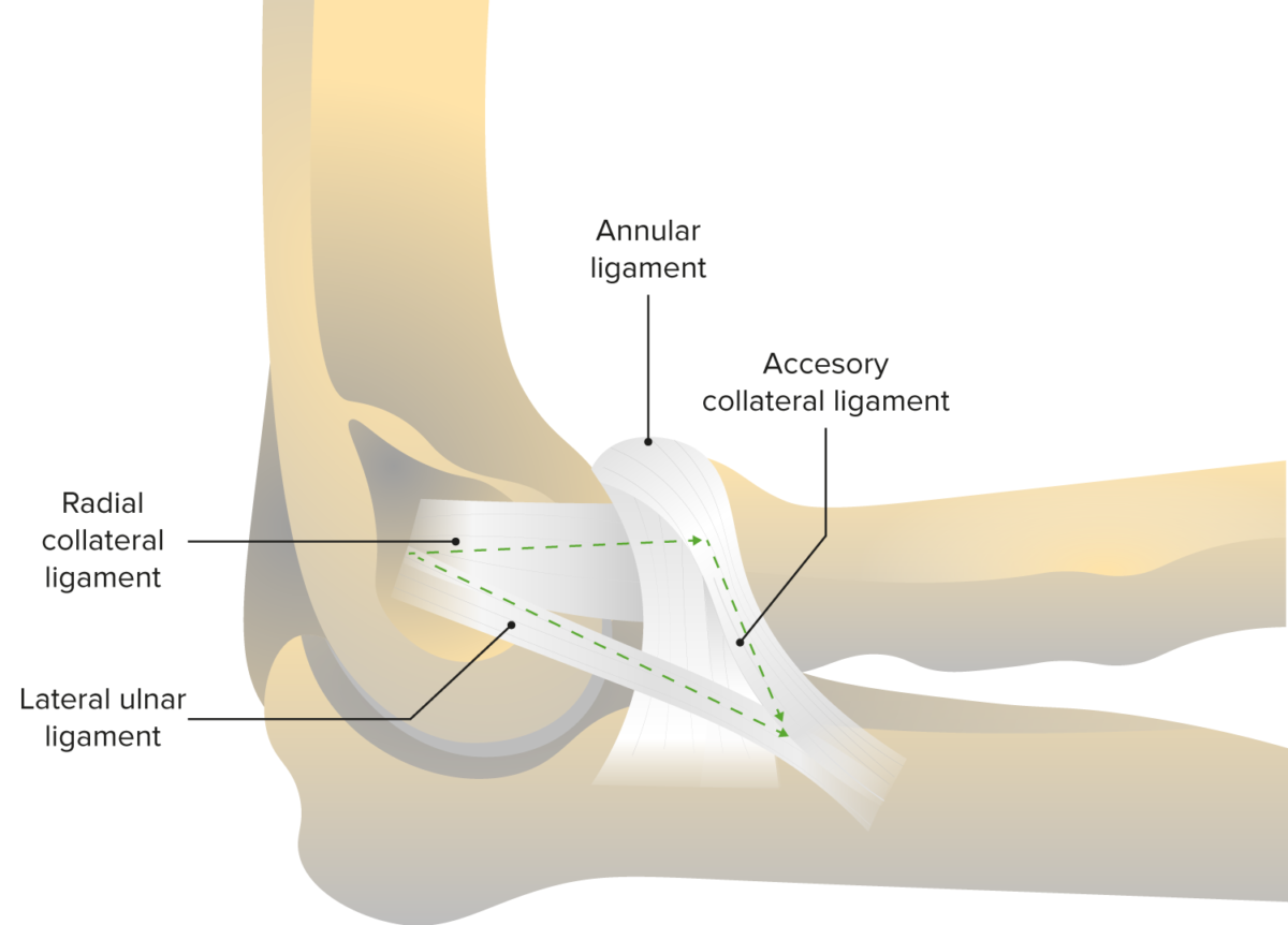
Lateral view of the elbow joint, featuring the annular ligament and the radial collateral ligament
Image by Lecturio.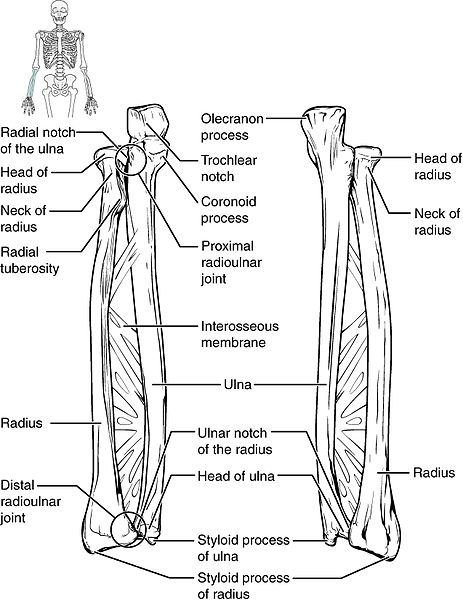
Interosseous membrane of the radius and ulna bones
Image: “Ulna and Radius” by OpenStax College. License: CC BY 3.0The functional anatomy of the elbow is unique secondary to the orientation Orientation Awareness of oneself in relation to time, place and person. Psychiatric Assessment and multiple articulations. The main function of the elbow is to link the shoulder and the hand Hand The hand constitutes the distal part of the upper limb and provides the fine, precise movements needed in activities of daily living. It consists of 5 metacarpal bones and 14 phalanges, as well as numerous muscles innervated by the median and ulnar nerves. Hand: Anatomy and position and stabilize the hand Hand The hand constitutes the distal part of the upper limb and provides the fine, precise movements needed in activities of daily living. It consists of 5 metacarpal bones and 14 phalanges, as well as numerous muscles innervated by the median and ulnar nerves. Hand: Anatomy during activities.
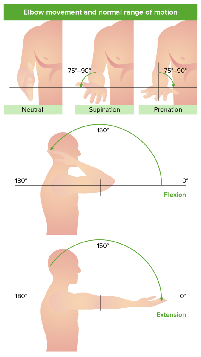
Range of motion and movements of the elbow joint
Image by Lecturio.Carrying angle:
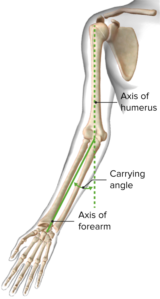
Carrying angle
Image by BioDigital, edited by LecturioThe muscles of the elbow originate in the upper arm Upper Arm The arm, or “upper arm” in common usage, is the region of the upper limb that extends from the shoulder to the elbow joint and connects inferiorly to the forearm through the cubital fossa. It is divided into 2 fascial compartments (anterior and posterior). Arm: Anatomy and insert into the forearm Forearm The forearm is the region of the upper limb between the elbow and the wrist. The term “forearm” is used in anatomy to distinguish this area from the arm, a term that is commonly used to describe the entire upper limb. The forearm consists of 2 long bones (the radius and the ulna), the interosseous membrane, and multiple arteries, nerves, and muscles. Forearm: Anatomy, producing flexion-extension of the elbow as well as supination-pronation of the forearm Forearm The forearm is the region of the upper limb between the elbow and the wrist. The term “forearm” is used in anatomy to distinguish this area from the arm, a term that is commonly used to describe the entire upper limb. The forearm consists of 2 long bones (the radius and the ulna), the interosseous membrane, and multiple arteries, nerves, and muscles. Forearm: Anatomy. The muscles also provide dynamic stabilization to the elbow joint.
| Muscle | Origin | Insertion | Nerve supply | Function |
|---|---|---|---|---|
| Brachialis Brachialis Arm: Anatomy muscle | Anterior aspect of the humerus Humerus Bone in humans and primates extending from the shoulder joint to the elbow joint. Arm: Anatomy lateral to the deltoid tuberosity Deltoid tuberosity Arm: Anatomy | Ulnar tuberosity | Musculocutaneous nerve Musculocutaneous Nerve A major nerve of the upper extremity. The fibers of the musculocutaneous nerve originate in the lower cervical spinal cord (usually C5 to C7), travel via the lateral cord of the brachial plexus, and supply sensory and motor innervation to the upper arm, elbow, and forearm. Axilla and Brachial Plexus: Anatomy (C5–C7) | Flexes the elbow and assists with supination Supination Applies to movements of the forearm in turning the palm forward or upward. When referring to the foot, a combination of adduction and inversion movements of the foot. Examination of the Upper Limbs |
| Brachioradialis Brachioradialis Forearm: Anatomy muscle | Proximal 2 thirds of lateral supracondylar ridge | Lateral surface of distal radius Radius The outer shorter of the two bones of the forearm, lying parallel to the ulna and partially revolving around it. Forearm: Anatomy and pre-styloid process | Radial nerve Radial Nerve A major nerve of the upper extremity. In humans the fibers of the radial nerve originate in the lower cervical and upper thoracic spinal cord (usually C5 to T1), travel via the posterior cord of the brachial plexus, and supply motor innervation to extensor muscles of the arm and cutaneous sensory fibers to extensor regions of the arm and hand. Axilla and Brachial Plexus: Anatomy (C6) | Weak flexor of the elbow, strong flexor when forearm Forearm The forearm is the region of the upper limb between the elbow and the wrist. The term “forearm” is used in anatomy to distinguish this area from the arm, a term that is commonly used to describe the entire upper limb. The forearm consists of 2 long bones (the radius and the ulna), the interosseous membrane, and multiple arteries, nerves, and muscles. Forearm: Anatomy mid-pronated |
| Biceps Biceps Arm: Anatomy brachii muscle | Short head: coracoid process; long head: supraglenoid tubercle | Tuberosity of the radius Radius The outer shorter of the two bones of the forearm, lying parallel to the ulna and partially revolving around it. Forearm: Anatomy | Musculocutaneous nerve Musculocutaneous Nerve A major nerve of the upper extremity. The fibers of the musculocutaneous nerve originate in the lower cervical spinal cord (usually C5 to C7), travel via the lateral cord of the brachial plexus, and supply sensory and motor innervation to the upper arm, elbow, and forearm. Axilla and Brachial Plexus: Anatomy (C5–C6) | Supinates the forearm Forearm The forearm is the region of the upper limb between the elbow and the wrist. The term “forearm” is used in anatomy to distinguish this area from the arm, a term that is commonly used to describe the entire upper limb. The forearm consists of 2 long bones (the radius and the ulna), the interosseous membrane, and multiple arteries, nerves, and muscles. Forearm: Anatomy and assists with elbow flexion Flexion Examination of the Upper Limbs |
| Triceps brachii | Long head: infraglenoid tubercle; lateral and medial heads: posterior humerus Humerus Bone in humans and primates extending from the shoulder joint to the elbow joint. Arm: Anatomy | Olecranon Olecranon A prominent projection of the ulna that articulates with the humerus and forms the outer protuberance of the elbow joint. Arm: Anatomy | Radial nerve Radial Nerve A major nerve of the upper extremity. In humans the fibers of the radial nerve originate in the lower cervical and upper thoracic spinal cord (usually C5 to T1), travel via the posterior cord of the brachial plexus, and supply motor innervation to extensor muscles of the arm and cutaneous sensory fibers to extensor regions of the arm and hand. Axilla and Brachial Plexus: Anatomy (C6–C8) | Extends the elbow |
| Anconeus | Inserts on the posterior aspect of lateral epicondyle Lateral epicondyle Arm: Anatomy | Lateral surface of the olecranon Olecranon A prominent projection of the ulna that articulates with the humerus and forms the outer protuberance of the elbow joint. Arm: Anatomy | Radial nerve Radial Nerve A major nerve of the upper extremity. In humans the fibers of the radial nerve originate in the lower cervical and upper thoracic spinal cord (usually C5 to T1), travel via the posterior cord of the brachial plexus, and supply motor innervation to extensor muscles of the arm and cutaneous sensory fibers to extensor regions of the arm and hand. Axilla and Brachial Plexus: Anatomy (C7, C8) | Assists in extension Extension Examination of the Upper Limbs of the elbow and stabilizes the joint |
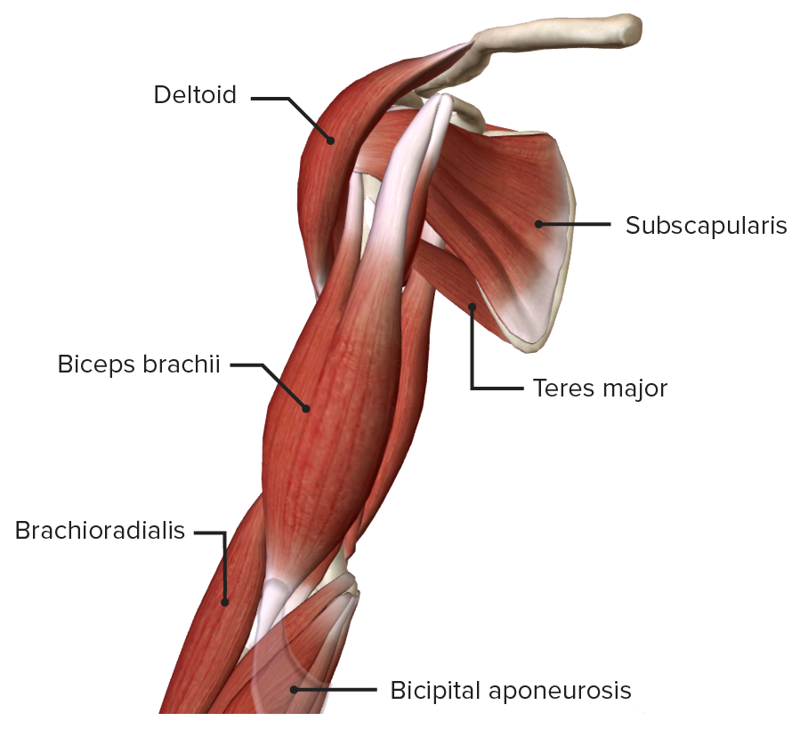
Anterior view of the right arm
Image by BioDigital, edited by Lecturio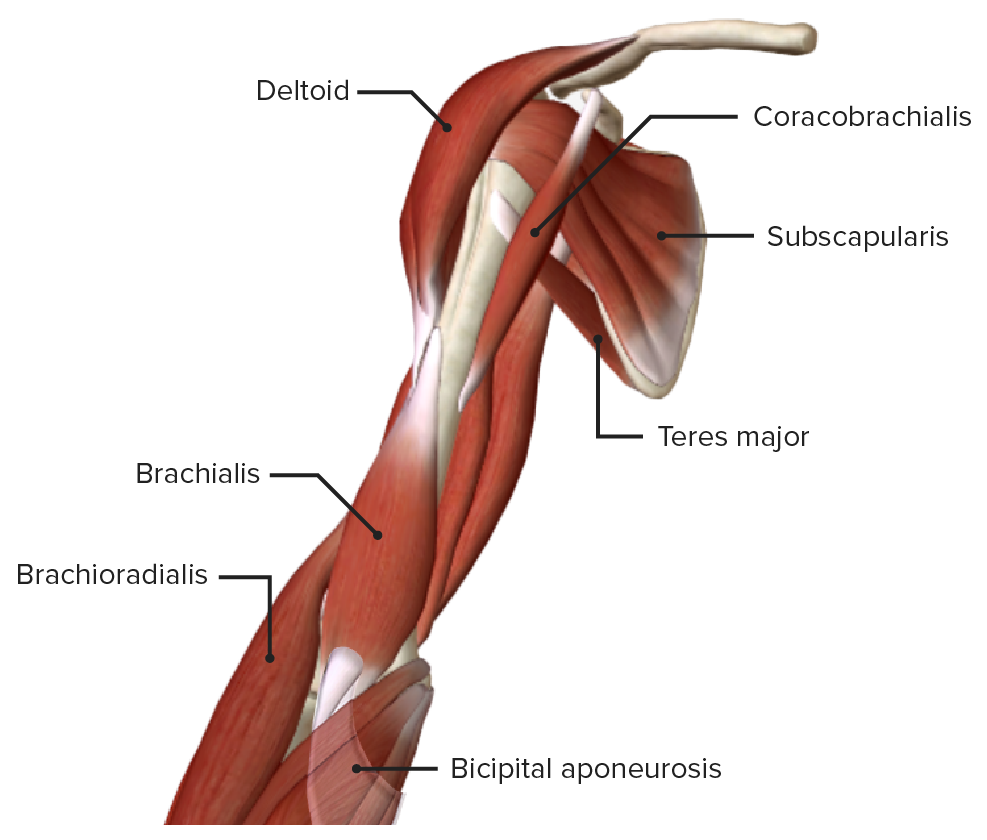
Anterior view of the right arm, featuring the muscles of the anterior compartment with the biceps brachii removed
Image by BioDigital, edited by Lecturio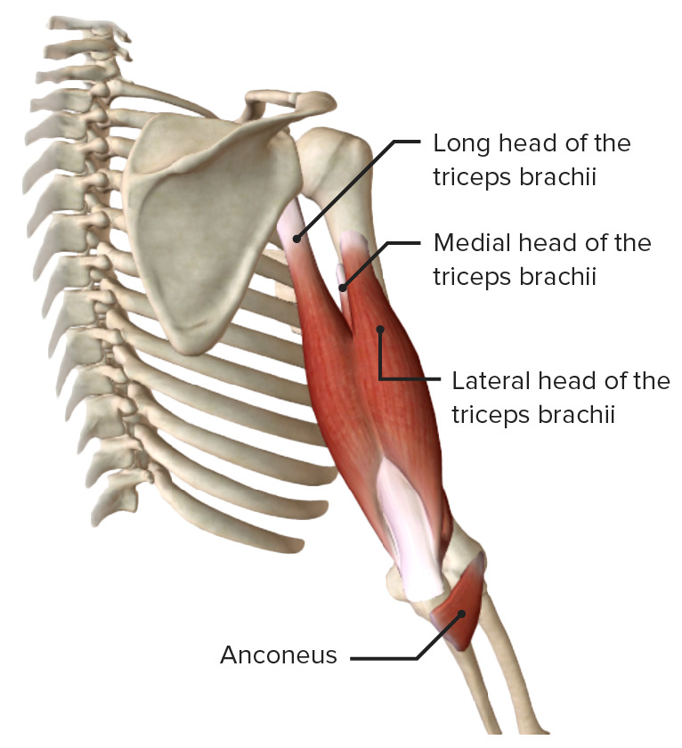
The extensor muscles of the elbow: the triceps brachii and anconeus
Image by BioDigital, edited by Lecturio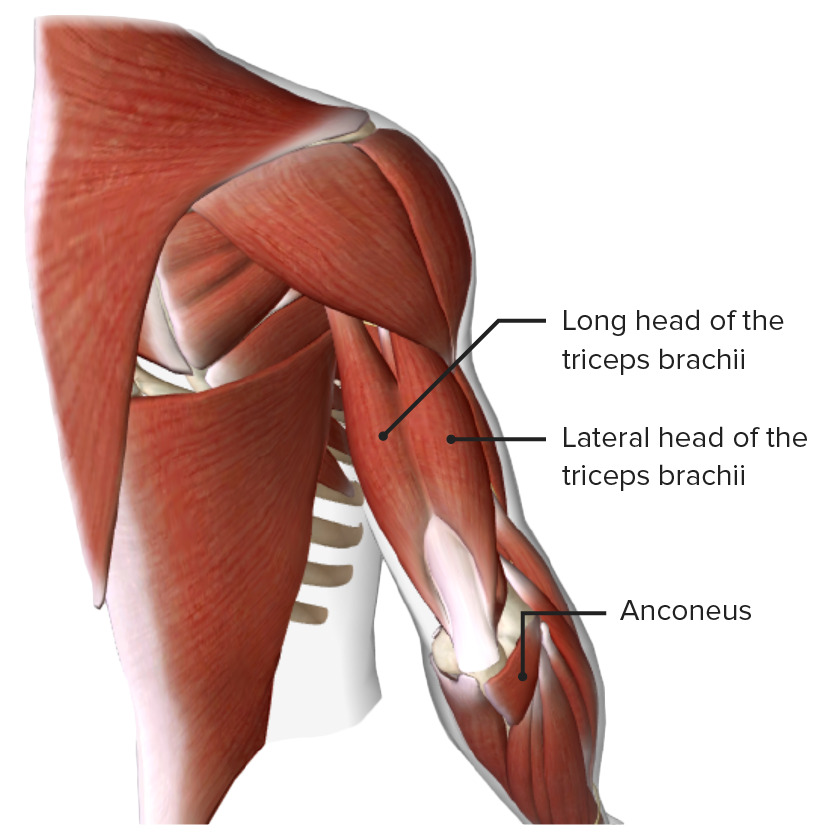
Posterior view of the upper arm, featuring the triceps brachii and anconeus muscles
Image by BioDigital, edited by Lecturio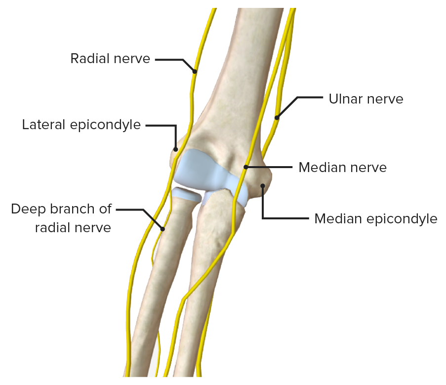
Nerves of the elbow
Image by BioDigital, edited by Lecturio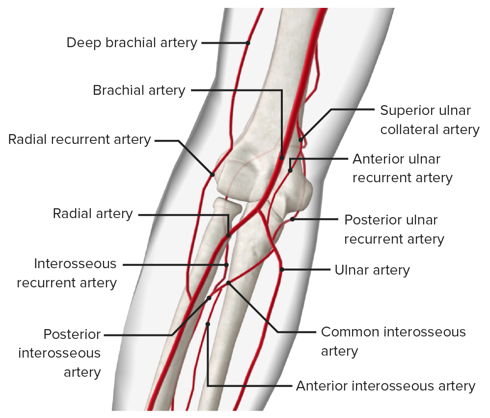
Arteries of the elbow
Image by BioDigital, edited by Lecturio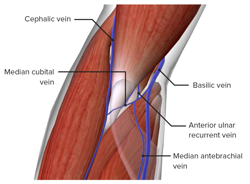
Superficial veins of the elbow
Image by BioDigital, edited by Lecturio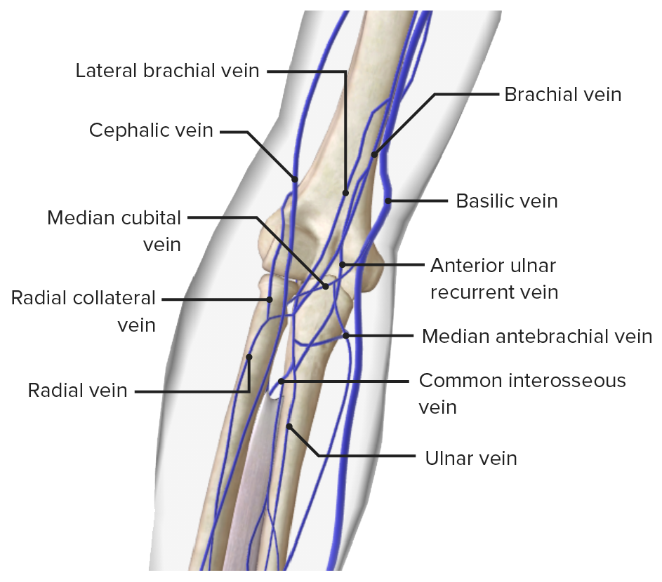
Veins of the elbow
Image by BioDigital, edited by Lecturio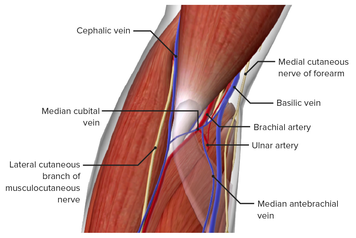
Neurovasculature of the elbow
Image by BioDigital, edited by LecturioEpitrochlear or cubital lymph nodes Cubital lymph nodes Cubital Fossa: Anatomy are found at the elbow and drain proximally to the axillary lymph nodes Lymph Nodes They are oval or bean shaped bodies (1 – 30 mm in diameter) located along the lymphatic system. Lymphatic Drainage System: Anatomy.
The following are common conditions related to the elbow: