Ehrlichiosis and Anaplasmosis

Ehrlichiosis and anaplasmosis are tick-borne bacterial infections Infections Invasion of the host organism by microorganisms or their toxins or by parasites that can cause pathological conditions or diseases. Chronic Granulomatous Disease. The most common causative species include Ehrlichia chaffeensis and Anaplasma phagocytophilum, which infect and multiply within monocytes Monocytes Large, phagocytic mononuclear leukocytes produced in the vertebrate bone marrow and released into the blood; contain a large, oval or somewhat indented nucleus surrounded by voluminous cytoplasm and numerous organelles. Innate Immunity: Phagocytes and Antigen Presentation and granulocytes Granulocytes Leukocytes with abundant granules in the cytoplasm. They are divided into three groups according to the staining properties of the granules: neutrophilic, eosinophilic, and basophilic. Mature granulocytes are the neutrophils; eosinophils; and basophils. White Myeloid Cells: Histology, respectively. The clinical presentation can vary widely, but often includes fever Fever Fever is defined as a measured body temperature of at least 38°C (100.4°F). Fever is caused by circulating endogenous and/or exogenous pyrogens that increase levels of prostaglandin E2 in the hypothalamus. Fever is commonly associated with chills, rigors, sweating, and flushing of the skin. Fever, malaise Malaise Tick-borne Encephalitis Virus, headache Headache The symptom of pain in the cranial region. It may be an isolated benign occurrence or manifestation of a wide variety of headache disorders. Brain Abscess, myalgia Myalgia Painful sensation in the muscles. Ion Channel Myopathy, and arthralgias. A maculopapular Maculopapular Dermatologic Examination or petechial rash Rash Rocky Mountain Spotted Fever occurs in some patients Patients Individuals participating in the health care system for the purpose of receiving therapeutic, diagnostic, or preventive procedures. Clinician–Patient Relationship. Gastrointestinal, neurologic, and respiratory symptoms are also possible. The diagnosis is based on clinical suspicion and confirmed PCR PCR Polymerase chain reaction (PCR) is a technique that amplifies DNA fragments exponentially for analysis. The process is highly specific, allowing for the targeting of specific genomic sequences, even with minuscule sample amounts. The PCR cycles multiple times through 3 phases: denaturation of the template DNA, annealing of a specific primer to the individual DNA strands, and synthesis/elongation of new DNA molecules. Polymerase Chain Reaction (PCR) or antibody testing. Management is with doxycycline.

Last updated: Oct 21, 2022

Editorial responsibility: Stanley Oiseth, Lindsay Jones, Evelin Maza

General Characteristics

Basic features of Ehrlichia and Anaplasma

  • Gram negative Gram negative Bacteria which lose crystal violet stain but are stained pink when treated by gram’s method. Yersinia spp./Yersiniosis
  • Obligate intracellular bacteria Bacteria Bacteria are prokaryotic single-celled microorganisms that are metabolically active and divide by binary fission. Some of these organisms play a significant role in the pathogenesis of diseases. Bacteriology
  • Coccobacilli
  • Grow in membrane-bound vacuoles in leukocytes Leukocytes White blood cells. These include granular leukocytes (basophils; eosinophils; and neutrophils) as well as non-granular leukocytes (lymphocytes and monocytes). White Myeloid Cells: Histology, particularly:
    • Monocytes Monocytes Large, phagocytic mononuclear leukocytes produced in the vertebrate bone marrow and released into the blood; contain a large, oval or somewhat indented nucleus surrounded by voluminous cytoplasm and numerous organelles. Innate Immunity: Phagocytes and Antigen Presentation
    • Granulocytes Granulocytes Leukocytes with abundant granules in the cytoplasm. They are divided into three groups according to the staining properties of the granules: neutrophilic, eosinophilic, and basophilic. Mature granulocytes are the neutrophils; eosinophils; and basophils. White Myeloid Cells: Histology 
  • Light microscopy:
    • Individual organisms are difficult to appreciate.
    • Frequently visualized as morulae (microcolony of organisms within a vacuole)
    • Stains: 
      • Wright
      • Giemsa
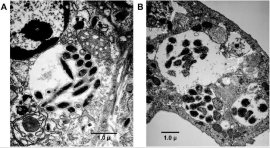
Giemsa stain of anaplasma

Microscopic images of Giemsa-stained cells infected with anaplasmosis:
Cell nuclei are labeled “N” and the arrows point to Anaplasma morulae.

Image: “Giemsa stained cells infected with Ap.” by University of Minnesota, Department of Entomology, Saint Paul, Minnesota 55108, USA. License: CC BY 2.0

Clinically relevant species

The most notable species are:

  • Ehrlichia chaffeensis human monocytic ehrlichiosis (HME)
  • Anaplasma phagocytophilum → human granulocytic anaplasmosis (HGA)

Rare causes of ehrlichiosis in humans:

  • E. ewingii
  • E. muris eauclairensis

Epidemiology and Risk Factors

Epidemiology

  • Incidence Incidence The number of new cases of a given disease during a given period in a specified population. It also is used for the rate at which new events occur in a defined population. It is differentiated from prevalence, which refers to all cases in the population at a given time. Measures of Disease Frequency in the United States:
    • HME: 3.2 cases per million per year
    • HGA: 6.3 cases per million per year
  • Endemic areas in the United States:
    • HME:
      • Southeast
      • South Central
      • Mid-Atlantic
    • HGA:
      • Northeast
      • Upper Midwest and Great Lakes
  • Endemic areas outside the United States:
  • Incidence Incidence The number of new cases of a given disease during a given period in a specified population. It also is used for the rate at which new events occur in a defined population. It is differentiated from prevalence, which refers to all cases in the population at a given time. Measures of Disease Frequency is higher in men than women.
  • Commonly occurs between April and September

Risk factors

For ehrlichiosis/anaplasmosis:

  • Travel to or living in an endemic area
  • Owning pets
  • Participating in outdoor activities in wooded areas:
    • Camping
    • Hiking
    • Hunting

For severe disease:

Pathogenesis

Transmission

  • Tick vectors:
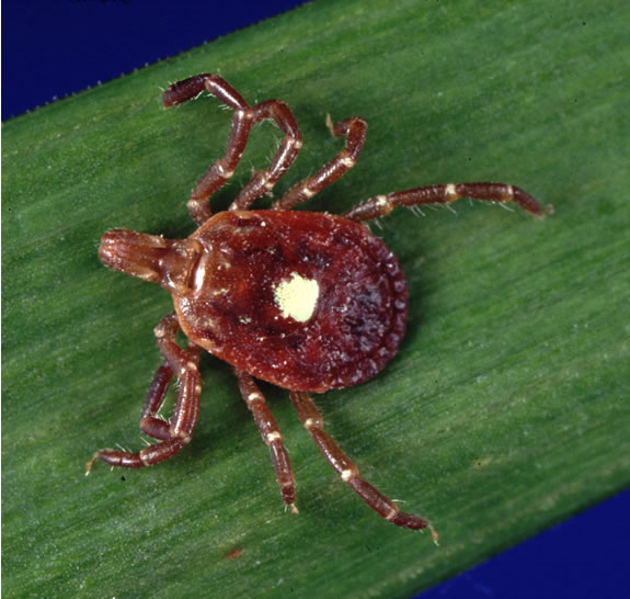
    • E. chaffeensis is transmitted by Amblyomma Amblyomma A genus of hardbacked ticks, in the family Ixodidae. It includes the lone star tick, Amblyomma americanum, and the Gulf Coast tick, Amblyomma maculatum, in the Americas. Amblyomma americanum is the primary vector of Ehrlichia chaffeensis and Ehrlichia ewingii which cause Ehrlichiosis. Other Amblyomma-associated diseases include tularemia and rickettsiosis. Rickettsia americanum ( Lone Star tick Lone Star Tick Rocky Mountain Spotted Fever).
    • A. phagocytophilum is transmitted by Ixodes ticks Ticks Blood-sucking acarid parasites of the order ixodida comprising two families: the softbacked ticks (argasidae) and hardbacked ticks (ixodidae). Ticks are larger than their relatives, the mites. They penetrate the skin of their host by means of highly specialized, hooked mouth parts and feed on its blood. Ticks attack all groups of terrestrial vertebrates. In humans they are responsible for many tick-borne diseases, including the transmission of rocky mountain spotted fever; tularemia; babesiosis; african swine fever; and relapsing fever. Coxiella/Q Fever.
  • Iatrogenic Iatrogenic Any adverse condition in a patient occurring as the result of treatment by a physician, surgeon, or other health professional, especially infections acquired by a patient during the course of treatment. Anterior Cord Syndrome (rare):
    • Blood transfusion 
    • Solid organ transplantation Organ Transplantation Transplantation is a procedure that involves the removal of an organ or living tissue and placing it into a different part of the body or into a different person. Organ transplantations have become the therapeutic option of choice for many individuals with end-stage organ failure. Organ Transplantation

Reservoirs

A wide range of wild and domestic animals Animals Unicellular or multicellular, heterotrophic organisms, that have sensation and the power of voluntary movement. Under the older five kingdom paradigm, animalia was one of the kingdoms. Under the modern three domain model, animalia represents one of the many groups in the domain eukaryota. Cell Types: Eukaryotic versus Prokaryotic can serve as reservoirs. The most notable are:

  • White-tailed deer (E. chaffeensis)
  • White-footed mice (A. phagocytophilum)

Virulence factors Virulence factors Those components of an organism that determine its capacity to cause disease but are not required for its viability per se. Two classes have been characterized: toxins, biological and surface adhesion molecules that affect the ability of the microorganism to invade and colonize a host. Haemophilus

  • Surface proteins Proteins Linear polypeptides that are synthesized on ribosomes and may be further modified, crosslinked, cleaved, or assembled into complex proteins with several subunits. The specific sequence of amino acids determines the shape the polypeptide will take, during protein folding, and the function of the protein. Energy Homeostasis:
    • Allow binding to host cells for entry
    • Antigenic variation helps with evading a host’s immune system Immune system The body’s defense mechanism against foreign organisms or substances and deviant native cells. It includes the humoral immune response and the cell-mediated response and consists of a complex of interrelated cellular, molecular, and genetic components. Primary Lymphatic Organs.
  • Bacterial effector proteins Proteins Linear polypeptides that are synthesized on ribosomes and may be further modified, crosslinked, cleaved, or assembled into complex proteins with several subunits. The specific sequence of amino acids determines the shape the polypeptide will take, during protein folding, and the function of the protein. Energy Homeostasis:
    • Translocate into a host cell
    • Modify the cell’s cytoskeleton Cytoskeleton The network of filaments, tubules, and interconnecting filamentous bridges which give shape, structure, and organization to the cytoplasm. The Cell: Cytosol and Cytoskeleton to allow bacterial entry
    • Allow bacteria-containing vacuoles to avoid lysosome fusion
    • Alter gene Gene A category of nucleic acid sequences that function as units of heredity and which code for the basic instructions for the development, reproduction, and maintenance of organisms. Basic Terms of Genetics expression in the host cell nucleus Nucleus Within a eukaryotic cell, a membrane-limited body which contains chromosomes and one or more nucleoli (cell nucleolus). The nuclear membrane consists of a double unit-type membrane which is perforated by a number of pores; the outermost membrane is continuous with the endoplasmic reticulum. A cell may contain more than one nucleus. The Cell: Organelles

Pathophysiology

Human monocytic ehrlichiosis:

  • E. chaffeensis is introduced into the skin Skin The skin, also referred to as the integumentary system, is the largest organ of the body. The skin is primarily composed of the epidermis (outer layer) and dermis (deep layer). The epidermis is primarily composed of keratinocytes that undergo rapid turnover, while the dermis contains dense layers of connective tissue. Skin: Structure and Functions during a tick bite.
  • Infects monocytes Monocytes Large, phagocytic mononuclear leukocytes produced in the vertebrate bone marrow and released into the blood; contain a large, oval or somewhat indented nucleus surrounded by voluminous cytoplasm and numerous organelles. Innate Immunity: Phagocytes and Antigen Presentation and macrophages Macrophages The relatively long-lived phagocytic cell of mammalian tissues that are derived from blood monocytes. Main types are peritoneal macrophages; alveolar macrophages; histiocytes; kupffer cells of the liver; and osteoclasts. They may further differentiate within chronic inflammatory lesions to epithelioid cells or may fuse to form foreign body giant cells or langhans giant cells. Innate Immunity: Phagocytes and Antigen Presentation
  • Spreads through the lymphatics or blood to:
    • Bone marrow Bone marrow The soft tissue filling the cavities of bones. Bone marrow exists in two types, yellow and red. Yellow marrow is found in the large cavities of large bones and consists mostly of fat cells and a few primitive blood cells. Red marrow is a hematopoietic tissue and is the site of production of erythrocytes and granular leukocytes. Bone marrow is made up of a framework of connective tissue containing branching fibers with the frame being filled with marrow cells. Bone Marrow: Composition and Hematopoiesis
    • Liver Liver The liver is the largest gland in the human body. The liver is found in the superior right quadrant of the abdomen and weighs approximately 1.5 kilograms. Its main functions are detoxification, metabolism, nutrient storage (e.g., iron and vitamins), synthesis of coagulation factors, formation of bile, filtration, and storage of blood. Liver: Anatomy
    • Lymph nodes Lymph Nodes They are oval or bean shaped bodies (1 – 30 mm in diameter) located along the lymphatic system. Lymphatic Drainage System: Anatomy
    • Spleen Spleen The spleen is the largest lymphoid organ in the body, located in the LUQ of the abdomen, superior to the left kidney and posterior to the stomach at the level of the 9th-11th ribs just below the diaphragm. The spleen is highly vascular and acts as an important blood filter, cleansing the blood of pathogens and damaged erythrocytes. Spleen: Anatomy
  • The clinical presentation is due to the effects of the host’s inflammatory response.

Human granulocytic anaplasmosis:

  • A. phagocytophilum is introduced into the skin Skin The skin, also referred to as the integumentary system, is the largest organ of the body. The skin is primarily composed of the epidermis (outer layer) and dermis (deep layer). The epidermis is primarily composed of keratinocytes that undergo rapid turnover, while the dermis contains dense layers of connective tissue. Skin: Structure and Functions during a tick bite → neutrophil recruitment Recruitment Skeletal Muscle Contraction 
  • A. phagocytophilum enters neutrophils Neutrophils Granular leukocytes having a nucleus with three to five lobes connected by slender threads of chromatin, and cytoplasm containing fine inconspicuous granules and stainable by neutral dyes. Innate Immunity: Phagocytes and Antigen Presentation:
    • Alters intracellular killing
    • Induces neutrophil activation and cytokine release Release Release of a virus from the host cell following virus assembly and maturation. Egress can occur by host cell lysis, exocytosis, or budding through the plasma membrane. Virology → contributes to tissue injury and clinical manifestations
  • Infection spreads hematogenously.

Clinical Presentation

Signs and symptoms

The incubation Incubation The amount time between exposure to an infectious agent and becoming symptomatic. Rabies Virus period is typically 1–2 weeks, and the clinical presentation can vary greatly.

Features of both HME and HGA:

  • Nonspecific symptoms: 
    • Fever Fever Fever is defined as a measured body temperature of at least 38°C (100.4°F). Fever is caused by circulating endogenous and/or exogenous pyrogens that increase levels of prostaglandin E2 in the hypothalamus. Fever is commonly associated with chills, rigors, sweating, and flushing of the skin. Fever 
    • Malaise Malaise Tick-borne Encephalitis Virus 
    • Headache Headache The symptom of pain in the cranial region. It may be an isolated benign occurrence or manifestation of a wide variety of headache disorders. Brain Abscess 
    • Myalgia Myalgia Painful sensation in the muscles. Ion Channel Myopathy
    • Arthralgia Arthralgia Pain in the joint. Rheumatic Fever
  • GI symptoms: 
    • Abdominal pain Abdominal Pain Acute Abdomen 
    • Nausea Nausea An unpleasant sensation in the stomach usually accompanied by the urge to vomit. Common causes are early pregnancy, sea and motion sickness, emotional stress, intense pain, food poisoning, and various enteroviruses. Antiemetics
    • Vomiting Vomiting The forcible expulsion of the contents of the stomach through the mouth. Hypokalemia
    • Diarrhea Diarrhea Diarrhea is defined as ≥ 3 watery or loose stools in a 24-hour period. There are a multitude of etiologies, which can be classified based on the underlying mechanism of disease. The duration of symptoms (acute or chronic) and characteristics of the stools (e.g., watery, bloody, steatorrheic, mucoid) can help guide further diagnostic evaluation. Diarrhea
  • Respiratory symptoms: 
    • Cough
    • Shortness of breath Shortness of breath Dyspnea is the subjective sensation of breathing discomfort. Dyspnea is a normal manifestation of heavy physical or psychological exertion, but also may be caused by underlying conditions (both pulmonary and extrapulmonary). Dyspnea

Findings more commonly seen in HME:

Complications

  • Seizures Seizures A seizure is abnormal electrical activity of the neurons in the cerebral cortex that can manifest in numerous ways depending on the region of the brain affected. Seizures consist of a sudden imbalance that occurs between the excitatory and inhibitory signals in cortical neurons, creating a net excitation. The 2 major classes of seizures are focal and generalized. Seizures
  • ARDS
  • Myocarditis Myocarditis Myocarditis is an inflammatory disease of the myocardium, which may occur alone or in association with a systemic process. There are numerous etiologies of myocarditis, but all lead to inflammation and myocyte injury, most often leading to signs and symptoms of heart failure. Myocarditis and heart failure Heart Failure A heterogeneous condition in which the heart is unable to pump out sufficient blood to meet the metabolic need of the body. Heart failure can be caused by structural defects, functional abnormalities (ventricular dysfunction), or a sudden overload beyond its capacity. Chronic heart failure is more common than acute heart failure which results from sudden insult to cardiac function, such as myocardial infarction. Total Anomalous Pulmonary Venous Return (TAPVR)
  • Pericardial effusion Pericardial effusion Fluid accumulation within the pericardium. Serous effusions are associated with pericardial diseases. Hemopericardium is associated with trauma. Lipid-containing effusion (chylopericardium) results from leakage of thoracic duct. Severe cases can lead to cardiac tamponade. Pericardial Effusion and Cardiac Tamponade and tamponade Tamponade Pericardial effusion, usually of rapid onset, exceeding ventricular filling pressures and causing collapse of the heart with a markedly reduced cardiac output. Pericarditis
  • Renal failure Renal failure Conditions in which the kidneys perform below the normal level in the ability to remove wastes, concentrate urine, and maintain electrolyte balance; blood pressure; and calcium metabolism. Renal insufficiency can be classified by the degree of kidney damage (as measured by the level of proteinuria) and reduction in glomerular filtration rate. Crush Syndrome
  • Coagulopathy
  • Septic shock Septic shock Sepsis associated with hypotension or hypoperfusion despite adequate fluid resuscitation. Perfusion abnormalities may include, but are not limited to lactic acidosis; oliguria; or acute alteration in mental status. Sepsis and Septic Shock
  • Hemophagocytic lymphohistiocytosis Hemophagocytic lymphohistiocytosis A group of related disorders characterized by lymphocytosis; histiocytosis; and hemophagocytosis. The two major forms are familial and reactive. Epstein-Barr Virus (rare, severe complication of HGA)
  • Opportunistic infections Infections Invasion of the host organism by microorganisms or their toxins or by parasites that can cause pathological conditions or diseases. Chronic Granulomatous Disease

Diagnosis and Management

Diagnosis

Definitive studies:

  • Indirect fluorescent antibody Indirect fluorescent antibody A form of fluorescent antibody technique commonly used to detect serum antibodies and immune complexes in tissues and microorganisms in specimens from patients with infectious diseases. The technique involves formation of an antigen-antibody complex which is labeled with fluorescein-conjugated anti-immunoglobulin antibody. Chikungunya Virus (IFA) testing
  • ELISA ELISA An immunoassay utilizing an antibody labeled with an enzyme marker such as horseradish peroxidase. While either the enzyme or the antibody is bound to an immunosorbent substrate, they both retain their biologic activity; the change in enzyme activity as a result of the enzyme-antibody-antigen reaction is proportional to the concentration of the antigen and can be measured spectrophotometrically or with the naked eye. Many variations of the method have been developed. St. Louis Encephalitis Virus antibody testing
  • PCR PCR Polymerase chain reaction (PCR) is a technique that amplifies DNA fragments exponentially for analysis. The process is highly specific, allowing for the targeting of specific genomic sequences, even with minuscule sample amounts. The PCR cycles multiple times through 3 phases: denaturation of the template DNA, annealing of a specific primer to the individual DNA strands, and synthesis/elongation of new DNA molecules. Polymerase Chain Reaction (PCR) to detect organism DNA DNA A deoxyribonucleotide polymer that is the primary genetic material of all cells. Eukaryotic and prokaryotic organisms normally contain DNA in a double-stranded state, yet several important biological processes transiently involve single-stranded regions. DNA, which consists of a polysugar-phosphate backbone possessing projections of purines (adenine and guanine) and pyrimidines (thymine and cytosine), forms a double helix that is held together by hydrogen bonds between these purines and pyrimidines (adenine to thymine and guanine to cytosine). DNA Types and Structure
  • Buffy coat or peripheral blood smear Peripheral Blood Smear Anemia: Overview and Types examination: Intracytoplasmic morulae are seen in peripheral blood leukocytes Leukocytes White blood cells. These include granular leukocytes (basophils; eosinophils; and neutrophils) as well as non-granular leukocytes (lymphocytes and monocytes). White Myeloid Cells: Histology.

Supporting laboratory studies:

  • Thrombocytopenia Thrombocytopenia Thrombocytopenia occurs when the platelet count is < 150,000 per microliter. The normal range for platelets is usually 150,000-450,000/µL of whole blood. Thrombocytopenia can be a result of decreased production, increased destruction, or splenic sequestration of platelets. Patients are often asymptomatic until platelet counts are < 50,000/µL. Thrombocytopenia
  • Leukopenia
  • Anemia Anemia Anemia is a condition in which individuals have low Hb levels, which can arise from various causes. Anemia is accompanied by a reduced number of RBCs and may manifest with fatigue, shortness of breath, pallor, and weakness. Subtypes are classified by the size of RBCs, chronicity, and etiology. Anemia: Overview and Types
  • AST AST Enzymes of the transferase class that catalyze the conversion of l-aspartate and 2-ketoglutarate to oxaloacetate and l-glutamate. Liver Function Tests and ALT ALT An enzyme that catalyzes the conversion of l-alanine and 2-oxoglutarate to pyruvate and l-glutamate. Liver Function Tests
  • Alkaline phosphatase Alkaline Phosphatase An enzyme that catalyzes the conversion of an orthophosphoric monoester and water to an alcohol and orthophosphate. Osteosarcoma
  • Lactate dehydrogenase Lactate Dehydrogenase Osteosarcoma
  • ↑ Creatinine
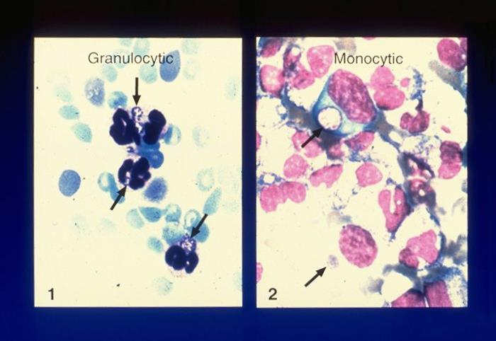
Granocytic and monocytic agents of ehrlichioses

Peripheral blood smear evaluations for ehrlichiosis and anaplasmosis:
These blood smears show intracellular morulae of A. phagocytophilum (1) and E. chaffeensis (2).

Image: “Etiologic agents of ehrlichioses” by CDC/NCID. License: Public Domain

Management

Doxycycline is the antibiotic of choice.

  • Treatment should be initiated in patients Patients Individuals participating in the health care system for the purpose of receiving therapeutic, diagnostic, or preventive procedures. Clinician–Patient Relationship with suspected HME or HGA while laboratory testing is pending.
  • If patients Patients Individuals participating in the health care system for the purpose of receiving therapeutic, diagnostic, or preventive procedures. Clinician–Patient Relationship do not improve with therapy, they should be evaluated for a concurrent Babesia Babesia Babesiosis is an infection caused by a protozoa belonging to the genus, Babesia. The most common Babesia seen in the United States is B. microti, which is transmitted by the Ixodes tick. The protozoa thrive and replicate within host erythrocytes. Lysis of erythrocytes and the body’s immune response result in clinical symptoms. Babesia/Babesiosis infection.

Prevention

Avoiding tick bites is key to preventing these diseases.

  • Wearing appropriate protective clothing
  • Using tick repellents
  • Inspecting for ticks Ticks Blood-sucking acarid parasites of the order ixodida comprising two families: the softbacked ticks (argasidae) and hardbacked ticks (ixodidae). Ticks are larger than their relatives, the mites. They penetrate the skin of their host by means of highly specialized, hooked mouth parts and feed on its blood. Ticks attack all groups of terrestrial vertebrates. In humans they are responsible for many tick-borne diseases, including the transmission of rocky mountain spotted fever; tularemia; babesiosis; african swine fever; and relapsing fever. Coxiella/Q Fever after outdoor activity
  • Removing any attached ticks Ticks Blood-sucking acarid parasites of the order ixodida comprising two families: the softbacked ticks (argasidae) and hardbacked ticks (ixodidae). Ticks are larger than their relatives, the mites. They penetrate the skin of their host by means of highly specialized, hooked mouth parts and feed on its blood. Ticks attack all groups of terrestrial vertebrates. In humans they are responsible for many tick-borne diseases, including the transmission of rocky mountain spotted fever; tularemia; babesiosis; african swine fever; and relapsing fever. Coxiella/Q Fever to reduce the risk of infection
  • Tick control on domestic animals Animals Unicellular or multicellular, heterotrophic organisms, that have sensation and the power of voluntary movement. Under the older five kingdom paradigm, animalia was one of the kingdoms. Under the modern three domain model, animalia represents one of the many groups in the domain eukaryota. Cell Types: Eukaryotic versus Prokaryotic
  • Doxycycline prophylaxis Doxycycline Prophylaxis Lyme Disease after a tick bite is not recommended.

Comparison of Gram-negative Tick-borne Bacteria

Table: Comparison of several clinically relevant gram-negative tick-borne bacteria Bacteria Bacteria are prokaryotic single-celled microorganisms that are metabolically active and divide by binary fission. Some of these organisms play a significant role in the pathogenesis of diseases. Bacteriology
Organism Ehrlichia chaffeensis Anaplasma phagocytophilum Rickettsia rickettsii Rickettsia rickettsii A species of gram-negative, aerobic bacteria that is the etiologic agent of rocky mountain spotted fever. Its cells are slightly smaller and more uniform in size than those of rickettsia prowazekii. Rickettsia Borrelia burgdorferi Borrelia burgdorferi A specific species of bacteria, part of the borrelia burgdorferi group, whose common name is lyme disease spirochete. Borrelia
Disease HME HGA Rocky Mountain spotted fever Fever Fever is defined as a measured body temperature of at least 38°C (100.4°F). Fever is caused by circulating endogenous and/or exogenous pyrogens that increase levels of prostaglandin E2 in the hypothalamus. Fever is commonly associated with chills, rigors, sweating, and flushing of the skin. Fever Lyme disease Lyme disease Lyme disease is a tick-borne infection caused by the gram-negative spirochete Borrelia burgdorferi. Lyme disease is transmitted by the black-legged Ixodes tick (known as a deer tick), which is only found in specific geographic regions. Patient presentation can vary depending on the stage of the disease and may include a characteristic erythema migrans rash. Lyme Disease
Micro
  • Obligate intracellular
  • Coccobacilli
  • Obligate intracellular
  • Coccobacilli
  • Microaerophilic Microaerophilic Helicobacter
  • Spirochete Spirochete Treponema is a gram-negative, microaerophilic spirochete. Owing to its very thin structure, it is not easily seen on Gram stain, but can be visualized using dark-field microscopy. This spirochete contains endoflagella, which allow for a characteristic corkscrew movement. Treponema
  • Difficult to visualize on Gram stain Gram stain Klebsiella
Vector Lone Star tick Lone Star Tick Rocky Mountain Spotted Fever Ixodes tick Dermacentor Dermacentor A widely distributed genus of ticks, in the family ixodidae, including a number that infest humans and other mammals. Several are vectors of diseases such as tularemia; rocky mountain spotted fever; colorado tick fever; and anaplasmosis. Rickettsia tick Ixodes tick
Reservoir Reservoir Animate or inanimate sources which normally harbor disease-causing organisms and thus serve as potential sources of disease outbreaks. Reservoirs are distinguished from vectors (disease vectors) and carriers, which are agents of disease transmission rather than continuing sources of potential disease outbreaks. Humans may serve both as disease reservoirs and carriers. Escherichia coli White-tailed deer White-footed mouse Dermacentor Dermacentor A widely distributed genus of ticks, in the family ixodidae, including a number that infest humans and other mammals. Several are vectors of diseases such as tularemia; rocky mountain spotted fever; colorado tick fever; and anaplasmosis. Rickettsia tick
  • Rodents
  • Deer
Geographical distribution in the United States Southeast and South Central states Northeast and upper Midwest states Southeast and South Central states Northeast and Midwest states
Diagnosis
  • PCR PCR Polymerase chain reaction (PCR) is a technique that amplifies DNA fragments exponentially for analysis. The process is highly specific, allowing for the targeting of specific genomic sequences, even with minuscule sample amounts. The PCR cycles multiple times through 3 phases: denaturation of the template DNA, annealing of a specific primer to the individual DNA strands, and synthesis/elongation of new DNA molecules. Polymerase Chain Reaction (PCR)
  • ELISA ELISA An immunoassay utilizing an antibody labeled with an enzyme marker such as horseradish peroxidase. While either the enzyme or the antibody is bound to an immunosorbent substrate, they both retain their biologic activity; the change in enzyme activity as a result of the enzyme-antibody-antigen reaction is proportional to the concentration of the antigen and can be measured spectrophotometrically or with the naked eye. Many variations of the method have been developed. St. Louis Encephalitis Virus
  • Immunofluorescent antibody (IFA)
  • Blood smear Blood smear Myeloperoxidase Deficiency
  • PCR PCR Polymerase chain reaction (PCR) is a technique that amplifies DNA fragments exponentially for analysis. The process is highly specific, allowing for the targeting of specific genomic sequences, even with minuscule sample amounts. The PCR cycles multiple times through 3 phases: denaturation of the template DNA, annealing of a specific primer to the individual DNA strands, and synthesis/elongation of new DNA molecules. Polymerase Chain Reaction (PCR)
  • ELISA ELISA An immunoassay utilizing an antibody labeled with an enzyme marker such as horseradish peroxidase. While either the enzyme or the antibody is bound to an immunosorbent substrate, they both retain their biologic activity; the change in enzyme activity as a result of the enzyme-antibody-antigen reaction is proportional to the concentration of the antigen and can be measured spectrophotometrically or with the naked eye. Many variations of the method have been developed. St. Louis Encephalitis Virus
  • IFA
  • Blood smear Blood smear Myeloperoxidase Deficiency
  • Clinical
  • Skin biopsy Skin Biopsy Secondary Skin Lesions
  • IFA
  • PCR PCR Polymerase chain reaction (PCR) is a technique that amplifies DNA fragments exponentially for analysis. The process is highly specific, allowing for the targeting of specific genomic sequences, even with minuscule sample amounts. The PCR cycles multiple times through 3 phases: denaturation of the template DNA, annealing of a specific primer to the individual DNA strands, and synthesis/elongation of new DNA molecules. Polymerase Chain Reaction (PCR)
  • Clinical
  • ELISA ELISA An immunoassay utilizing an antibody labeled with an enzyme marker such as horseradish peroxidase. While either the enzyme or the antibody is bound to an immunosorbent substrate, they both retain their biologic activity; the change in enzyme activity as a result of the enzyme-antibody-antigen reaction is proportional to the concentration of the antigen and can be measured spectrophotometrically or with the naked eye. Many variations of the method have been developed. St. Louis Encephalitis Virus
  • Western blot
Management Doxycycline
  • Doxycycline
  • Amoxicillin Amoxicillin A broad-spectrum semisynthetic antibiotic similar to ampicillin except that its resistance to gastric acid permits higher serum levels with oral administration. Penicillins
  • Ceftriaxone Ceftriaxone A broad-spectrum cephalosporin antibiotic and cefotaxime derivative with a very long half-life and high penetrability to meninges, eyes and inner ears. Cephalosporins

IFA: immunofluorescent antibody

Differential Diagnosis

  • Babesiosis Babesiosis Babesiosis is an infection caused by a protozoa belonging to the genus, Babesia. The most common Babesia seen in the United States is B. microti, which is transmitted by the Ixodes tick. The protozoa thrive and replicate within host erythrocytes. Lysis of erythrocytes and the body’s immune response result in clinical symptoms. Babesia/Babesiosis: a tick-borne infection caused by Babesia Babesia Babesiosis is an infection caused by a protozoa belonging to the genus, Babesia. The most common Babesia seen in the United States is B. microti, which is transmitted by the Ixodes tick. The protozoa thrive and replicate within host erythrocytes. Lysis of erythrocytes and the body’s immune response result in clinical symptoms. Babesia/Babesiosis. Patients Patients Individuals participating in the health care system for the purpose of receiving therapeutic, diagnostic, or preventive procedures. Clinician–Patient Relationship can be asymptomatic or develop fever Fever Fever is defined as a measured body temperature of at least 38°C (100.4°F). Fever is caused by circulating endogenous and/or exogenous pyrogens that increase levels of prostaglandin E2 in the hypothalamus. Fever is commonly associated with chills, rigors, sweating, and flushing of the skin. Fever, fatigue Fatigue The state of weariness following a period of exertion, mental or physical, characterized by a decreased capacity for work and reduced efficiency to respond to stimuli. Fibromyalgia, malaise Malaise Tick-borne Encephalitis Virus, and arthralgias. Asplenic, immunocompromised immunocompromised A human or animal whose immunologic mechanism is deficient because of an immunodeficiency disorder or other disease or as the result of the administration of immunosuppressive drugs or radiation. Gastroenteritis, and elderly patients Patients Individuals participating in the health care system for the purpose of receiving therapeutic, diagnostic, or preventive procedures. Clinician–Patient Relationship are at risk for severe disease, causing hemolytic anemia Hemolytic Anemia Hemolytic anemia (HA) is the term given to a large group of anemias that are caused by the premature destruction/hemolysis of circulating red blood cells (RBCs). Hemolysis can occur within (intravascular hemolysis) or outside the blood vessels (extravascular hemolysis). Hemolytic Anemia, thrombocytopenia Thrombocytopenia Thrombocytopenia occurs when the platelet count is < 150,000 per microliter. The normal range for platelets is usually 150,000-450,000/µL of whole blood. Thrombocytopenia can be a result of decreased production, increased destruction, or splenic sequestration of platelets. Patients are often asymptomatic until platelet counts are < 50,000/µL. Thrombocytopenia, hepatosplenomegaly Hepatosplenomegaly Cytomegalovirus, renal failure Renal failure Conditions in which the kidneys perform below the normal level in the ability to remove wastes, concentrate urine, and maintain electrolyte balance; blood pressure; and calcium metabolism. Renal insufficiency can be classified by the degree of kidney damage (as measured by the level of proteinuria) and reduction in glomerular filtration rate. Crush Syndrome, and death. Diagnosis is confirmed with a peripheral blood smear Peripheral Blood Smear Anemia: Overview and Types, serologic testing, and PCR PCR Polymerase chain reaction (PCR) is a technique that amplifies DNA fragments exponentially for analysis. The process is highly specific, allowing for the targeting of specific genomic sequences, even with minuscule sample amounts. The PCR cycles multiple times through 3 phases: denaturation of the template DNA, annealing of a specific primer to the individual DNA strands, and synthesis/elongation of new DNA molecules. Polymerase Chain Reaction (PCR). Management includes antimicrobials such as atovaquone Atovaquone A hydroxynaphthoquinone that has antimicrobial activity and is being used in antimalarial protocols. Antimalarial Drugs plus azithromycin Azithromycin A semi-synthetic macrolide antibiotic structurally related to erythromycin. It has been used in the treatment of Mycobacterium avium intracellulare infections, toxoplasmosis, and cryptosporidiosis. Macrolides and Ketolides.
  • Rocky Mountain spotted fever Fever Fever is defined as a measured body temperature of at least 38°C (100.4°F). Fever is caused by circulating endogenous and/or exogenous pyrogens that increase levels of prostaglandin E2 in the hypothalamus. Fever is commonly associated with chills, rigors, sweating, and flushing of the skin. Fever: a disease caused by Rickettsia rickettsii Rickettsia rickettsii A species of gram-negative, aerobic bacteria that is the etiologic agent of rocky mountain spotted fever. Its cells are slightly smaller and more uniform in size than those of rickettsia prowazekii. Rickettsia that presents with fever Fever Fever is defined as a measured body temperature of at least 38°C (100.4°F). Fever is caused by circulating endogenous and/or exogenous pyrogens that increase levels of prostaglandin E2 in the hypothalamus. Fever is commonly associated with chills, rigors, sweating, and flushing of the skin. Fever, fatigue Fatigue The state of weariness following a period of exertion, mental or physical, characterized by a decreased capacity for work and reduced efficiency to respond to stimuli. Fibromyalgia, headache Headache The symptom of pain in the cranial region. It may be an isolated benign occurrence or manifestation of a wide variety of headache disorders. Brain Abscess, and a rash Rash Rocky Mountain Spotted Fever following a tick bite. However, this disease is associated with the Dermacentor Dermacentor A widely distributed genus of ticks, in the family ixodidae, including a number that infest humans and other mammals. Several are vectors of diseases such as tularemia; rocky mountain spotted fever; colorado tick fever; and anaplasmosis. Rickettsia tick, and the rash Rash Rocky Mountain Spotted Fever presents more frequently than in HME or HGA. Diagnosis is made based on the clinical features, biopsy Biopsy Removal and pathologic examination of specimens from the living body. Ewing Sarcoma of the rash Rash Rocky Mountain Spotted Fever, and serologic testing. Management involves antibiotics, including doxycycline.
  • Viral hepatitis: liver Liver The liver is the largest gland in the human body. The liver is found in the superior right quadrant of the abdomen and weighs approximately 1.5 kilograms. Its main functions are detoxification, metabolism, nutrient storage (e.g., iron and vitamins), synthesis of coagulation factors, formation of bile, filtration, and storage of blood. Liver: Anatomy inflammation Inflammation Inflammation is a complex set of responses to infection and injury involving leukocytes as the principal cellular mediators in the body’s defense against pathogenic organisms. Inflammation is also seen as a response to tissue injury in the process of wound healing. The 5 cardinal signs of inflammation are pain, heat, redness, swelling, and loss of function. Inflammation caused by an infection with the hepatitis virus Virus Viruses are infectious, obligate intracellular parasites composed of a nucleic acid core surrounded by a protein capsid. Viruses can be either naked (non-enveloped) or enveloped. The classification of viruses is complex and based on many factors, including type and structure of the nucleoid and capsid, the presence of an envelope, the replication cycle, and the host range. Virology. Patients Patients Individuals participating in the health care system for the purpose of receiving therapeutic, diagnostic, or preventive procedures. Clinician–Patient Relationship may present with a viral prodrome Prodrome Symptoms that appear 24–48 hours prior to migraine onset. Migraine Headache of fever Fever Fever is defined as a measured body temperature of at least 38°C (100.4°F). Fever is caused by circulating endogenous and/or exogenous pyrogens that increase levels of prostaglandin E2 in the hypothalamus. Fever is commonly associated with chills, rigors, sweating, and flushing of the skin. Fever, anorexia Anorexia The lack or loss of appetite accompanied by an aversion to food and the inability to eat. It is the defining characteristic of the disorder anorexia nervosa. Anorexia Nervosa, and nausea Nausea An unpleasant sensation in the stomach usually accompanied by the urge to vomit. Common causes are early pregnancy, sea and motion sickness, emotional stress, intense pain, food poisoning, and various enteroviruses. Antiemetics. Right upper quadrant Right upper quadrant Anterior Abdominal Wall: Anatomy abdominal pain Abdominal Pain Acute Abdomen, jaundice Jaundice Jaundice is the abnormal yellowing of the skin and/or sclera caused by the accumulation of bilirubin. Hyperbilirubinemia is caused by either an increase in bilirubin production or a decrease in the hepatic uptake, conjugation, or excretion of bilirubin. Jaundice, and transaminitis Transaminitis Tick-borne Encephalitis Virus also occur. The diagnosis is made with viral serologic testing and will differentiate hepatitis from HME or HGA. Management of acute hepatitis Acute Hepatitis Autoimmune Hepatitis is supportive.
  • Mononucleosis Mononucleosis Infectious mononucleosis (IM), also known as “the kissing disease,” is a highly contagious viral infection caused by the Epstein-Barr virus. Its common name is derived from its main method of transmission: the spread of infected saliva via kissing. Clinical manifestations of IM include fever, tonsillar pharyngitis, and lymphadenopathy. Mononucleosis: a disease caused by the Epstein-Barr virus Epstein-Barr Virus Epstein-Barr virus (EBV) is a linear, double-stranded DNA virus belonging to the Herpesviridae family. This highly prevalent virus is mostly transmitted through contact with oropharyngeal secretions from an infected individual. The virus can infect epithelial cells and B lymphocytes, where it can undergo lytic replication or latency. Epstein-Barr Virus that is characterized by fever Fever Fever is defined as a measured body temperature of at least 38°C (100.4°F). Fever is caused by circulating endogenous and/or exogenous pyrogens that increase levels of prostaglandin E2 in the hypothalamus. Fever is commonly associated with chills, rigors, sweating, and flushing of the skin. Fever, fatigue Fatigue The state of weariness following a period of exertion, mental or physical, characterized by a decreased capacity for work and reduced efficiency to respond to stimuli. Fibromyalgia, lymphadenopathy Lymphadenopathy Lymphadenopathy is lymph node enlargement (> 1 cm) and is benign and self-limited in most patients. Etiologies include malignancy, infection, and autoimmune disorders, as well as iatrogenic causes such as the use of certain medications. Generalized lymphadenopathy often indicates underlying systemic disease. Lymphadenopathy, and pharyngitis Pharyngitis Pharyngitis is an inflammation of the back of the throat (pharynx). Pharyngitis is usually caused by an upper respiratory tract infection, which is viral in most cases. It typically results in a sore throat and fever. Other symptoms may include a runny nose, cough, headache, and hoarseness. Pharyngitis. These latter 2 features are less commonly seen in HME or HGA. Diagnosis is based on clinical features and testing, such as a heterophile antibody test or serology Serology The study of serum, especially of antigen-antibody reactions in vitro. Yellow Fever Virus. Management is supportive. 
  • Lyme disease Lyme disease Lyme disease is a tick-borne infection caused by the gram-negative spirochete Borrelia burgdorferi. Lyme disease is transmitted by the black-legged Ixodes tick (known as a deer tick), which is only found in specific geographic regions. Patient presentation can vary depending on the stage of the disease and may include a characteristic erythema migrans rash. Lyme Disease: an infection caused by B. burgdorferi, which is transmitted by the Ixodes tick. Presentation depends on the stage of the disease and may include a characteristic erythema Erythema Redness of the skin produced by congestion of the capillaries. This condition may result from a variety of disease processes. Chalazion migrans rash Rash Rocky Mountain Spotted Fever. Neurological, cardiac, ocular, and joint manifestations are also common in later stages. Diagnosis relies on clinical findings and tick exposure and is supported by serological testing. Antibiotics are used for treatment. 
  • Brucellosis Brucellosis Brucellosis (also known as undulant fever, Mediterranean fever, or Malta fever) is a zoonotic infection that spreads predominantly through ingestion of unpasteurized dairy products or direct contact with infected animal products. Clinical manifestations include fever, arthralgias, malaise, lymphadenopathy, and hepatosplenomegaly. Brucella/Brucellosis: an infection caused by Brucella Brucella Brucellosis (also known as undulant fever, Mediterranean fever, or Malta fever) is a zoonotic infection that spreads predominantly through ingestion of unpasteurized dairy products or direct contact with infected animal products. Clinical manifestations include fever, arthralgias, malaise, lymphadenopathy, and hepatosplenomegaly. Brucella/Brucellosis, which spreads predominantly after the ingestion of unpasteurized dairy products or direct contact with infected animal products. Clinical manifestations include fever Fever Fever is defined as a measured body temperature of at least 38°C (100.4°F). Fever is caused by circulating endogenous and/or exogenous pyrogens that increase levels of prostaglandin E2 in the hypothalamus. Fever is commonly associated with chills, rigors, sweating, and flushing of the skin. Fever, arthralgias, malaise Malaise Tick-borne Encephalitis Virus, lymphadenopathy Lymphadenopathy Lymphadenopathy is lymph node enlargement (> 1 cm) and is benign and self-limited in most patients. Etiologies include malignancy, infection, and autoimmune disorders, as well as iatrogenic causes such as the use of certain medications. Generalized lymphadenopathy often indicates underlying systemic disease. Lymphadenopathy, and hepatosplenomegaly Hepatosplenomegaly Cytomegalovirus. Diagnosis is based on clinical manifestations, exposure history, serology Serology The study of serum, especially of antigen-antibody reactions in vitro. Yellow Fever Virus, and culture studies. Management involves a combination of antibiotics, including doxycycline, rifampin Rifampin A semisynthetic antibiotic produced from streptomyces mediterranei. It has a broad antibacterial spectrum, including activity against several forms of Mycobacterium. In susceptible organisms it inhibits dna-dependent RNA polymerase activity by forming a stable complex with the enzyme. It thus suppresses the initiation of RNA synthesis. Rifampin is bactericidal, and acts on both intracellular and extracellular organisms. Epiglottitis, and aminoglycosides Aminoglycosides Aminoglycosides are a class of antibiotics including gentamicin, tobramycin, amikacin, neomycin, plazomicin, and streptomycin. The class binds the 30S ribosomal subunit to inhibit bacterial protein synthesis. Unlike other medications with a similar mechanism of action, aminoglycosides are bactericidal. Aminoglycosides.

References

  1. Summary of notifiable diseases–United States, 2010. (2010). Centers for Disease Control and Prevention (CDC). MMWR Morb Mortal Wkly Rep. 2012;59(53):1. https://pubmed.ncbi.nlm.nih.gov/22647710/
  2. Everett ED, Evans KA, Henry RB, McDonald G. Human ehrlichiosis in adults after tick exposure. Diagnosis using polymerase chain reaction. (1994). Ann Intern Med. https://pubmed.ncbi.nlm.nih.gov/8147545/
  3. Dawson JE, Fishbein DB, Eng TR, Redus MA, Green NR. Diagnosis of human ehrlichiosis with the indirect fluorescent antibody test: kinetics and specificity. (1990). https://pubmed.ncbi.nlm.nih.gov/2192013/
  4. Dumler JS, Dawson JE, Walker DH. Human ehrlichiosis: hematopathology and immunohistologic detection of Ehrli Humchia chaffeensis. (1993). Hum Pathol. https://pubmed.ncbi.nlm.nih.gov/8491479/
  5. Snowden, J., Bartman, M., Kong, E.L., and Simonsen, K.A. (2020). Ehrlichiosis. [online] StatPearls. Retrieved March 18, 2021, from https://www.ncbi.nlm.nih.gov/books/NBK441966/
  6. Guzman, N., Naga, S., Yarrarapu, S., and Beidas, S.O. (2021). Anaplasma phagocytophilum. [online] StatPearls. Retrieved March 18, 2021, from https://www.ncbi.nlm.nih.gov/books/NBK513341/
  7. Petri, W.A. (2020). Ehrlichiosis and anaplasmosis. [online] MSD Manual Professional Version. Retrieved March 18, 2021, from https://www.msdmanuals.com/professional/infectious-diseases/rickettsiae-and-related-organisms/ehrlichiosis-and-anaplasmosis
  8. Abuhammour, W. (2018). Ehrlichiosis. In Bronze, M.S. (Ed.), Medscape. Retrieved March 18, 2021, from https://emedicine.medscape.com/article/235839-overview
  9. Sexton, D.J., and McClain, M.T. (2020). Human ehrlichiosis and anaplasmosis. In Mitty, J. (Ed.), UpToDate. Retrieved March 18, 2021, from https://www.uptodate.com/contents/human-ehrlichiosis-and-anaplasmosis
  10. Sexton, D.J., and Dulmer, J.S. (2021). Biology of ehrlichiae. In Mitty, J. (Ed.), UpToDate. Retrieved March 18, 2021, from https://www.uptodate.com/contents/biology-of-ehrlichiae

Create your free account or log in to continue reading!

Sign up now and get free access to Lecturio with concept pages, medical videos, and questions for your medical education.

User Reviews

Details