Advertisement

Advertisement

Advertisement

Advertisement

Edwards Syndrome (Trisomy 18)

Edwards syndrome, or trisomy Trisomy The possession of a third chromosome of any one type in an otherwise diploid cell. Types of Mutations 18, is a genetic syndrome caused by the presence of an extra chromosome Chromosome In a prokaryotic cell or in the nucleus of a eukaryotic cell, a structure consisting of or containing DNA which carries the genetic information essential to the cell. Basic Terms of Genetics 18. The extra chromosome Chromosome In a prokaryotic cell or in the nucleus of a eukaryotic cell, a structure consisting of or containing DNA which carries the genetic information essential to the cell. Basic Terms of Genetics is either from 3 full copies of chromosome Chromosome In a prokaryotic cell or in the nucleus of a eukaryotic cell, a structure consisting of or containing DNA which carries the genetic information essential to the cell. Basic Terms of Genetics 18 or an additional segment of chromosome Chromosome In a prokaryotic cell or in the nucleus of a eukaryotic cell, a structure consisting of or containing DNA which carries the genetic information essential to the cell. Basic Terms of Genetics 18. As the 2nd most common trisomy Trisomy The possession of a third chromosome of any one type in an otherwise diploid cell. Types of Mutations, Edwards syndrome is seen in 1 out of every 5,500 live births and increases with maternal age. Many cases are detected prenatally with maternal screening Screening Preoperative Care and ultrasound findings. Abnormalities include intrauterine growth restriction (IUGR), overlapping fingers, typical craniofacial features, rocker-bottom feet, and congenital Congenital Chorioretinitis heart defects. Trisomy Trisomy The possession of a third chromosome of any one type in an otherwise diploid cell. Types of Mutations 18 frequently results in fetal loss. For term pregnancies, most deaths occur during the 1st 6 months of life. Delivery in a specialized center is recommended for full-term pregnancies and intervention is based on associated abnormalities.

Last updated: Jul 26, 2023

Editorial responsibility: Stanley Oiseth, Lindsay Jones, Evelin Maza

Advertisement

Advertisement

Advertisement

Advertisement

Advertisement

Advertisement

Overview

Definition

Edwards syndrome, or trisomy Trisomy The possession of a third chromosome of any one type in an otherwise diploid cell. Types of Mutations 18, is defined as the presence of 3 copies of chromosome Chromosome In a prokaryotic cell or in the nucleus of a eukaryotic cell, a structure consisting of or containing DNA which carries the genetic information essential to the cell. Basic Terms of Genetics 18.

Epidemiology

  • 2nd most common trisomy Trisomy The possession of a third chromosome of any one type in an otherwise diploid cell. Types of Mutations (1 per 5,500 live births)
  • Affects girls more than boys
  • In utero death rate: 90%
  • If full-term pregnancy Pregnancy The status during which female mammals carry their developing young (embryos or fetuses) in utero before birth, beginning from fertilization to birth. Pregnancy: Diagnosis, Physiology, and Care survival, the death rate is 50% within the first 2 weeks of life (most within the first year of life).
  • Incidence Incidence The number of new cases of a given disease during a given period in a specified population. It also is used for the rate at which new events occur in a defined population. It is differentiated from prevalence, which refers to all cases in the population at a given time. Measures of Disease Frequency increases with advanced maternal age.

Etiology

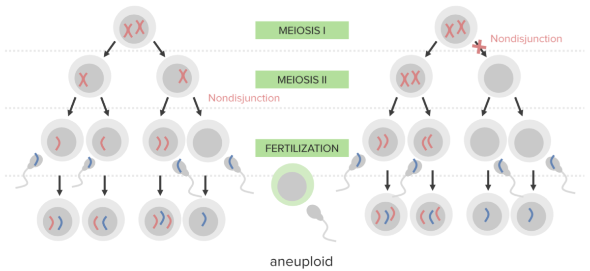
  • In 90% of trisomy Trisomy The possession of a third chromosome of any one type in an otherwise diploid cell. Types of Mutations 18 cases, the presence of 3 copies of the 18th chromosome Chromosome In a prokaryotic cell or in the nucleus of a eukaryotic cell, a structure consisting of or containing DNA which carries the genetic information essential to the cell. Basic Terms of Genetics is due to nondisjunction Nondisjunction The failure of homologous chromosomes or chromatids to segregate during mitosis or meiosis with the result that one daughter cell has both of a pair of parental chromosomes or chromatids and the other has none. Basic Terms of Genetics.
  • Types:
    • Trisomy Trisomy The possession of a third chromosome of any one type in an otherwise diploid cell. Types of Mutations 18 due to nondisjunction Nondisjunction The failure of homologous chromosomes or chromatids to segregate during mitosis or meiosis with the result that one daughter cell has both of a pair of parental chromosomes or chromatids and the other has none. Basic Terms of Genetics
    • Translocation involving chromosome Chromosome In a prokaryotic cell or in the nucleus of a eukaryotic cell, a structure consisting of or containing DNA which carries the genetic information essential to the cell. Basic Terms of Genetics 18
    • Trisomy Trisomy The possession of a third chromosome of any one type in an otherwise diploid cell. Types of Mutations 18 mosaicism Mosaicism The occurrence in an individual of two or more cell populations of different chromosomal constitutions, derived from a single zygote, as opposed to chimerism in which the different cell populations are derived from more than one zygote. Chromosome Testing

Mnemonic

Mnemonic for Edwards syndrome (trisomy 18): “At 18 you can vote in an election.”
Pathophysiology

Genetics Genetics Genetics is the study of genes and their functions and behaviors. Basic Terms of Genetics

  • Chromosomes Chromosomes In a prokaryotic cell or in the nucleus of a eukaryotic cell, a structure consisting of or containing DNA which carries the genetic information essential to the cell. DNA Types and Structure contain genetic material.
  • Human cells:
    • 46 chromosomes Chromosomes In a prokaryotic cell or in the nucleus of a eukaryotic cell, a structure consisting of or containing DNA which carries the genetic information essential to the cell. DNA Types and Structure: 23 pairs with 1 homolog from the mother/egg and 1 from the father/sperm
    • Chromosomes Chromosomes In a prokaryotic cell or in the nucleus of a eukaryotic cell, a structure consisting of or containing DNA which carries the genetic information essential to the cell. DNA Types and Structure 1–22: autosomes
    • Chromosome Chromosome In a prokaryotic cell or in the nucleus of a eukaryotic cell, a structure consisting of or containing DNA which carries the genetic information essential to the cell. Basic Terms of Genetics 23: 2 sex chromosomes Sex chromosomes The homologous chromosomes that are dissimilar in the heterogametic sex. There are the X chromosome, the Y chromosome, and the w, z chromosomes (in animals in which the female is the heterogametic sex (the silkworm moth bombyx mori, for example)). In such cases the w chromosome is the female-determining and the male is zz. Basic Terms of Genetics
      • Female: X, X
      • Male: X, Y
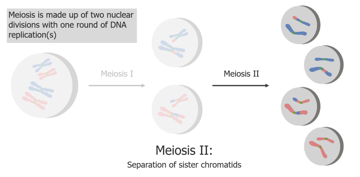
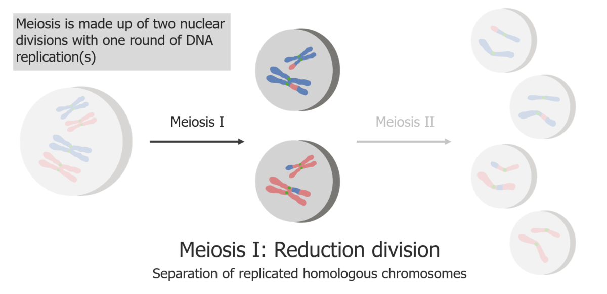
  • Meiosis Meiosis The creation of eukaryotic gametes involves a DNA replication phase followed by 2 cellular division stages: meiosis I and meiosis II. Meiosis I separates homologous chromosomes into separate cells (1n, 2c), while meiosis II separates sister chromatids into gametes (1n, 1c). Meiosis:
    • DNA replication DNA replication The entire DNA of a cell is replicated during the S (synthesis) phase of the cell cycle. The principle of replication is based on complementary nucleotide base pairing: adenine forms hydrogen bonds with thymine (or uracil in RNA) and guanine forms hydrogen bonds with cytosine. DNA Replication is followed by reproductive cell division Cell Division A type of cell nucleus division by means of which the two daughter nuclei normally receive identical complements of the number of chromosomes of the somatic cells of the species. Cell Cycle (cells separate chromosomes Chromosomes In a prokaryotic cell or in the nucleus of a eukaryotic cell, a structure consisting of or containing DNA which carries the genetic information essential to the cell. DNA Types and Structure before reproduction from 46 → 23 chromosomes Chromosomes In a prokaryotic cell or in the nucleus of a eukaryotic cell, a structure consisting of or containing DNA which carries the genetic information essential to the cell. DNA Types and Structure).
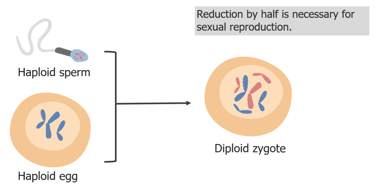
    • Diploid Diploid The chromosomal constitution of cells, in which each type of chromosome is represented twice. Symbol: 2n or 2x. Basic Terms of Genetics (2 sets of chromosomes Chromosomes In a prokaryotic cell or in the nucleus of a eukaryotic cell, a structure consisting of or containing DNA which carries the genetic information essential to the cell. DNA Types and Structure) cells divide into an egg or sperm gamete Gamete Gametogenesis and become a haploid Haploid The chromosomal constitution of cells, in which each type of chromosome is represented once. Symbol: n. Basic Terms of Genetics (1 set of chromosomes Chromosomes In a prokaryotic cell or in the nucleus of a eukaryotic cell, a structure consisting of or containing DNA which carries the genetic information essential to the cell. DNA Types and Structure) cell.
  • In fertilization Fertilization To undergo fertilization, the sperm enters the uterus, travels towards the ampulla of the fallopian tube, and encounters the oocyte. The zona pellucida (the outer layer of the oocyte) deteriorates along with the zygote, which travels towards the uterus and eventually forms a blastocyst, allowing for implantation to occur. Fertilization and First Week, a diploid Diploid The chromosomal constitution of cells, in which each type of chromosome is represented twice. Symbol: 2n or 2x. Basic Terms of Genetics zygote Zygote The fertilized ovum resulting from the fusion of a male and a female gamete. Fertilization and First Week forms when a haploid Haploid The chromosomal constitution of cells, in which each type of chromosome is represented once. Symbol: n. Basic Terms of Genetics egg and sperm unite.

Nondisjunction Nondisjunction The failure of homologous chromosomes or chromatids to segregate during mitosis or meiosis with the result that one daughter cell has both of a pair of parental chromosomes or chromatids and the other has none. Basic Terms of Genetics

  • Most common mechanism
  • In nondisjunction Nondisjunction The failure of homologous chromosomes or chromatids to segregate during mitosis or meiosis with the result that one daughter cell has both of a pair of parental chromosomes or chromatids and the other has none. Basic Terms of Genetics:
    • Failure of proper separation of 2 homologous chromosomes Homologous chromosomes Basic Terms of Genetics or sister chromatids
    • A diploid Diploid The chromosomal constitution of cells, in which each type of chromosome is represented twice. Symbol: 2n or 2x. Basic Terms of Genetics cell with a pair of chromosomes Chromosomes In a prokaryotic cell or in the nucleus of a eukaryotic cell, a structure consisting of or containing DNA which carries the genetic information essential to the cell. DNA Types and Structure divides → 1 gamete Gamete Gametogenesis ends up with 2 chromosomes Chromosomes In a prokaryotic cell or in the nucleus of a eukaryotic cell, a structure consisting of or containing DNA which carries the genetic information essential to the cell. DNA Types and Structure while the other cell has none (not viable)
    • Results in aneuploidy (a state of chromosomal imbalance)
  • Trisomy Trisomy The possession of a third chromosome of any one type in an otherwise diploid cell. Types of Mutations 18:
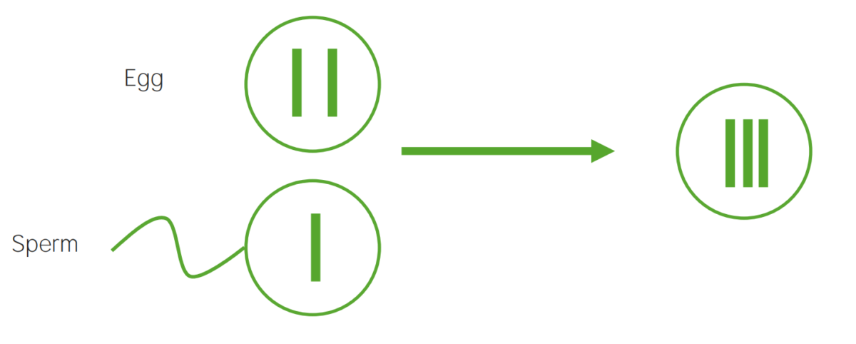
    • Chromosome Chromosome In a prokaryotic cell or in the nucleus of a eukaryotic cell, a structure consisting of or containing DNA which carries the genetic information essential to the cell. Basic Terms of Genetics 18 is present 3 times (or has 3 copies).
    • Occurrence:
      • The egg with a chromosome Chromosome In a prokaryotic cell or in the nucleus of a eukaryotic cell, a structure consisting of or containing DNA which carries the genetic information essential to the cell. Basic Terms of Genetics 18 pair is fertilized by a normal haploid Haploid The chromosomal constitution of cells, in which each type of chromosome is represented once. Symbol: n. Basic Terms of Genetics cell (2 from the egg and 1 from the sperm = 3).
      • The sperm with a chromosome Chromosome In a prokaryotic cell or in the nucleus of a eukaryotic cell, a structure consisting of or containing DNA which carries the genetic information essential to the cell. Basic Terms of Genetics 18 pair fertilizes a normal haploid Haploid The chromosomal constitution of cells, in which each type of chromosome is represented once. Symbol: n. Basic Terms of Genetics cell (2 from the sperm and 1 from the egg = 3).

Translocation

  • Translocation occurs when a part of the chromosome Chromosome In a prokaryotic cell or in the nucleus of a eukaryotic cell, a structure consisting of or containing DNA which carries the genetic information essential to the cell. Basic Terms of Genetics either attaches to or interchanges with a segment of another chromosome Chromosome In a prokaryotic cell or in the nucleus of a eukaryotic cell, a structure consisting of or containing DNA which carries the genetic information essential to the cell. Basic Terms of Genetics.
  • Translocation trisomy Trisomy The possession of a third chromosome of any one type in an otherwise diploid cell. Types of Mutations 18 or partial trisomy Trisomy The possession of a third chromosome of any one type in an otherwise diploid cell. Types of Mutations 18:
    • 2% of cases
    • A piece of chromosome Chromosome In a prokaryotic cell or in the nucleus of a eukaryotic cell, a structure consisting of or containing DNA which carries the genetic information essential to the cell. Basic Terms of Genetics 18 attaches to another chromosome Chromosome In a prokaryotic cell or in the nucleus of a eukaryotic cell, a structure consisting of or containing DNA which carries the genetic information essential to the cell. Basic Terms of Genetics.
    • Symptoms are less severe as only a segment of chromosome Chromosome In a prokaryotic cell or in the nucleus of a eukaryotic cell, a structure consisting of or containing DNA which carries the genetic information essential to the cell. Basic Terms of Genetics 18 long or short arm Arm The arm, or “upper arm” in common usage, is the region of the upper limb that extends from the shoulder to the elbow joint and connects inferiorly to the forearm through the cubital fossa. It is divided into 2 fascial compartments (anterior and posterior). Arm: Anatomy is present in the 3 copies.
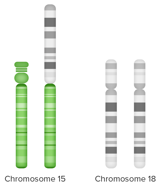
Unbalanced translocation

Unbalanced translocation:
Extra chromosome 18 material (long arm) attached to chromosome 15 (on the left) and
partial trisomy 18 from 2 sets of chromosome 18 + extra long arm chromosome 18 (on the right)
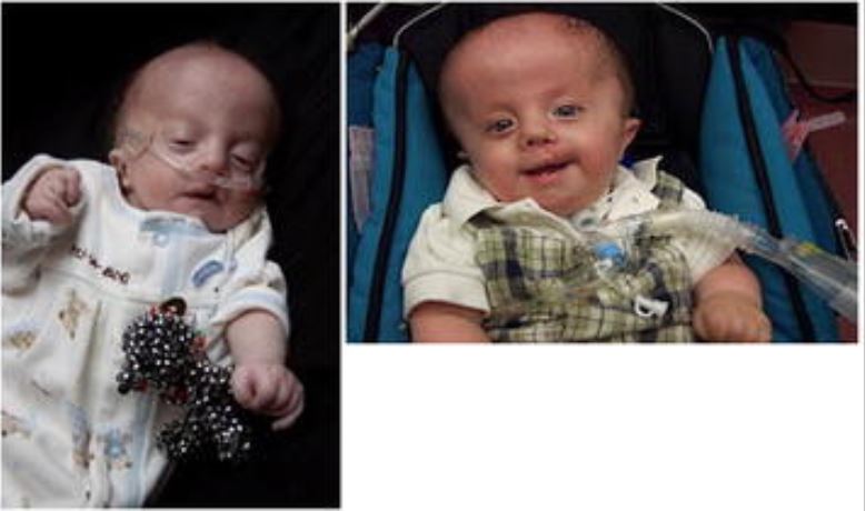
Image by Lecturio.

Mosaicism Mosaicism The occurrence in an individual of two or more cell populations of different chromosomal constitutions, derived from a single zygote, as opposed to chimerism in which the different cell populations are derived from more than one zygote. Chromosome Testing

  • Occurrence < 5% 
  • Extra chromosome Chromosome In a prokaryotic cell or in the nucleus of a eukaryotic cell, a structure consisting of or containing DNA which carries the genetic information essential to the cell. Basic Terms of Genetics 18 is not carried by all cells.
  • Less severe clinical manifestations

Related videos

Clinical Presentation

  • Major findings:
    • Intrauterine growth restriction (IUGR) with polyhydramnios Polyhydramnios Polyhydramnios is a pathological excess of amniotic fluid. Common causes of polyhydramnios include fetal anomalies, gestational diabetes, multiple gestations, and congenital infections. Patients are often asymptomatic but may present with dyspnea, extremity swelling, or abdominal distention. Polyhydramnios on ultrasound
    • Prominent occiput
    • Micrognathia Micrognathia Abnormally small jaw. Pierre Robin Sequence
    • Microcephaly Microcephaly A congenital abnormality in which the cerebrum is underdeveloped, the fontanels close prematurely, and, as a result, the head is small. (desk reference for neuroscience, 2nd ed. ). Fetal Alcohol Spectrum Disorder
    • Low-set, pointy ears
    • Cleft lip Cleft lip The embryological development of craniofacial structures is an intricate sequential process involving tissue growth and directed cell apoptosis. Disruption of any step in this process may result in the formation of a cleft lip alone or in combination with a cleft palate. As the most common craniofacial malformation of the newborn, the diagnosis of a cleft is clinical and usually apparent at birth. Cleft Lip and Cleft Palate and palate Palate The palate is the structure that forms the roof of the mouth and floor of the nasal cavity. This structure is divided into soft and hard palates. Palate: Anatomy
    • Short sternum Sternum A long, narrow, and flat bone commonly known as breastbone occurring in the midsection of the anterior thoracic segment or chest region, which stabilizes the rib cage and serves as the point of origin for several muscles that move the arms, head, and neck. Chest Wall: Anatomy
    • Horseshoe kidney Horseshoe Kidney Congenital Renal Abnormalities
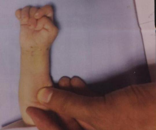
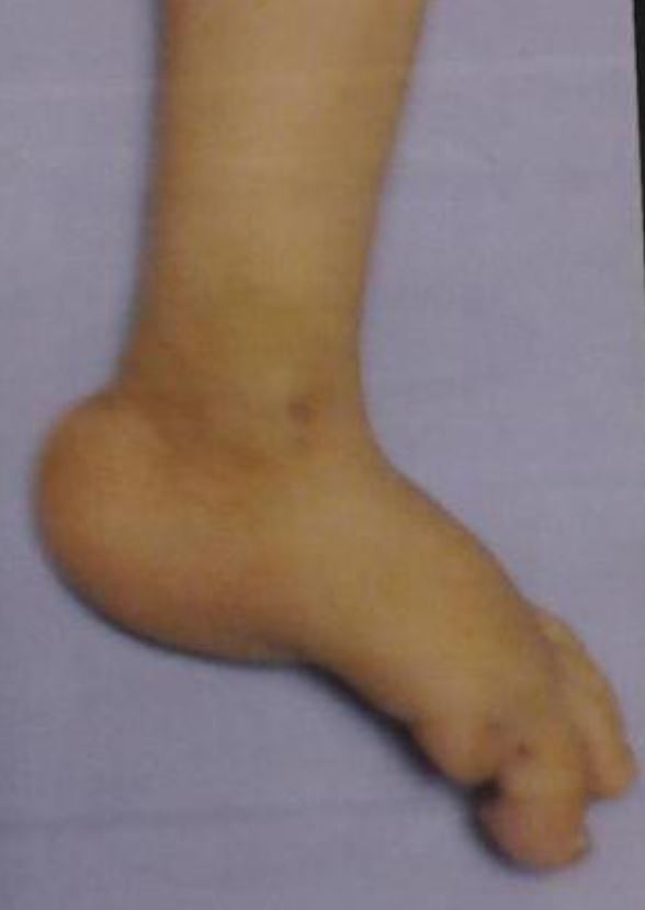
    • Rocker-bottom feet and clenched, overlapping fingers
  • Cardiac malformations in > 50% of patients Patients Individuals participating in the health care system for the purpose of receiving therapeutic, diagnostic, or preventive procedures. Clinician–Patient Relationship:
    • Most common: ventricular septal defect Ventricular Septal Defect Tetralogy of Fallot (VSD)
    • Patent ductus arteriosus Ductus arteriosus A fetal blood vessel connecting the pulmonary artery with the descending aorta. Patent Ductus Arteriosus (PDA) ( PDA PDA The ductus arteriosus (DA) allows blood to bypass pulmonary circulation. After birth, the DA remains open for up to 72 hours and then constricts and involutes, becoming the ligamentum arteriosum. Failure of this process to occur results in patent ductus arteriosus (PDA), a condition that causes up to 10% of congenital heart defects. Patent Ductus Arteriosus (PDA))
    • Pulmonary stenosis Stenosis Hypoplastic Left Heart Syndrome (HLHS)
    • Atrial septal defect Atrial Septal Defect Atrial septal defects (ASDs) are benign acyanotic congenital heart defects characterized by an opening in the interatrial septum that causes blood to flow from the left atrium (LA) to the right atrium (RA) (left-to-right shunt). Atrial Septal Defect (ASD) ( ASD ASD Autism spectrum disorder (ASD) is a neurodevelopmental disorder marked by poor social skills, restricted interests/social interactions, and repetitive/stereotyped behaviors. The condition is termed a “spectrum” because of the wide variability in the severity of symptoms exhibited. Autism Spectrum Disorder)
  • GI involvement in approximately 75% of cases:
  • Severe intellectual disability Disability Determination of the degree of a physical, mental, or emotional handicap. The diagnosis is applied to legal qualification for benefits and income under disability insurance and to eligibility for social security and workman’s compensation benefits. ABCDE Assessment in survivors

Diagnosis and Prognosis

Maternal screening Screening Preoperative Care

  • 1st-trimester combined test:
    • 11th–14th week of gestation
    • Maternal serum testing:
      • ↓ ꞵ-hCG
      • ↓ Pregnancy-associated plasma Plasma The residual portion of blood that is left after removal of blood cells by centrifugation without prior blood coagulation. Transfusion Products protein A
    • Fetal nuchal translucency Nuchal translucency A prenatal ultrasonography measurement of the soft tissue behind the fetal neck. Either the translucent area below the skin in the back of the fetal neck (nuchal translucency) or the distance between occipital bone to the outer skin line (nuchal fold) is measured. Obstetric Imaging:
      • On ultrasound: increased hypoechoic Hypoechoic A structure that produces a low-amplitude echo (darker grays) Ultrasound (Sonography) area in the posterior neck Neck The part of a human or animal body connecting the head to the rest of the body. Peritonsillar Abscess
      • 11th–13th week of gestation
  • 2nd-trimester triple test (triple screen):
    • 15th–20th week of gestation; ideally up to week 18
    • ↓ 𝛼-fetoprotein, β-hCG, free estriol Estriol A hydroxylated metabolite of estradiol or estrogen that has a hydroxyl group at C3, 16-alpha, and 17-beta position. Estriol is a major urinary estrogen. During pregnancy, a large amount of estriol is produced by the placenta. Isomers with inversion of the hydroxyl group or groups are called epiestriol. Noncontraceptive Estrogen and Progestins
  • 2nd-trimester quadruple test ( quad screen Quad Screen Prenatal Care):
    • 15th–21st week of gestation; ideally up to week 18
    • The best option if the mother is presenting for prenatal care Prenatal care Prenatal care is a systematic and periodic assessment of pregnant women during gestation to assure the best health outcome for the mother and her fetus. Prenatal care prevents and identifies maternal and fetal problems that adversely affect the pregnancy outcome. Prenatal Care in the 2nd trimester.
    • Findings:
      • Unchanged inhibin A Inhibin A Glycoproteins that inhibit pituitary follicle stimulating hormone secretion. Inhibins are secreted by the sertoli cells of the testes, the granulosa cells of the ovarian follicles, the placenta, and other tissues. Inhibins and activins are modulators of follicle stimulating hormone secretions; both groups belong to the TGF-beta superfamily, as the transforming growth factor beta. Inhibins consist of a disulfide-linked heterodimer with a unique alpha linked to either a beta a or a beta B subunit to form inhibin a or inhibin b, respectively. Menstrual Cycle
      • ↓ Free estriol Estriol A hydroxylated metabolite of estradiol or estrogen that has a hydroxyl group at C3, 16-alpha, and 17-beta position. Estriol is a major urinary estrogen. During pregnancy, a large amount of estriol is produced by the placenta. Isomers with inversion of the hydroxyl group or groups are called epiestriol. Noncontraceptive Estrogen and Progestins, 𝛼-fetoprotein, β-hCG
  • Full integrated test: 
    • Combination of 1st-trimester pregnancy-associated plasma Plasma The residual portion of blood that is left after removal of blood cells by centrifugation without prior blood coagulation. Transfusion Products protein A and 2nd-trimester quadruple test 
    • Ultrasound fetal nuchal translucency Nuchal translucency A prenatal ultrasonography measurement of the soft tissue behind the fetal neck. Either the translucent area below the skin in the back of the fetal neck (nuchal translucency) or the distance between occipital bone to the outer skin line (nuchal fold) is measured. Obstetric Imaging
  • Serum integrated test: full integrated test without ultrasound fetal nuchal translucency Nuchal translucency A prenatal ultrasonography measurement of the soft tissue behind the fetal neck. Either the translucent area below the skin in the back of the fetal neck (nuchal translucency) or the distance between occipital bone to the outer skin line (nuchal fold) is measured. Obstetric Imaging
  • Sequential Sequential Computed Tomography (CT) screening Screening Preoperative Care:
    • 1st-trimester screening Screening Preoperative Care: The patient is informed of the results.
    • Above a specific threshold Threshold Minimum voltage necessary to generate an action potential (an all-or-none response) Skeletal Muscle Contraction, counseling is provided and diagnostic testing (i.e., chorionic villus sampling) is offered.
  • Cell-free DNA DNA A deoxyribonucleotide polymer that is the primary genetic material of all cells. Eukaryotic and prokaryotic organisms normally contain DNA in a double-stranded state, yet several important biological processes transiently involve single-stranded regions. DNA, which consists of a polysugar-phosphate backbone possessing projections of purines (adenine and guanine) and pyrimidines (thymine and cytosine), forms a double helix that is held together by hydrogen bonds between these purines and pyrimidines (adenine to thymine and guanine to cytosine). DNA Types and Structure test:
    • Noninvasive fetal procedure
    • Anytime after the 10th week of gestation
    • Fetal DNA DNA A deoxyribonucleotide polymer that is the primary genetic material of all cells. Eukaryotic and prokaryotic organisms normally contain DNA in a double-stranded state, yet several important biological processes transiently involve single-stranded regions. DNA, which consists of a polysugar-phosphate backbone possessing projections of purines (adenine and guanine) and pyrimidines (thymine and cytosine), forms a double helix that is held together by hydrogen bonds between these purines and pyrimidines (adenine to thymine and guanine to cytosine). DNA Types and Structure is isolated from maternal blood and evaluated for chromosomal abnormalities.
    • More accurate and specific than a traditional screening Screening Preoperative Care test
    • Tests for fetal sex Sex The totality of characteristics of reproductive structure, functions, phenotype, and genotype, differentiating the male from the female organism. Gender Dysphoria
Table: Maternal screening Screening Preoperative Care 1st and 2nd trimesters
1st trimester 2nd trimester
NT PAPP-A hCG AFP AFP The first alpha-globulins to appear in mammalian sera during fetal development and the dominant serum proteins in early embryonic life. Hepatocellular Carcinoma (HCC) and Liver Metastases Estriol Estriol A hydroxylated metabolite of estradiol or estrogen that has a hydroxyl group at C3, 16-alpha, and 17-beta position. Estriol is a major urinary estrogen. During pregnancy, a large amount of estriol is produced by the placenta. Isomers with inversion of the hydroxyl group or groups are called epiestriol. Noncontraceptive Estrogen and Progestins hCG Inhibin A Inhibin A Glycoproteins that inhibit pituitary follicle stimulating hormone secretion. Inhibins are secreted by the sertoli cells of the testes, the granulosa cells of the ovarian follicles, the placenta, and other tissues. Inhibins and activins are modulators of follicle stimulating hormone secretions; both groups belong to the TGF-beta superfamily, as the transforming growth factor beta. Inhibins consist of a disulfide-linked heterodimer with a unique alpha linked to either a beta a or a beta B subunit to form inhibin a or inhibin b, respectively. Menstrual Cycle
Trisomy Trisomy The possession of a third chromosome of any one type in an otherwise diploid cell. Types of Mutations 13 ↓↓ Unchanged Unchanged Unchanged Unchanged
Trisomy Trisomy The possession of a third chromosome of any one type in an otherwise diploid cell. Types of Mutations 18 ↑↑ ↓↓ ↓↓ ↓↓ ↓↓ Unchanged
Trisomy 21 Trisomy 21 Down syndrome, or trisomy 21, is the most common chromosomal aberration and the most frequent genetic cause of developmental delay. Both boys and girls are affected and have characteristic craniofacial and musculoskeletal features, as well as multiple medical anomalies involving the cardiac, gastrointestinal, ocular, and auditory systems. Down syndrome (Trisomy 21) ↑↑ ↓↓
NT: nuchal translucency
PAPP-A: pregnancy-associated plasma protein A
hCG: human chorionic gonadotropin
AFP: ?-fetoprotein

Diagnostic tests Diagnostic tests Diagnostic tests are important aspects in making a diagnosis. Some of the most important epidemiological values of diagnostic tests include sensitivity and specificity, false positives and false negatives, positive and negative predictive values, likelihood ratios, and pre-test and post-test probabilities. Epidemiological Values of Diagnostic Tests

  • Diagnostic invasive tests:
    • Indications:
    • Risks include bleeding, infection, fetal injury, and fetal loss (rare)
    • Procedures:
      • Chorionic villus sampling: sample of the placenta Placenta A highly vascularized mammalian fetal-maternal organ and major site of transport of oxygen, nutrients, and fetal waste products. It includes a fetal portion (chorionic villi) derived from trophoblasts and a maternal portion (decidua) derived from the uterine endometrium. The placenta produces an array of steroid, protein and peptide hormones (placental hormones). Placenta, Umbilical Cord, and Amniotic Cavity for testing (10th–13th week of gestation)
      • Amniocentesis Amniocentesis Percutaneous transabdominal puncture of the uterus during pregnancy to obtain amniotic fluid. It is commonly used for fetal karyotype determination in order to diagnose abnormal fetal conditions. Polyhydramnios: sample of the amniotic fluid Amniotic fluid A clear, yellowish liquid that envelopes the fetus inside the sac of amnion. In the first trimester, it is likely a transudate of maternal or fetal plasma. In the second trimester, amniotic fluid derives primarily from fetal lung and kidney. Cells or substances in this fluid can be removed for prenatal diagnostic tests (amniocentesis). Placenta, Umbilical Cord, and Amniotic Cavity for testing (15th–20th week of gestation)
      • Percutaneous umbilical blood sampling: blood sample from the umbilical cord Umbilical cord The flexible rope-like structure that connects a developing fetus to the placenta in mammals. The cord contains blood vessels which carry oxygen and nutrients from the mother to the fetus and waste products away from the fetus. Placenta, Umbilical Cord, and Amniotic Cavity for testing (18th–22nd week of gestation); fetal loss rate of 2%
  • Fetal karyotyping Karyotyping Mapping of the karyotype of a cell. Chromosome Testing: a prenatal and postnatal confirmatory test
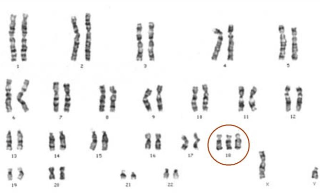
Trisomy 18

Karyotype of person affected with Edward Syndrome:
Three copies of chromosome 18 can be seen

Image: “Trisomia 18” by Serra Amoros. License: CC BY 4.0

Management and Prognosis

Management

  • Delivery in a specialized center with a neonatologist
  • Supportive management:
    • Intervention: dependent on the type of abnormalities
    • Hospice care
  • Genetic counseling Genetic Counseling An educational process that provides information and advice to individuals or families about a genetic condition that may affect them. The purpose is to help individuals make informed decisions about marriage, reproduction, and other health management issues based on information about the genetic disease, the available diagnostic tests, and management programs. Psychosocial support is usually offered. Myotonic Dystrophies and chromosome Chromosome In a prokaryotic cell or in the nucleus of a eukaryotic cell, a structure consisting of or containing DNA which carries the genetic information essential to the cell. Basic Terms of Genetics studies for parents (especially if planning future pregnancies)
  • Supportive resources (e.g., Support Organization for Trisomy Trisomy The possession of a third chromosome of any one type in an otherwise diploid cell. Types of Mutations, or SOFT)

Prognosis Prognosis A prediction of the probable outcome of a disease based on a individual’s condition and the usual course of the disease as seen in similar situations. Non-Hodgkin Lymphomas

  • The majority of babies will die in utero.
  • Of the babies born alive, 50% will die in the firsts 2 weeks of life, and 80%–90% will die in the first 6 months of life.
  • Death is usually due to central apnea and heart failure Heart Failure A heterogeneous condition in which the heart is unable to pump out sufficient blood to meet the metabolic need of the body. Heart failure can be caused by structural defects, functional abnormalities (ventricular dysfunction), or a sudden overload beyond its capacity. Chronic heart failure is more common than acute heart failure which results from sudden insult to cardiac function, such as myocardial infarction. Total Anomalous Pulmonary Venous Return (TAPVR).

Differential Diagnosis

  • Patau’s syndrome ( trisomy Trisomy The possession of a third chromosome of any one type in an otherwise diploid cell. Types of Mutations 13): the presence of 3 copies of the 13th chromosome Chromosome In a prokaryotic cell or in the nucleus of a eukaryotic cell, a structure consisting of or containing DNA which carries the genetic information essential to the cell. Basic Terms of Genetics. Many clinical findings of trisomy Trisomy The possession of a third chromosome of any one type in an otherwise diploid cell. Types of Mutations 18 and 13 overlap, but cleft lip Cleft lip The embryological development of craniofacial structures is an intricate sequential process involving tissue growth and directed cell apoptosis. Disruption of any step in this process may result in the formation of a cleft lip alone or in combination with a cleft palate. As the most common craniofacial malformation of the newborn, the diagnosis of a cleft is clinical and usually apparent at birth. Cleft Lip and Cleft Palate and/or palate Palate The palate is the structure that forms the roof of the mouth and floor of the nasal cavity. This structure is divided into soft and hard palates. Palate: Anatomy, coloboma Coloboma Congenital anomaly in which some of the structures of the eye are absent due to incomplete fusion of the fetal intraocular fissure during gestation. Esophageal Atresia and Tracheoesophageal Fistula of the iris, scalp defects, and polydactyly Polydactyly A congenital anomaly of the hand or foot, marked by the presence of supernumerary digits. Physical Examination of the Newborn are more commonly seen in trisomy Trisomy The possession of a third chromosome of any one type in an otherwise diploid cell. Types of Mutations 13. Both trisomy Trisomy The possession of a third chromosome of any one type in an otherwise diploid cell. Types of Mutations 18 and 13 have poor prognoses.
  • Thrombocytopenia-absent radius Radius The outer shorter of the two bones of the forearm, lying parallel to the ulna and partially revolving around it. Forearm: Anatomy syndrome: a microdeletion syndrome causing thrombocytopenia Thrombocytopenia Thrombocytopenia occurs when the platelet count is < 150,000 per microliter. The normal range for platelets is usually 150,000-450,000/µL of whole blood. Thrombocytopenia can be a result of decreased production, increased destruction, or splenic sequestration of platelets. Patients are often asymptomatic until platelet counts are < 50,000/µL. Thrombocytopenia, congenital Congenital Chorioretinitis heart defects, and the absence of bilateral radii in the forearms Forearms Part of the upper extremity in humans and primates extending from the elbow to the wrist. Bowen Disease and Erythroplasia of Queyrat. Bleeding may occur in the brain Brain The part of central nervous system that is contained within the skull (cranium). Arising from the neural tube, the embryonic brain is comprised of three major parts including prosencephalon (the forebrain); mesencephalon (the midbrain); and rhombencephalon (the hindbrain). The developed brain consists of cerebrum; cerebellum; and other structures in the brain stem. Nervous System: Anatomy, Structure, and Classification and other organs. Normal intellectual abilities can be expected in patients Patients Individuals participating in the health care system for the purpose of receiving therapeutic, diagnostic, or preventive procedures. Clinician–Patient Relationship without bleeding issues. Children with trisomy Trisomy The possession of a third chromosome of any one type in an otherwise diploid cell. Types of Mutations 18 may present with absent radii and thrombocytopenia Thrombocytopenia Thrombocytopenia occurs when the platelet count is < 150,000 per microliter. The normal range for platelets is usually 150,000-450,000/µL of whole blood. Thrombocytopenia can be a result of decreased production, increased destruction, or splenic sequestration of platelets. Patients are often asymptomatic until platelet counts are < 50,000/µL. Thrombocytopenia.
  • Fetal akinesia deformation sequence (also known as Pena-Shokeir syndrome type 1 Type 1 Spinal Muscular Atrophy): Characteristics of fetal akinesia deformation sequence include fetal akinesia, pulmonary hypoplasia Hypoplasia Hypoplastic Left Heart Syndrome (HLHS), IUGR, facial anomalies, other developmental abnormalities, and multiple joint contractures Contractures Prolonged shortening of the muscle or other soft tissue around a joint, preventing movement of the joint. Wound Healing (arthrogryposis), including overlapping fingers. Approximately 30% of affected individuals are stillborn. Infants born alive have limited survival time due to complications of pulmonary hypoplasia Hypoplasia Hypoplastic Left Heart Syndrome (HLHS)
  • Colobomas, heart defects, atresia Atresia Hypoplastic Left Heart Syndrome (HLHS) of the choanae, restriction of growth/developmental delay, genitourinary abnormalities, and ear anomalies (CHARGE) syndrome: In over 50% of cases, a genetic mutation Mutation Genetic mutations are errors in DNA that can cause protein misfolding and dysfunction. There are various types of mutations, including chromosomal, point, frameshift, and expansion mutations. Types of Mutations in the CHD7 gene Gene A category of nucleic acid sequences that function as units of heredity and which code for the basic instructions for the development, reproduction, and maintenance of organisms. Basic Terms of Genetics is involved. No specific treatment is recommended as management is individualized based on the degree of major and minor abnormalities.

References

  1. Cereda, A., Carey, J. C. (2012). The trisomy 18 syndrome. Orphanet journal of rare diseases. 7(81). https://doi.org/10.1186/1750-1172-7-81
  2. Edwards, J. H., et al. (1960). A New Trisomic Syndrome. Lancet. 1(7128), 787–790. https://pubmed.ncbi.nlm.nih.gov/13819419/
  3. Giersch, A. (2019). Congenital cytogenetic abnormalities. UpToDate. Retrieved March 23, 2021, from https://www.uptodate.com/contents/congenital-cytogenetic-abnormalities
  4. Jones, K. L., et al. (2013). Smith’s Recognizable Patterns of Human Malformation. Seventh edition, Elsevier Saunders.
  5. Meeks, N. J. L., et al. (2018). Genetics & Dysmorphology. In Hay William Jr., W., et al. Current Diagnosis & Treatment: Pediatrics. 24th ed., McGraw-Hill Education. accessmedicine.mhmedical.com/content.aspx?aid=1153315258
  6. Genetic and Rare Diseases Information Center (GARD). (2012). Fetal Akinesia Deformation Sequence. https://rarediseases.info.nih.gov/diseases/9634/fetal-akinesia-deformation-sequence
  7. National Organization for Rare Disorders (NORD). (2020). Trisomy 18. https://rarediseases.org/rare-diseases/trisomy-18-syndrome/

Create your free account or log in to continue reading!

Sign up now and get free access to Lecturio with concept pages, medical videos, and questions for your medical education.

User Reviews

Details