Advertisement

Advertisement

Advertisement

Advertisement

Drowning

Drowning occurs due to respiratory impairment from submersion or immersion in a liquid medium. Aspiration of water leads to hypoxemia Hypoxemia Neonatal Respiratory Distress Syndrome, which affects all organ systems, resulting in respiratory insufficiency and acute respiratory distress syndrome Acute Respiratory Distress Syndrome Acute respiratory distress syndrome is characterized by the sudden onset of hypoxemia and bilateral pulmonary edema without cardiac failure. Sepsis is the most common cause of ARDS. The underlying mechanism and histologic correlate is diffuse alveolar damage (DAD). Acute Respiratory Distress Syndrome (ARDS) (ARDS), cardiac arrhythmias, and neuronal damage. The management of drowning focuses initially on ventilatory support followed by cardiopulmonary resuscitation Resuscitation The restoration to life or consciousness of one apparently dead. . Neonatal Respiratory Distress Syndrome. As drowning is most often preventable, prevention is the focus of most interventions.

Last updated: Mar 29, 2023

Editorial responsibility: Stanley Oiseth, Lindsay Jones, Evelin Maza

Advertisement

Advertisement

Advertisement

Advertisement

Advertisement

Advertisement

Overview

Definition

  • Drowning: death secondary to asphyxia caused by submersion or immersion in a liquid medium
  • Non-fatal drowning: survival, at least temporarily, after suffocation (or loss of consciousness) by submersion in a liquid medium

Epidemiology

  • 3rd-most common cause of injury-related deaths worldwide
  • 370,000 drowning deaths worldwide each year
  • Peak incidence Incidence The number of new cases of a given disease during a given period in a specified population. It also is used for the rate at which new events occur in a defined population. It is differentiated from prevalence, which refers to all cases in the population at a given time. Measures of Disease Frequency of drowning in the United States occurs between May and August.
  • Important cause of childhood fatalities worldwide: Children aged 1–4 have the highest drowning rates.
  • Non-accidental drowning deaths: male:female ratio of 2:1
  • Most often affects low- and middle-income patients Patients Individuals participating in the health care system for the purpose of receiving therapeutic, diagnostic, or preventive procedures. Clinician–Patient Relationship (91% of unintentional drowning deaths)

Risk factors

  • Inadequate adult supervision
  • Inability to swim (or overestimation of swimming ability)
  • High-risk behaviors (especially in ages 15–25): use of alcohol and other drugs
  • Hypothermia Hypothermia Hypothermia can be defined as a drop in the core body temperature below 35°C (95°F) and is classified into mild, moderate, severe, and profound forms based on the degree of temperature decrease. Hypothermia
  • Concomitant trauma, stroke, or myocardial infarction Myocardial infarction MI is ischemia and death of an area of myocardial tissue due to insufficient blood flow and oxygenation, usually from thrombus formation on a ruptured atherosclerotic plaque in the epicardial arteries. Clinical presentation is most commonly with chest pain, but women and patients with diabetes may have atypical symptoms. Myocardial Infarction
  • Seizure disorder

Pathophysiology

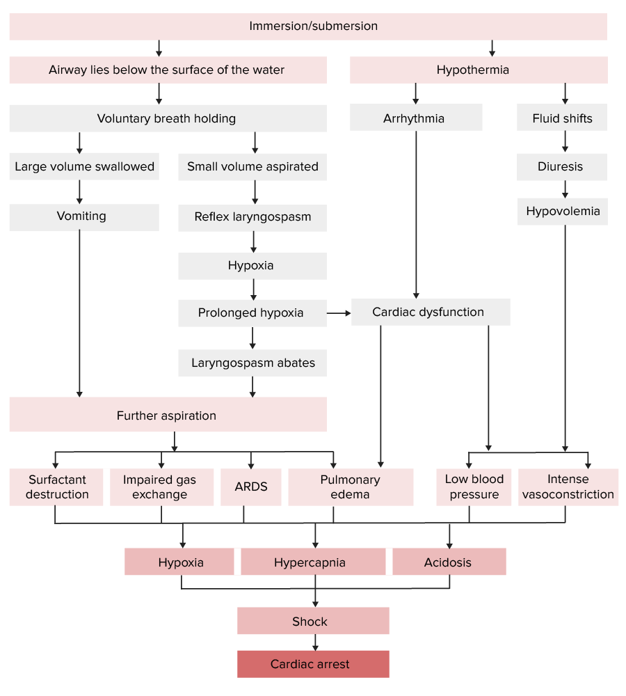
Pathophysiology of drowning

Pathophysiology of drowning:
Note that the respiratory and cardiovascular systems are both affected, leading to respiratory failure and shock.
ARDS: acute respiratory distress syndrome

Image by Lecturio.

Diagnosis

Phyical exam

Clinical manifestations of drowning are due to the effects of hypoxemia Hypoxemia Neonatal Respiratory Distress Syndrome.

Vital signs:

  • Hypotension Hypotension Hypotension is defined as low blood pressure, specifically < 90/60 mm Hg, and is most commonly a physiologic response. Hypotension may be mild, serious, or life threatening, depending on the cause. Hypotension 
  • Hypothermia Hypothermia Hypothermia can be defined as a drop in the core body temperature below 35°C (95°F) and is classified into mild, moderate, severe, and profound forms based on the degree of temperature decrease. Hypothermia
  • Hypoxemia Hypoxemia Neonatal Respiratory Distress Syndrome 

Respiratory changes:

  • Water aspiration causes surfactant Surfactant Substances and drugs that lower the surface tension of the mucoid layer lining the pulmonary alveoli. Acute Respiratory Distress Syndrome (ARDS) washout from the alveoli Alveoli Small polyhedral outpouchings along the walls of the alveolar sacs, alveolar ducts and terminal bronchioles through the walls of which gas exchange between alveolar air and pulmonary capillary blood takes place. Acute Respiratory Distress Syndrome (ARDS). This leads to:
    • Pulmonary edema Pulmonary edema Pulmonary edema is a condition caused by excess fluid within the lung parenchyma and alveoli as a consequence of a disease process. Based on etiology, pulmonary edema is classified as cardiogenic or noncardiogenic. Patients may present with progressive dyspnea, orthopnea, cough, or respiratory failure. Pulmonary Edema
    • Respiratory insufficiency
    • Acute respiratory distress syndrome Acute Respiratory Distress Syndrome Acute respiratory distress syndrome is characterized by the sudden onset of hypoxemia and bilateral pulmonary edema without cardiac failure. Sepsis is the most common cause of ARDS. The underlying mechanism and histologic correlate is diffuse alveolar damage (DAD). Acute Respiratory Distress Syndrome (ARDS) (ARDS) 
  • May experience shortness of breath Shortness of breath Dyspnea is the subjective sensation of breathing discomfort. Dyspnea is a normal manifestation of heavy physical or psychological exertion, but also may be caused by underlying conditions (both pulmonary and extrapulmonary). Dyspnea, crackles, and wheezing Wheezing Wheezing is an abnormal breath sound characterized by a whistling noise that can be relatively high-pitched and shrill (more common) or coarse. Wheezing is produced by the movement of air through narrowed or compressed small (intrathoracic) airways. Wheezing
  • Even if the patient is initially stabilized, ARDS can develop insidiously over the next 72 hours.

Cardiovascular changes:

  • Arrhythmias often occur secondary to hypothermia Hypothermia Hypothermia can be defined as a drop in the core body temperature below 35°C (95°F) and is classified into mild, moderate, severe, and profound forms based on the degree of temperature decrease. Hypothermia and hypoxemia Hypoxemia Neonatal Respiratory Distress Syndrome.
  • Tachycardia Tachycardia Abnormally rapid heartbeat, usually with a heart rate above 100 beats per minute for adults. Tachycardia accompanied by disturbance in the cardiac depolarization (cardiac arrhythmia) is called tachyarrhythmia. Sepsis in Children followed by sinus bradycardia Sinus bradycardia Bradyarrhythmias
  • May progress to atrial fibrillation Atrial fibrillation Atrial fibrillation (AF or Afib) is a supraventricular tachyarrhythmia and the most common kind of arrhythmia. It is caused by rapid, uncontrolled atrial contractions and uncoordinated ventricular responses. Atrial Fibrillation 

Neurologic changes:

Neuronal damage from hypoxia Hypoxia Sub-optimal oxygen levels in the ambient air of living organisms. Ischemic Cell Damage and ischemia Ischemia A hypoperfusion of the blood through an organ or tissue caused by a pathologic constriction or obstruction of its blood vessels, or an absence of blood circulation. Ischemic Cell Damage causes:

  • Cerebral edema Cerebral edema Increased intracellular or extracellular fluid in brain tissue. Cytotoxic brain edema (swelling due to increased intracellular fluid) is indicative of a disturbance in cell metabolism, and is commonly associated with hypoxic or ischemic injuries. An increase in extracellular fluid may be caused by increased brain capillary permeability (vasogenic edema), an osmotic gradient, local blockages in interstitial fluid pathways, or by obstruction of CSF flow (e.g., obstructive hydrocephalus). Increased Intracranial Pressure (ICP)
  • Elevated intracranial pressure Intracranial Pressure Idiopathic Intracranial Hypertension
  • Hypoxic encephalopathy Encephalopathy Hyper-IgM Syndrome
  • Seizures Seizures A seizure is abnormal electrical activity of the neurons in the cerebral cortex that can manifest in numerous ways depending on the region of the brain affected. Seizures consist of a sudden imbalance that occurs between the excitatory and inhibitory signals in cortical neurons, creating a net excitation. The 2 major classes of seizures are focal and generalized. Seizures

Laboratory testing

  • Arterial blood gas Arterial blood gas Respiratory Alkalosis (ABG):
    • Metabolic and/or respiratory acidosis Acidosis A pathologic condition of acid accumulation or depletion of base in the body. The two main types are respiratory acidosis and metabolic acidosis, due to metabolic acid build up. Respiratory Acidosis
    • Electrolyte abnormalities
  • Blood alcohol level and drug toxicology panel:
    • If warranted by context
    • To determine if the cause was not organic

Imaging

Imaging is done via X-ray X-ray Penetrating electromagnetic radiation emitted when the inner orbital electrons of an atom are excited and release radiant energy. X-ray wavelengths range from 1 pm to 10 nm. Hard x-rays are the higher energy, shorter wavelength x-rays. Soft x-rays or grenz rays are less energetic and longer in wavelength. The short wavelength end of the x-ray spectrum overlaps the gamma rays wavelength range. The distinction between gamma rays and x-rays is based on their radiation source. Pulmonary Function Tests.
  • Usually normal initially
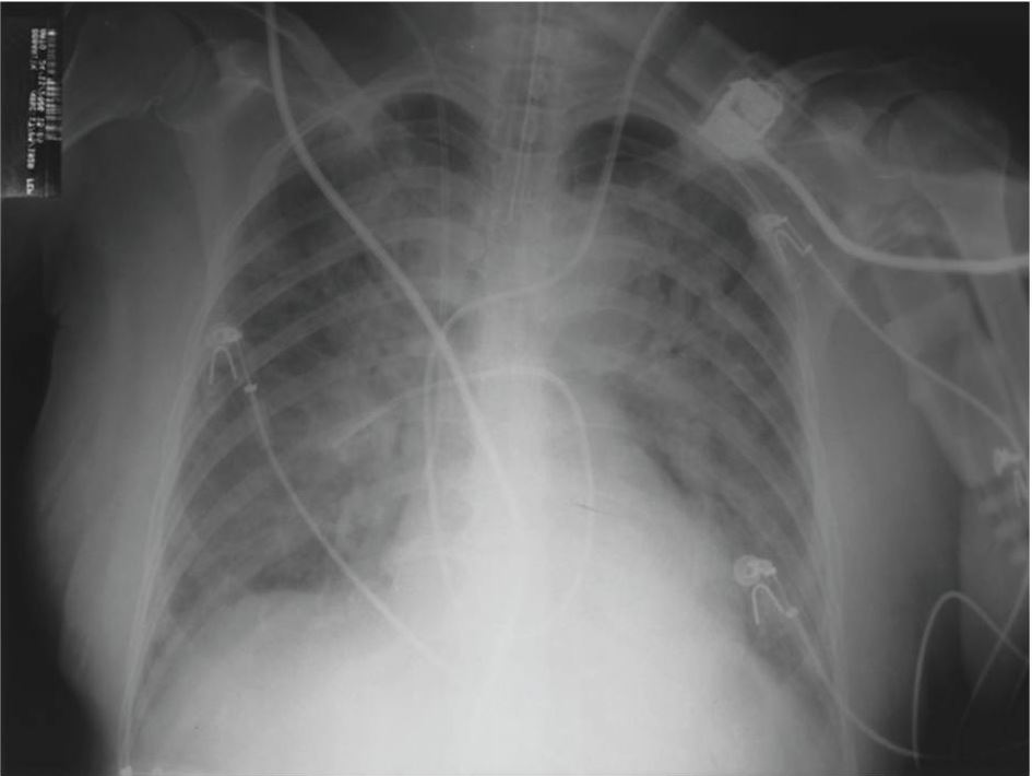
  • ARDS usually does not show on X-ray X-ray Penetrating electromagnetic radiation emitted when the inner orbital electrons of an atom are excited and release radiant energy. X-ray wavelengths range from 1 pm to 10 nm. Hard x-rays are the higher energy, shorter wavelength x-rays. Soft x-rays or grenz rays are less energetic and longer in wavelength. The short wavelength end of the x-ray spectrum overlaps the gamma rays wavelength range. The distinction between gamma rays and x-rays is based on their radiation source. Pulmonary Function Tests until later in course.
Bilateral pulmonary parenchymal infiltrate compatible with ards

Chest radiograph showing bilateral pulmonary parenchymal infiltrate compatible with ARDS

Image: “Fulminant nonocclusive mesenteric ischemia just after hip arthroplasty” by Auxiliadora-Martins M, Alkmin-Teixeira GC, Feres O, Martins-Filho OA, Basile-Filho A. License: CC BY 3.0

Management

While prevention is the most effective intervention, rapid resuscitation Resuscitation The restoration to life or consciousness of one apparently dead. . Neonatal Respiratory Distress Syndrome on-site is essential to improving patient prognosis Prognosis A prediction of the probable outcome of a disease based on a individual’s condition and the usual course of the disease as seen in similar situations. Non-Hodgkin Lymphomas.

1. Safely remove patient from the water and begin primary survey Primary Survey Thoracic Trauma in Children and resuscitation Resuscitation The restoration to life or consciousness of one apparently dead. . Neonatal Respiratory Distress Syndrome:

  • Check for breathing:
    • Normal breathing → supplement with oxygen (goal: oxygen saturation Oxygen Saturation Basic Procedures (SpO₂) > 90%)
    • If no effective breathing → begin rescue breathing. (Note: This is the priority! Begin rescue breathing immediately and then check for pulse to determine if cardiopulmonary resuscitation Resuscitation The restoration to life or consciousness of one apparently dead. . Neonatal Respiratory Distress Syndrome ( CPR CPR The artificial substitution of heart and lung action as indicated for heart arrest resulting from electric shock, drowning, respiratory arrest, or other causes. The two major components of cardiopulmonary resuscitation are artificial ventilation and closed-chest cardiac massage. Cardiac Arrest) is needed.)
  • Check for pulse:
    • No pulse → begin immediate CPR CPR The artificial substitution of heart and lung action as indicated for heart arrest resulting from electric shock, drowning, respiratory arrest, or other causes. The two major components of cardiopulmonary resuscitation are artificial ventilation and closed-chest cardiac massage. Cardiac Arrest 
    • If pulse is found but breathing is not normal → continue rescue breathing/oxygen supplementation.
  • Intubate if:
    • Patient is apneic or in respiratory distress
    • Patient is unable to protect their airways
    • Inability to maintain partial pressure Partial pressure The pressure that would be exerted by one component of a mixture of gases if it were present alone in a container. Gas Exchange of oxygen (PaO₂) > 60 mm MM Multiple myeloma (MM) is a malignant condition of plasma cells (activated B lymphocytes) primarily seen in the elderly. Monoclonal proliferation of plasma cells results in cytokine-driven osteoclastic activity and excessive secretion of IgG antibodies. Multiple Myeloma Hg or SpO₂ > 90% despite the use of high-flow oxygen or noninvasive ventilation Ventilation The total volume of gas inspired or expired per unit of time, usually measured in liters per minute. Ventilation: Mechanics of Breathing
  • If hypothermic (core temperature < 33℃):
    • Remove wet clothing.
    • Begin rewarming.

2. Once patient has been resuscitated and stabilized, initiate secondary survey Secondary Survey ABCDE Assessment or transport to the nearest medical facility:

  • Head-to-toe assessment for signs of trauma
  • Asses for signs of substance use disorder
  • Interview witnesses.

3. Once the patient has arrived at a medical facility, perform the following:

  • Repeat or initial trauma evaluation
  • Frequent vital signs and clinical reassessment
  • Continuous oxygen supplementation and end-tidal carbon dioxide End-Tidal Carbon Dioxide Malignant Hyperthermia (CO₂) monitoring
  • Deliver appropriate IV fluids IV fluids Intravenous fluids are one of the most common interventions administered in medicine to approximate physiologic bodily fluids. Intravenous fluids are divided into 2 categories: crystalloid and colloid solutions. Intravenous fluids have a wide variety of indications, including intravascular volume expansion, electrolyte manipulation, and maintenance fluids. Intravenous Fluids to correct electrolyte disturbances.
  • Continuous telemetry Telemetry Transmission of the readings of instruments to a remote location by means of wires, radio waves, or other means. Crush Syndrome

Related videos

Outcome and Prevention

Outcome

The following are associated with poor prognosis Prognosis A prediction of the probable outcome of a disease based on a individual’s condition and the usual course of the disease as seen in similar situations. Non-Hodgkin Lymphomas:

  • Submersion duration > 5 minutes
  • Initiation of basic life support Basic Life Support Airway Management > 10 minutes
  • Resuscitation Resuscitation The restoration to life or consciousness of one apparently dead. . Neonatal Respiratory Distress Syndrome duration > 25 minutes
  • Age > 14 years
  • Glasgow Coma Coma Coma is defined as a deep state of unarousable unresponsiveness, characterized by a score of 3 points on the GCS. A comatose state can be caused by a multitude of conditions, making the precise epidemiology and prognosis of coma difficult to determine. Coma Scale Scale Dermatologic Examination ( GCS GCS A scale that assesses the response to stimuli in patients with craniocerebral injuries. The parameters are eye opening, motor response, and verbal response. Coma) < 5
  • Arterial blood pH pH The quantitative measurement of the acidity or basicity of a solution. Acid-Base Balance < 7.1 on presentation

Prevention

The best prevention is education on water safety:

  • Gating access to swimming pools
  • Providing adult supervision
  • Not swimming alone
  • Use of personal flotation devices
  • Avoidance of alcohol and drugs while swimming

Differential Diagnosis

  • ARDS: a severe inflammatory reaction of the lung that is characterized by the presence of pulmonary infiltrates due to alveolar fluid accumulation (without evidence suggestive of a cardiogenic etiology). The main finding of ARDS is respiratory failure Respiratory failure Respiratory failure is a syndrome that develops when the respiratory system is unable to maintain oxygenation and/or ventilation. Respiratory failure may be acute or chronic and is classified as hypoxemic, hypercapnic, or a combination of the two. Respiratory Failure. Chest X-ray X-ray Penetrating electromagnetic radiation emitted when the inner orbital electrons of an atom are excited and release radiant energy. X-ray wavelengths range from 1 pm to 10 nm. Hard x-rays are the higher energy, shorter wavelength x-rays. Soft x-rays or grenz rays are less energetic and longer in wavelength. The short wavelength end of the x-ray spectrum overlaps the gamma rays wavelength range. The distinction between gamma rays and x-rays is based on their radiation source. Pulmonary Function Tests usually shows diffuse bilateral lung infiltrates (“butterfly opacity Opacity Imaging of the Lungs and Pleura”). Management depends mainly on treating the underlying etiology and maintaining adequate oxygenation, which may require intubation Intubation Peritonsillar Abscess and mechanical ventilation Ventilation The total volume of gas inspired or expired per unit of time, usually measured in liters per minute. Ventilation: Mechanics of Breathing.
  • Pulmonary edema Pulmonary edema Pulmonary edema is a condition caused by excess fluid within the lung parenchyma and alveoli as a consequence of a disease process. Based on etiology, pulmonary edema is classified as cardiogenic or noncardiogenic. Patients may present with progressive dyspnea, orthopnea, cough, or respiratory failure. Pulmonary Edema: a condition caused by excess fluid in the lungs Lungs Lungs are the main organs of the respiratory system. Lungs are paired viscera located in the thoracic cavity and are composed of spongy tissue. The primary function of the lungs is to oxygenate blood and eliminate CO2. Lungs: Anatomy. Pulmonary edema Pulmonary edema Pulmonary edema is a condition caused by excess fluid within the lung parenchyma and alveoli as a consequence of a disease process. Based on etiology, pulmonary edema is classified as cardiogenic or noncardiogenic. Patients may present with progressive dyspnea, orthopnea, cough, or respiratory failure. Pulmonary Edema is a consequence of a disease process rather than a primary pathology. The condition is classified into cardiogenic and noncardiogenic based on the cause of the edema Edema Edema is a condition in which excess serous fluid accumulates in the body cavity or interstitial space of connective tissues. Edema is a symptom observed in several medical conditions. It can be categorized into 2 types, namely, peripheral (in the extremities) and internal (in an organ or body cavity). Edema. Pulmonary edema Pulmonary edema Pulmonary edema is a condition caused by excess fluid within the lung parenchyma and alveoli as a consequence of a disease process. Based on etiology, pulmonary edema is classified as cardiogenic or noncardiogenic. Patients may present with progressive dyspnea, orthopnea, cough, or respiratory failure. Pulmonary Edema is visible on chest X-ray X-ray Penetrating electromagnetic radiation emitted when the inner orbital electrons of an atom are excited and release radiant energy. X-ray wavelengths range from 1 pm to 10 nm. Hard x-rays are the higher energy, shorter wavelength x-rays. Soft x-rays or grenz rays are less energetic and longer in wavelength. The short wavelength end of the x-ray spectrum overlaps the gamma rays wavelength range. The distinction between gamma rays and x-rays is based on their radiation source. Pulmonary Function Tests.

References

  1. Drowning. (2020). WHO. https://www.who.int/news-room/fact-sheets/detail/drowning
  2. Tyler, Matthew D., et al. (2017). The Epidemiology of Drowning in Low- and Middle-Income Countries: A Systematic Review. BMC Public Health, 17(413). PubMed Central. https://doi.org/10.1186/s12889-017-4239-2

Create your free account or log in to continue reading!

Sign up now and get free access to Lecturio with concept pages, medical videos, and questions for your medical education.

User Reviews

Details