Disorders of Amino Acid Metabolism

Amino acids Amino acids Organic compounds that generally contain an amino (-NH2) and a carboxyl (-COOH) group. Twenty alpha-amino acids are the subunits which are polymerized to form proteins. Basics of Amino Acids are the building blocks of proteins Proteins Linear polypeptides that are synthesized on ribosomes and may be further modified, crosslinked, cleaved, or assembled into complex proteins with several subunits. The specific sequence of amino acids determines the shape the polypeptide will take, during protein folding, and the function of the protein. Energy Homeostasis, whose production is a tightly regulated enzymatic cascade. Disorders of the enzymes Enzymes Enzymes are complex protein biocatalysts that accelerate chemical reactions without being consumed by them. Due to the body's constant metabolic needs, the absence of enzymes would make life unsustainable, as reactions would occur too slowly without these molecules. Basics of Enzymes involved in amino acid Amino acid Amino acids (AAs) are composed of a central carbon atom attached to a carboxyl group, an amino group, a hydrogen atom, and a side chain (R group). Basics of Amino Acids metabolism are often serious and present early in life. Congenital Congenital Chorioretinitis errors in amino acid Amino acid Amino acids (AAs) are composed of a central carbon atom attached to a carboxyl group, an amino group, a hydrogen atom, and a side chain (R group). Basics of Amino Acids metabolism are due to either impaired synthesis Synthesis Polymerase Chain Reaction (PCR) or degradation. In the United States, neonates are routinely screened at birth for common disorders of amino acid Amino acid Amino acids (AAs) are composed of a central carbon atom attached to a carboxyl group, an amino group, a hydrogen atom, and a side chain (R group). Basics of Amino Acids metabolism, which include phenylketonuria (PKU), maple syrup urine disease (MSUD), homocystinuria, tyrosinemia Tyrosinemia A group of disorders which have in common elevations of tyrosine in the blood and urine secondary to an enzyme deficiency. Type I tyrosinemia features episodic weakness, self-mutilation, hepatic necrosis, renal tubular injury, and seizures and is caused by a deficiency of the enzyme fumarylacetoacetase. Type II tyrosinemia features intellectual disability, painful corneal ulcers, and keratosis of the palms and plantar surfaces and is caused by a deficiency of the enzyme tyrosine transaminase. Type III tyrosinemia features intellectual disability and is caused by a deficiency of the enzyme 4-hydroxyphenylpyruvate dioxygenase. Renal Tubular Acidosis, and alkaptonuria. Symptoms of these conditions often present early in life. Although treatment varies, most of these conditions require dietary changes, and some require protein supplementation or other medication regimens.

Last updated: Mar 29, 2023

Editorial responsibility: Stanley Oiseth, Lindsay Jones, Evelin Maza

Phenylketonuria

Overview

Phenylketonuria (PKU) is an inherited disease that causes an increase in phenylalanine Phenylalanine An essential aromatic amino acid that is a precursor of melanin; dopamine; noradrenalin (norepinephrine), and thyroxine. Synthesis of Nonessential Amino Acids levels in the body due to the inability to metabolize this amino acid Amino acid Amino acids (AAs) are composed of a central carbon atom attached to a carboxyl group, an amino group, a hydrogen atom, and a side chain (R group). Basics of Amino Acids.
Pathophysiology

  • Mutations in the phenylalanine hydroxylase Phenylalanine hydroxylase An enzyme of the oxidoreductase class that catalyzes the formation of l-tyrosine, dihydrobiopterin, and water from l-phenylalanine, tetrahydrobiopterin, and oxygen. Synthesis of Nonessential Amino Acids ( PAH PAH The glycine amide of 4-aminobenzoic acid. Its sodium salt is used as a diagnostic aid to measure effective renal plasma flow (ERPF) and excretory capacity. Glomerular Filtration) gene Gene A category of nucleic acid sequences that function as units of heredity and which code for the basic instructions for the development, reproduction, and maintenance of organisms. Basic Terms of Genetics, which encodes PAH PAH The glycine amide of 4-aminobenzoic acid. Its sodium salt is used as a diagnostic aid to measure effective renal plasma flow (ERPF) and excretory capacity. Glomerular Filtration, an enzyme that converts phenylalanine Phenylalanine An essential aromatic amino acid that is a precursor of melanin; dopamine; noradrenalin (norepinephrine), and thyroxine. Synthesis of Nonessential Amino Acids to tyrosine Tyrosine A non-essential amino acid. In animals it is synthesized from phenylalanine. It is also the precursor of epinephrine; thyroid hormones; and melanin. Synthesis of Nonessential Amino Acids
  • A defect in PAH PAH The glycine amide of 4-aminobenzoic acid. Its sodium salt is used as a diagnostic aid to measure effective renal plasma flow (ERPF) and excretory capacity. Glomerular Filtration leads to accumulation of phenylalanine Phenylalanine An essential aromatic amino acid that is a precursor of melanin; dopamine; noradrenalin (norepinephrine), and thyroxine. Synthesis of Nonessential Amino Acids (in most cases tyrosine Tyrosine A non-essential amino acid. In animals it is synthesized from phenylalanine. It is also the precursor of epinephrine; thyroid hormones; and melanin. Synthesis of Nonessential Amino Acids levels are normal or slightly low).
  • Elevated phenylalanine Phenylalanine An essential aromatic amino acid that is a precursor of melanin; dopamine; noradrenalin (norepinephrine), and thyroxine. Synthesis of Nonessential Amino Acids levels cause damage to white matter White Matter The region of central nervous system that appears lighter in color than the other type, gray matter. It mainly consists of myelinated nerve fibers and contains few neuronal cell bodies or dendrites. Brown-Séquard Syndrome tracts and myelin (mechanism unknown), leading to neurological deficits.
Classic phenylketonuria

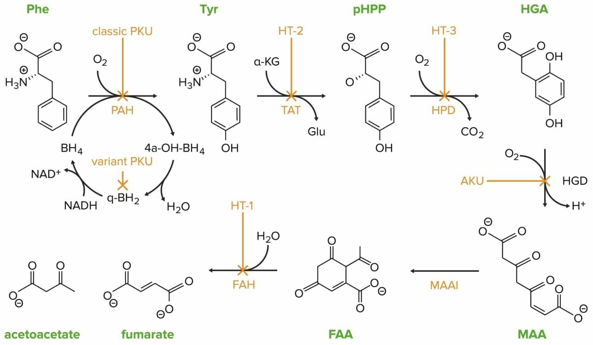
Classic phenylketonuria (PKU) is caused by a mutation in the phenylalanine hydroxylase (PAH) gene.
Phe: phenylalanine
PAH: phenylalanine hydroxylase
Tyr: tyrosine
HT-2: tyrosinemia II
TAT: tyrosine transaminase
pHPP: p-hydroxyphenylpyruvate
HPD: p-hydroxyphenylpyruvate dioxygenase
HT-3: tyrosinemia III
HGA: homogentisate
HGD: homogentisate oxidase
AKU: alcaptonuria
MAA: 4-maleylacetoacetate
MAAI: 4-maleylacetoacetate isomerase
FAA: 4-fumarylacetoacetate
FAH: 4-fumarylacetoacetase
HT-1: tyrosinemia I
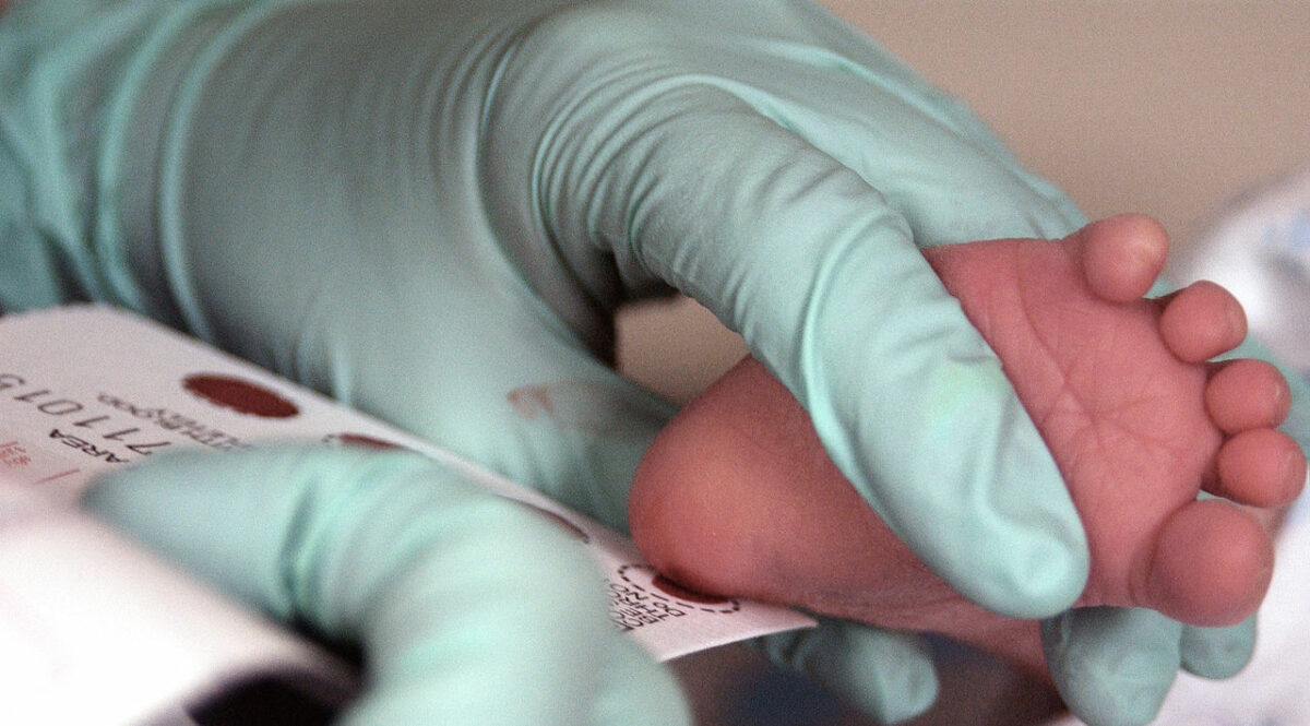
Image by Lecturio.

Classification

  • Classical PKU: very little PAH PAH The glycine amide of 4-aminobenzoic acid. Its sodium salt is used as a diagnostic aid to measure effective renal plasma flow (ERPF) and excretory capacity. Glomerular Filtration gene Gene A category of nucleic acid sequences that function as units of heredity and which code for the basic instructions for the development, reproduction, and maintenance of organisms. Basic Terms of Genetics activity and significant accumulation of phenylalanine Phenylalanine An essential aromatic amino acid that is a precursor of melanin; dopamine; noradrenalin (norepinephrine), and thyroxine. Synthesis of Nonessential Amino Acids
  • Mild PKU: less severe phenylalanine Phenylalanine An essential aromatic amino acid that is a precursor of melanin; dopamine; noradrenalin (norepinephrine), and thyroxine. Synthesis of Nonessential Amino Acids accumulation, some PAH PAH The glycine amide of 4-aminobenzoic acid. Its sodium salt is used as a diagnostic aid to measure effective renal plasma flow (ERPF) and excretory capacity. Glomerular Filtration gene Gene A category of nucleic acid sequences that function as units of heredity and which code for the basic instructions for the development, reproduction, and maintenance of organisms. Basic Terms of Genetics activity present

Symptoms

  • Newborns do not present with symptoms; it takes time for phenylalanine Phenylalanine An essential aromatic amino acid that is a precursor of melanin; dopamine; noradrenalin (norepinephrine), and thyroxine. Synthesis of Nonessential Amino Acids to accumulate.
  • Neurologic:
    • Developmental delays
    • Intellectual disability Disability Determination of the degree of a physical, mental, or emotional handicap. The diagnosis is applied to legal qualification for benefits and income under disability insurance and to eligibility for social security and workman’s compensation benefits. ABCDE Assessment
    • Seizures Seizures A seizure is abnormal electrical activity of the neurons in the cerebral cortex that can manifest in numerous ways depending on the region of the brain affected. Seizures consist of a sudden imbalance that occurs between the excitatory and inhibitory signals in cortical neurons, creating a net excitation. The 2 major classes of seizures are focal and generalized. Seizures
    • Microcephaly Microcephaly A congenital abnormality in which the cerebrum is underdeveloped, the fontanels close prematurely, and, as a result, the head is small. (desk reference for neuroscience, 2nd ed. ). Fetal Alcohol Spectrum Disorder
  • Behavioral and psychiatric:
  • Orthopedic: low bone Bone Bone is a compact type of hardened connective tissue composed of bone cells, membranes, an extracellular mineralized matrix, and central bone marrow. The 2 primary types of bone are compact and spongy. Bones: Structure and Types density
  • Dermatologic
    • Skin Skin The skin, also referred to as the integumentary system, is the largest organ of the body. The skin is primarily composed of the epidermis (outer layer) and dermis (deep layer). The epidermis is primarily composed of keratinocytes that undergo rapid turnover, while the dermis contains dense layers of connective tissue. Skin: Structure and Functions rashes Rashes Rashes are a group of diseases that cause abnormal coloration and texture to the skin. The etiologies are numerous but can include irritation, allergens, infections, or inflammatory conditions. Rashes that present in only 1 area of the body are called localized rashes. Generalized rashes occur diffusely throughout the body. Generalized and Localized Rashes ( eczema Eczema Atopic dermatitis, also known as eczema, is a chronic, relapsing, pruritic, inflammatory skin disease that occurs more frequently in children, although adults can also be affected. The condition is often associated with elevated serum levels of IgE and a personal or family history of atopy. Skin dryness, erythema, oozing, crusting, and lichenification are present. Atopic Dermatitis (Eczema))
    • Fair skin Skin The skin, also referred to as the integumentary system, is the largest organ of the body. The skin is primarily composed of the epidermis (outer layer) and dermis (deep layer). The epidermis is primarily composed of keratinocytes that undergo rapid turnover, while the dermis contains dense layers of connective tissue. Skin: Structure and Functions (due to lack of melanin Melanin Insoluble polymers of tyrosine derivatives found in and causing darkness in skin (skin pigmentation), hair, and feathers providing protection against sunburn induced by sunlight. Carotenes contribute yellow and red coloration. Seborrheic Keratosis)
  • Other: a musty odor in breath or urine

Diagnosis

  • PKU screening Screening Preoperative Care via heel prick within the 1st week of life
    • Measurement of amino acid Amino acid Amino acids (AAs) are composed of a central carbon atom attached to a carboxyl group, an amino group, a hydrogen atom, and a side chain (R group). Basics of Amino Acids levels showing high phenylalanine Phenylalanine An essential aromatic amino acid that is a precursor of melanin; dopamine; noradrenalin (norepinephrine), and thyroxine. Synthesis of Nonessential Amino Acids and low/normal tyrosine Tyrosine A non-essential amino acid. In animals it is synthesized from phenylalanine. It is also the precursor of epinephrine; thyroid hormones; and melanin. Synthesis of Nonessential Amino Acids 
    • Positive results must be confirmed with follow-up testing.
  • Genetic testing Genetic Testing Detection of a mutation; genotype; karyotype; or specific alleles associated with genetic traits, heritable diseases, or predisposition to a disease, or that may lead to the disease in descendants. It includes prenatal genetic testing. Myotonic Dystrophies
Phenylketonuria testing

The blood of a 2-week-old infant is collected for a phenylketonuria (PKU) screening.

Image: “Phenylketonuria testing” by USAF Photographic Archives. License: Public Domain

Management

This condition cannot be cured and must be managed with dietary modification and medication/supplementation.

  • Regular Regular Insulin blood testing to evaluate the level of phenylalanine Phenylalanine An essential aromatic amino acid that is a precursor of melanin; dopamine; noradrenalin (norepinephrine), and thyroxine. Synthesis of Nonessential Amino Acids in the blood
  • Low-phenylalanine diet, eliminating foods such as soybean, chicken, shrimp, nuts, turkey, and legumes
  • Avoid aspartame-containing products, as these are converted to phenylalanine Phenylalanine An essential aromatic amino acid that is a precursor of melanin; dopamine; noradrenalin (norepinephrine), and thyroxine. Synthesis of Nonessential Amino Acids.
  • Protein supplementation formula:
    • Includes the amino acid Amino acid Amino acids (AAs) are composed of a central carbon atom attached to a carboxyl group, an amino group, a hydrogen atom, and a side chain (R group). Basics of Amino Acids tyrosine Tyrosine A non-essential amino acid. In animals it is synthesized from phenylalanine. It is also the precursor of epinephrine; thyroid hormones; and melanin. Synthesis of Nonessential Amino Acids
    • Includes large neutral amino acids Amino acids Organic compounds that generally contain an amino (-NH2) and a carboxyl (-COOH) group. Twenty alpha-amino acids are the subunits which are polymerized to form proteins. Basics of Amino Acids (e.g., valine)
  • For adults with poor control of their phenylalanine Phenylalanine An essential aromatic amino acid that is a precursor of melanin; dopamine; noradrenalin (norepinephrine), and thyroxine. Synthesis of Nonessential Amino Acids, the drug pegvaliase is available and acts as an enzyme that can metabolize phenylalanine Phenylalanine An essential aromatic amino acid that is a precursor of melanin; dopamine; noradrenalin (norepinephrine), and thyroxine. Synthesis of Nonessential Amino Acids.

Cystinuria

Overview

Cystinuria Cystinuria An inherited disorder due to defective reabsorption of cystine and other basic amino acids by the proximal renal tubules. This form of aminoaciduria is characterized by the abnormally high urinary levels of cystine; lysine; arginine; and ornithine. Mutations involve the amino acid transport protein gene slc3a1. Nephrolithiasis is a genetic cause of kidney stones Kidney stones Nephrolithiasis is the formation of a stone, or calculus, anywhere along the urinary tract caused by precipitations of solutes in the urine. The most common type of kidney stone is the calcium oxalate stone, but other types include calcium phosphate, struvite (ammonium magnesium phosphate), uric acid, and cystine stones. Nephrolithiasis due to impairment of renal cystine Cystine A covalently linked dimeric nonessential amino acid formed by the oxidation of cysteine. Two molecules of cysteine are joined together by a disulfide bridge to form cystine. Nephrolithiasis transport.  There is decreased proximal tubular reabsorption of filtered cystine Cystine A covalently linked dimeric nonessential amino acid formed by the oxidation of cysteine. Two molecules of cysteine are joined together by a disulfide bridge to form cystine. Nephrolithiasis resulting in increased urinary cystine Cystine A covalently linked dimeric nonessential amino acid formed by the oxidation of cysteine. Two molecules of cysteine are joined together by a disulfide bridge to form cystine. Nephrolithiasis excretion and cystine Cystine A covalently linked dimeric nonessential amino acid formed by the oxidation of cysteine. Two molecules of cysteine are joined together by a disulfide bridge to form cystine. Nephrolithiasis stone formation.

Pathophysiology

  • Genetic defects:
    • SLC3A1 gene mutation Gene Mutation Myotonic Dystrophies: codes rBAT protein
    • SLC7A9 gene mutation Gene Mutation Myotonic Dystrophies: codes b⁰,+AT protein
    • Under nonpathological conditions, the proteins Proteins Linear polypeptides that are synthesized on ribosomes and may be further modified, crosslinked, cleaved, or assembled into complex proteins with several subunits. The specific sequence of amino acids determines the shape the polypeptide will take, during protein folding, and the function of the protein. Energy Homeostasis form a heterodimer, which is responsible for small intestinal transport and renal proximal tubule Proximal tubule The renal tubule portion that extends from the bowman capsule in the kidney cortex into the kidney medulla. The proximal tubule consists of a convoluted proximal segment in the cortex, and a distal straight segment descending into the medulla where it forms the u-shaped loop of henle. Tubular System transport of:
      • Cystine Cystine A covalently linked dimeric nonessential amino acid formed by the oxidation of cysteine. Two molecules of cysteine are joined together by a disulfide bridge to form cystine. Nephrolithiasis
      • Ornithine Ornithine An amino acid produced in the urea cycle by the splitting off of urea from arginine. Urea Cycle
      • Arginine Arginine An essential amino acid that is physiologically active in the l-form. Urea Cycle
      • Lysine
  • Inheritance pattern: autosomal recessive Autosomal recessive Autosomal inheritance, both dominant and recessive, refers to the transmission of genes from the 22 autosomal chromosomes. Autosomal recessive diseases are only expressed when 2 copies of the recessive allele are inherited. Autosomal Recessive and Autosomal Dominant Inheritance
  • Gross pathology:
    • Bellini ducts with cystine Cystine A covalently linked dimeric nonessential amino acid formed by the oxidation of cysteine. Two molecules of cysteine are joined together by a disulfide bridge to form cystine. Nephrolithiasis crystal deposition
    • Medullary collecting ducts with calcium Calcium A basic element found in nearly all tissues. It is a member of the alkaline earth family of metals with the atomic symbol ca, atomic number 20, and atomic weight 40. Calcium is the most abundant mineral in the body and combines with phosphorus to form calcium phosphate in the bones and teeth. It is essential for the normal functioning of nerves and muscles and plays a role in blood coagulation (as factor IV) and in many enzymatic processes. Electrolytes phosphate Phosphate Inorganic salts of phosphoric acid. Electrolytes deposition
    • Renal tubular interstitial fibrosis Fibrosis Any pathological condition where fibrous connective tissue invades any organ, usually as a consequence of inflammation or other injury. Bronchiolitis Obliterans
  • Cystine Cystine A covalently linked dimeric nonessential amino acid formed by the oxidation of cysteine. Two molecules of cysteine are joined together by a disulfide bridge to form cystine. Nephrolithiasis crystals form at alkaline urine concentrations

Symptoms

  • Individuals present in childhood or early adolescence with nephrolithiasis Nephrolithiasis Nephrolithiasis is the formation of a stone, or calculus, anywhere along the urinary tract caused by precipitations of solutes in the urine. The most common type of kidney stone is the calcium oxalate stone, but other types include calcium phosphate, struvite (ammonium magnesium phosphate), uric acid, and cystine stones. Nephrolithiasis:
  • Predisposition for the development of chronic kidney disease Chronic Kidney Disease Chronic kidney disease (CKD) is kidney impairment that lasts for ≥ 3 months, implying that it is irreversible. Hypertension and diabetes are the most common causes; however, there are a multitude of other etiologies. In the early to moderate stages, CKD is usually asymptomatic and is primarily diagnosed by laboratory abnormalities. Chronic Kidney Disease

Diagnosis

Clinical suspicion:

  • Nephrolithiasis Nephrolithiasis Nephrolithiasis is the formation of a stone, or calculus, anywhere along the urinary tract caused by precipitations of solutes in the urine. The most common type of kidney stone is the calcium oxalate stone, but other types include calcium phosphate, struvite (ammonium magnesium phosphate), uric acid, and cystine stones. Nephrolithiasis presenting in childhood or adolescence
  • Family history Family History Adult Health Maintenance of nephrolithiasis Nephrolithiasis Nephrolithiasis is the formation of a stone, or calculus, anywhere along the urinary tract caused by precipitations of solutes in the urine. The most common type of kidney stone is the calcium oxalate stone, but other types include calcium phosphate, struvite (ammonium magnesium phosphate), uric acid, and cystine stones. Nephrolithiasis
  • Parental history of cystine Cystine A covalently linked dimeric nonessential amino acid formed by the oxidation of cysteine. Two molecules of cysteine are joined together by a disulfide bridge to form cystine. Nephrolithiasis stone formation/ cystinuria Cystinuria An inherited disorder due to defective reabsorption of cystine and other basic amino acids by the proximal renal tubules. This form of aminoaciduria is characterized by the abnormally high urinary levels of cystine; lysine; arginine; and ornithine. Mutations involve the amino acid transport protein gene slc3a1. Nephrolithiasis
  • Large kidney stones Kidney stones Nephrolithiasis is the formation of a stone, or calculus, anywhere along the urinary tract caused by precipitations of solutes in the urine. The most common type of kidney stone is the calcium oxalate stone, but other types include calcium phosphate, struvite (ammonium magnesium phosphate), uric acid, and cystine stones. Nephrolithiasis ( staghorn calculi Staghorn calculi Renal calculi with a characteristic shape like antlers of a deer, formed as it extends into multiple calyces of the renal pelvis. A large number of staghorn calculi as well as other urinary calculi are composed of struvite. Proteus)
  • Recurrent kidney stones Kidney stones Nephrolithiasis is the formation of a stone, or calculus, anywhere along the urinary tract caused by precipitations of solutes in the urine. The most common type of kidney stone is the calcium oxalate stone, but other types include calcium phosphate, struvite (ammonium magnesium phosphate), uric acid, and cystine stones. Nephrolithiasis

Confirmatory testing:

  • Radiologic testing to detect stones:
    • Kidney ultrasound
    • Noncontrast CT of the abdomen/ pelvis Pelvis The pelvis consists of the bony pelvic girdle, the muscular and ligamentous pelvic floor, and the pelvic cavity, which contains viscera, vessels, and multiple nerves and muscles. The pelvic girdle, composed of 2 “hip” bones and the sacrum, is a ring-like bony structure of the axial skeleton that links the vertebral column with the lower extremities. Pelvis: Anatomy
  • Cyanide-nitroprusside test:
    • When added to urine that contains cystine Cystine A covalently linked dimeric nonessential amino acid formed by the oxidation of cysteine. Two molecules of cysteine are joined together by a disulfide bridge to form cystine. Nephrolithiasis → purple color change
    • Indicates urinary cystine Cystine A covalently linked dimeric nonessential amino acid formed by the oxidation of cysteine. Two molecules of cysteine are joined together by a disulfide bridge to form cystine. Nephrolithiasis levels > 75 mg/L
  • Kidney stone analysis → cystine Cystine A covalently linked dimeric nonessential amino acid formed by the oxidation of cysteine. Two molecules of cysteine are joined together by a disulfide bridge to form cystine. Nephrolithiasis crystal predominance
  • Urine microscopy → characteristic hexagonal cystine Cystine A covalently linked dimeric nonessential amino acid formed by the oxidation of cysteine. Two molecules of cysteine are joined together by a disulfide bridge to form cystine. Nephrolithiasis crystals
  • Genetic testing Genetic Testing Detection of a mutation; genotype; karyotype; or specific alleles associated with genetic traits, heritable diseases, or predisposition to a disease, or that may lead to the disease in descendants. It includes prenatal genetic testing. Myotonic Dystrophies for SLC3A and/or 1SLC7A9 mutation Mutation Genetic mutations are errors in DNA that can cause protein misfolding and dysfunction. There are various types of mutations, including chromosomal, point, frameshift, and expansion mutations. Types of Mutations

Management

  • Conservative measures:
    • Hydration > 2–3 L/day
    • Low-sodium diet
    • Diet low in animal-based protein
  • Urinary alkalinization:
    • Used as an adjunct to conservative measures in resistant cases
    • Potassium Potassium An element in the alkali group of metals with an atomic symbol k, atomic number 19, and atomic weight 39. 10. It is the chief cation in the intracellular fluid of muscle and other cells. Potassium ion is a strong electrolyte that plays a significant role in the regulation of fluid volume and maintenance of the water-electrolyte balance. Hyperkalemia citrate/ potassium Potassium An element in the alkali group of metals with an atomic symbol k, atomic number 19, and atomic weight 39. 10. It is the chief cation in the intracellular fluid of muscle and other cells. Potassium ion is a strong electrolyte that plays a significant role in the regulation of fluid volume and maintenance of the water-electrolyte balance. Hyperkalemia bicarbonate Bicarbonate Inorganic salts that contain the -HCO3 radical. They are an important factor in determining the ph of the blood and the concentration of bicarbonate ions is regulated by the kidney. Levels in the blood are an index of the alkali reserve or buffering capacity. Electrolytes supplementation:
      • Must be taken 3x per day
      • Around-the-clock alkalinization is required to maintain urine pH pH The quantitative measurement of the acidity or basicity of a solution. Acid-Base Balance < 7.
    • Home urine-pH testing is used initially to ensure therapeutic supplementation.
  • Thiol-containing drugs:
    • Tiopronin and D-penicillamine D-penicillamine The most characteristic degradation product of the penicillin antibiotics. It is used as an antirheumatic and as a chelating agent in wilson’s disease. Movement Disorder Drugs are options.
    • Tiotropin is preferred (better tolerated)
    • Indicated after failure of conservative measures
    • Conservative measures must be continued in addition to thiol-containing drugs
  • Biochemical monitoring:
    • Efficacy of therapy is monitored with 24-hour urine collection every 3–6 months until stable on the therapeutic regimen.
    • Urine microscopy may be performed to examine for cystine Cystine A covalently linked dimeric nonessential amino acid formed by the oxidation of cysteine. Two molecules of cysteine are joined together by a disulfide bridge to form cystine. Nephrolithiasis crystals.
  • Radiologic monitoring:
    • Kidney ultrasound every 3–6 months in asymptomatic individuals:
      • To detect new stone formation
      • To monitor existing stones for progression
    • Noncontrast CT is reserved for symptomatic individuals:
      • To detect stones missed by ultrasound
      • To determine stone size/location if surgical intervention is considered
  • Surgical intervention:
    • Emergent indications:
      • Acute urinary obstruction with clinical suspicion or urinary tract Urinary tract The urinary tract is located in the abdomen and pelvis and consists of the kidneys, ureters, urinary bladder, and urethra. The structures permit the excretion of urine from the body. Urine flows from the kidneys through the ureters to the urinary bladder and out through the urethra. Urinary Tract: Anatomy infection
      • Bilateral obstruction with acute renal failure Renal failure Conditions in which the kidneys perform below the normal level in the ability to remove wastes, concentrate urine, and maintain electrolyte balance; blood pressure; and calcium metabolism. Renal insufficiency can be classified by the degree of kidney damage (as measured by the level of proteinuria) and reduction in glomerular filtration rate. Crush Syndrome
      • Unilateral obstruction if the individual has a solitary-functioning kidney
    • Elective indications:
      • Ureteral stone size > 10 mm MM Multiple myeloma (MM) is a malignant condition of plasma cells (activated B lymphocytes) primarily seen in the elderly. Monoclonal proliferation of plasma cells results in cytokine-driven osteoclastic activity and excessive secretion of IgG antibodies. Multiple Myeloma
      • Intractable pain Pain An unpleasant sensation induced by noxious stimuli which are detected by nerve endings of nociceptive neurons. Pain: Types and Pathways
      • Pregnancy Pregnancy The status during which female mammals carry their developing young (embryos or fetuses) in utero before birth, beginning from fertilization to birth. Pregnancy: Diagnosis, Physiology, and Care
      • Persistent kidney obstruction
      • Recurrent urinary tract Urinary tract The urinary tract is located in the abdomen and pelvis and consists of the kidneys, ureters, urinary bladder, and urethra. The structures permit the excretion of urine from the body. Urine flows from the kidneys through the ureters to the urinary bladder and out through the urethra. Urinary Tract: Anatomy infection
    • Surgical approaches:
      • Shockwave lithotripsy is reserved for stones < 1.5 cm.
      • Ureteroscopy with extraction is reserved for stones < 1.5 cm.
      • Percutaneous nephrolithotomy with ultrasonic or laser lithotripsy is preferred for:
        • Larger stones (> 1.5 cm)
        • Stones resistant to shockwave lithotripsy or ureteroscopy

Homocystinuria

Overview

Homocystinuria is an inherited metabolic disorder of methionine metabolism that results in elevated levels of homocysteine in the plasma Plasma The residual portion of blood that is left after removal of blood cells by centrifugation without prior blood coagulation. Transfusion Products and urine and causes an array of clinical manifestations.

Pathophysiology

There are 2 normal homocysteine metabolic pathways:

  • Transsulfuration:
    • Catalyzed by cystathionine β-synthase
    • Requires pyridoxal phosphate Phosphate Inorganic salts of phosphoric acid. Electrolytes (vitamin B6) as a cofactor
  • Remethylation:
    • Catalyzed either by methionine synthase or by betaine-homocysteine methyltransferase
  • Requires cobalamin Cobalamin A cobalt-containing coordination compound produced by intestinal microorganisms and found also in soil and water. Higher plants do not concentrate vitamin B 12 from the soil and so are a poor source of the substance as compared with animal tissues. Intrinsic factor is important for the assimilation of vitamin B 12. Folate and Vitamin B12 (vitamin B12, the precursor of methylcobalamin)

Elevations in plasma Plasma The residual portion of blood that is left after removal of blood cells by centrifugation without prior blood coagulation. Transfusion Products homocysteine levels primarily result from:

  • Genetic defects:
    • Methylenetetrahydrofolate reductase (MTHFR) mutation Mutation Genetic mutations are errors in DNA that can cause protein misfolding and dysfunction. There are various types of mutations, including chromosomal, point, frameshift, and expansion mutations. Types of Mutations:
      • Approximately 30% of the U.S. population is heterozygous for MTHFR mutation Mutation Genetic mutations are errors in DNA that can cause protein misfolding and dysfunction. There are various types of mutations, including chromosomal, point, frameshift, and expansion mutations. Types of Mutations.
      • Approximately 10% of the U.S. population is homozygous for MTHFR mutation Mutation Genetic mutations are errors in DNA that can cause protein misfolding and dysfunction. There are various types of mutations, including chromosomal, point, frameshift, and expansion mutations. Types of Mutations
      • Often occurs with low serum folate Folate Folate and vitamin B12 are 2 of the most clinically important water-soluble vitamins. Deficiencies can present with megaloblastic anemia, GI symptoms, neuropsychiatric symptoms, and adverse pregnancy complications, including neural tube defects. Folate and Vitamin B12 levels 
      • Even with supplemented folate Folate Folate and vitamin B12 are 2 of the most clinically important water-soluble vitamins. Deficiencies can present with megaloblastic anemia, GI symptoms, neuropsychiatric symptoms, and adverse pregnancy complications, including neural tube defects. Folate and Vitamin B12 intake, homozygotes have lower plasma Plasma The residual portion of blood that is left after removal of blood cells by centrifugation without prior blood coagulation. Transfusion Products folate Folate Folate and vitamin B12 are 2 of the most clinically important water-soluble vitamins. Deficiencies can present with megaloblastic anemia, GI symptoms, neuropsychiatric symptoms, and adverse pregnancy complications, including neural tube defects. Folate and Vitamin B12 levels than nonhomozygotes.
  • Inheritance pattern: autosomal recessive Autosomal recessive Autosomal inheritance, both dominant and recessive, refers to the transmission of genes from the 22 autosomal chromosomes. Autosomal recessive diseases are only expressed when 2 copies of the recessive allele are inherited. Autosomal Recessive and Autosomal Dominant Inheritance

Other causes of elevated plasma Plasma The residual portion of blood that is left after removal of blood cells by centrifugation without prior blood coagulation. Transfusion Products homocysteine:

  • Vitamin deficiencies:
    • The majority of individuals with elevated homocysteine levels have low plasma Plasma The residual portion of blood that is left after removal of blood cells by centrifugation without prior blood coagulation. Transfusion Products levels of:
      • Folate Folate Folate and vitamin B12 are 2 of the most clinically important water-soluble vitamins. Deficiencies can present with megaloblastic anemia, GI symptoms, neuropsychiatric symptoms, and adverse pregnancy complications, including neural tube defects. Folate and Vitamin B12
      • Vitamin B6
      • Vitamin B12 
    • More common in individuals with alcohol use disorder Alcohol use disorder Alcohol is one of the most commonly used addictive substances in the world. Alcohol use disorder (AUD) is defined as pathologic consumption of alcohol leading to impaired daily functioning. Acute alcohol intoxication presents with impairment in speech and motor functions and can be managed in most cases with supportive care. Alcohol Use Disorder
  • Chronic kidney disease Chronic Kidney Disease Chronic kidney disease (CKD) is kidney impairment that lasts for ≥ 3 months, implying that it is irreversible. Hypertension and diabetes are the most common causes; however, there are a multitude of other etiologies. In the early to moderate stages, CKD is usually asymptomatic and is primarily diagnosed by laboratory abnormalities. Chronic Kidney Disease:
    • Decreased renal removal of homocysteine
    • Impaired renal metabolism of homocysteine
  • Drugs:
    • Fibrates Fibrates Lipid Control Drugs
    • Nicotinic acid Nicotinic acid A water-soluble vitamin of the B complex occurring in various animal and plant tissues. It is required by the body for the formation of coenzymes nad and nadp. It has pellagra-curative, vasodilating, and antilipemic properties. Lipid Control Drugs
    • Metformin Metformin A biguanide hypoglycemic agent used in the treatment of non-insulin-dependent diabetes mellitus not responding to dietary modification. Metformin improves glycemic control by improving insulin sensitivity and decreasing intestinal absorption of glucose. Non-insulinotropic Diabetes Drugs
    • Methotrexate Methotrexate An antineoplastic antimetabolite with immunosuppressant properties. It is an inhibitor of tetrahydrofolate dehydrogenase and prevents the formation of tetrahydrofolate, necessary for synthesis of thymidylate, an essential component of DNA. Antimetabolite Chemotherapy
  • Smoking Smoking Willful or deliberate act of inhaling and exhaling smoke from burning substances or agents held by hand. Interstitial Lung Diseases

Symptoms

  • Ocular abnormalities:
    • Lens Lens A transparent, biconvex structure of the eye, enclosed in a capsule and situated behind the iris and in front of the vitreous humor (vitreous body). It is slightly overlapped at its margin by the ciliary processes. Adaptation by the ciliary body is crucial for ocular accommodation. Eye: Anatomy dislocation
    • Nearsightedness Nearsightedness Refractive Errors
  • Osteopenia Osteopenia Osteoporosis/ osteoporosis Osteoporosis Osteoporosis refers to a decrease in bone mass and density leading to an increased number of fractures. There are 2 forms of osteoporosis: primary, which is commonly postmenopausal or senile; and secondary, which is a manifestation of immobilization, underlying medical disorders, or long-term use of certain medications. Osteoporosis
  • Learning disabilities Learning disabilities Conditions characterized by a significant discrepancy between an individual’s perceived level of intellect and their ability to acquire new language and other cognitive skills. These may result from organic or psychological conditions. Relatively common subtypes include dyslexia, dyscalculia, and dysgraphia. DiGeorge Syndrome/developmental delay
  • Musculoskeletal abnormalities (marfanoid features):
  • Thrombophilia Thrombophilia A disorder of hemostasis in which there is a tendency for the occurrence of thrombosis. Hypercoagulable States
  • Premature Premature Childbirth before 37 weeks of pregnancy (259 days from the first day of the mother’s last menstrual period, or 245 days after fertilization). Necrotizing Enterocolitis atherosclerosis Atherosclerosis Atherosclerosis is a common form of arterial disease in which lipid deposition forms a plaque in the blood vessel walls. Atherosclerosis is an incurable disease, for which there are clearly defined risk factors that often can be reduced through a change in lifestyle and behavior of the patient. Atherosclerosis

Diagnosis

Clinical suspicion:

  • 1st-degree relatives of individuals with homocystinuria
  • Children/adolescents presenting with:
    • Characteristic physical findings
    • Developmental delay
    • Premature Premature Childbirth before 37 weeks of pregnancy (259 days from the first day of the mother’s last menstrual period, or 245 days after fertilization). Necrotizing Enterocolitis cardiovascular disease
    • Thromboembolism Thromboembolism Obstruction of a blood vessel (embolism) by a blood clot (thrombus) in the blood stream. Systemic Lupus Erythematosus

Confirmatory testing:

  • Newborn Newborn An infant during the first 28 days after birth. Physical Examination of the Newborn screening Screening Preoperative Care in regions with a high prevalence Prevalence The total number of cases of a given disease in a specified population at a designated time. It is differentiated from incidence, which refers to the number of new cases in the population at a given time. Measures of Disease Frequency
  • Elevated levels of methionine in the urine or blood
  • Elevated levels of homocysteine in the urine or blood
  • MTHFR genetic testing Genetic Testing Detection of a mutation; genotype; karyotype; or specific alleles associated with genetic traits, heritable diseases, or predisposition to a disease, or that may lead to the disease in descendants. It includes prenatal genetic testing. Myotonic Dystrophies

Management

  • Vitamin supplementation:
    • Folate Folate Folate and vitamin B12 are 2 of the most clinically important water-soluble vitamins. Deficiencies can present with megaloblastic anemia, GI symptoms, neuropsychiatric symptoms, and adverse pregnancy complications, including neural tube defects. Folate and Vitamin B12
    • Vitamin B6
    • Vitamin B12
  • Discontinue offending drugs.
  • Smoking Smoking Willful or deliberate act of inhaling and exhaling smoke from burning substances or agents held by hand. Interstitial Lung Diseases cessation
  • Treat/minimize risk factors for thromboembolic disease and coronary atherosclerosis Atherosclerosis Atherosclerosis is a common form of arterial disease in which lipid deposition forms a plaque in the blood vessel walls. Atherosclerosis is an incurable disease, for which there are clearly defined risk factors that often can be reduced through a change in lifestyle and behavior of the patient. Atherosclerosis.

Tyrosinemia

Overview

A genetic disorder manifesting in the abnormal metabolism of tyrosine Tyrosine A non-essential amino acid. In animals it is synthesized from phenylalanine. It is also the precursor of epinephrine; thyroid hormones; and melanin. Synthesis of Nonessential Amino Acids, an amino acid Amino acid Amino acids (AAs) are composed of a central carbon atom attached to a carboxyl group, an amino group, a hydrogen atom, and a side chain (R group). Basics of Amino Acids involved in the synthesis Synthesis Polymerase Chain Reaction (PCR) of thyroid Thyroid The thyroid gland is one of the largest endocrine glands in the human body. The thyroid gland is a highly vascular, brownish-red gland located in the visceral compartment of the anterior region of the neck. Thyroid Gland: Anatomy hormones Hormones Hormones are messenger molecules that are synthesized in one part of the body and move through the bloodstream to exert specific regulatory effects on another part of the body. Hormones play critical roles in coordinating cellular activities throughout the body in response to the constant changes in both the internal and external environments. Hormones: Overview and Types, catecholamines Catecholamines A general class of ortho-dihydroxyphenylalkylamines derived from tyrosine. Adrenal Hormones, and melanin Melanin Insoluble polymers of tyrosine derivatives found in and causing darkness in skin (skin pigmentation), hair, and feathers providing protection against sunburn induced by sunlight. Carotenes contribute yellow and red coloration. Seborrheic Keratosis.

Pathophysiology

Under nonpathological conditions, tyrosine Tyrosine A non-essential amino acid. In animals it is synthesized from phenylalanine. It is also the precursor of epinephrine; thyroid hormones; and melanin. Synthesis of Nonessential Amino Acids is metabolized by hepatocytes Hepatocytes The main structural component of the liver. They are specialized epithelial cells that are organized into interconnected plates called lobules. Liver: Anatomy and renal proximal tubules into acetoacetate Acetoacetate Salts and derivatives of acetoacetic acid. Ketone Body Metabolism (ketogenic) and fumarate Fumarate Citric Acid Cycle (glucogenic).

Genetic defects:

  • Hereditary tyrosinemia Tyrosinemia A group of disorders which have in common elevations of tyrosine in the blood and urine secondary to an enzyme deficiency. Type I tyrosinemia features episodic weakness, self-mutilation, hepatic necrosis, renal tubular injury, and seizures and is caused by a deficiency of the enzyme fumarylacetoacetase. Type II tyrosinemia features intellectual disability, painful corneal ulcers, and keratosis of the palms and plantar surfaces and is caused by a deficiency of the enzyme tyrosine transaminase. Type III tyrosinemia features intellectual disability and is caused by a deficiency of the enzyme 4-hydroxyphenylpyruvate dioxygenase. Renal Tubular Acidosis 1 (HT1), aka hepatorenal tyrosinemia Tyrosinemia A group of disorders which have in common elevations of tyrosine in the blood and urine secondary to an enzyme deficiency. Type I tyrosinemia features episodic weakness, self-mutilation, hepatic necrosis, renal tubular injury, and seizures and is caused by a deficiency of the enzyme fumarylacetoacetase. Type II tyrosinemia features intellectual disability, painful corneal ulcers, and keratosis of the palms and plantar surfaces and is caused by a deficiency of the enzyme tyrosine transaminase. Type III tyrosinemia features intellectual disability and is caused by a deficiency of the enzyme 4-hydroxyphenylpyruvate dioxygenase. Renal Tubular Acidosis:
    • Deficiency of fumarylacetoacetate hydrolase → accumulation of fumarylacetoacetate (FAA)*
    • *FAA has a short intracellular half-life Half-Life The time it takes for a substance (drug, radioactive nuclide, or other) to lose half of its pharmacologic, physiologic, or radiologic activity. Pharmacokinetics and Pharmacodynamics and, thus, is not found in body fluids of HT1-affected individuals
    • The principal metabolites of FAA, succinylacetoacetate and succinylacetone, are released into the circulation Circulation The movement of the blood as it is pumped through the cardiovascular system. ABCDE Assessment and can be measured for diagnosis:
      • Fumarylacetoacetate accumulates in hepatocytes Hepatocytes The main structural component of the liver. They are specialized epithelial cells that are organized into interconnected plates called lobules. Liver: Anatomy and proximal renal tubular cells
      • Reacts with glutathione and sulfhydryl groups of proteins Proteins Linear polypeptides that are synthesized on ribosomes and may be further modified, crosslinked, cleaved, or assembled into complex proteins with several subunits. The specific sequence of amino acids determines the shape the polypeptide will take, during protein folding, and the function of the protein. Energy Homeostasis → oxidative damage
      • Results in severe, progressive liver Liver The liver is the largest gland in the human body. The liver is found in the superior right quadrant of the abdomen and weighs approximately 1.5 kilograms. Its main functions are detoxification, metabolism, nutrient storage (e.g., iron and vitamins), synthesis of coagulation factors, formation of bile, filtration, and storage of blood. Liver: Anatomy and kidney damage
      • Increased risk of hepatocellular carcinoma Hepatocellular carcinoma Hepatocellular carcinoma (HCC) typically arises in a chronically diseased or cirrhotic liver and is the most common primary liver cancer. Diagnosis may include ultrasound, CT, MRI, biopsy (if inconclusive imaging), and/or biomarkers. Hepatocellular Carcinoma (HCC) and Liver Metastases
      • Autosomal recessive inheritance Autosomal recessive inheritance Autosomal Recessive and Autosomal Dominant Inheritance pattern
  • Hereditary tyrosinemia Tyrosinemia A group of disorders which have in common elevations of tyrosine in the blood and urine secondary to an enzyme deficiency. Type I tyrosinemia features episodic weakness, self-mutilation, hepatic necrosis, renal tubular injury, and seizures and is caused by a deficiency of the enzyme fumarylacetoacetase. Type II tyrosinemia features intellectual disability, painful corneal ulcers, and keratosis of the palms and plantar surfaces and is caused by a deficiency of the enzyme tyrosine transaminase. Type III tyrosinemia features intellectual disability and is caused by a deficiency of the enzyme 4-hydroxyphenylpyruvate dioxygenase. Renal Tubular Acidosis 2 (HT2), aka oculocutaneous tyrosinemia Tyrosinemia A group of disorders which have in common elevations of tyrosine in the blood and urine secondary to an enzyme deficiency. Type I tyrosinemia features episodic weakness, self-mutilation, hepatic necrosis, renal tubular injury, and seizures and is caused by a deficiency of the enzyme fumarylacetoacetase. Type II tyrosinemia features intellectual disability, painful corneal ulcers, and keratosis of the palms and plantar surfaces and is caused by a deficiency of the enzyme tyrosine transaminase. Type III tyrosinemia features intellectual disability and is caused by a deficiency of the enzyme 4-hydroxyphenylpyruvate dioxygenase. Renal Tubular Acidosis or Richner-Hanhart syndrome: deficiency of tyrosine Tyrosine A non-essential amino acid. In animals it is synthesized from phenylalanine. It is also the precursor of epinephrine; thyroid hormones; and melanin. Synthesis of Nonessential Amino Acids aminotransferase → elevated plasma Plasma The residual portion of blood that is left after removal of blood cells by centrifugation without prior blood coagulation. Transfusion Products tyrosine Tyrosine A non-essential amino acid. In animals it is synthesized from phenylalanine. It is also the precursor of epinephrine; thyroid hormones; and melanin. Synthesis of Nonessential Amino Acids and tyrosine Tyrosine A non-essential amino acid. In animals it is synthesized from phenylalanine. It is also the precursor of epinephrine; thyroid hormones; and melanin. Synthesis of Nonessential Amino Acids metabolites:
  • Hereditary tyrosinemia Tyrosinemia A group of disorders which have in common elevations of tyrosine in the blood and urine secondary to an enzyme deficiency. Type I tyrosinemia features episodic weakness, self-mutilation, hepatic necrosis, renal tubular injury, and seizures and is caused by a deficiency of the enzyme fumarylacetoacetase. Type II tyrosinemia features intellectual disability, painful corneal ulcers, and keratosis of the palms and plantar surfaces and is caused by a deficiency of the enzyme tyrosine transaminase. Type III tyrosinemia features intellectual disability and is caused by a deficiency of the enzyme 4-hydroxyphenylpyruvate dioxygenase. Renal Tubular Acidosis 3 (HT3): deficiency of 4-hydroxyphenylpyruvate dioxygenase (HPPD):
  • Alkaptonuria (AKU): deficiency of homogentisic acid dioxygenase → elevated plasma Plasma The residual portion of blood that is left after removal of blood cells by centrifugation without prior blood coagulation. Transfusion Products levels of homogentisic acid:

Symptoms

HT1:

  • Affected individuals present in early infancy with:
    • Failure to thrive Failure to Thrive Failure to thrive (FTT), or faltering growth, describes suboptimal weight gain and growth in children. The majority of cases are due to inadequate caloric intake; however, genetic, infectious, and oncological etiologies are also common. Failure to Thrive
    • Hepatomegaly
    • Neurologic abnormalities
  • Manifestations of hepatic dysfunction:
    • Conjugated hyperbilirubinemia Conjugated hyperbilirubinemia Hyperbilirubinemia of the Newborn
    • Hypoglycemia Hypoglycemia Hypoglycemia is an emergency condition defined as a serum glucose level ≤ 70 mg/dL (≤ 3.9 mmol/L) in diabetic patients. In nondiabetic patients, there is no specific or defined limit for normal serum glucose levels, and hypoglycemia is defined mainly by its clinical features. Hypoglycemia
    • Coagulation abnormalities/hemorrhage
    • Jaundice Jaundice Jaundice is the abnormal yellowing of the skin and/or sclera caused by the accumulation of bilirubin. Hyperbilirubinemia is caused by either an increase in bilirubin production or a decrease in the hepatic uptake, conjugation, or excretion of bilirubin. Jaundice
    • Ascites Ascites Ascites is the pathologic accumulation of fluid within the peritoneal cavity that occurs due to an osmotic and/or hydrostatic pressure imbalance secondary to portal hypertension (cirrhosis, heart failure) or non-portal hypertension (hypoalbuminemia, malignancy, infection). Ascites
    • Cirrhosis Cirrhosis Cirrhosis is a late stage of hepatic parenchymal necrosis and scarring (fibrosis) most commonly due to hepatitis C infection and alcoholic liver disease. Patients may present with jaundice, ascites, and hepatosplenomegaly. Cirrhosis can also cause complications such as hepatic encephalopathy, portal hypertension, portal vein thrombosis, and hepatorenal syndrome. Cirrhosis
  • Manifestations of neurologic dysfunction:

HT2:

  • Affected individuals present in early infancy with:
    • Ophthalmologic abnormalities:
    • Dermatologic abnormalities:
      • Hyperkeratotic plaques
      • Erythematous papular lesions
    • Cognitive impairment
  • Some individuals may not manifest symptoms until adulthood.

HT3:

  • Affected individuals present  in childhood with neurologic dysfunction:
    • Ataxia Ataxia Impairment of the ability to perform smoothly coordinated voluntary movements. This condition may affect the limbs, trunk, eyes, pharynx, larynx, and other structures. Ataxia may result from impaired sensory or motor function. Sensory ataxia may result from posterior column injury or peripheral nerve diseases. Motor ataxia may be associated with cerebellar diseases; cerebral cortex diseases; thalamic diseases; basal ganglia diseases; injury to the red nucleus; and other conditions. Ataxia-telangiectasia
    • Seizures Seizures A seizure is abnormal electrical activity of the neurons in the cerebral cortex that can manifest in numerous ways depending on the region of the brain affected. Seizures consist of a sudden imbalance that occurs between the excitatory and inhibitory signals in cortical neurons, creating a net excitation. The 2 major classes of seizures are focal and generalized. Seizures
    • Psychomotor retardation
    • Intellectual disability Disability Determination of the degree of a physical, mental, or emotional handicap. The diagnosis is applied to legal qualification for benefits and income under disability insurance and to eligibility for social security and workman’s compensation benefits. ABCDE Assessment
  • No other systemic manifestations

AKU:

  • Often asymptomatic in childhood
  • Urine in diaper may turn dark or black after several hours
  • Ochronosis manifests in 3rd decade of life:
  • Ochronotic arthritis Arthritis Acute or chronic inflammation of joints. Osteoarthritis:
    • Progressive calcification, pigment deposition, and ankyloses of joints
    • Progressive loss of joint function
  • Pigmented perspiration → stained clothing

Diagnosis

Clinical suspicion:

  • Unexplained liver Liver The liver is the largest gland in the human body. The liver is found in the superior right quadrant of the abdomen and weighs approximately 1.5 kilograms. Its main functions are detoxification, metabolism, nutrient storage (e.g., iron and vitamins), synthesis of coagulation factors, formation of bile, filtration, and storage of blood. Liver: Anatomy/kidney disease
  • Unexplained neurological abnormalities
  • Hyperpigmented urine/perspiration (AKU)
  • Unexplained arthritis Arthritis Acute or chronic inflammation of joints. Osteoarthritis (AKU)

Confirmatory testing:

  • Newborn Newborn An infant during the first 28 days after birth. Physical Examination of the Newborn screening Screening Preoperative Care in regions with high prevalence Prevalence The total number of cases of a given disease in a specified population at a designated time. It is differentiated from incidence, which refers to the number of new cases in the population at a given time. Measures of Disease Frequency
  • Quantitative measurement of plasma Plasma The residual portion of blood that is left after removal of blood cells by centrifugation without prior blood coagulation. Transfusion Products amino acids Amino acids Organic compounds that generally contain an amino (-NH2) and a carboxyl (-COOH) group. Twenty alpha-amino acids are the subunits which are polymerized to form proteins. Basics of Amino Acids
  • Elevated plasma Plasma The residual portion of blood that is left after removal of blood cells by centrifugation without prior blood coagulation. Transfusion Products or urine levels:
  • Genetic testing Genetic Testing Detection of a mutation; genotype; karyotype; or specific alleles associated with genetic traits, heritable diseases, or predisposition to a disease, or that may lead to the disease in descendants. It includes prenatal genetic testing. Myotonic Dystrophies

Management

  • Conservative measures:
    • Treatment is with a low-protein diet and a supplemental protein formula low in tyrosine Tyrosine A non-essential amino acid. In animals it is synthesized from phenylalanine. It is also the precursor of epinephrine; thyroid hormones; and melanin. Synthesis of Nonessential Amino Acids and/or phenylalanine Phenylalanine An essential aromatic amino acid that is a precursor of melanin; dopamine; noradrenalin (norepinephrine), and thyroxine. Synthesis of Nonessential Amino Acids.
    • Nitisinone, an inhibitor of 4-hydroxyphenylpyruvate dioxygenase, is often prescribed in addition to dietary changes and protein supplementation for HT1 and AKU.
  • Liver transplantation Liver transplantation The transference of a part of or an entire liver from one human or animal to another. Hepatocellular Carcinoma (HCC) and Liver Metastases in HT1 individuals refractory to conservative measures

Organic Acidemia

Overview

A group of genetic disorders of amino acid Amino acid Amino acids (AAs) are composed of a central carbon atom attached to a carboxyl group, an amino group, a hydrogen atom, and a side chain (R group). Basics of Amino Acids metabolism involving branched-chain amino acids Amino acids Organic compounds that generally contain an amino (-NH2) and a carboxyl (-COOH) group. Twenty alpha-amino acids are the subunits which are polymerized to form proteins. Basics of Amino Acids such as leucine, isoleucine, and valine. These disorders are caused by disruption of metabolism of these amino acids Amino acids Organic compounds that generally contain an amino (-NH2) and a carboxyl (-COOH) group. Twenty alpha-amino acids are the subunits which are polymerized to form proteins. Basics of Amino Acids, resulting in accumulation of toxic metabolites that often spill into urine. 

There are 4 main types of organic acidemia Acidemia Respiratory Acidosis:
Pathophysiology

Organic acidemia

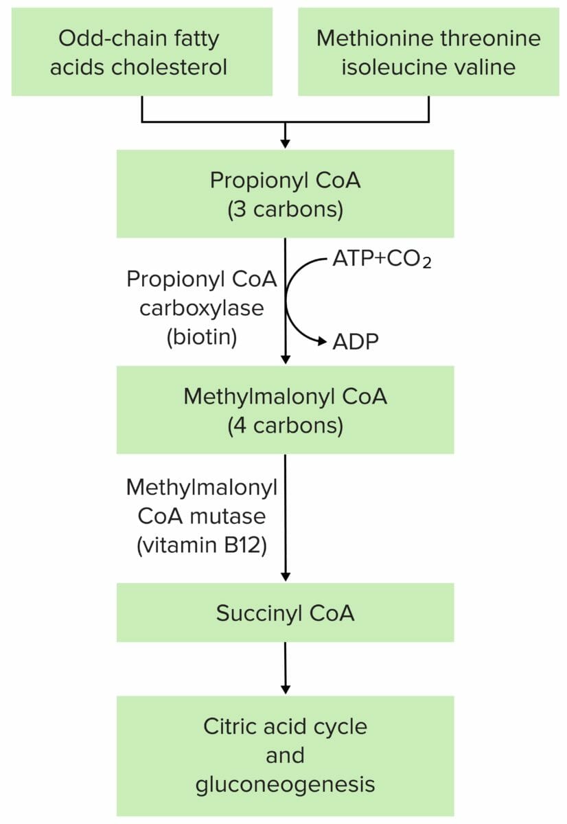
The pathophysiology of methylmalonic acidemia is most often due to mutation at either methylmalonyl CoA mutase or methylmalonyl CoA epimerase.

Image by Lecturio.

Symptoms

  • MSUD
    • Symptoms present within the first 2 days of life.
    • Neurologic:
      • Developmental delay
      • Poor feeding
      • Lethargy Lethargy A general state of sluggishness, listless, or uninterested, with being tired, and having difficulty concentrating and doing simple tasks. It may be related to depression or drug addiction. Hyponatremia
      • Irritability
      • Hypertonia Hypertonia Abnormal increase in skeletal or smooth muscle tone. Skeletal muscle hypertonicity may be associated with pyramidal tract lesions or basal ganglia diseases. Neurological Examination
      • Spasticity Spasticity Spinal Disk Herniation
      • Convulsions Convulsions Seizures
      • Coma Coma Coma is defined as a deep state of unarousable unresponsiveness, characterized by a score of 3 points on the GCS. A comatose state can be caused by a multitude of conditions, making the precise epidemiology and prognosis of coma difficult to determine. Coma
    • Orthopedic: osteoporosis Osteoporosis Osteoporosis refers to a decrease in bone mass and density leading to an increased number of fractures. There are 2 forms of osteoporosis: primary, which is commonly postmenopausal or senile; and secondary, which is a manifestation of immobilization, underlying medical disorders, or long-term use of certain medications. Osteoporosis
    • GI: pancreatitis Pancreatitis Inflammation of the pancreas. Pancreatitis is classified as acute unless there are computed tomographic or endoscopic retrograde cholangiopancreatographic findings of chronic pancreatitis. The two most common forms of acute pancreatitis are alcoholic pancreatitis and gallstone pancreatitis. Acute Pancreatitis
    • GU: sweet-smelling urine
  • Isovaleric acidemia Acidemia Respiratory Acidosis
    • Characterized by a distinct odor described as “sweaty feet”
    • GI:
      • Nausea Nausea An unpleasant sensation in the stomach usually accompanied by the urge to vomit. Common causes are early pregnancy, sea and motion sickness, emotional stress, intense pain, food poisoning, and various enteroviruses. Antiemetics
      • Vomiting Vomiting The forcible expulsion of the contents of the stomach through the mouth. Hypokalemia
    • Neurologic:
      • Seizures Seizures A seizure is abnormal electrical activity of the neurons in the cerebral cortex that can manifest in numerous ways depending on the region of the brain affected. Seizures consist of a sudden imbalance that occurs between the excitatory and inhibitory signals in cortical neurons, creating a net excitation. The 2 major classes of seizures are focal and generalized. Seizures
      • Apathy Apathy Lack of emotion or emotional expression; a disorder of motivation that persists over time. Wernicke Encephalopathy and Korsakoff Syndrome
      • Coma Coma Coma is defined as a deep state of unarousable unresponsiveness, characterized by a score of 3 points on the GCS. A comatose state can be caused by a multitude of conditions, making the precise epidemiology and prognosis of coma difficult to determine. Coma 
      • Hypotonia Hypotonia Duchenne Muscular Dystrophy
  • Methylmalonic acidemia Acidemia Respiratory Acidosis
    • GI:
      • Nausea Nausea An unpleasant sensation in the stomach usually accompanied by the urge to vomit. Common causes are early pregnancy, sea and motion sickness, emotional stress, intense pain, food poisoning, and various enteroviruses. Antiemetics
      • Vomiting Vomiting The forcible expulsion of the contents of the stomach through the mouth. Hypokalemia 
    • Respiratory: respiratory distress
  • Propionic acidemia Acidemia Respiratory Acidosis
    • Neurologic:
      • Poor feeding
      • Hypotonia Hypotonia Duchenne Muscular Dystrophy
      • Seizures Seizures A seizure is abnormal electrical activity of the neurons in the cerebral cortex that can manifest in numerous ways depending on the region of the brain affected. Seizures consist of a sudden imbalance that occurs between the excitatory and inhibitory signals in cortical neurons, creating a net excitation. The 2 major classes of seizures are focal and generalized. Seizures
    • GI:
      • Nausea Nausea An unpleasant sensation in the stomach usually accompanied by the urge to vomit. Common causes are early pregnancy, sea and motion sickness, emotional stress, intense pain, food poisoning, and various enteroviruses. Antiemetics
      • Vomiting Vomiting The forcible expulsion of the contents of the stomach through the mouth. Hypokalemia
      • Dehydration Dehydration The condition that results from excessive loss of water from a living organism. Volume Depletion and Dehydration
    • Genitourinary:  CKD CKD Chronic kidney disease (CKD) is kidney impairment that lasts for ≥ 3 months, implying that it is irreversible. Hypertension and diabetes are the most common causes; however, there are a multitude of other etiologies. In the early to moderate stages, CKD is usually asymptomatic and is primarily diagnosed by laboratory abnormalities. Chronic Kidney Disease 
    • Cardiovascular:
      • Cardiomyopathy Cardiomyopathy Cardiomyopathy refers to a group of myocardial diseases associated with structural changes of the heart muscles (myocardium) and impaired systolic and/or diastolic function in the absence of other heart disorders (coronary artery disease, hypertension, valvular disease, and congenital heart disease). Cardiomyopathy: Overview and Types (dilated and hypertrophic)
      • Conduction disorders

Diagnosis

  • MSUD:
    • Newborn Newborn An infant during the first 28 days after birth. Physical Examination of the Newborn screening Screening Preoperative Care to measure the amount of branched-chain amino acids Amino acids Organic compounds that generally contain an amino (-NH2) and a carboxyl (-COOH) group. Twenty alpha-amino acids are the subunits which are polymerized to form proteins. Basics of Amino Acids 
    • Urine organic acid levels to confirm
    • Genetic testing Genetic Testing Detection of a mutation; genotype; karyotype; or specific alleles associated with genetic traits, heritable diseases, or predisposition to a disease, or that may lead to the disease in descendants. It includes prenatal genetic testing. Myotonic Dystrophies for further confirmation and prenatal counseling
  • Isovaleric acidemia Acidemia Respiratory Acidosis:
    • Newborn Newborn An infant during the first 28 days after birth. Physical Examination of the Newborn urinary screening Screening Preoperative Care
    • Organic acid level measurements in older patients Patients Individuals participating in the health care system for the purpose of receiving therapeutic, diagnostic, or preventive procedures. Clinician–Patient Relationship
    • Assay of isovaleryl-CoA dehydrogenase activity in cultured skin Skin The skin, also referred to as the integumentary system, is the largest organ of the body. The skin is primarily composed of the epidermis (outer layer) and dermis (deep layer). The epidermis is primarily composed of keratinocytes that undergo rapid turnover, while the dermis contains dense layers of connective tissue. Skin: Structure and Functions fibroblasts Fibroblasts Connective tissue cells which secrete an extracellular matrix rich in collagen and other macromolecules. Sarcoidosis as confirmatory testing
  • Methylmalonic acidemia Acidemia Respiratory Acidosis:
    • Newborn Newborn An infant during the first 28 days after birth. Physical Examination of the Newborn screening Screening Preoperative Care showing elevation of propionylcarnitine
    • Screening Screening Preoperative Care looks for a high level of methylmalonic acid: Urine organic acids Acids Chemical compounds which yield hydrogen ions or protons when dissolved in water, whose hydrogen can be replaced by metals or basic radicals, or which react with bases to form salts and water (neutralization). An extension of the term includes substances dissolved in media other than water. Acid-Base Balance, plasma Plasma The residual portion of blood that is left after removal of blood cells by centrifugation without prior blood coagulation. Transfusion Products amino acids Amino acids Organic compounds that generally contain an amino (-NH2) and a carboxyl (-COOH) group. Twenty alpha-amino acids are the subunits which are polymerized to form proteins. Basics of Amino Acids, plasma Plasma The residual portion of blood that is left after removal of blood cells by centrifugation without prior blood coagulation. Transfusion Products acylcarnitine, and plasma Plasma The residual portion of blood that is left after removal of blood cells by centrifugation without prior blood coagulation. Transfusion Products total homocysteine should be obtained.
  • Propionic acidemia Acidemia Respiratory Acidosis: diagnosed by detection of elevated levels of propionic acid metabolites (e.g., 3-hydroxypropionate) in urine and serum

Management

  • MSUD:
    • Dietary modification
      • Protein-free diet 
      • Use of a diet that is low in the amino acids Amino acids Organic compounds that generally contain an amino (-NH2) and a carboxyl (-COOH) group. Twenty alpha-amino acids are the subunits which are polymerized to form proteins. Basics of Amino Acids leucine, valine, and isoleucine, which aren’t broken down in patients Patients Individuals participating in the health care system for the purpose of receiving therapeutic, diagnostic, or preventive procedures. Clinician–Patient Relationship
    • Acute metabolic decompensation:
      • Glucose Glucose A primary source of energy for living organisms. It is naturally occurring and is found in fruits and other parts of plants in its free state. It is used therapeutically in fluid and nutrient replacement. Lactose Intolerance infusions are needed.
      • Insulin Insulin Insulin is a peptide hormone that is produced by the beta cells of the pancreas. Insulin plays a role in metabolic functions such as glucose uptake, glycolysis, glycogenesis, lipogenesis, and protein synthesis. Exogenous insulin may be needed for individuals with diabetes mellitus, in whom there is a deficiency in endogenous insulin or increased insulin resistance. Insulin injections promoting anabolism
    • Surgical therapy: Liver transplantation Liver transplantation The transference of a part of or an entire liver from one human or animal to another. Hepatocellular Carcinoma (HCC) and Liver Metastases is successful in classic MSUD with no neurologic symptoms.
  • Isovaleric acidemia Acidemia Respiratory Acidosis:
    • Dietary changes to reduce the amount of leucine
    • Patients Patients Individuals participating in the health care system for the purpose of receiving therapeutic, diagnostic, or preventive procedures. Clinician–Patient Relationship with acute manifestations of disease are given glycine Glycine A non-essential amino acid. It is found primarily in gelatin and silk fibroin and used therapeutically as a nutrient. It is also a fast inhibitory neurotransmitter. Synthesis of Nonessential Amino Acids or carnitine Carnitine A constituent of striated muscle and liver. It is an amino acid derivative and an essential cofactor for fatty acid metabolism. Fatty Acid Metabolism
  • Methylmalonic acidemia Acidemia Respiratory Acidosis:
  • Propionic acidemia Acidemia Respiratory Acidosis:
    • Low-protein diet 
    • Protein supplementation with methionine, threonine, valine, isoleucine, and carnitine Carnitine A constituent of striated muscle and liver. It is an amino acid derivative and an essential cofactor for fatty acid metabolism. Fatty Acid Metabolism
    • Antibiotics for ⅓ of each month (removes flora that may increase protein levels)

References

  1. Williams, RA, Mamotte, CD, & Burnett, JR. (2008). Phenylketonuria: An inborn error of phenylalanine metabolism. The Clinical Biochemist. Reviews, 29(1), 31–41.
  2. Blackburn, P.R, Gass, JM, Vairo, F, Farnham, KM, Atwal, HK, Macklin, S, Klee, EW, & Atwal, PS. (2017). Maple syrup urine disease: Mechanisms and management. The Application of Clinical Genetics, 10, 57–66.
  3. Loots DT. (2009). Isovaleric acidemia. In: Lang F. (eds) Encyclopedia of Molecular Mechanisms of Disease. Springer, Berlin, Heidelberg. https://doi.org/10.1007/978-3-540-29676-8_983.
  4. Fraser, JL, & Venditti, CP. (2016). Methylmalonic and propionic acidemias: Clinical management update. Current Opinion in Pediatrics, 28(6), 682–693. https://doi.org/10.1097/MOP.0000000000000422.
  5. Grünert, SC, Müllerleile, S, De Silva, L, Barth, M, Walter, M, Walter, K, Meissner, T, Lindner, M, Ensenauer, R, Santer, R, Bodamer, OA, Baumgartner, MR, Brunner-Krainz, M, Karall, D, Haase, C, Knerr, I, Marquardt, T, Hennermann, JB, Steinfeld, & R, Beblo. (2013). Propionic acidemia: Clinical course and outcome in 55 pediatric and adolescent patients. Orphanet Journal of Rare Diseases, 8, 6. https://doi.org/10.1186/1750-1172-8-6.

Create your free account or log in to continue reading!

Sign up now and get free access to Lecturio with concept pages, medical videos, and questions for your medical education.

User Reviews

Details